Article

Propagation of Hinoki Cypress (Chamaecyparis obtusa) Through Tissue Culture Technique as a Sustainable Method for Mass Cloning of Selected Trees

1
Department of Forest Molecular Genetics and Biotechnology, Forestry and Forest Products Research Institute, Matsunosato 1, Tsukuba 305-8687, Japan
2
Tama Forest Science Garden, Forestry and Forest Products Research Institute, Todori 1833-81, Hachioji 193-0843, Japan
3
Kumamoto Prefectural Forestry Research and Instruction Center, 8-222-2, Kurokami, Kumamoto 860-0862, Japan
4
Shizuoka Prefectural Research Institute of Agriculture and Forestry, 2542-8, Negata, Hamamatsu 434-0016, Japan
*
Author to whom correspondence should be addressed.
Sustainability 2025, 17(7), 3039; https://doi.org/10.3390/su17073039
Submission received: 21 February 2025 / Revised: 25 March 2025 / Accepted: 27 March 2025 / Published: 29 March 2025
(This article belongs to the Section Sustainable Forestry)

Abstract

:
Propagation of hinoki cypress (Japanese cypress, Chamaecyparis obtusa, Cupressaceae) through adventitious bud multiplication was performed using leaf-segment explants from cutting plants of selected adult trees. Explants were successfully surface-sterilized (>90% asepsis) by agitating them in 2.5% (w/v available chlorine) sodium hypochlorite solution for 15 min and then rinsed with sterile distilled water. Explants approximately 2 cm long were cultured on plates containing medium supplemented with 6-benzylaminopurine (BAP) and 2,4-dichlorophenoxyacetic acid (2,4-D), 20 g/L sucrose, and 7 g/L agar. The cultures were kept at 25 ± 1 °C under a 16-h photoperiod with a photon flux density of approximately 65 µmol m−2 s−1. The optimal adventitious bud multiplication (31.5 buds per explant) was obtained on a medium supplemented with 10 µM BAP in combination with 1 µM 2,4-D. Proliferated adventitious buds were elongated better on medium supplemented with 1 µM trans-zeatin. The best rooting result (86%) was achieved on a rooting medium supplemented with 1 µM 3-indolebutyric acid in combination with 0.1 µM 1-naphthaleneacetic acid. However, rooting response varied according to genotypes. Clones related to the cultivar ‘Nangouhi’ (Na18, Na14 x Isa, Na14-14, Isa x Na14, and NaS) were easier to root than those derived from the cultivar ‘ShizuokaKenZairai’ (SKZ5 and SKZ8). Regenerated plantlets did not show morphological abnormalities and showed a high survival rate after acclimatization (>90%).

1. Introduction

Hinoki cypress (Japanese cypress, Chamaecyparis obtusa (Sieb et Zucc.) Endl., Cupressaceae) is one of six species belonging to the genus distributed throughout the world [1]. In Japan, due to its high wood quality, hinoki cypress is one of the most important commercial timber trees and the second most reforested species, accounting for about 25% of the country’s plantation area with approximately 2.6 million ha [2]. It is used for housing materials, structural timbers, interior joinery, furniture, flooring boards, traditional bathtubs, and many other articles [3]. Hinoki cypress has a natural distribution from Fukushima Prefecture to Kyushu Island and is cultivated in almost the same area [4]. However, plantation areas are subject to pest and disease attacks [5,6,7]. In addition, the large amount of pollen released every spring from hinoki cypress forests is reported to be the cause of one of the most serious allergies in Japan [8]. Currently, over one thousand elite trees have been selected throughout the country from national and private forests for genetic improvement purposes [3]. Although hinoki cypress is generally propagated by seed, difficulty of obtaining strobili each year and low seed fertility from some crosses among selected clones or between reported hybrids [9,10,11,12] make it difficult to produce seedlings for practical reforestation [7]. Furthermore, because hinoki cypress is rarely propagated by cuttings and seed production in seed orchards is scarce [13], plant regeneration through tissue culture is an optimal alternative for the efficient and sustainable propagation of seedlings.
Propagation protocols for hinoki cypress through somatic embryogenesis have been established from seed explants [14,15]. However, to date, attempts to induce somatic embryogenesis and plant regeneration from the vegetative material of adult trees have not been successful. Efficient clonal propagation from vegetative explants of selected adult trees is a powerful tool to accelerate genetic improvement programs and a key factor for sustainable forestry. An efficient propagation system is also important for genetic engineering to develop improved trees for faster growth, better wood quality, disease resistance, pollen-free plants, and climate change adaptation. Information on in vitro organogenesis of hinoki cypress from vegetative materials is mainly reported using germinants or juvenile seedlings [3,16,17,18,19,20], in vitro microcuttings [21], and encapsulated shoot explants [22]. Although some studies on plant regeneration using vegetative explants from adult trees have been reported [3,23,24], the information is still scarce and requires improvement for practical application. This report describes an improved protocol for sustainable micropropagation of selected hinoki cypress trees.

2. Materials and Methods

2.1. Plant Materials and Surface Sterilization of Explants

Samples were taken from selected trees of eleven clones propagated by cuttings for the purpose of rejuvenating the explants to promote micropropagation of adult trees. All of these clones (one tree per clone) represent different genotypes selected for their useful characteristics (shape, growth, wood quality) and used as breeding stock. Short branches used for cutting propagation were obtained from 30 to 40 years old individuals grown in Kumamoto Prefectural Forestry Research and Instruction Center (Kurokami, Kumamoto, Japan), Shizuoka Prefectural Research Institute of Agriculture and Forestry (Hamamatsu, Shizuoka, Japan), and Ibaraki Prefectural Government Forestry Technology Center (Naka, Ibaraki, Japan). About 10 cm of the shoots were cut off and prepared for cuttings, which were inserted into a cutting bed filled with Kanuma soil (Akagi Engei Co., Ltd., Isezaki, Japan) in the greenhouse to promote rooting. The temperature and humidity in the greenhouse were uncontrolled and varied throughout the year between 10 °C and 35 °C and 30% and 75% relative humidity, respectively. The cuttings were watered twice daily with an automatic irrigation system to keep the soil sufficiently moist and were grown in approximately 50% shaded conditions by covering the top of the cutting bed with a shading net. The onset of rooting of the cuttings was observed approximately six weeks after their placement into the cutting bed. Leaf segments collected from rooted cuttings grown for approximately three years were cut into 3–4 cm lengths and surface-sterilized by agitating them in 2.5% (w/v available chlorine) sodium hypochlorite (FUJIFILM Wako Chemicals, Co., Ltd., Osaka, Japan) solution for 15 min and then rinsed with sterile distilled water five times for 3 min each. Subsequently, the leaf segments were adjusted into about 2 cm long explants by cutting their basal end before being transferred to the adventitious bud induction medium. Data were collected two weeks after inoculation.

2.2. Adventitious Bud Induction

For the initial culture, surface-sterilized explants were placed horizontally on plates (90 mm × 20 mm) (Asahi Glass Co., Ltd., Tokyo, Japan) with basal WP medium [25] containing 20 g/L sucrose (FUJIFILM Wako Chemicals Co., Ltd.) and 7 g/L agar (FUJIFILM Wako Chemicals Co., Ltd.). To determine the effect of plant growth regulators (PGRs) on adventitious bud induction, the addition of 6-benzylaminopurine (BAP) (FUJIFILM Wako Chemicals Co., Ltd.) or trans-zeatin (ZEA) (FUJIFILM Wako Chemicals Co., Ltd.) alone at different concentrations and in combination with 2,4-dichlorophenoxyacetic Acid (2,4-D) (FUJIFILM Wako Chemicals Co., Ltd.) or 1-naphthaleneacetic Acid (NAA) (FUJIFILM Wako Chemicals Co., Ltd.) were tested with clone No7-5. Each treatment has 10 replicates (10 explants per plate). The cultures were kept at 25 ± 1 °C under a 16-h photoperiod with photon flux density of approximately 65 µmol m−2 s−1. Data were collected after 10 weeks of culture. To examine the response of different clones on adventitious bud induction, explants were cultured at the same conditions described for initial culture on a medium supplemented with 10 µM BAP and 1 µM 2,4-D. Data were collected after 10 weeks of culture from 10 replicates per clone. For subsequent adventitious bud proliferation, 5–10 mm diameter cluster buds (10 pieces per plate) were transferred onto the same fresh medium and subcultured at 3-month intervals in the same culture conditions described above.

2.3. Shoot Elongation from Induced Adventitious Buds

To promote shoot elongation from induced adventitious buds, bud clusters of clone Na14 x 14, about 10 mm diameter, were transferred to plates containing MS medium [26] with basal salts reduced to half-concentration (1/2 MS), 20 g/L sucrose, 7 g/L agar, and supplemented with ZEA or NAA alone or in combination, at concentrations of 1 µM and 0.05 µM, respectively. Ten explants were used for each treatment. The cultures were kept at 25 ± 1 °C under a 16-h photoperiod with a photon flux density of approximately 65 µmol m−2 s−1. Data were collected after 16 weeks of culture. To promote shoot elongation from adventitious buds induced in the other clones, bud clusters were cultured on a medium supplemented with 1 µM ZEA at the same culture conditions described above. All shoot elongation studies were performed using bud clusters previously obtained on medium supplemented with 10 μM BAP and 1 μM 2,4-D.

2.4. Rooting of Elongated Shoots

To determine the effect of PGRs on rooting, elongated shoots of clone Na14 x Isa, longer than 20 mm, were isolated from the adventitious bud clusters and cultured in bio-flasks (450 mL) (Nihon Yamamura Glass Co., Ltd., Hyogo, Japan) containing 1/2 MS medium supplemented with 10 g/L sucrose, 7 g/L agar, and 1–5 µM 3-indolebutyric acid (IBA) (FUJIFILM Wako Chemicals Co., Ltd.) alone, or in combination with 0.1–0.5 µM NAA. Ten flasks containing ten shoots per flask were used for each medium condition. The cultures were kept at 25 ± 1 °C under a 16-h photoperiod with a photon flux density of approximately 65 µmol m−2 s−1. Data were collected after 12 weeks of culture. In our experiments, we consider an explant rooted if it shows both emergence and subsequent growing roots of at least 10 mm. To examine the response of different clones on root induction, elongated shoots were cultured under the same conditions on a medium supplemented with 1 µM IBA and 0.1 µM NAA. Data were collected after 12 weeks of culture from ten flasks containing ten shoots per clone. All rooting studies were performed using shoots previously elongated on a medium supplemented with 1 µM ZEA.

2.5. In Vitro Growth and Ex Vitro Acclimatization of Regenerated Plants

To promote the growth of rooted shoots, they were transferred to bio-flasks containing 1/2 MS medium supplemented with 30 g/L sucrose, 5 g/L activated charcoal (AC) (FUJIFILM Wako Chemicals Co., Ltd.), and 11.5 g/L agar. The cultures were kept at 25 ± 1 °C under a 16-h photoperiod with a photon flux density of about 65 µmol m−2 s−1 for approximately 3 months before ex vitro acclimatization. Developed plantlets were transplanted into plastic pots filled with vermiculite (Akagi Engei Co., Ltd.) and acclimatized in plastic boxes (Shinkigosei Co., Ltd., Tokyo, Japan) inside a growth cabinet according to the method described elsewhere for embling acclimatization of related Chamaecyparis pisifera (Sieb et Zucc.) Endl., sawara cypress [27]. Ex vitro survival data were collected eight weeks after acclimatization.

2.6. Statistical Analyses

Differences in the PGRs composition of the culture medium for adventitious bud induction, shoot elongation (total and >1 cm shoot), and rooting were examined in generalized linear models (GLMs) or generalized linear mixed models (GLMMs). The error distribution for the objective variable was assumed to follow a Poisson distribution for the number of adventitious buds induced and shoots elongated and a binomial distribution for rooting, with the composition of PGRs used as an explanatory variable. In addition, flasks were used for random effects in the rooting response model. Clonal differences in surface-sterilization of adult leaf explants, adventitious bud induction, and rooting responses were also evaluated by constructing GLMs or GLMMs. The objective variables surface-sterilization status (aseptic or non-aseptic) and rooting were assumed to have binomial error distributions, while the number of adventitious buds had a Poisson error distribution. The clone was used as an explanatory variable, and the flask was used as a random effect in the rooting response model. Tukey’s multiple comparisons with BH adjustment were used to determine the difference between treatments. The ‘lme4’ [28] and ‘multcomp’ [29] packages in R ver. 4.4.0 [30] were used for the analysis.

3. Results

3.1. Surface Sterilization of Explants for In Vitro Culture Initiation

The results of the surface sterilization response of leaf-segment explants from cutting plants of different selected clones are shown in Table 1. The average percentage of aseptic explants two weeks after inoculation onto induction media was 91.10%, varying from a minimal rate of 70.59% for the explants from the No7-5 clone to a maximum rate of 100.00% asepsis for the explants from the clones Na14-14 and NaS. The GLM model (N = 1074) fit was better when a clone was used as a variable, but there were no significant differences among clones (p > 0.05).

3.2. Effect of PGRs on Adventitious Bud Induction and Response from Different Clones

Adventitious bud induction from the leaf segments was evident approximately 3–4 weeks after the explant inoculation. After 10 weeks of culture, an average of approximately 17 adventitious buds per explant was recorded in the highest response among the PGRs tested (10 µM BAP in combination with 1 µM 2,4-D), which differed significantly from other treatments at the 5% level (Figure 1). Except for the lowest response recorded on the medium supplemented with 1 µM ZEA (approximately four buds per explant), no significant statistical differences were observed among the remaining PGR treatments, with average responses ranging from around 9 to 12 shoots per explant.
Additionally, as shown in Figure 2, significant differences (p < 0.05) in the number of adventitious buds per explant were observed among the tested clones, with a wide variation from an average of 4.8 to 31.5 induced buds. High adventitious bud induction responses (around 20–30 buds per explant) were achieved in the clones Na14 x Isa, Isa x Na14, and Na14-14, whereas low induction responses (less than 10 buds per explant) were registered in the clones Isa, SKZ3, and SKZ6. The other clones showed an induction response that varied around 10–15 adventitious buds per explant.

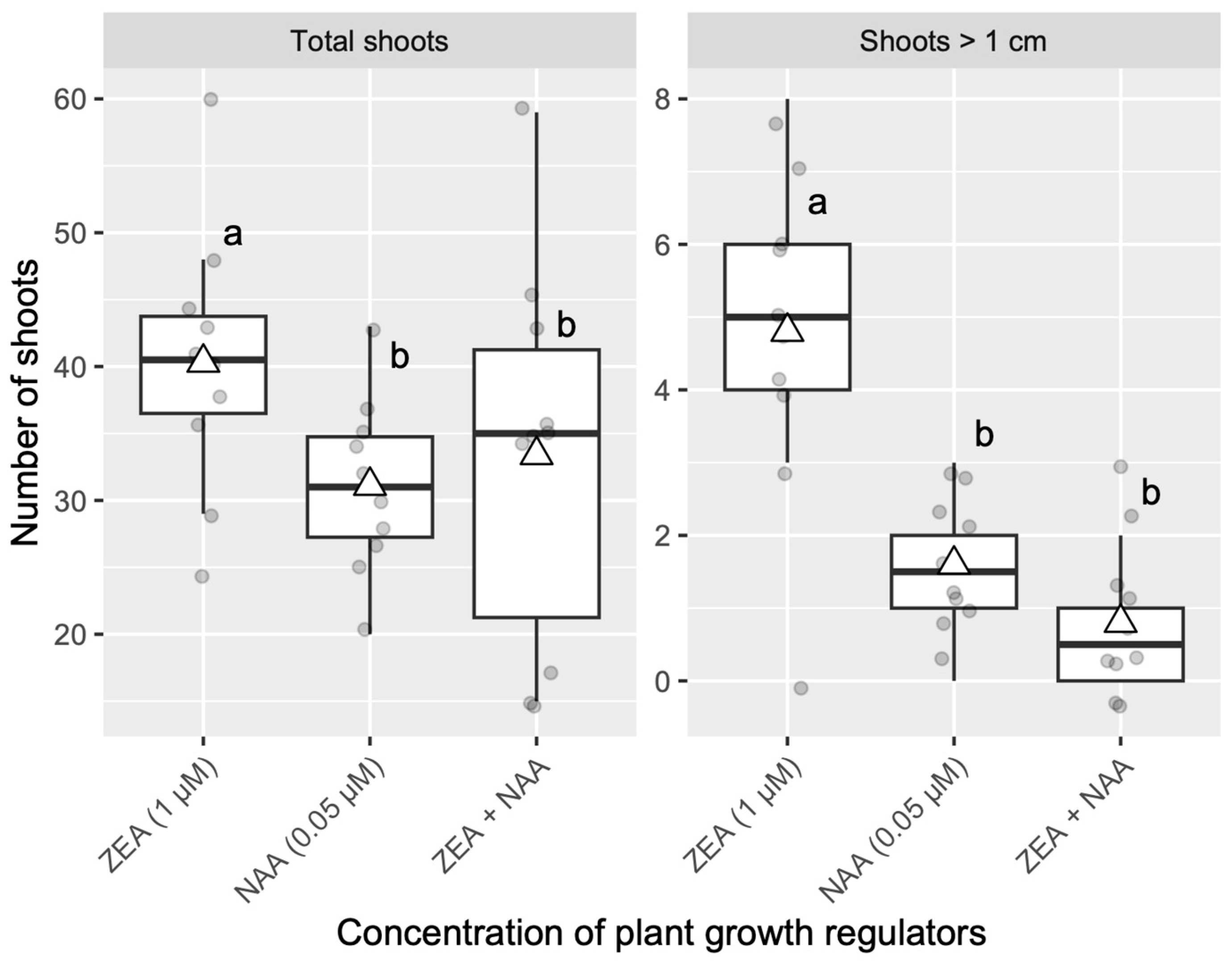
3.3. Effect of PGRs on Shoot Elongation from Adventitious Buds

The results of the experiment to determine the effect of PGRs on shoot elongation from adventitious buds are shown in Figure 3. The best elongation response after 16 weeks of culture was recorded in an elongation medium supplemented with 1 µM ZEA. Here, the total number of shoots was the highest and was also significantly greater when limited to shoots longer than 1 cm (p < 0.05 by GLM). Around five elongated shoots more than 1 cm long were obtained per bud cluster, compared to around two and one elongated shoots from bud clusters cultured in elongation medium supplemented with 0.05 µM NAA and 1 µM ZEA + 0.05 µM NAA, respectively. Shoot elongation from adventitious buds induced in the other clones was promoted by transferring the bud clusters to a medium supplemented with 1 µM ZEA. No morphological differences in shoot elongation among clones were observed.

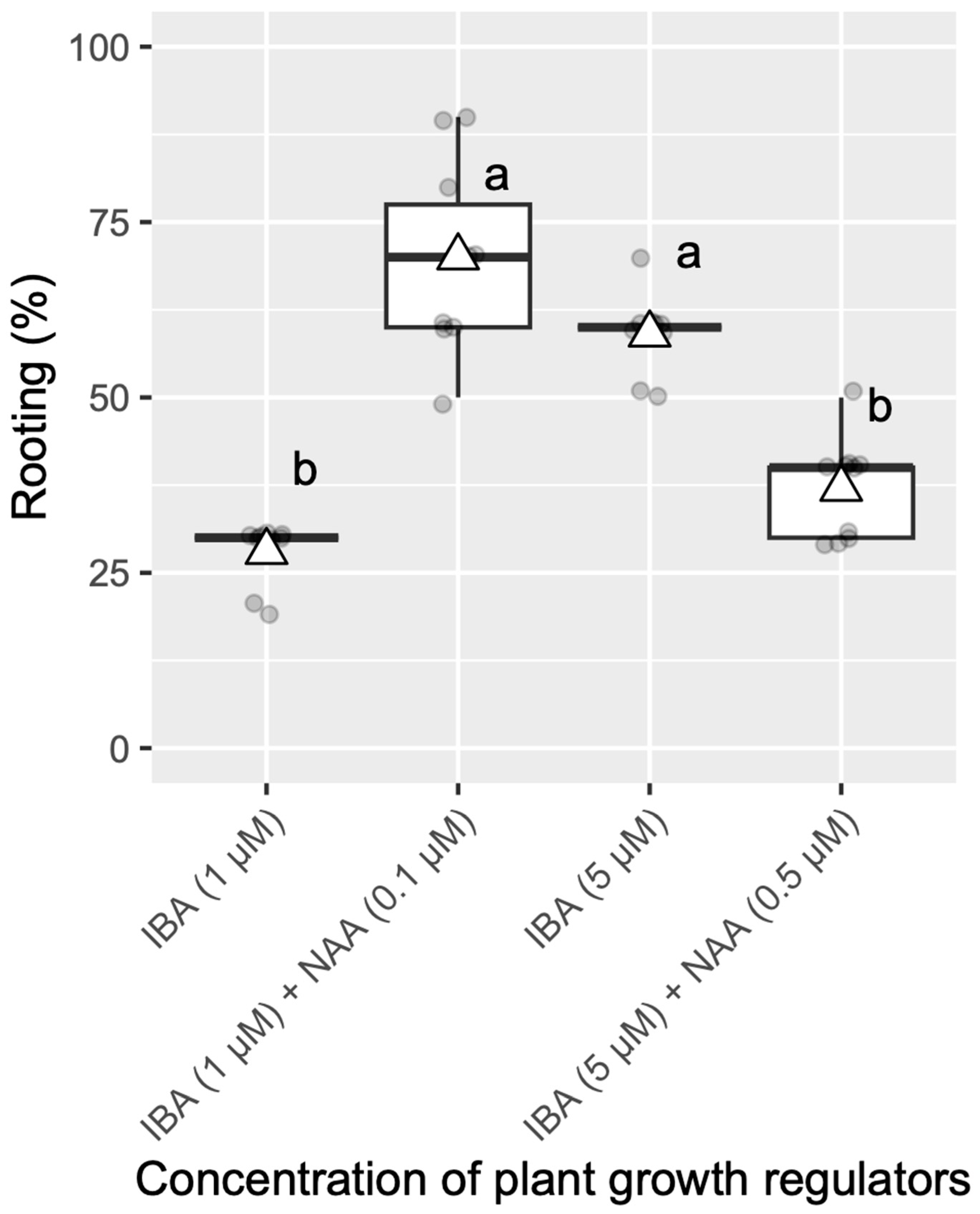
3.4. Effect of PGRs on Rooting and Response from Different Clones

After 12 weeks of culture, the best rooting response (p < 0.05) was achieved in the medium supplemented with 1 µM IBA and 0.1 µM NAA (Figure 4). The effect of IBA alone (1 or 5 µM) or in combination with a high concentration of NAA (0.5 µM) on root induction was considerably reduced compared to the best rooting response of 70% and decreased at frequencies ranging from 59 to 28%. The results of the clonal differences in rooting rates are shown in Figure 5. Clonal differences at the 5% significance level on the root induction rates were observed among the clones, with rooting variation rates ranging from around 22% to 86%. High rooting responses (more than 70%) were achieved in the clones Na18, Na14 x Isa, and No7-5; moderate rooting responses (around 50–60%) were observed in the clones Na14-14, Isa x Na14, and NaS, whereas low rooting responses (less than 30%) were registered in the clones SKZ5 and SKZ8.

3.5. In Vitro Growth and Ex Vitro Acclimatization of Regenerated Plants

About three months after culturing in a growth medium, rooted shoots that developed into plantlets were ready for their ex vitro acclimatization. The regenerated plantlets did not show morphological abnormalities (Figure 6I) and showed a survival rate of >90% eight weeks after acclimatization.

4. Discussion

Vegetative propagation methods are important to retain the desirable characteristics of selected trees and to propagate plants true-to-type. Among clonal propagation methods, cutting- or grafting-based techniques are the most popular. However, for sustainable large-scale production, one of the most desirable methods targets organogenesis by in vitro culturing of vegetative materials [31]. In our study, tissue culture was initiated from leaf-segment explants of cutting plants derived from selected adult trees (Figure 6A). Although microbial contamination is reported as one of the most critical and frequent problems in plant tissue culture initiation, especially when materials are taken from older trees [32,33], the high asepsis rates achieved in all clones evaluated (mean 91.10%) throughout the experiments suggest that the method applied to the explants was appropriate for the initiation of in vitro culture of hinoki cypress from leaf-segment explants. This result is consistent with previous reports on surface sterilization of hinoki cypress explants from adult material [3,24] and from cuttings grown in a nursery [21].
Adventitious bud initiation and proliferation from the leaf-segment explants were stimulated by the cytokinin supplementation but more efficiently in combination with a low concentration of auxins (Figure 6B,C). BAP alone or in combination with NAA, IBA, or 3-indoleacetic acid (IAA) has been reported as effective for the multiplication of C. obtusa [3,16,19], Chamaecyparis nootkatensis (D. Don) Spach [34], Chamaecyparis pisifera var. filifera Beissn. et Hochst. [35], Cupressus sempervirens L. and Chamaecyparis lawsoniana (A. Murr.) Par. [36], Cupressus sempervirens var. horizontalis (Mill.) [37], and C. pisifera [24,38]. The cytokinin N6-(2-isopentenyl) adenine (2iP) was reported to be more suitable than BAP as a morphogenetic agent for in vitro multiplication and elongation of Thuja plicata D. Don ex Lambert [39]. Additionally, the use of ZEA has been reported in the protocols for clonal propagation of Thuja occidentalis L. [40] and C. obtusa [20]. Overall, although adventitious bud induction rates vary according to species, explant age, and culture conditions, in the present study, the large variation recorded in the efficiency of adventitious bud induction among clones can be attributed to the genetic origin of the explant (Figure 2).
Subsequent proliferation routines were performed by subculturing the bud clusters every three months without observing a decrease in the multiplication rate five years after the induction. After continuous subculture routines on multiplication medium, proliferated adventitious buds, with intermediate levels of vitrification, were observed (Figure 6D,E). However, vitrified buds reverted to normal after being transferred to an elongation medium containing low levels of PGRs (Figure 6F). Although vitrification is a common problem in micropropagation, in many cases, it can be minimized by lowering the PGR concentration and/or increasing the concentration of the solidifying agent in the medium [32,41,42]. In our experiments, most of the elongated shoots became normal after three months of culturing in a medium supplemented with low cytokinin concentration, maintaining their normal appearance in the subsequent phases of rooting (Figure 6G) and growth (Figure 6H). Similar results were reported for the elongation of non-vitrified shoots of hinoki cypress, but only after transferring adventitious bud clusters to a medium containing a high concentration of agar [18]. Although supplements of low concentrations of PGRs in the medium are used for shoot elongation of several cypress trees [3,16,19,37,40], PGR-free medium was reported as beneficial for Mediterranean cypress and Lawson cypress [36]. Additionally, adding AC to the medium was reported to effectively elongate bud shoots of eastern white cedar [40] and adventitious buds of sawara cypress [38].
In contrast to the lower rooting frequencies of less than 30% and 15% reported for shoot explants derived from adult trees of hinoki cypress [3] and Mediterranean cypress [37,43,44], respectively, the highest percentage of rooting (86%) in this study was achieved by adding low concentrations of IBA (1 μM) and NAA (0.1 μM) to the medium (Figure 5). This result was consistent with the high rooting rate reported for shoots from 18 months old seedlings of C. sempervirens and C. lawsoniana [36], germinated juvenile seedlings of C. obtusa [3], and for shoots derived from somatic embryos of C. pisifera [38]. Similarly, Capuana and Giannini (1997) [43] reported 65%, 11%, and 5% rooting for 7-day seedlings, 15 years old trees, and 150 years old trees of C. sempervirevirens, respectively. On the other hand, Misson et al. (1991) [39] reported rooting in shoots from a 183 years old T. plicata but only after their rejuvenation by successive micrograftings and successive 2iP cytokinin pulse treatments. These results confirm that explants taken from young plants root easily, whereas explants taken from older trees have a very low rooting rate and that root induction is the most problematic step in micropropagation of mature trees, becoming even more difficult as the plant ages. Although several interacting factors such as the kind of explants, ontogenetic age, age of the organ, and culture methods and conditions may significantly affect the rooting response of shoots [45], the results achieved in this study suggest that differences in rooting ability among clones were strictly genotype-dependent. The large clonal differences registered among the tested genotypes (Figure 5), ranging from the highest rooting response in the clone Na18 (86%) to the lowest in the clone SKZ8 (22%), were consistent with the rooting rates achieved in a nursery with cuttings from the same clones. Hinoki cypress is traditionally propagated by seeds and rarely by cuttings [3]. One of the few exceptions is the use of the ‘Nangouhi’ cultivar, which is famous for being easy to propagate by cuttings. In our study, clone Na18, which originated from the ‘Nangouhi’ cultivar, showed the highest rooting frequency. Conversely, the clone SKZ8, derived from the ‘ShizuokaKenZairai’ cultivar, is known to be recalcitrant to propagation by cuttings, which was consistent with the lowest in vitro rooting rate obtained. In general, clones related to the cultivar ‘Nangouhi’ (Na18, Na14 x Isa, Na14-14, Isa x Na14, and NaS) were easier to root than those derived from the cultivar ‘ ShizuokaKenZairai ‘ (SKZ5 and SKZ8) (Figure 5).
Regenerated plantlets showed a high survival rate after acclimatization, similar to the results reported elsewhere for the emblings of sawara [27] and hinoki cypress [15]. The methodology applied in our study was also shown to be successful for the plant acclimatization of Japanese pines [46] and Japanese cedar [47]. In contrast, lower survival rates, no greater than 40%, were reported for somatic plants of Mediterranean cypress derived from clones resistant to the bark canker disease [48].

5. Conclusions

In this study, we developed an updated protocol for the efficient and sustainable propagation of hinoki cypress through the in vitro culture of leaf segments from cutting plants of selected adult trees. We believe that the described methodology, which covered all the micropropagation phases with a large number of genotypes, will not only contribute to the clonal propagation of this species but will also provide valuable information for the development of efficient protocols for other species of the Cupressaceae family. The developed system could also be used as a powerful tool for genetic engineering and inter-specific hybridization breeding to develop improved trees for sustainable forestry.

Author Contributions

Conceptualization, T.E.M., M.T. and A.M.; Formal analysis, M.T.; Funding acquisition, T.E.M.; Methodology, T.E.M., M.T., A.M., R.K. and T.H.; Project administration, T.E.M.; Supervision, T.E.M.; Writing—original draft, T.E.M.; Writing—review and editing, T.E.M., M.T., A.M., R.K. and T.H. All authors have read and agreed to the published version of the manuscript.

Funding

This work was supported in part by JSPS KAKENHI (Grant No. 21K19154).

Institutional Review Board Statement

Not applicable.

Informed Consent Statement

Not applicable.

Data Availability Statement

All the data presented have been made available as tables and figures. The data presented in this study are available on request from the corresponding author.

Acknowledgments

The authors would like to thank the Ibaraki Prefectural Government Forestry Technology Center and Kyushu Regional Breeding Office for their logistical support in the preparation of plant materials.

Conflicts of Interest

The authors declare that this research was conducted without any commercial or financial relationships that could be construed as potential conflicts of interest.

References

  1. Wang, Y.; Ruhsam, M.; Milne, R.; Graham, S.W.; Li, J.; Tao, T.; Zhang, Y.; Mao, K. Incomplete lineage sorting and local extinction shaped the complex evolutionary history of the Paleogene relict conifer genus, Chamaecyparis (Cupressaceae). Molec. Phylogen. Evolut. 2022, 172, 107485. [Google Scholar]
  2. Forestry Agency (Ed.) Statistical Directory of Forest and Forestry of Japan 2024 (Shinrin Ringyo Tokei Yoran); Nihon Shinrin Ringyo Shinkokai: Tokyo, Japan, 2024; pp. 1–261. (In Japanese) [Google Scholar]
  3. Ishi, K. Hinoki cypress (Chamaecyparis obtusa). In Biotechnology in Agriculture and Forestry, Trees III; Bajaj, Y.P.S., Ed.; Springer: Berlin/Heidelberg, Germany, 1991; Volume 16, pp. 471–478. [Google Scholar]
  4. Asakawa, S.; Katsuta, M.; Yokoyama, T. Seeds of Woody Plants in Japanese Gymnospermae; Japan Forest Tree Breeding Association: Tokyo, Japan, 1981; 150p. (In Japanese) [Google Scholar]
  5. Kobayashi, K. Cone and seed insects of Japanese conifers. In Proceedings of the 17th IUFRO World Congress, Kyoto, Japan, 6–17 September 1981; Div. 2. pp. 455–461. [Google Scholar]
  6. Maeta, T. Effects of gamma-rays irradiation on interspecific hybridization between Chamaecyparis obtusa S. et Z. and C. pisifera S. et Z. Hoshasen Ikussyujo Kenkkyu Hokoku 1982, 5, 1–87. (In Japanese) [Google Scholar]
  7. Ishii, K.; Maruyama, E.; Hosoi, Y. Plant regeneration by somatic embryogenesis from in vitro-cultured shoots of hinoki cypress (Chamaecyparis obtusa Sieb. et Zucc.). Propag. Ornam. Plants. 2003, 3, 19–22. [Google Scholar]
  8. Osada, T.; Okano, M. Japanese cedar and cypress pollinosis updated: New allergens, cross-reactivity, and treatment. Allergol. Int. 2021, 70, 281–290. [Google Scholar] [PubMed]
  9. Yamamoto, C.; Fukuhara, N. Cone and seed yields after open-, self-, intraspecific-, and interspecific-pollinations in Chamaecyparis obtusa (Sieb. et Zucc.) Endl. and C. pisifera (Sieb. et Zucc.). Endl. Bull. For. Forest Prod. Res. Inst. 1980, 311, 65–92. [Google Scholar]
  10. Maeta, T.; Yamamoto, C. Interspecific hybridization among Chamaecyparis species. In Proceedings of the 17th IUFRO World Congress, Kyoto, Japan, 6–17 September 1981; Div. 2. pp. 169–176. [Google Scholar]
  11. Ohguro, T.; Okamura, M. Morphological and cytological studies on hybridization among Chamaecyparis obtusa Sieb. et Zucc., C pisifera Sieb. et Zucc., and C. lawsoniana Parl. Bull. For. Tree. Breed. Inst. 1997, 5, 59–87. (In Japanese) [Google Scholar]
  12. Fukuhara, N. Fertility in interspecific-crossing between hinoki (Chamaecyparis obtusa Endl.) and sawara (C. pisifera Endl.) and the identification of the hybrids. Bull. For. Prod. Res. Inst. 1989, 354, 1–38. [Google Scholar]
  13. Fukuhara, N. Meiotic observation in the pollen mother cell of interspecific hybrid between Chamaecyparis obtusa and C. pisifera. J. Jpn. For. Soc. 1978, 60, 437–441. [Google Scholar]
  14. Taniguchi, T.; Kurita, M.; Itahana, N.; Kondo, T. Somatic embryogenesis and plant regeneration from immature zygotic embryos of hinoki cypress (Chamaecyparis obtusa Sieb. et Zucc.). Plant Cell Rep. 2004, 23, 26–31. [Google Scholar] [CrossRef]
  15. Maruyama, E.; Ishii, K.; Hosoi, Y. Efficient plant regeneration of hinoki cypress (Chamaecyparis obtusa Sieb. et Zucc.) via somatic embryogenesis. J. For. Res. 2005, 10, 73–77. [Google Scholar] [CrossRef]
  16. Ishii, K. In vitro plantlet formation from adventitious buds on juvenile seedlings of hinoki cypress (Chamaecyparis obtusa). Plant Cell Tiss. Org. Cult. 1986, 7, 247–255. [Google Scholar] [CrossRef]
  17. Okamura, M.; Senda, M.; Kondo, T. Tissue culture of hinoki (Chamaecyparis obtusa). Bull. National For. Tree. Breed. Center 1995, 13, 119–129. [Google Scholar]
  18. Ishii, K. Liquid culture and transformation of hinoki cypress (Chamaecyparis obtusa Sieb. et Zucc.). J. For. Res. 2002, 7, 99–104. [Google Scholar] [CrossRef]
  19. Min, J.Y.; Park, D.J.; Jeong, M.J.; Song, H.J.; Kim, Y.D.; Kang, Y.M.; Karigar, C.S.; Choi, M.S. In vitro propagation of Chamaecyparis obtusa Sieb. et Zucc. Prop. Ornam. Plants 2010, 10, 117–121. [Google Scholar]
  20. Kim, J.A.; Lee, N.-N.; Kim, Y.W. In vitro plantlets regeneration by multi-shoots induction and rooting in Chamaecyparis obtusa. J. Plant Biotechnol. 2019, 46, 303–309. [Google Scholar] [CrossRef]
  21. Ide, Y.; Yamamoto, S. Adventitious root formation on in vitro microcuttings of hinoki. J. Jpn. For. Soc. 1986, 68, 296–298. [Google Scholar]
  22. Okamura, M.; Senda, M.; Kondo, T. Rooting from the encapsulated adventitious buds of hinoki, Chamaecyparis obtusa. J. Jpn. For. Soc. 1994, 76, 601–603. [Google Scholar]
  23. Ishii, K.; Yoshioka, H.; Takijiri, F. Regeneration of hinoki cypress by tissue culture from adult tree. Trans. 100th Mtg. Jpn. For. Soc. 1989, 523–524. (In Japanese) [Google Scholar]
  24. Hosoi, Y.; Maruyama, E.T. Multiple buds formation and plant regeneration from leaflets of Chamaecyparis obtusa and multiple buds formation from leaflets of Chamaecyparis pisifera. Kanto Shinrin Kenkyu 2018, 69, 7–10. [Google Scholar]
  25. Lloyd, G.; McCown, B. Commercially-feasible micropropagation of mountain laurel, Kalmia latifolia, by use of shoot-tip culture. Comb. Proc. Int. Plant Prop. Soc. 1980, 30, 421–427. [Google Scholar]
  26. Murashige, T.; Skoog, F. A revised medium for rapid growth and bioassays with tobacco cultures. Physiol. Plant. 1962, 15, 473–497. [Google Scholar]
  27. Maruyama, E.; Hosoi, Y.; Ishii, K. Somatic embryogenesis in sawara cypress (Chamaecyparis pisifera Sieb. et Zucc.) for stable and efficient plant regeneration, propagation and protoplast culture. J. For. Res. 2002, 7, 23–34. [Google Scholar]
  28. Bates, D.; Maechler, M.; Bolker, B.; Walker, S. Fitting linear mixed-effects models using lme4. J. Stat. Softw. 2015, 67, 1–48. [Google Scholar]
  29. Hothorn, T.; Bretz, F.; Westfall, P. Simultaneous inference in general parametric models. Biom. J. 2008, 50, 346–363. [Google Scholar] [CrossRef]
  30. R Core Team. R: A Language and Environment for Statistical Computing; R Foundation for Statistical Computing: Vienna, Austria, 2024. [Google Scholar]
  31. Maruyama, E.T.; Tsuruta, M.; Katsuki, T. Tissue culture response and in vitro plant regeneration of ‘Haruka’ (Cerasus Sato-zakura Group ‘Haruka’), a new cultivar of Japanese flowering cherry. In Vitro Cell. Dev. Biol.-Plant 2024, 60, 183–193. [Google Scholar]
  32. Bonga, J.M.; von Aderkas, P. (Eds.) In Vitro Culture of Trees; Kluwer Academic Publishers: Dordrecht, The Netherlands, 1992; pp. 55–71. [Google Scholar]
  33. Szendrák, E.; Read, P.E.; Yang, G. Prevention and elimination of contamination for in vitro culture of several woody species. In Pathogen and Microbial Contamination Management in Micropropagation; Cassells, A.C., Ed.; Kluwer Academic Publishers: Dordrecht, The Netherlands, 1997; pp. 233–236. [Google Scholar]
  34. Kurz, M.L.; Webb, D.T.; Vidaver, W.E. Micropropagation of yellow cedar (Chamaecyparis nootkatensis). Plant Cell Tiss. Org. Cult. 1989, 18, 297–312. [Google Scholar]
  35. Yokoyama, T.; Arai, M.; Takeuchi, M. Induction and subculture of bud forming tissues from twig sections of Chamaecyparis pisifera var. filifera. Plant Tiss. Cult. Letters 1988, 6, 63–67. [Google Scholar]
  36. Spanos, K.A.; Pirrie, A.; Woodward, S. Micropropagation of Cupressus sempervirens L. and Chamaecyparis lawsoniana (A. Murr.) Par. Silvae Genet. 1997, 46, 291–295. [Google Scholar]
  37. Khamushi, M.; Dehestani, M.; Zarei, A.; Kamali, K. An efficient protocol for micropropagation of old cypress of Abarkuh (Cupressus sempervirens var. horizontalis [Mill.]) under in vitro conditions. Plant Cell Tiss. Org. Cult. 2019, 138, 597–601. [Google Scholar]
  38. Maruyama, E.; Hosoi, Y.; Ishii, K. Somatic embryo culture for propagation, artificial seed production, and conservation of sawara cypress (Chamaecyparis pisifera Sieb. et Zucc.). J. For. Res. 2003, 8, 1–8. [Google Scholar]
  39. Misson, J.P.; de Cannière, C.; Andre, P. Western red cedar (Thuja plicata D. Don ex Lambert). In Biotechnology in Agriculture and Forestry, Trees III; Bajaj, Y.P.S., Ed.; Springer: Berlin/Heidelberg, Germany, 1991; Volume 16, pp. 479–490. [Google Scholar]
  40. Nour, K.A.; Thorpe, T. In vitro shoot multiplication of easter white cedar (Thuja occidentalis). In Vitro Cell. Dev. Biol.-Plant 1993, 29, 65–71. [Google Scholar]
  41. Gaspar, T.; Revers, C.; Debergh, P.; Maene, L.; Paques, M.; Boxus, P. Vitrification: Morphological and ecological aspects. In Cell and Tissue Culture in Forestry; Bonga, J.M., Durzan, D.J., Eds.; Martinus Nijhoff Publishers: Dordrecht, The Netherlands, 1987; Volume 1, pp. 152–166. [Google Scholar]
  42. Ziv, M. Vitrification: Morphological and physiological disorders of in vitro plants. In Micropropagation, Technology and Application; Debergh, P.C., Zimmerman, R.H., Eds.; Kluwer Academic Publishers: Dordrecht, The Netherlands, 1991; pp. 45–69. [Google Scholar]
  43. Capuana, M.; Giannini, R. Micropropagation of young and adult plants of cypress (Cupressus sempervirens L.). J. Hortic. Sci. 1997, 72, 453–460. [Google Scholar] [CrossRef]
  44. Giovanelli, A.; de Carlo, A. Micropropagation of Mediterranean cypress (Cupressus sempervirens L.). In Protocols for Micropropagation of Woody Trees and Fruits; Jain, S.M., Häggman, H., Eds.; Springer: Dordrecht, The Netherlands, 2007; pp. 93–105. [Google Scholar]
  45. George, E.F.; Hall, M.A.; de Klerk, G.-J. (Eds.) Plant Propagation by Tissue Culture 3rd Edition; Springer: Dordrecht, The Netherlands, 2008; pp. 355–401. [Google Scholar]
  46. Maruyama, T.E.; Hosoi, Y. Progress in somatic embryogenesis of Japanese pines. Front. Plant Sci. 2019, 10, 31. [Google Scholar]
  47. FFPRI (Forestry and Forest Products Research Institute); Department of Forest Molecular Genetics and Biotechnology (Eds.) Manual for the Propagation of Pollen-Free Sugi (Cryptomeria japonica) via Somatic Embryogenesis; Asahi Publishers: Ibaraki, Japan, 2024; Version 1.2; pp. 11–15. (In Japanese) [Google Scholar]
  48. Barberini, S.; Danti, R.; Lambardi, M. Somatic plant regeneration from selected common cypress (Cupressus sempervirens L.) clones resistant to the bark canker disease. Plant Cell Tiss. Organ Cult. 2016, 124, 393–403. [Google Scholar]
Figure 1. Effect of PGRs on adventitious bud induction from leaf-segment explants of Chamaecyparis obtusa. The bold line in the box represents the median, and the triangle represents the average. Different letters indicate significant differences (p < 0.05) based on generalized linear model analysis.
Sustainability 17 03039 g001
Figure 2. Clonal differences in adventitious bud induction from leaf-segment explants of Chamaecyparis obtusa. All clones were cultured on the previously selected optimal medium containing 10 μM BAP and 1 μM 2,4-D. The bold line in the box represents the median, and the triangle represents the average. Different letters indicate a 5% level of significance.
Sustainability 17 03039 g002
Figure 3. Effect of PGRs on shoot elongation from adventitious buds induced from leaf-segment explants of Chamaecyparis obtusa. (Left) Total number of shoots; (Right) number of elongated shoots longer than one cm. The bold line in the box represents the median, and the triangle represents the average. Different letters indicate a 5% level significance based on GLMs.
Sustainability 17 03039 g003
Figure 4. Effect of PGRs on rooting of elongated shoot from adventitious buds induced from leaf-segment explants of Chamaecyparis obtusa. The bold line in the box represents the median, and the triangle represents the average. Different letters indicate the significance of the differences among treatments at the 5% level based on GLMM.
Sustainability 17 03039 g004
Figure 5. Clonal differences in rooting of elongated shoots from adventitious buds induced from leaf-segment explants of Chamaecyparis obtusa. All clones were cultured on the previously selected optimal medium containing 1 μM IBA and 0.1 μM NAA. The bold line in the box represents the median, and the triangle represents the average. Different letters indicate a 5% level of significance.
Sustainability 17 03039 g005
Figure 6. Micropropagation of selected elite trees of hinoki cypress (Chamaecyparis obtusa). (A) Leaf-segment explants for culture initiation; (B,C) Adventitious bud induction; (D,E) Adventitious bud proliferation; (F) Elongation of shoots; (G) Rooting of shoots; (H) In vitro growth of regenerated plantlets; (I) Ex vitro acclimatized plants. Bars (AH) 1 cm; (I) 5 cm.
Sustainability 17 03039 g006
Table 1. Surface-sterilization response of leaf-segment explants from different clones of hinoki cypress (Chamaecyparis obtusa) for in vitro culture initiation.
CLONE CODEClone NameNumber
of Tested Explants
Number
of Aseptic Explants
Percentage
of Aseptic Explants
Number of Non-Aseptic ExplantsPercentage of Non-Aseptic Explants
Na14-14‘Nangouhi 14-14’6868 100.00 0 0.00
Na18‘Nangouhi 18’124116 93.55 8 6.45
NaS‘Nangouhi S’112112 100.00 0 0.00
Isa‘Kenisahaya 1’8058 72.50 22 27.50
Na14 x Isa‘Nangouhi 14’ x ‘Kenisahaya 1’112104 92.86 8 7.14
Isa x Na14‘Kenisahaya 1’ x ‘Nangouhi 14’ 116112 96.55 4 3.45
SKZ3‘‘ShizuokaKenZairai 3’124113 91.13 11 8.87
SKZ5‘ShizuokaKenZairai 5’10089 89.00 11 11.00
SKZ6‘ShizuokaKenZairai 6’8079 98.75 1 1.25
SKZ8‘ShizuokaKenZairai 8’7367 91.78 6 8.22
No7-5‘Nojiri 7-5’8560 70.59 25 29.41
Total 1074978 91.1096 8.90
Disclaimer/Publisher’s Note: The statements, opinions and data contained in all publications are solely those of the individual author(s) and contributor(s) and not of MDPI and/or the editor(s). MDPI and/or the editor(s) disclaim responsibility for any injury to people or property resulting from any ideas, methods, instructions or products referred to in the content.

