Article

Biotechnological Applications of the Ubiquitous Fungus Penicillium sp. 8L2: Biosorption of Zn(II) and Synthesis of ZnO Nanoparticles as Biocidal Agents

by
Antonio Jesús Muñoz Cobo
1,*,
Francisco Espínola Lozano
1,2,
Manuel Moya Vilar
1,2,
Celia Martín Valenzuela
1 and
Encarnación Ruiz Ramos
1,2
1
Department of Chemical, Environmental and Materials Engineering, University of Jaen, Campus Las Lagunillas, 23071 Jaén, Spain
2
Institute of Biorefineries Research (I3B), University of Jaen, Campus Las Lagunillas, 23071 Jaén, Spain
*
Author to whom correspondence should be addressed.
Sustainability 2025, 17(6), 2379; https://doi.org/10.3390/su17062379
Submission received: 5 February 2025 / Revised: 3 March 2025 / Accepted: 6 March 2025 / Published: 8 March 2025

Abstract

:
In this study, the capacity of the ubiquitous filamentous fungus Penicillium sp. 8L2 to remove Zn(II) ions present in synthetic solutions was studied and the optimal operating conditions were obtained based on a response surface methodology (RSM). The contact time was optimized through kinetic tests. Equilibrium tests were then carried out, which allowed biosorption isotherms to be obtained for several mathematical models. At the same time, the capacity of the fungal cell extract to transform metal ions into ZnO nanoparticles with a biocidal capacity was evaluated. Its inhibitory capacity for five microbial strains was then determined. The biosorption mechanisms and nanoparticle synthesis were characterized by different crystallographic, spectrophotometric and microscopic analytical techniques. It was confirmed that the metal was bound superficially but also in the periplasmic space with a strong bond to phosphate groups, both in the biosorption stage and during the synthesis and consolidation of the nanoparticles. In addition, the presence of hydroxyl, amino, carbonyl and methylene groups was identified, which could promote the synthesis of nanoparticles, since some of them have a reducing nature. The kinetics showed that the biosorption of Zn(II) occurred in two stages, the first very fast and the second slower. Equilibrium tests identified a maximum biosorption capacity of 52.14 mg/g for the Langmuir model under optimized conditions: a contact time of 5 days, pH 5.6 and a 0.2 g/L biomass dose. The success of the biological synthesis route was confirmed and ZnO nanoparticles with an average size of 18 nm were obtained. The data showed that the nanoparticles showed a good inhibition ability against the tested microorganisms, with values ranging from 62.5 to 1000 µg/mL. Penicillium sp. 8L2 is a promising ubiquitous microorganism in the field of heavy metal biosorption and applied biotechnology.

1. Introduction

Heavy metal pollution is a growing global problem that poses a significant threat to human and animal health as well as the environment [1,2,3], and they are considered to be the most dangerous toxins at very low levels. These inorganic pollutants are being dumped into water, soil and the atmosphere due to the rapid growth of agriculture, metallurgical industries, mining, improper waste disposal, fertilizers and pesticides [4]. One of these metals is zinc, classified as a transition metal and considered to be one of the less abundant elements, forming part of the earth’s crust at 0.0005–0.02%. It is classified as very toxic to aquatic organisms and has long-term adverse effects on the environment [5]. The U.S. Environmental Protection Agency (US-EPA) describes the risk of elevated zinc in aquatic ecosystems and sets limits between 180 and 570 µg/L, depending on water hardness. In addition, zinc accumulates in aquatic organisms and can reach bioconcentration values of 1000 to 2000 units for freshwater and saltwater fish, respectively, with a value greater than one considered indicative of bioaccumulation. Zinc is a common contaminant found in industrial effluents, mining, phosphate fertilizers, zinc plating, landfill leachate, urban stormwater, poultry effluent, compost, waste and galvanizing waste [5,6]. As such, it poses a significant threat to soil quality, water, plant growth and human and animal health [7].
Zinc is an essential micronutrient for plant growth and development, playing a critical role in photosynthesis, hormone regulation and nutrient uptake. It is also a mineral necessary for cell stabilization and proliferation with an important role in the defense response against pathogens and pests [8]. However, excessive levels of zinc in soil can reduce its fertility by acting as a toxicant to both plants and microorganisms [9]. This is observed because growth is affected and there is an overall decrease in dry matter along with damage to the leaf and root systems [10]. In addition, plants that accumulate it pose risks to human and animal health due to its bioaccumulative nature and its transfer into the food chain [11]. The aquatic environment is another common sink for contamination with zinc and its compounds from industrial and wastewater treatment plants. This results in the presence of contaminated sludge on river banks, from which the metal leaches when the pH of the water changes [5]. Zinc is highly mobile in aquatic ecosystems, so it tends to accumulate at different levels in the trophic chain and can alter the microbiota and the functions of some living organisms [12]. Despite its importance in human health, high levels have serious consequences: it can induce cell death, limit the absorption of copper, iron, and other essential nutrients, inhibit immune function and alter high-density lipoprotein levels [13,14]. It also affects the nervous system, digestive system, child development and numerous organs [15].
On the other hand, zinc is a metal with excellent properties and multiple uses, so its elimination from waste water can be combined with the implementation of technological strategies aimed at its recovery and reuse. The sustainable use of heavy metals is a goal that still needs to be achieved. Physicochemical techniques have usually been used to eliminate zinc and other heavy metals, and are effective when the metals are present in high concentrations (>100 mg/L) but lose effectiveness and profitability when the metal ions are very diluted. The biosorption of heavy metals using microbial biomass is very effective in these cases and is also cheap, ecological and in line with current sustainable policies. Microorganisms are capable of using various mechanisms to immobilize or transform metal ions. In general, they can interact in two ways: (1) at the surface level, promoting chemisorption, physisorption, mineralization and complexation phenomena from the initial precipitates, and (2) at the intracellular level, promoting phenomena in which cellular metabolism is involved together with different organelles and biomolecules such as siderophores, metallothioneins and other enzymes, acidocalcisomes, vacuoles, polyphosphate granules, etc. [16]. These mechanisms offer important advantages for heavy metal bioremediation. Among them is a great versatility that allows the biomass to retain metal ions under very different conditions, both alive and dead. In addition, many microorganisms express genes that promote the formation of biofilms, allowing them to be applied to different types of biofilters that can be used to treat contaminated wastewater in different types of industries [16]. These microorganisms also have a high potential to be used as catalysts capable of promoting the synthesis of nanomaterials from these metals, which is in line with the circular economy concepts included in these environmental policies [17]. The ability of microorganisms to synthesize ZnO nanoparticles (ZnO-NPs) has already been demonstrated and these nanomaterials have been shown to have additional properties in numerous technological fields [18,19]. Not all microorganisms have optimal characteristics for use in metal bioremediation, as there is great variability at different levels, such as (1) the type of microorganism (fungus, bacteria or yeast), (2) differences in membrane structure according to the type of bacteria (Gram-positive/Gram-negative), (3) the presence or absence of an S-layer, (4) the ability to form biofilms, (5) the amount and type of functional groups, (6) growth conditions, (7) the presence of adaptation mechanisms to contaminated media, (8) level of pathogenicity, etc. There are also unknown concepts to be identified regarding the mechanisms involved in the microorganism–metal relationship. Therefore, (1) the identification of microorganisms with potential for heavy metal biosorption, (2) the identification and description of the mechanisms involved and (3) the analysis of their future viability in continuous bioremediation systems remain topics of great interest. If these microorganisms also promote the recovery of metals and their transformation into nanomaterials that can be used in areas such as biomedicine, the future benefits could be considerable. These aspects justify the efforts and financial resources devoted to this field.
This work investigated the ability of Penicillium sp. 8L2 to retain Zn(II) ions from synthetic solutions, while also exploring the ability of its cell extract to promote the green synthesis of ZnO-NPs. This is a fungus isolated from urban wastewater that has already shown potential to bio-absorb other heavy metals and also to promote the synthesis of nanoparticles [20,21,22]. Finally, our aim was to investigate whether these nanoparticles had applications in the field of biomedicine.

2. Materials and Methods

2.1. Biomass Preparation for Testing

As a heavy metal biosorbent and catalyst in the synthesis of metal nanoparticles, the ubiquitous fungus Penicillium sp. 8L2 was tested. The fungus was isolated from urban wastewater in a previous work [20]. The fungus was maintained at −80 °C in a glycerol solution and activated in liquid YPG (yeast, peptone, glucose) medium at 27 °C for 48 h, and then maintained through periodontal reseeding on solid PDA (potato, dextrose, agar) medium at 5 °C. The fungal biomass for the biosorption assays and ZnO nanoparticles (ZnO-NPs) synthesis was prepared as described in Muñoz et al., 2024 [22]. In brief, the biomass was grown for 24 h and collected and washed with 0.1 M NaCl electrolyte to remove excess nutrient medium, and was then (1) used directly for biosorption assays and (2) mixed with sterile ultrapure water under stirring conditions at 27 °C for several days to induce the release of exopolysaccharides and proteins; then, the supernatant was recovered by filtration.

2.2. Biosorption Tests

Three types of tests were performed: (1) experimental design tests aimed at obtaining the optimal pH and biomass dosage conditions (see Section 2.3), (2) kinetic tests aimed at optimizing the contact time, in which a time interval between 0 and 8600 min was studied, and (3) equilibrium tests aimed at obtaining biosorption isotherms for the Zn(II) biosorption process carried out by Penicillium sp. 8L2 and in which metal concentrations between 20 and 450 mg/L were used. An initial metal concentration of 50 mg/L was used for the kinetic and equilibrium studies.
The biosorption tests were performed by combining the fresh biomass obtained as described in Section 2.1 with a zinc sulfate solution (ZnSO4·7H2O) at different concentrations, prepared in distilled water from a 1 g/L stock solution. All tests were performed in duplicate with the appropriate controls in 100 mL flasks with a working volume of 50 mL. The tests were performed at a controlled temperature of 27 °C and 200 rpm in a shaker model SI-600R (Lab Companion Warpsgrove Ln, Chalgrove, Oxfordshire, UK). Solutions of 0.1 N NaOH and 0.1 M HNO3 were used to adjust the pH of the metal solution for the different test conditions. Samples taken before and after the test were filtered (PES, 0.22 µm) and measured by flame atomic absorption (AAS) in a Perkin Elmer Analyst 800 instrument (Midland, ON, Canada). Finally, the biosorption capacity (q) of each biomass was obtained from the results obtained. This parameter was expressed in mg of metal per gram of biomass and was obtained using Equation (1), where V is the working volume expressed in L, m is the grams of dry biomass used and Ci and Cf are the initial and final metal concentrations (mg/L), respectively.
q = C i C f V m
The experimental results obtained for the kinetic and equilibrium tests were fitted to different mathematical models whose equations and parameters are described in a previous work [23]. In the case of kinetics, the Ho and Langergreen models were studied, while for equilibrium studies the well-known Freundlich and Langmuir models were chosen. The models are shown in Tables S1 and S2.

2.3. Experimental Design

The response surface methodology was applied to a rotatable composite central design, carried out in duplicate, in order to obtain the optimum operating conditions. Table 1 shows the pH and biomass dose conditions for the experimental design tests. The range of values to be studied was determined based on the existing bibliography and the study of species distribution diagrams as a function of the pH of the solution; then, the software provided the conditions to be tested. In this table, the replicates appear as independent data. The Design Expert 8.0.7.1 software from Stat-Ease, Inc. (Minneapolis, MN, USA) was used to design the experiment and for the final analysis.

2.4. Biosorption Mechanisms Study

Fourier transform infrared spectroscopy (FT-IR) analysis was used to determine the functional groups present in the initial biomass and their involvement in the Zn(II) biosorption process. For this purpose, samples of the fungal biomass were taken before and after the biosorption step and then washed repeatedly with ultrapure water. The samples were completely dried at 60 °C and, after grinding in a ceramic mortar, were analyzed with an Attenuated Total Reflectance (ATR) detector in the range of 400 to 4000 cm−1. A VERTEX 70 (Bruker Corporation, Billerica, MA, USA) was used. The samples obtained and those prepared as described in our previous work [22] were analyzed by scanning electron microscopy (FESEM-EDX) on a MERLIN instrument (Göttingen, Germany).

2.5. Nanoparticle Synthesis: Recovery and Identification

To obtain ZnO-NPs, two protocols were designed based on the previous work of other authors [24] and optimized for this occasion. The two protocols differed only in that in one of them (protocol 2), the cell extract obtained as described in Section 2.1 was filtered with 22 µm PVDF filters. In general, the synthesis protocol consisted of the 6 steps described below: (1) 125 mL of the cellular extract of Penicillium sp. 8L2 was treated dropwise with 500 mL of a 0. 1 N Zn(CH3COO)2 solution; (2) the mixture was stirred at the same temperature for 1 h; (3) the reaction product was repeatedly washed with ultrapure water in cycles of 5500 rpm/4 min until a concentrate free of residues was obtained; (4) the washed and drained concentrate was stored at 60 °C for 48 h until completely dry; (5) the dry residue was ground in a ceramic mortar and then calcined at 500 °C for 2 h; (6) finally, the solid was ground in an agate mortar and stored in a desiccator until use. To characterize the nanoparticles, three techniques were used: (1) UV-vis spectrophotometry using a Shimadzu UV-1800 High Resolution instrument (Römer-strasse 3, Reinach BL, Switzerland), (2) SEM-EDX microscopy, and (3) X-ray diffraction (XRD) using a Malvern Panalytical Empyrean instrument (Malvern, UK) and as described in a previous paper [23]. In the first case, the solid obtained was suspended in ultrapure water and analyzed in the range of 200 to 700 nm. In the case of the SEM analysis, different conditions were used starting with the solid obtained, as described in the corresponding images. The XRD analysis was performed with exposure times of 20 min, using a k-alpha2/k-alpha1 wavelength ratio of 0.5, a scan range of 2θ angles from 10° to 90° with a step size of 0.013 and a voltage of 45 V [23]. Finally, from the obtained XRD spectra, the average crystal size was calculated using the Debye–Scherrer equation (Equation (2)), where k is a constant that takes the value of 0.9, λ is the wavelength of the incident beam (1.5406 Å), θ is the Braag diffraction angle and β is the width at half height of the most intense peak. Finally, the actual average size of the nanoparticles was obtained by analyzing the SEM images using ImageJ software (version 1.53e).
d = k λ β c o s θ

2.6. Biocide Tests: Protocols

For comparative purposes, the biocidal activity of ZnO-NPs was studied against five microorganisms previously studied [22,25,26]. Two types of biocidal tests were performed: (1) in solid Müller–Hinton medium doped with different concentrations of nanoparticles, and (2) on liquid medium in 96-well plates containing a 10% dispersant (polyvinyl alcohol, PVA). The procedures are described in detail in Muñoz et al. [22]. All assays included positive and negative controls, were performed in triplicate and the minimum inhibitory concentration (MIC) was determined. In this work, the MIC was expressed as an interval in which the upper limit was the concentration value at which the microorganism was inhibited from growing and the lower limit was the immediately preceding value at which it was able to grow. In the second protocol, results were obtained by analyzing the growth curve at 24 h by reading the absorbance at 630 nm every 30 min in two spectrophotometric devices: BioTek Synergy HT (Santa Clara, CA, USA) and TECAN Infinite M Plex (Männedorf, Switzerland).

3. Results and Discussion

3.1. Optimal Operating Conditions for Zn(II) Biosorption

Table 1 shows the results (experimental data) obtained in the experimental design tests for the biosorption of Zn(II) by Penicillium sp. 8L2. Good biosorption values between 32.04 and 55.55 mg/g were obtained. The experimental data were fitted to a quadratic model represented by Equation (3) in coded factors. When the values of pH (A) and biomass dose (B) were optimized with this equation, the following optimal operating conditions were obtained: 0.2 g/L biomass dose and a pH of 5.6.
q e = 41.94 + 2.12 A 6.91 B 2.62 A 2 + 1.84 B 2 ± 1.68
The model fit the experimental data well and presented a coefficient of determination (R2) of 0.95 and a standard deviation of 1.68 mg/g. At the same time, Figure 1a shows the response surface obtained from Equation (3) for the factors studied. This figure shows the biosorption capacity obtained for each combination of the two factors and, as can be seen, the best response was obtained for a pH value (A) of 5.6 and a biomass dose (B) of 0.2 g/L. In parallel, Figure 1b shows the perturbation diagram obtained from the coded values of both factors, and in it a significant negative influence of the biomass dose is observed, while pH shows a slight positive influence until it stabilizes.

3.2. Kinetic and Equilibrium Tests

Figure 2a shows the curve obtained by fitting the experimental data obtained in the kinetic test described in Section 2.2 to the Lagergren model. In this case, the two kinetic models studied fit the experimental data well, but the Lagergren or pseudo first-order model fitted them better with an R2 value of 0.93, a standard deviation of 4.29 mg/g and a p-value for the variable qe less than 0.0001 and 0.0037 for the constant k. The pseudo first-order model assumes that adsorption is controlled by the mass transfer of metal ions to the surface of the adsorbent, whereas in the pseudo second-order model, the adsorption phase would be controlled by the presence of chemisorption phenomena. In the case of Zn(II) biosorption by Penicillium sp. 8L2, the data seem to indicate that physisorption and chemisorption phenomena can coexist. The kinetic curve allows the identification of two phases in the Zn(II) biosorption process: (1) a rapid phase that occurs in the first few minutes and lasts about 24 h, and (2) a slower phase that lasts almost four days.
Figure 2b shows the isotherm of the Langmuir model after fitting the experimental data obtained in the equilibrium test. This model showed a good fit with an adjusted R2 of 0.98, a standard deviation of 0.92 mg/g and p-values below 0.0001 for the values of qm and b (equilibrium constant). The model predicted a theoretical value of qm of 52.14 mg/g, which is very close to the experimental value. Although the Langmuir model assumes a predominance of monolayer adsorption, the fitting of the data to the Freundlich model, with an adjusted R2 of 0.80, a standard deviation of 2.69 mg/g and significant statistical parameters, indicates that the microorganism involves different mechanisms, which will be discussed in Section 3.3. In general, the maximum equilibrium Zn(II) biosorption capacity of Penicillium sp. 8L2 was higher than that reported by other authors. Table 2 shows the values obtained in different previous studies. As can be seen, except for the values obtained by Fan et al. with a filamentous fungus of the same genus [27], all others are much lower than those found in this work. Saravanan et al. found similar values, but worked with modified plant biomass in combination with Aspergillus tamarii [28].

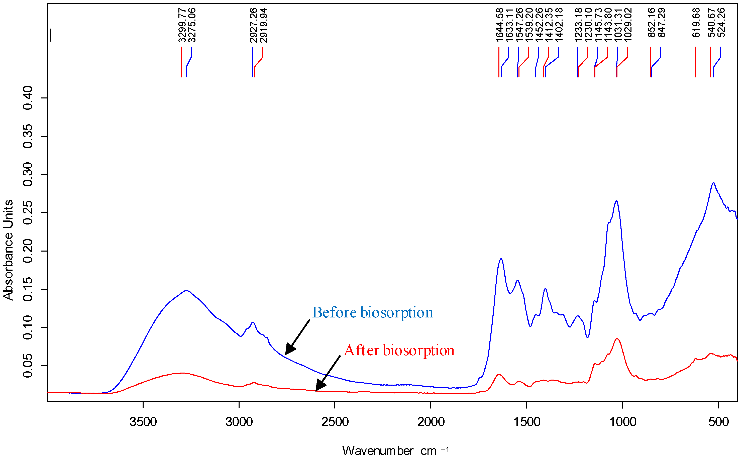
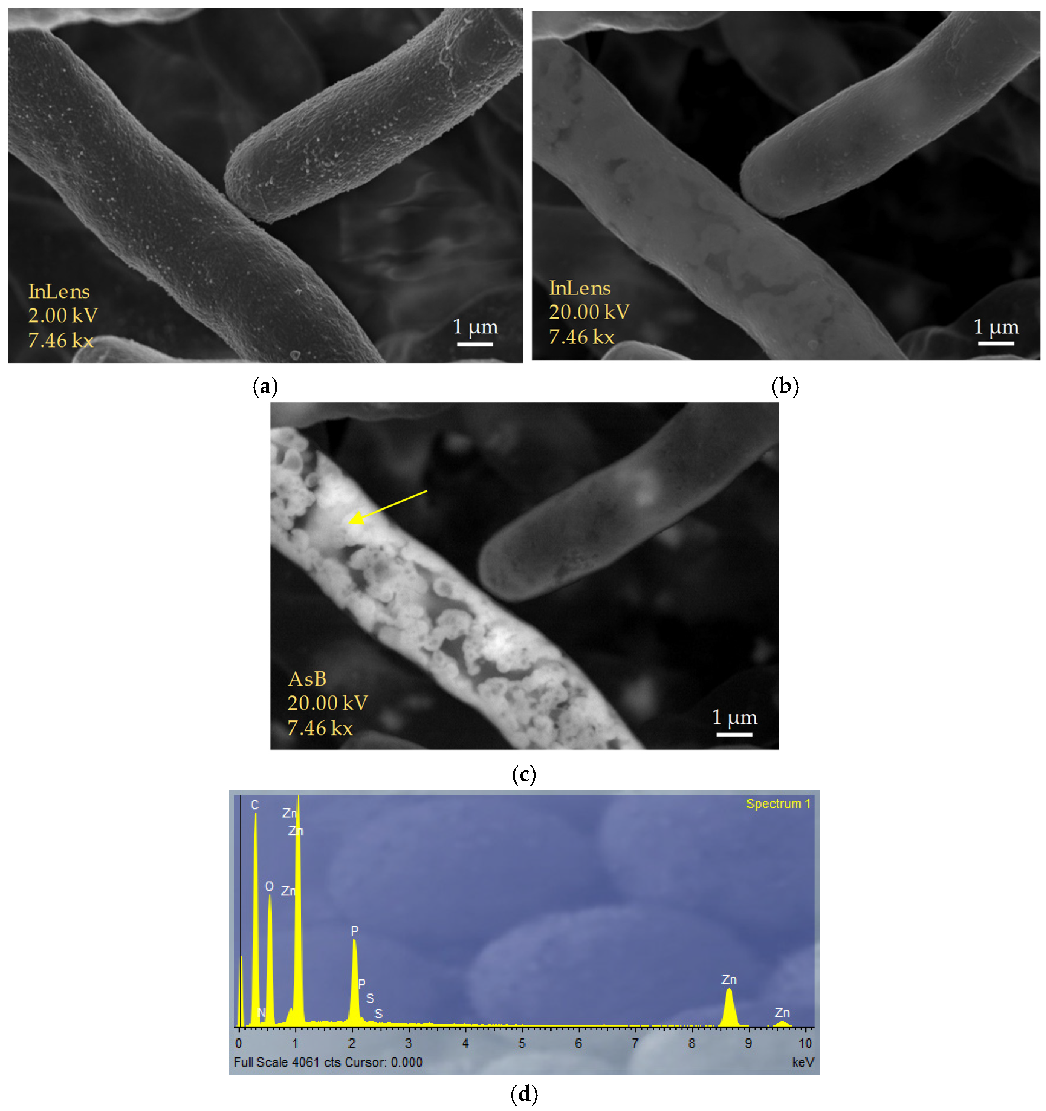
3.3. Biosorption Mechanisms

Figure 3 shows the spectra obtained from the samples of Penicillium sp. 8L2 biomass collected before and after their contact with the Zn(II) solutions. The first thing that can be observed is a notable loss of intensity in the signal, indicating a generalized involvement of the functional groups present in the biomass. Important shifts in the representative bands of the different functional groups were also observed. The band located at 3275 cm−1 shifted to 3300 cm−1, which identified the presence of stretching vibrations from the amino (N-H) and hydroxyl (O-H) groups. In this region, the characteristic band of the methylene groups (-CH2) located at 2927 cm−1 shifted to 2920 cm−1. The amide I and amide II regions were also affected with strong shifts in the 1633 cm−1 and 1644 cm−1 bands to 1644 cm−1 and 1539 cm−1, respectively [2,33]. The changes in the amide bands indicate the involvement of C=O bond stretching (amide I), N-H bond twisting and C-N bond stretching (amide II). In parallel, a strong change was observed in the region between 1500 and 1400 cm−1: the bands originally located at 1452 and 1402 cm−1 shifted to 1412 cm−1 after Zn(II) biosorption, confirming the involvement of methylene groups (-CH2) [34]. The involvement of carbonyl groups was also confirmed by the strong loss of intensity of the band at 1233 cm−1, which also suffers a slight shift to 1230 cm−1. This band is usually assigned to stretching vibrations in the C-O bonds of carbohydrates and alcohols, but also to the stretching of phosphorus bonds, which could indicate the additional involvement of phosphate groups (POO-). Something similar happens with the band at 1031 cm−1, which suffers a strong loss of intensity which is also related to the stretching of C-O bonds [35]. In conclusion, the biomass of Penicillium sp. 8L2 contains amino, carbonyl, hydroxyl, methylene and possibly phosphate groups involved in the retention of Zn(II) ions.
Figure 4 shows images of the biomass of Penicillium sp. 8L2 obtained after the Zn(II) biosorption step. Figure 4a shows the characteristic hyphae of the fungus taken with the secondary electron lens detector (InLens) at low kilovoltage. As the kilovoltage increased (Figure 4b), it was observed that the retained metal was mostly located in a subsurface space of the fungal hyphae. To confirm this, the AsB backscattered electron detector was switched on and this allowed us to clearly identify that the location of the zinc precipitates seemed to be mainly between the membrane and the cell wall of the hyphae (Figure 4c). EDX spectra show that zinc, although dispersed throughout the hypha, is concentrated in well-defined precipitates. In addition, intense phosphorus peaks are observed, indicating that this element is involved in the formation of these precipitates. The results of this SEM analysis are compatible with those observed in the kinetic tests, where two different phases were observed in the biosorption of Zn(II) by Penicillium sp. 8L2. The first stage was rapid and dominated by physicochemical mechanisms (bioadsorption), which would explain the homogeneous distribution of zinc on the cell surface. The second stage was slower and involved active metabolism (bioaccumulation) leading to the formation of zinc phosphate precipitates and possibly ZnO-NPs at the intracellular level. This hypothesis would also be supported because the kinetic data fit better to a pseudo second-order model in which the process is dominated by chemical adsorption [36]. A recent meta-analysis of 56 previous studies evaluated the response of different suspended bacterial strains to various metals, including zinc, and found differences in biosorption efficiency depending on whether these metals were non-essential or essential. It was observed that non-essential metals required short contact times of only 2 h, while essential metals, including Cu(II) or Zn(II), required long contact times (24 h on average) to optimize the process [37]. It is possible that the micronutrient nature of both metals prevents microbial cells from initially recognizing the risk of toxicity, and this leads them to act in a natural way aimed at incorporating these ions into the cytoplasm and then triggering metal elimination mechanisms that could involve precipitation phenomena such as those observed in the SEM images.
To gain a deeper understanding of the morphology and composition of the precipitates, elemental mappings were performed on some specific regions. Figure 5 shows one of them, where these precipitates are perfectly identified (Figure 5a). EDX spectra 1 and 2 (Figure 5g,h, respectively), taken at the location indicated by the arrows, show that the metal is concentrated in nanometric aggregates with a spherical shape. The different elemental maps clearly show that O and P are involved in the formation of these precipitates, which is consistent with what was shown in the FT-IR analysis, indicating that the hydroxyl and phosphate groups are involved in the biosorption of Zn(II) by Penicillium sp. 8L2. Spectra 1 and 2 are also consistent with this evidence, showing that where there is more zinc, there is also more phosphorus. In parallel, the active participation of oxygen also shows that the precipitates could contain aggregates of ZnO nanoparticles.

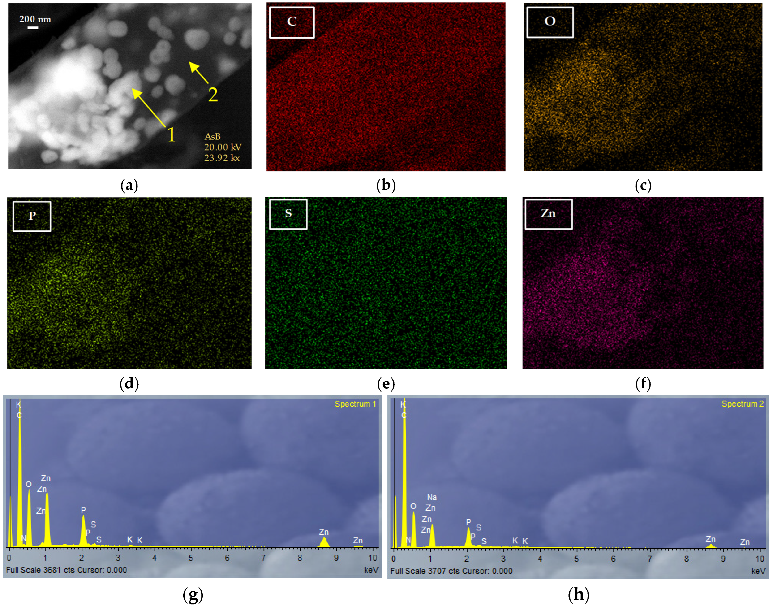
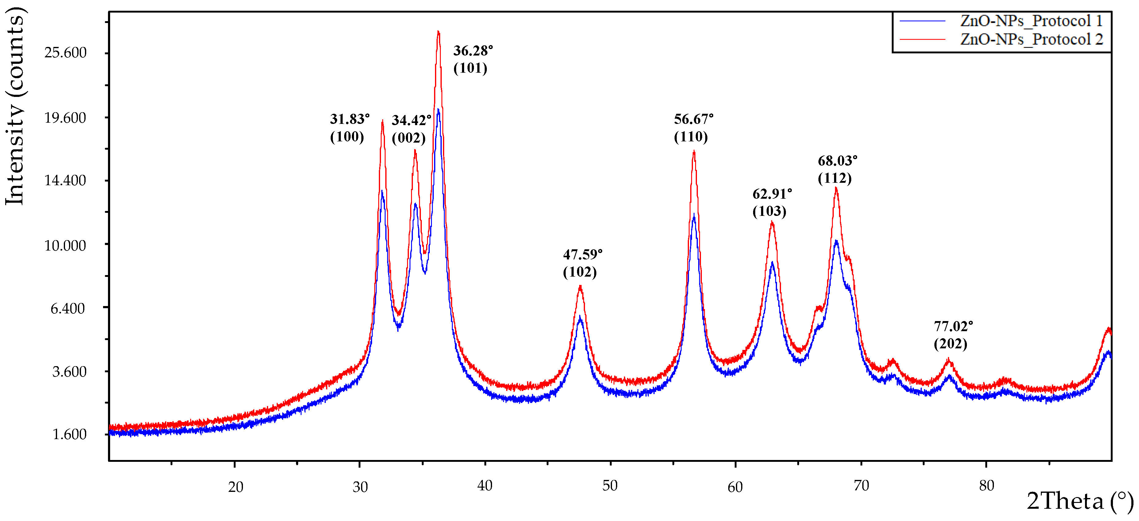
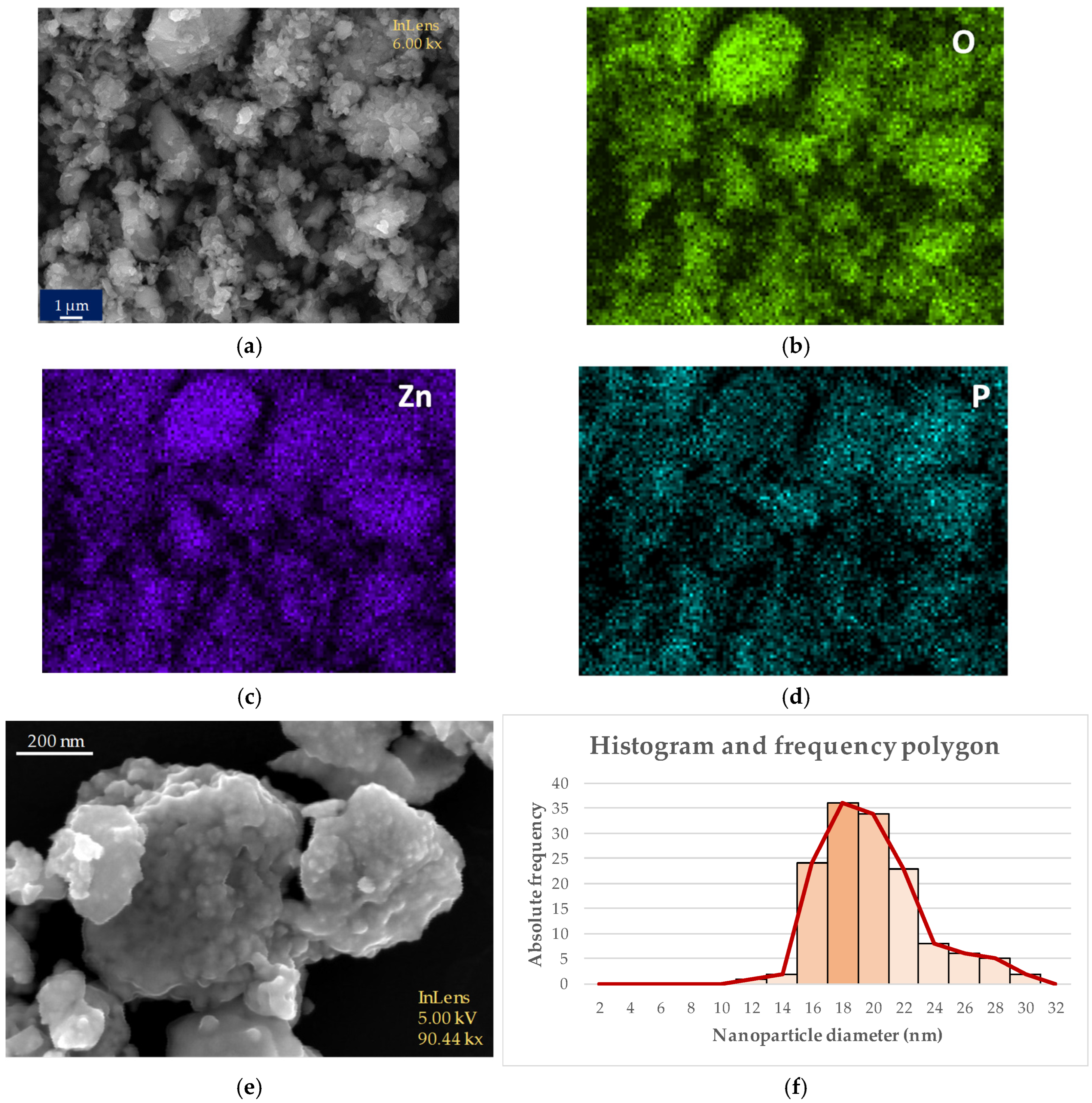
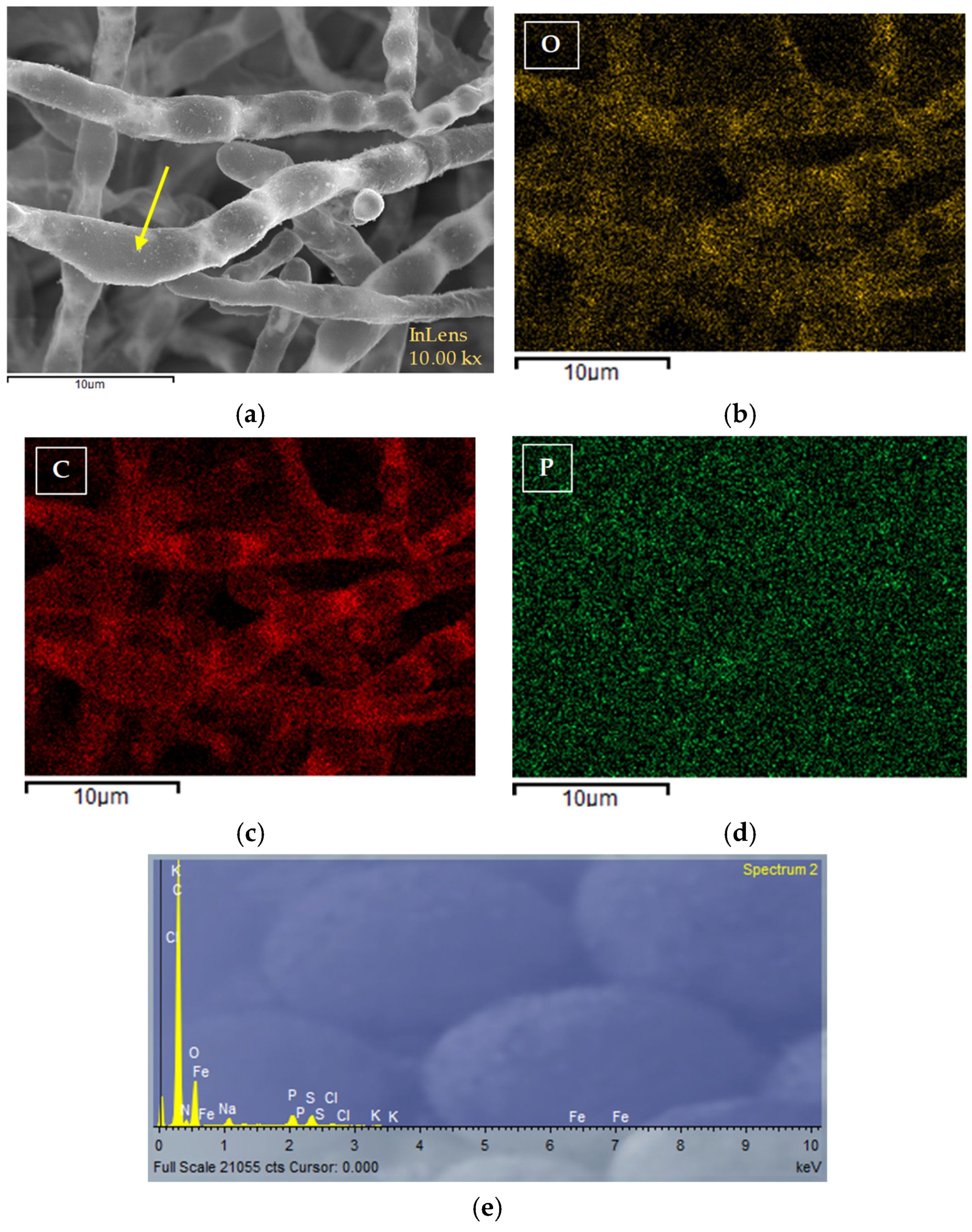
3.4. Characterization of Nanoparticles

The first confirmation of the formation of ZnO-NPs was obtained during the synthesis stage itself, where a fraction of the solid obtained was resuspended in ultrapure water and analyzed by UV-Vis spectroscopy. Figure 6 shows the spectrum obtained, where a peak at 374 nm is observed. Other authors have synthesized ZnO-NPs and identified the characteristic peak of these nanoparticles in this region of the spectrum. Assefa et al. recently performed a detailed study of this type of nanoparticles obtained from a plant extract and found that pure ZnO-NPs exhibited a characteristic peak at 380 nm [38]. Similar data were obtained by Ma et al. with nanoparticles obtained from different plant extracts with peaks between 370 and 373 nm [39].
The solid obtained at the synthesis stage was also subjected to XRD analysis and Figure 7 shows the spectra obtained with the material resulting from the two synthesis protocols. Both spectra show identical peaks, only varying in intensity. In both cases, the bands are perfectly related to what was expected and confirm that they are ZnO-NPs, although in protocol 2 (red color) more intense peaks were obtained, indicating a higher purity and crystallinity of the nanoparticles. The positions for the 2Theta angle and their respective Miller indices were as follows: 31.83° (100), 34.42° (002), 36.28° (101), 47.59° (102), 56.67° (110), 62.91° (103), 68.03° (112), 77.02° (202). These data are consistent with those obtained by other authors and confirm the success of the synthesis and the high purity of the nanoparticles obtained [38,40]. When the most intense peak was analyzed using the Debye–Scherrer equation, an average crystal size of 9.14 nm was obtained, which is slightly smaller than that obtained by the SEM analysis. Figure 8 shows images of the nanoparticles obtained before and after calcining at 500 °C. Figure 8a corresponds to the nanoparticles obtained in protocol 1 (unfiltered extract) before passing through the calcination stage. The image already shows a significant presence of nanoparticles, which are very heterogeneous in size and shape. A strong presence of aggregates is also observed. The elemental map shown in Figure 8b–d of the same figure confirms the participation of oxygen, phosphorus and zinc in its formation, since these elements appear to be associated throughout the solid sample obtained from the synthesis process. The same occurred during the Zn(II) biosorption process, where the precipitates involved the same elements, as shown in Figure 5. The same elements also appear in the pure nanoparticles that have undergone the calcination stage. In parallel, the image in Figure 8e shows the appearance of the nanoparticles obtained in protocol 2 (filtered extract) after the calcination step at 500 °C, in which small nanoparticles with predominantly spherical shapes can be observed. Figure S1 shows a detailed image of these nanoparticles.
These XRD spectra, images and elemental maps allowed us to affirm that the nanoparticles obtained by protocol 2 have better morphological characteristics and for this reason they were selected for use in the subsequent biocidal tests (see Section 3.5). In this context, the SEM-NP-e Figure 8e was chosen to perform the analysis of the average particle size using the ImageJ software. Likewise, the image in Figure 8f shows a histogram and a frequency polygon that reveal an average crystal size of 18 nm. This graph was obtained from Figure 8e of the same figure by counting the nanoparticles present with the ImageJ software, allowing a realistic approximation of the average nanoparticle size. The observed difference between the theoretical and real size is within the expected range, taking into account the tendency of the obtained ZnO-NPs to aggregate. For this reason, two types of biocidal tests were carried out; in the second of which, a dispersing agent was added, as described in the following section. Abdelsattar et al. found slightly larger sizes (20–25 nm) for the ZnO-NPs obtained from Origanum majorana extract, with mostly spherical sizes [41]. On the other hand, Ma et al. found similar sizes between 10 and 20 nm with nanoparticles obtained from different plant extracts [39]. Dejene carried out extensive work on the different types of ZnO-NPs synthesized using different extracts of bacteria, fungi, algae and plants as precursors. This author found that, in most cases, the sizes were larger than those reported in this research. He also found that spherical shapes predominated over the rest and that changes in the precursors of the synthesis reaction affected the final result [42]. This statement is consistent with the results of this research since, as mentioned above, it was observed that the filtering of the initial extract significantly affected the purity, crystallinity, morphology and size of the final nanoparticles. This fact indicates that small changes, which do not involve excessive additional costs, can lead to significant improvements in the nanoparticles obtained.
A final relevant fact is the presence of phosphorus in the elemental maps (Figure 8d image) and in the EDX spectra obtained from the ZnO-NPs. The data show that there is a residual fraction of this element involved in the synthesis of the nanoparticles, which is not reflected in the XRD spectra. As explained, these spectra show the characteristic peaks of pure ZnO-NPs. In the analysis of the mechanisms (Section 3.3), the involvement of phosphate groups in the retention of Zn(II) was demonstrated, and everything indicates that they are also involved in the formation and consolidation of the nanoparticles. This is a little studied phenomenon, but some authors have proposed biomeralization models to explain it. Working with yeast, He et al. identified a chemical precipitation mechanism in aqueous medium that gives rise to zinc phosphate nanoparticles that aggregate to form mesoporous aggregates with a high specific surface area [43]. In this case, the authors added Na3PO4 to the synthesis reaction and this influenced the final result, in which the XRD spectra differ significantly from the characteristic spectra of ZnO-NPs, although they also show some coinciding peaks. It is possible that cellular phosphorus plays a relevant role in the molecular structure of ZnO-NPs obtained from cell extracts, which should be studied in the future. Finally, and to provide additional information on this interesting aspect, Figure 9 has been included, which shows an SEM image of the Penicillium sp. 8L2 biomass obtained before the biosorption process. The elemental maps of C, O and P are also shown, as well as the EDX spectrum obtained on the fungal hyphae. As can be seen, before the biosorption process, phosphorus appears at a very low concentration distributed throughout the biomass and this means that it is not detected in the elemental mapping, so its signal is confused with background noise (Figure 9d). On the other hand, a small peak is detected in the EDX spectrum (Figure 9e), revealing its presence. Analyzing all the data provided so far, it is clear that phosphorus is strongly involved in the zinc biosorption process and also in the formation of ZnO-NPs, since in both situations phosphorus concentrates and increases its signal up to a limit that is perfectly detectable in the elemental maps and also in the EDX spectra.

3.5. Biocidal Tests

Table 3 shows the results obtained in the biocidal tests performed with the ZnO-NPs synthesized in this work. The MIC values are presented as a range, which refers to the value at which microorganisms did not grow (the highest) and the value immediately preceding it. In the case of the yeast R. mucilaginosa 1S1, tests were performed only with the PVA-adjuvanted nanoparticles. The preliminary tests carried out on four bacteria yielded MIC values between 125 and 2000 µg/mL. Subsequently, the addition of 10% PVA resulted in a significant improvement in the biocidal response with values between 62.5 and 1000 µg/mL. The most sensitive microorganisms were the Gram-positive bacterium S. epidermidis, the Gram-negative bacterium E. coli and the yeast R. mucilaginosa, with an MIC between 62.5 and 125 µg/mL in all three cases. On the other hand, the Gram-negative bacterium P. fluorescens showed a higher resistance with an MIC between 500 and 1000 µg/mL. Similarly, the Gram-positive bacterium B. cereus showed an MIC between 250 and 500 µg/mL. In each case, the values obtained were good and indicated that the nanoparticles had consistent biocidal activity. ZnO-NPs are known to exhibit several potential mechanisms of biocidal action, such as the release of Zn(II) ions or the production of reactive oxygen species (ROS) [44]. There are not many studies that directly determine the MIC of biologically synthesized ZnO-NPs; most studies use the disk diffusion technique on plates, which does not allow for a direct reading of this parameter. Kamaraj et al. tested two types of nanoparticles synthesized from fruit extracts against different microorganisms such as the bacteria S. aureus, S. typhi, B. cereus, S. mutans, P. aeruginosa and E. coli or the yeast Candida albicans. The results showed that at a concentration of 100 µg/mL, inhibition zones between 10 and 18 mm were obtained, indicating that the microorganisms were sensitive to the nanoparticles [45]. Another study determined the MIC of green chemistry-derived ZnO-NPs against Bacillus cereus, Streptococcus pneumoniae, Salmonella typhi and Shigella sp. and found values of 100 µg/mL against the first two strains and 200 µg/mL for the rest [46]. Similar values were found by Marques et al. against a battery of collection microorganisms: Staphylococcus epidermidis ATCC35984, S. aureus ATCC25923, S. aureus ATCC8095, Enterococcus faecalis ATCC29212, Enterococcus faecium ATCC700221, Klebsiella pneumoniae ATCC700603, Escherichia coli ATCC25922, Acinetobacter baumannii ATCC19606 and Pseudomonas aeruginosa ATCC27853, with values ranging from 256 to 512 μg/mL [47]. The above confirms that the biocidal capacity of ZnO-NPs is in similar or better ranges than those obtained by other authors and reinforces the importance of continuing to study the medical applications of this type of nanoparticles in a context of increasing resistance to antibiotics by pathogenic microorganisms. In line with the contributions of other authors, we hypothesize that ZnO-NPs act at several levels when they act as biocidal agents. First, they can release Zn(II) ions, which act at two levels, (1) facilitating the production of reactive oxygen species (ROS) with the capacity to generate oxidative stress, and (2) interacting electrostatically with the functional groups of the membrane, which would affect its structure and function. Secondly, the small size of the ZnO NPs synthesized in this work would allow direct access of the nanoparticles to the microbial cytoplasm, where again the direct action of both Zn(II) ions and ROS on essential enzymes and cellular organelles would be provoked [48]. All of this would lead to cell inactivation. Although specific assays to determine the minimum bactericidal concentration (MBC) were not developed in this work, some of the cell suspensions were tested after the MIC assays and a significant loss of cellular activity, in some cases complete, was observed. These preliminary findings need to be developed in future work, but suggest that the mechanisms of cell damage described above may be occurring.

4. Conclusions

Penicillium sp. 8L2 showed good potential for biotechnological application in the field of heavy metal removal from contaminated wastewater, and also offered good potential for use in the green synthesis of ZnO-NPs. The results obtained in this work showed that the ubiquitous microorganism had a good biosorption capacity for Zn(II) (qm = 52.14 mg/g) under the optimal conditions obtained by the experimental design. The novel methodology used in the FESEM-EDX analysis made it possible to identify the metal retained at the subsurface level without resorting to TEM analysis. The synthesized nanoparticles presented spherical shapes and an average size of 18 nm and exhibited a high biocidal capacity against four bacteria and one yeast, with MIC values ranging from 62.5 to 1000 µg/mL. These nanoparticles were obtained using a simple method with good performance. The inclusion of a dispersing agent, PVA, improved the biocidal efficacy and indicates that the addition of different non-toxic agents can help to enhance the efficacy of the nanoparticles. In the future, this line of work should be further explored, as it may allow for a reduction in nanoparticle doses while obtaining the same effect, which could be a step towards their use in in vivo applications.

Supplementary Materials

The following supporting information can be downloaded at: https://www.mdpi.com/article/10.3390/su17062379/s1, Figure S1: SEM images obtained on the ZnO-NPs synthesized with the cell extract of Penicillium sp. 8L2. Image a shows an overview of the nanoparticles, while image b was obtained by zooming in on the area marked in yellow. The nanoparticles were metallized with carbon to improve image sharpness; Table S1: Kinetic models and parameters of Zn(II) biosorption with Penicillium sp. 8L2; Table S2: Isotherm models and biosorption equilibrium parameters for the different isotherm models tested for the biosorption of Zn(II) by Penicillium sp. 8L2.

Author Contributions

Conceptualization, A.J.M.C. and F.E.L.; Methodology, A.J.M.C. and F.E.L.; Validation, A.J.M.C. and F.E.L.; Formal analysis, A.J.M.C., F.E.L. and M.M.V.; Investigation, A.J.M.C. and C.M.V.; Resources, F.E.L. and M.M.V.; Data curation, A.J.M.C., F.E.L. and M.M.V.; Writing—original draft, A.J.M.C., C.M.V., M.M.V. and F.E.L.; Writing—review and editing, A.J.M.C., F.E.L., M.M.V., E.R.R. and C.M.V.; Supervision, F.E.L., M.M.V. and E.R.R.; Project administration, F.E.L.; Funding acquisition, F.E.L.; All authors have read and agreed to the published version of the manuscript.

Funding

Ministerio de Ciencia e Innovación. Plan estatal de Investigación Científica, Técnica y de Innovación 2021–2023. Ref. TED2021-129552B-100.

Institutional Review Board Statement

Not applicable.

Informed Consent Statement

Not applicable.

Data Availability Statement

The raw data supporting the conclusions of this article will be made available by the authors on request.

Acknowledgments

CICT technical staff of the University of Jaén.

Conflicts of Interest

The authors declare no conflicts of interest.

References

  1. Priya, A.K.; Gnanasekaran, L.; Dutta, K.; Rajendran, S.; Balakrishnan, D.; Soto-Moscoso, M. Biosorption of heavy metals by microorganisms: Evaluation of different underlying mechanisms. Chemosphere 2022, 307, 135957. [Google Scholar] [CrossRef]
  2. Gu, S.; Lan, C.Q. Effects of culture pH on cell surface properties and biosorption of Pb (II), Cd (II), Zn (II) of green alga Neochloris oleoabundans. J. Chem. Eng. 2023, 468, 143579. [Google Scholar] [CrossRef]
  3. Al Tanjil, H.; Akter, S.; Hossain, M.S.; Iqbal, A. Evaluation of physical and heavy metal contamination and their distribution in waters around Maddhapara Granite Mine, Bangladesh. Water Cycle 2024, 5, 286–296. [Google Scholar] [CrossRef]
  4. Briffa, J.; Sinagra, E.; Blundell, R. Heavy metal pollution in the environment and their toxicological effects on humans. Heliyon 2020, 6, e04691. [Google Scholar] [CrossRef] [PubMed]
  5. Registro Estatal de Emisiones y Fuentes Contaminantes (PRTR-España). Available online: https://prtr-es.es/ (accessed on 24 January 2025).
  6. Chen, M.; Wang, D.; Ding, S.; Fan, X.; Jin, Z.; Wu, Y.; Wang, Y.; Zhang, C. Zinc pollution in zones dominated by algae and submerged macrophytes in Lake Taihu. Sci. Total Environ. 2019, 670, 361–368. [Google Scholar] [CrossRef]
  7. Hu, J.; Yang, X.; Chi, H.; Liu, X.; Lu, N.; Liu, Y.; Yang, S.; Wen, X. Transport, pollution, and health risk of heavy metals in “soil-medicinal and edible plant-human” system: A case study of farmland around the Beiya mining area in Yunnan, China. Microchem. J. 2024, 207, 111958. [Google Scholar] [CrossRef]
  8. Chen, F.; Saqlain, L.; Ma, J.; Khan, Z.I.; Ahmad, K.; Ashfaq, A.; Sultana, R.; Muhammad, F.G.; Magsood, A.; Naeem, M.; et al. Evaluation of potential ecological risk and prediction of zinc accumulation and its transfer in soil plants and ruminants: Public health implications. Environ. Sci. Pollut. Res. 2022, 29, 3386–3393. [Google Scholar] [CrossRef] [PubMed]
  9. Van, H.T.; Hoang, V.H.; Nga, L.T.Q.; Nguyen, V.Q. Effects of Zn pollution on soil: Pollution sources, impacts and solutions. Results Surf. Interfaces 2024, 17, 100360. [Google Scholar] [CrossRef]
  10. Kalousek, P.; Holátko, J.; Schreiber, P.; Pluháček, T.; Širůčková Lónová, K.; Radziemska, M.; Tarkowski, P.; Vyhnánek, T.; Hammerschmiedt, T.; Brtnický, M. The effect of chelating agents on the Zn-phytoextraction potential of hemp and soil microbial activity. Chem. Biol. Technol. Agric. 2024, 11, 23. [Google Scholar] [CrossRef]
  11. Guarino, F.; Improta, G.; Triassi, M.; Cicatelli, A.; Castiglione, S. Effects of zinc pollution and compost amendment on the root microbiome of a metal tolerant poplar clone. Front. Microbiol. 2020, 11, 1677. [Google Scholar] [CrossRef]
  12. Lobo, H.; Méndez-Fernández, L.; Martinez-Madrid, M.; Rodríguez, P.; Daam, M.A.; Espíndola, E.L.G. Bioaccumulation and chronic toxicity of arsenic and zinc in the aquatic oligochaetes Branchiura sowerbyi and Tubifex tubifex (Annelida, Clitellata). Aquat. Toxicol. 2021, 239, 105955. [Google Scholar] [CrossRef] [PubMed]
  13. Harmanescu, M.; Alda, L.M.; Bordean, D.M.; Gogoasa, I.; Gergen, I. Heavy metals health risk assessment for population via consumption of vegetables grown in old mining area; a case study: Banat County, Romania. Chem. Cent. J. 2011, 5, 64. [Google Scholar] [CrossRef]
  14. Anjum, A.; Garg, R.; Garg, R.; Gupta, D.; Eddy, N.O. Efficient sequestration of zinc and copper from aqueous media: Exploring strategies, mechanisms, and challenges. Int. J. Environ. Sci. Technol. 2024, 22, 5105–5126. [Google Scholar] [CrossRef]
  15. Dehkordi, M.M.; Nodeh, Z.P.; Dehkordi, K.S.; Salmanvandi, H.; Khorjestan, R.R.; Ghaffarzadeh, M. Soil, air, and water pollution from mining and industrial activities: Sources of pollution, environmental impacts, and prevention and control methods. Results Eng. 2024, 23, 102729. [Google Scholar] [CrossRef]
  16. Fomina, M.; Gadd, G.M. Biosorption: Current perspectives on concept, definition and application. Bioresour. Technol. 2014, 160, 3–14. [Google Scholar] [CrossRef]
  17. Kumar, D.; Kumar, S.; Gholap, A.D.; Kumari, R.; Tanwar, R.; Kumar, V.; Khalid, M.; Faiyazuddin, M. A comprehensive review on the biomass-mediated synthesis of silver nanoparticles: Opportunities and challenges. J. Environ. Chem. Eng. 2025, 13, 115133. [Google Scholar] [CrossRef]
  18. Bordin, E.R.; Ramsdorf, W.A.; Domingos, L.M.L.; de Souza Miranda, L.P.; Mattoso Filho, N.P.; Cestari, M.M. Ecotoxicological effects of zinc oxide nanoparticles (ZnO-NPs) on aquatic organisms: Current research and emerging trends. J. Environ. Manag. 2024, 349, 119396. [Google Scholar] [CrossRef]
  19. Zango, Z.U.; Garba, A.; Shittu, F.B.; Iman, S.S.; Haruna, A.; Zango, M.U.; Wadi, I.A.; Bello, U.; Adamu, H.; Keshta, B.E.; et al. A state-of-the-art review on green synthesis and modifications of ZnO nanoparticles for organic pollutants decomposition and CO2 conversion. J. Hazard. Mater. Adv. 2025, 17, 100588. [Google Scholar] [CrossRef]
  20. Muñoz, A.J.; Ruiz, E.; Abriouel, H.; Gálvez, A.; Ezzouhri, L.; Lairini, k.; Espínola, F. Heavy metal tolerance of microorganisms isolated from wastewaters: Identification and evaluation of its potential for biosorption. J. Chem. Eng. 2012, 210, 325–332. [Google Scholar] [CrossRef]
  21. Muñoz, A.J.; Espínola, F.; Ruiz, E.; Cuartero, M.; Castro, E. Biotechnological use of the ubiquitous fungus Penicillium sp. 8L2: Biosorption of Ag(I) and synthesis of silver nanoparticles. J. Environ. Manag. 2022, 316, 115281. [Google Scholar] [CrossRef]
  22. Muñoz, A.J.; Espínola, F.; Moya, M.; Martín, C.; Ruiz, E. Biosorption, Recovery and Reuse of Cu(II) by Penicillium sp. 8L2: A Proposal Framed Within Environmental Regeneration and the Sustainability of Mineral Resources. Sustainability 2024, 16, 11001. [Google Scholar] [CrossRef]
  23. Muñoz, A.J.; Espínola, F.; Ruiz, E.; Moya, M.; Castro, E. Ag(I) Biosorption and Green Synthesis of Silver/Silver Chloride Nanoparticles by Rhodotorula mucilaginosa 1S1. Nanomaterials 2023, 13, 295. [Google Scholar] [CrossRef]
  24. Kumar, R.V.; Vinoth, S.; Baskar, V.; Arun, M.; Gurusaravanan, P. Synthesis of zinc oxide nanoparticles mediated by Dictyota dichotoma endophytic fungi and its photocatalytic degradation of fast green dye and antibacterial applications. S. Afr. J. Bot. 2022, 151, 337–344. [Google Scholar] [CrossRef]
  25. Muñoz, A.J.; Espínola, F.; Ruiz, E.; Moya, M.; Castro, E. Biocidal and synergistic effect of three types of biologically synthesised silver/silver chloride nanoparticles. World J. Microbiol. Biotechnol. 2024, 40, 18. [Google Scholar] [CrossRef] [PubMed]
  26. Muñoz, A.J.; Espinola, F.; Moya, M.; Martín, C.; Ruiz, E. Cu(II) Biosorption and Synthesis of CuO Nanoparticles by Staphylococcus epidermidis CECT 4183: Evaluation of the Biocidal Effect. Appl. Sci. 2024, 14, 7623. [Google Scholar] [CrossRef]
  27. Fan, T.; Liu, Y.; Feng, B.; Zeng, G.; Yang, C.; Zhou, M.; Zhou, H.; Tan, Z.; Wang, X. Biosorption of cadmium(II), zinc(II) and lead(II) by Penicillium simplicissimum: Isotherms, kinetics and thermodynamics. J. Hazard. Mater. 2008, 160, 655–661. [Google Scholar] [CrossRef]
  28. Saravanan, A.; Jeevanantham, S.; Kumar, P.S.; Varjani, S.; Yaashikaa, P.R.; Karishma, S. Enhanced Zn(II) ion adsorption on surface modified mixed biomass—Borassus flabellifer and Aspergillus tamarii: Equilibrium, kinetics and thermodynamics study. Ind. Crops. Prod. 2020, 153, 112613. [Google Scholar] [CrossRef]
  29. Tunali, S.; Akar, T. Zn(II) biosorption properties of Botrytis cinerea biomass. J. Hazard. Mater. 2006, 131, 137–145. [Google Scholar] [CrossRef]
  30. Areco, M.M.; Hanela, S.; Duran, J.; Afonso, M.S. Biosorption of Cu(II), Zn(II), Cd(II) and Pb(II) by dead biomasses of green alga Ulva lactuca and the development of a sustainable matrix for adsorption implementation. J. Hazard. Mater. 2012, 213–214, 123–132. [Google Scholar] [CrossRef]
  31. Kumar, Y.P.; King, P.; Prasad, V.S.R.K. Zinc biosorption on Tectona grandis L.f. leaves biomass: Equilibrium and kinetic studies. J. Chem. Eng. 2006, 124, 63–70. [Google Scholar] [CrossRef]
  32. Zhou, J.L. Zn biosorption by Rhizopus arrhizus and other fungi. Appl. Microbiol. Biotechnol. 1999, 51, 686–693. [Google Scholar] [CrossRef]
  33. Tao, Q.; Liu, Z.; Dai, Y.; Zhan, X. Biosorption properties of extracellular polymeric substances towards Zn(II) and Cu(II). Desalin. Water Treat. 2012, 45, 40–47. [Google Scholar] [CrossRef]
  34. Garip, S.; Bozoglu, F.; Servercan, F. Differentiation of Mesophilic and Thermophilic Bacteria with Fourier Transform Infrared Spectroscopy. Appl. Spectrosc. 2007, 61, 186–192. [Google Scholar] [CrossRef] [PubMed]
  35. Qu, Y.; Meng, Q.; Zhao, Q.; Ye, Z. Biosorption of Pb(II) and Zn(II) from aqueous solutions by living B350 biomass. Desalin. Water Treat. 2015, 55, 1832–1839. [Google Scholar] [CrossRef]
  36. Joshi, H.K.; Vishwakarma, M.C.; Kumar, R.; Sharma, H.; Bhandari, N.S.; Joshi, S.K. The biosorption of Zn2+ by various biomasses from wastewater: A review. J. Water Process Eng. 2023, 56, 104389. [Google Scholar] [CrossRef]
  37. Fathollahi, A.; Khasteganan, N.; Coupe, S.J.; Newman, A.P. A meta-analysis of metal biosorption by suspended bacteria from three phyla. Chemosphere 2021, 268, 129290. [Google Scholar] [CrossRef] [PubMed]
  38. Assefa, A.G.; Demeke, T.; Tefera, M.; Mulu, M.; Tesfaye, S. Biosynthesis and characterization of ZnO NPs using aqueous extract of Zehneria scabra L. leaf for comparing antibacterial activities and the efficacies of antioxidant activities. S. Afr. J. Chem. Eng. 2025, in press. [Google Scholar] [CrossRef]
  39. Ma, W.; Zhu, G.; Zhang, Y.; Guo, J. Green synthesis of ZnO NPs with long-lasting and ultra-high antimicrobial activity. Surf. Interfaces 2024, 50, 104506. [Google Scholar] [CrossRef]
  40. Subba, B.; Rai, G.B.; Bhandary, R.; Parajuli, P.; Thapa, N.; Kandel, D.R.; Mulmi, S.; Shrestha, S.; Malla, S. Antifungal activity of zinc oxide nanoparticles (ZnO NPs) on Fusarium equiseti phytopathogen isolated from tomato plant in Nepal. Heliyon 2024, 10, e40198. [Google Scholar] [CrossRef]
  41. Abdelsattar, A.S.; Kamel, A.G.; Eita, M.A.; Elbermawy, Y.; El-Shibiny, A. The cytotoxic potency of green synthesis of zinc oxide nanoparticles (ZnO-NPs) using Origanum majorana. Mater. Lett. 2024, 367, 136654. [Google Scholar] [CrossRef]
  42. Dejene, B.K. Biosynthesized ZnO nanoparticle-functionalized fabrics for antibacterial and biocompatibility evaluations in medical applications: A critical review. Mater. Today Chem. 2024, 42, 102421. [Google Scholar] [CrossRef]
  43. He, W.; Yan, S.; Wang, Y.; Zhang, X.; Zhou, W.; Tian, X.; Sol, X.; Han, X. Biomimetic synthesis of mesoporous zinc phosphate nanoparticles. J. Alloys Compd. 2009, 477, 657–660. [Google Scholar] [CrossRef]
  44. Janani, M.; Viswanathan, D.; Pandiaraj, S.; Govindasamy, R.; Gomathi, T.; Vijayakumar, S. Review on phyto-extract methodologies for procuring ZnO NPs and its pharmacological functionalities. Process Biochem. 2024, 147, 186–212. [Google Scholar] [CrossRef]
  45. Kamaraj, C.; Naveenkumar, S.; Kumar, R.C.S.; Al-Ghanim, K.A.; Natesan, K.; Priyadharsan, A. Ice apple fruit peel assisted bio-synthesis of zinc oxide nanoparticles (ZnO NPs): An anticancer, antimicrobial, and larvicidal applications. J. Drug Deliv. Sci. Technol. 2025, 105, 106585. [Google Scholar] [CrossRef]
  46. Elankathirselvan, K.; Fathima H, A.; K, P.; Al-Ansari, M.M. Synthesis and characterization of Pyrus communis fruit extract synthesized ZnO NPs and assessed their anti-diabetic and anti-microbial potential. Environ. Res. 2024, 258, 119450. [Google Scholar] [CrossRef] [PubMed]
  47. Marques, G.L.; Moreira, A.J.; Nóbrega, E.T.D.; Braga, S.; Argentin, M.N.; Da Cunha Camargo, I.L.B.; Azevedo, E.; Pereira, E.C.; Bernardi, M.I.B.; Mascaro, L.H. Selective inhibitory activity of multidrug-resistant bacteria by zinc oxide nanoparticles. J. Environ. Chem. Eng. 2024, 12, 111870. [Google Scholar] [CrossRef]
  48. Tran, G.T.; Nguyen, N.T.H.; Nguyen, N.T.T.; Nguyen, T.T.T.; Nguyen, D.T.C.; Tran, T.V. Formation, properties and applications of microalgae-based ZnO nanoparticles: A review. J. Environ. Chem. Eng. 2023, 11, 110939. [Google Scholar] [CrossRef]
Figure 1. (a): Response surface obtained after fitting the data obtained in the experimental design for the biosorption of Zn(II) by Penicillium sp. 8L2. The graph identifies factor A (pH) and factor B (biomass dose) for an initial metal concentration of 50 mg/L. (b): Perturbation diagram in coded units obtained for the following conditions: A = 5.2 and B = 0.5. The diagram shows the influence of changes in the value of the variables on the response (q, mg/g).
Sustainability 17 02379 g001
Figure 2. (a) Biosorption kinetics obtained for the Lagergren model. (b): Biosorption isotherm obtained for the Langmuir model.
Sustainability 17 02379 g002
Figure 3. FT-IR spectra obtained before (blue color) and after (red color) the Zn(II) biosorption stage by Penicillium sp. 8L2.
Sustainability 17 02379 g003
Figure 4. Images (ac): FESEM image sequence of Penicillium sp. 8L2 hyphae after the Zn(II) biosorption step. The images show that by changing the kilovoltage and the detector of the instrument, information about the location of the retained metal can be obtained. Image (d): EDX spectrum obtained in the area indicated by the arrow.
Sustainability 17 02379 g004
Figure 5. (a) SEM image of Penicillium sp. 8L2 biomass obtained after the Zn(II) biosorption process, where the arrows indicate the location where the EDX spectra were recorded (images (g,h)). (bf) Elemental maps of image a for the elements C, O, P, S and Zn, respectively.
Sustainability 17 02379 g005
Figure 6. UV-vis spectrum of a suspension of ZnO-NPs nanoparticles synthesized with cell extract of Penicillium sp. 8L2. A characteristic peak at 374 nm is identified.
Sustainability 17 02379 g006
Figure 7. XRD spectra of ZnO-NPs. The blue spectrum corresponds to the NPs obtained from the unfiltered cell extract (protocol 1). The red spectrum was obtained from the NPs obtained from the filtered cell extract (protocol 2).
Sustainability 17 02379 g007
Figure 8. (a): SEM image of ZnO-NPs obtained from the cell extract of Penicillium sp. 8L2 using protocol 1 (before calcination step). (bd): Elemental maps obtained from image (a). (e): SEM image of ZnO-NPs obtained from the cell extract of Penicillium sp. 8L2 using protocol 2 (after calcination step). (f): Histogram and frequency polygon obtained from image (d).
Sustainability 17 02379 g008
Figure 9. (a) FESEM image of the biomass of Penicillium sp. 8L2 obtained before exposure to Zn(II) ions. (bd) Relative to image a, elemental maps of O, C and P, respectively. (e) EDX spectrum obtained at the location indicated by the arrow (a).
Sustainability 17 02379 g009
Table 1. Experimental design conditions for the biosorption of Zn(II) by Penicillium sp. 8L2 and the biosorption capacity data obtained in each case.
RunF. 1F. 2R F. 1F. 2R
A: pHB: (g/L)qe (mg/g)RunA: pHB: (g/L)qe (mg/g)
16.210.2044.77146.210.2052.49
25.190.5043.56155.190.5041.02
35.190.0855.55165.190.0853.32
46.620.5036.50176.620.5041.89
55.190.5040.03185.190.5043.15
66.210.8034.21196.210.8037.40
75.190.5040.87205.190.5042.74
85.190.5040.72215.190.5036.39
94.240.8033.30224.240.8032.04
104.240.2038.43234.240.2035.35
115.190.9036.54245.190.9035.95
123.830.5033.72253.830.5034.33
135.190.5038.98265.190.5043.16
F and R refer to the factors examined and the response obtained, respectively. A and B are pH and biomass dose, respectively.
Table 2. Maximum Zn(II) biosorption capacity for different types of biomass.
Type of BiomassBiomassqm (mg/g) *Reference
Plant/fungiBorassus flabellifer/Aspergillus tamarii49.79
54.74
[28]
FungiBotrytis cinerea12.98[29]
Green algaUlva lactuva22.88[30]
PlantTectona grandis16.42[31]
FungiRhizopus arrhizus
Mucor racemosus
Mycotypha africana
Aspergillus nidulans
Aspergillus niger
Schizosaccharomyces pombe
13.92
13.01
12.55
11.90
11.90
10.10
[32]
FungiPenicillium simplicissimum52.50
65.60
76.90
[27]
FungiPenicillium sp. 8L252.14This work
* qm: Maximum biosorption capacity expressed in mg of metal per g of biomass.
Table 3. MIC values (μg/mL) obtained for the microorganisms tested against pure ZnO-NPs and those adjuvanted with 10% PVA.
BacteriaNPsNPs + PVA *
B. cereus1000–2000250–500
S. epidermidis125–25062.5–125
E. coli250–50062.5–125
P. fluorescens1000–2000500–1000
R. mucilaginosa 1S1-62.5–125
* PVA: polyvinyl alcohol.
Disclaimer/Publisher’s Note: The statements, opinions and data contained in all publications are solely those of the individual author(s) and contributor(s) and not of MDPI and/or the editor(s). MDPI and/or the editor(s) disclaim responsibility for any injury to people or property resulting from any ideas, methods, instructions or products referred to in the content.

