Article

Development and Characterization of Sustainable Antimicrobial Food Packaging Films with Incorporated Silver Nanoparticles Synthesized from Olive Oil Mill By-Products

by
Christina M. Gkaliouri
1,
Nikolas Rigopoulos
1,*,
Zacharias Ioannou
1,*,
Efstathios Giaouris
1,
Konstantinos P. Giannakopoulos
2 and
Kosmas Ellinas
1
1
Department of Food Science and Nutrition, School of the Environment, University of the Aegean, Mitrop. Ioakeim 2, Myrina, 81400 Lemnos, Greece
2
Institute of Nanoscience and Nanotechnology, National Center for Scientific Research “Demokritos”, Patriarchou Gregoriou E & Neapoleos 27, 15341 Athens, Greece
*
Authors to whom correspondence should be addressed.
Sustainability 2025, 17(19), 8916; https://doi.org/10.3390/su17198916
Submission received: 8 September 2025 / Revised: 1 October 2025 / Accepted: 4 October 2025 / Published: 8 October 2025

Abstract

The growing accumulation of non-biodegradable petrochemical plastics and increasing food waste present urgent environmental and public health challenges. This study addresses both issues by developing biodegradable food packaging films from agar and starch, enhanced with antimicrobial properties by incorporating silver nanoparticles. The innovation of this work is the synthesis of novel agar–starch–silver nanoparticle coatings, where the contained nanoparticles were produced via green methods using two agro-industrial by-products of Greek olive oil production—olive stone extract and olive mill wastewater—as reducing agents. The morphology of the novel coatings was confirmed using transmission electron microscopy combined with energy-dispersive X-ray spectroscopy, revealing nanoscale particles with variable sizes. Additional film characterization was performed through Fourier-transform infrared spectroscopy, scanning electron microscopy coupled with energy-dispersive spectroscopy, and surface profilometry. Infrared spectroscopy analysis suggested the presence of functional groups responsible for nanoparticle stabilization, while energy-dispersive X-ray spectroscopy revealed silver aggregation in both olive stone extract and olive mill wastewater-derived films. Profilometry showed that films with olive mill wastewater-based nanoparticles had a rougher surface than those synthesized from olive stone extract. Antibacterial efficacy was tested against Escherichia coli (Gram-negative) and Staphylococcus epidermidis (Gram-positive) using a spot-on-film assay with high (106 CFU/film) and low (103 CFU/film) bacterial loads. After 72 h of incubation at 4 °C, both film types showed strong antibacterial activity at high bacterial concentrations, demonstrating their potential for active food packaging. These findings highlight a promising approach to sustainable food packaging within the circular economy, utilizing agricultural waste to create biodegradable materials with effective antimicrobial functionality.

1. Introduction

Food packaging is crucial for maintaining food safety, quality, and sensory attributes, including color, texture, and flavor. It prevents spoilage and extends shelf life. Despite growing environmental concerns, petroleum-based plastics remain the dominant choice in packaging due to their low cost, mechanical strength, stability, and sufficient barrier properties [1,2]. However, their widespread use leads to environmental pollution and bioaccumulation in ecosystems. Additionally, synthetic plastics may pose health risks, as toxic compounds like bisphenol, antimony, and phthalates can migrate into food products [3,4]. Between 2009 and 2020, food packaging waste increased significantly—from 149.9 kg to 177.9 kg per person [3].
As a sustainable alternative, biopolymer-based packaging materials have gained attention. Derived from living organisms or biomass, these materials are non-toxic, biodegradable, compostable, and renewable. Unlike synthetic polymers, biopolymers decompose into harmless by-products such as carbon dioxide, water, and biomass [5,6]. However, they often have inferior mechanical properties and higher permeability to moisture and gases [4]. To address these drawbacks, biopolymers can be combined with plasticizers, other biopolymers, or small amounts of synthetic additives [2]. Antimicrobial agents—such as essential oils, enzymes like lysozyme, and nanomaterials—can also be incorporated to prevent microbial spoilage and enhance their antimicrobial functionality [7,8].
Starch-based edible films (ST) are widely used in the food packaging industry due to their unique properties such as good processability, intrinsic biodegradability, and biocompatibility, and they are moderate-to-good barriers against oxygen and carbon dioxide [9,10]. A variety of films have been produced from starch based on plants such as potato, corn, cassava, rice, and yam. Starch films present severe drawbacks such as low mechanical strength, high water vapor permeability, and water sensitivity [11]. The introduction of agar (AG) led to the production of a dense three-dimensional network structure with sulfated functional groups and hydrophilic colloid molecules, ideal for film formation [11,12]. Agar enhanced the tensile strength of potato starch films in both normal and high-moisture environments [12], ameliorating the mechanical properties of starch coatings [11,13] and extending their shelf life. The presence of agar in starch films increased the hygroscopic characteristic of films due to its hydrophilicity. Specifically, in high-moisture environments (RH > 80%), AG-ST films have similar water vapor permeability (WVP) values to those of individual AG-based films but lower WVP values than those of individual ST-based films. In low-moisture environments (22 < RH < 57%), AG-ST films have lower WVP values than those of both AG- and ST-based films [12,13]. The above results depend on the plasticizer used (frequently glycerol or mannitol), the characteristics of films formed, e.g., thickness, and the ratio of agar and starch used for film production. Agar acts as a good cohesive matrix that can enhance the mechanical as well as the barrier properties of other films manufactured from polysaccharide-based materials. Moreover, agar, when combined with starch, affects its swelling, and therefore, it modifies the properties (e.g., crystallinity, physical, thermomechanical, and barrier properties) of the starch-based films [13,14]. These films offer significant shelf life benefits by regulating moisture and oxygen, but their UV stability is inherently low and requires the addition of protective additives.
Nanoparticles (NPs), particularly silver nanoparticles (AgNPs), alter the agar starch films’ properties, exhibiting broad-spectrum antimicrobial activity—even against resistant pathogens [15,16]. According to Roy and Rhim [17], the incorporation of AgNPs in the starch–agar matrix had a positive effect on hydrophobicity of the films, as well as on their water vapor barrier. The water vapor permeability of the starch/agar film was 0.75 × 10⁻⁹ g∙m/m2∙Pa∙s, and it was significantly decreased (p < 0.05) when AgNPs were added due to the difficulties of water vapor diffusion from the impermeable silver nanoparticles and the presence of hydrogen bonds in the starch–agar film. However, adding AgNPs to AG-ST film did not affect the film’s mechanical strength (slightly lower tensile strength and stiffness and higher flexibility compared to AG-ST film without AgNPs) and thermal stability, while other research supports the opposite finding that incorporating AgNPs improved the mechanical properties of starch-based films [18]. Moreover, the UV light barrier properties of the starch films were also slightly enhanced by the addition of AgNPs up to 28.6 ppm. Higher concentrations of AgNPs led to films with similar UV barrier capacity with the control sample. According to Roy et al. [19], the films produced by carrageenan and combined with AgNPs showed excellent UV light barrier property. In addition, an increase in the concentration of Ag and ZnO nanoparticles in the starch/agar film matrix turned out to significantly diminish its moisture content and water solubility and slightly decrease the water vapor permeability [20]. Although the exact antimicrobial mechanisms of NPs remain unclear, proposed actions include cell membrane disruption, the production of reactive oxygen species (ROS), interference with protein and nucleic acids, and the release of silver ions [20,21].

Literature Review

Green synthesis employs renewable resources and therefore supports sustainability [22]. Leaf, peal, and other plant parts can be used for NP synthesis [23]. In addition, vegetable waste can also be employed [23]. Agro-industrial by-products, such as whey, fruit peels, and olive mill residues, are rich in bioactive compounds and represent a sustainable source of raw materials [24,25]. A comprehensive review of this subject has been published recently [23]. Carotenoids, flavonoids, and phenolic acids are a few of the biomolecules present in plants and vegetable waste extracts, acting as reducing and capping agents on AgNP synthesis [23]. Potato [26] and cucumber [27] peels, Alpinia Nigra [28], and basil waste [29], for instance, have been employed for AgNPs synthesis. Recently, agri-food waste from the olive oil industry—olive mill wastewater (OMW) and olive stone extracts (OSEs)—has been employed for AgNP synthesis by our group [30].
Nanocomposite films, prepared by embedding AgNPs into biodegradable polymers, have been thoroughly investigated [31]. Agar nanocomposite films, employing AgNPs synthesized from fruit extract of Lagersroemia speciosa, showed good antibacterial activity [32]. In a different approach, soybean polysaccharide solution was used both as the reducing agent for AgNP synthesis and the casting solution for nanocomposite film formation [33]. Cellulose was also used as the reducing agent and casting solution [34]. Agro-waste can produce micro-crystalline cellulose, which is reinforced with starch and whey protein, and it has been demonstrated to be suitable for food packaging [35]. Polysaccharide- based nanocomposites for food safety are an active area of research [36,37]. Utilizing agro-waste, both for biopolymer and metallic NP synthesis, offers a sustainable route for valorization of food industry by-products [38].
The development of new food packaging based on AgNPs is a reality at the research level. However, its development on an industrial scale requires additional measures, especially regarding the management of waste from such industries. Furthermore, increased amounts of AgNPs are present in domestic and industrial wastewater originating from various sectors such as cosmetics, textiles and clothing, filters, detergents, dietary supplements, and electronic devices. Wastewater treatment plants contain large quantities of the above silver in their sludge (>98% of AgNPs are retained insoluble in the sediment), and the remaining 2% will end up in the soil or water bodies [39]. The implementation of silver (Ag) in soil, primarily in the form of silver nanoparticles (AgNPs) or ionic silver, depends on factors like soil conditions, NP properties, and the presence of organic matter or other chemicals [39]. Nanoparticle mobility in soil is higher in soil with low clay content and in slightly acidic conditions [39]. According to Benoit et al. [40], the oxidation of silver nanoparticles or the dissociation of adsorbed silver from soil surfaces leads to the release of silver into the soil solution. The transportation of AgNPs to soil depends on different soil processes such as adsorption/desorption to/from soil, ion exchange, dissolution–precipitation, transformation, and heteroaggregation [3]. The uptake of Ag from plant species presents large variations with spinach and silver beet to accumulate sufficient Ag causing health problems after consumption [41]. The growth of plants such as monocot (wheat) and dicot (rape) crop species was reduced with the presence of Ag in the soil [42]. Such activities raise concerns about potential accumulation in soil, plant uptake, and leaching, especially due to the use of sewage sludge as fertilizer in agriculture in the EU, which necessitates further research into the long-term ecological effects and risks of soil–plant systems.
In agar–starch–AgNP coatings, the highest cost comes from the manufacture of AgNPs. The cost of industrial-scale AgNP production varies significantly based on the production method, i.e., physical, chemical, or biological method, purity, and specific particle characteristics, i.e., size and shape, but generally involves substantial investment in equipment, raw materials, energy, and quality control processes. Chemical and physical methods such as electrochemical processes, chemical reduction, sonochemical reduction, and photochemical reduction are expensive methods for AgNPs production [43,44] due to the high-purity reagents, chemicals, expensive equipment, and high energy inputs. In contrast, green synthesis of NPs presents lower costs due to the use of cheaper and biodegradable raw materials. According to the literature [45], the global AgNP market size reached USD 4 billion in 2024 and is forecast to be valued at USD 12.1 billion in 2034. The basic reagent for AgNPs is silver nitrate of high purity (>99.8%) with a cost range from USD 550 to USD 820 per kg [46,47]. However, agar and starch are low-cost, abundant, and renewable materials compared to AgNPs. The price of starch begins at USD 1.08 per kg [48] for cassava starch for industrial use, while the respective price for agar begins at USD 8.31 per kg [49]. The increased purity of the agar and starch materials also increases their price per kg in food packaging processes. Moreover, the price of different reagents used to produce films is as follows: the sodium hydroxide price is equal to USD 0.28 per kg [50], the industrial glycerol price ranges from USD 0.73 to USD 0.98/kg [51], and the price of industrial grade distilled water is approximately USD 0.5/L [52]. Production at a larger industrial scale generally lowers the cost per unit compared to production at a laboratory scale. Finally, energy, labor, and equipment amortization add to the overall cost of film production. Other studies [43] have shown that the production of AgNPs on a large scale is feasible and profitable. Two main factors should be covered: the governmental or industrial foundation support for sustainable production and prevention actions concerning the pollution of industrial waste enriched with Ag.
Multiple studies in the literature have dealt with the production of food packaging using nanotechnology to maintain quality and enforce the safety of products [53,54,55,56,57,58]. Nevertheless, regulations for the use of nanomaterials in food packaging pose some concerns relative to the potential risks for human health by using these nanoscale particles in materials that are in direct contact with food products. Globally, regulations for the use of AgNPs in food packaging vary. For example, in Canada there are no limitations on NPs as additives, with other countries also having finite regulations that regard food-contact materials [59].
In contrast, in European countries, the scientific panel on food additives, flavorings, processing aids, and materials in contact with food (AFC) of the European Food Safety Authority (EFSA, 2004) has proposed a threshold of 0.05 mg Ag/kg food, taking into account the toxicological results provided and the report of the World Health Organization [60] that a total oral intake of 10 g of Ag over a human lifetime is unlikely to cause any adverse effects [59,61,62]. Based on [62] the panel reported that the nanoscale Ag particles remain embedded in the polymer, do not migrate, and resist release upon abrasion; therefore, they do not cause food exposure and toxicological concerns under the tested conditions of uses. Other studies have also been conducted to examine the potential for NPs used in food packaging to migrate into food products [54,61,63]. These studies indicated that food type determines the migration level, with acidic products promoting higher migration. Nevertheless, analysis showed that even in the higher migration levels, the migrating amounts of AgNPs were less than the concentration limit (2002/72/EC). Other studies suggest the use of such materials to wrap fat-rich foodstuffs, as the migration level was less in this type of product [53]. Marchiore et al. [64] also mentioned that common techniques like washing or cooking before eating tend to diminish the amount of AgNPs on the product surface. Other studies have shown similar results [54].
In addition, the Food and Drug Administration/Centre for Food Safety and Applied Nutrition (FDA/CFSAN—USA) accepts the use of silver compounds as food additives, such as silver nitrate in bottled water and silver zeolites in all types of food-contact polymers (FDA, 2010), while in the European Regulation, the use of silver is accepted as a coloring agent (E-174) under 94/36/EC Directive with no restrictions [53]. The framework rule EC 1935/2004, which regulates active and intelligent packaging, declares that the only released compounds into the food by packaging must be substances authorized as food additives or food flavorings [65].
Nonetheless, the use of AgNPs in food packages is not authorized by the European Food Safety Authority (EFSA), due to insufficient safety data. In 2021, the EFSA determined the general principles for any material that is in contact with food, with no specific mention of nanotechnology [56]. Generally, the EFSA has determined that the greater the exposure through migration, the more toxicological data is required to assess the safety of a substance. Currently, there are three migration levels that define the need for the provision of more toxicological information. The first one is between 5.0 and 60.0 mg/kg of food, the second one is between 0.05 and 5.0 mg/kg of food, and the third one is less than 0.05 mg/kg of food [62] Therefore, further research on incorporated nanoparticles is needed in food packaging applications to ensure their safety. The use of metal NPs in packaging is really promising due to their superior mechanical, physicochemical, thermal, antimicrobial, barrier, and other properties that can enhance the biopolymer matrix [63].
This study aims to develop biodegradable, antimicrobial food packaging films using agar and starch as biopolymers and AgNPs synthesized from olive stone extract (OSE) and olive mill wastewater (OMW) as active agents. By valorizing olive mill by-products, this research contributes to waste reduction, resource efficiency, and sustainable packaging innovation in the framework of the circular economy.

2. Materials and Methods

2.1. Silver Nanoparticle (AgNP) Synthesis

Silver nanoparticles were synthesized by using two different reducing agents derived from olive industry activities: OSE and OMW. Both materials were obtained from a local olive oil mill located on the island of Lemnos, Greece. Olive mill wastewater was stored at 4 °C in amber glass bottles. To prepare the olive stone extract, 0.3 g of olive stone powder was added to 10 mL of distilled water (dH2O). The solution was heated until the boiling point, and then it was left to cool down at 25 °C. Afterwards, the solution was centrifuged at 9000× g (Z 383, Hermle Labortechnik, Wehingen, Germany) for 5 min at 25 °C, and the supernatant was collected and stored at 4 °C for AgNP synthesis. The chemical reagents of silver nitrate 0.1 M and the pellets of sodium hydroxide were purchased from Sigma Aldrich Co. (Steinheim, Germany) and used for AgNP synthesis.
The process to produce silver nanoparticles from olive stone extract (AgNPs-OSE) and olive mill wastewater (AgNPs-OMW) has been described in previous works [30,66]. In brief, AgNPs were formed at the following ratios and conditions: silver nitrate 2 mM, olive stone (OSE) 2% v/v, and sodium hydroxide 0.2 mM for AgNPs-OSE, and silver nitrate 1 mM, olive mill wastewater (OMW) 2% v/v, and sodium hydroxide 7.9 mM for AgNPs-OMW. The two mixtures were placed in a water bath for 30 min at a constant temperature of 60 °C. Centrifugation was performed for both obtained NP suspensions at 20,000× g for a duration of 30 min and at constant temperature of 4 °C. The derived pellets were collected and stored at 4 °C in the dark for the following experiments.

2.2. Film Preparation and AgNP Incorporation

The film solution was prepared by using starch and agar in a 1:1 ratio. More specifically, 0.5 g of agar and 0.5 g of starch were mixed in powder form, and then they were added to 50 mL of dH2O, which contained glycerol, in a proportion of 30% by weight of agar and starch [17]. Agar was purchased from Fisher Scientific (Thermofisher Scientific, Vantaa, Finland), while starch and glycerol were purchased from Merck (Merck KGaA, Darmstadt, Germany).
The mixture was then heated at a temperature of 90 °C for 1 h with constant magnetic stirring. Before cooling down the biopolymer film, the film mixture was separated into three different volumetric cylinders, one for adding AgNPs-OSE, one for adding AgNPs-OMW, while in the third, no NPs were added (reference sample, RS). Silver nanoparticle pellets were added to each mixture in a ratio of 1:1 and were blended manually using a glass rod. Finally, AgNPs-OSE film, AgNPs-OMW film, and RS film were spread on glass Petri dishes and left to cool down at room temperature.

2.3. Silver Nanoparticles and Film Characterization

The main color parameters (L, a, and b) of the film samples were measured using a chromameter (Lovibond LC100) against a white standard plate as a background. These measurements were performed in triplicate.
The AgNPs-OSE were prepared for TEM-EDS analysis according to the literature [30]. The observation was conducted on a Philips CM-20 with High Resolution (HRTEM), Electron Energy Loss Spectroscopy (EELS), and EDS capabilities (Philips, Amsterdam, Holland).
The three types of films were characterized by attenuated total reflection (ATR-FT-IR) spectroscopy. For this, the films were placed in the ATR diamond holder of a Jasco spectrometer (Jasco FT-IR-6700, Jasco Corporation, Tokyo, Japan). The spectra were collected in the range of 4000–400 cm−1 by 160 scans with 4 cm−1 resolution. The baseline spectrum was calculated by Spectra Manager 2.15.12 software (Jasco Corporation, Japan).
The SEM-EDS analysis of the three types of films was carried out in a JEOL JSM-IT500 scanning electron microscope (JEOL Ltd., Boston, MA, USA) and simultaneously, elemental analysis was determined by EDS with the use of x-Act detector of Oxford Instruments (High Wicombe, UK). Before these analyses, an AGAR Sputter Coater apparatus (AGAR scientific company, Rotherham, UK) was used to deposit a thin layer of gold (Au) onto the samples to enhance image quality and conductivity. Open-source software for processing and analyzing scientific images, ImageJ (Version 1.54p), was used for the calculation of the mean diameter of the nanoparticles [67].
The 3D optical profilometer Profilm3D (KLA, Villach, Austria) was used for the profilometry analysis of the three types of films. The profilometer was used with a camera of 2592 × 1944 (5 megapixels) and with a thickness range (WLI) between 50 nm and 10 mm, an XY range of 100 × 100 mm, and a Z stage range equal to 100 mm. The Profilm-Analysis Software for stitching XY grid of images was utilized for the analysis and visualization of the surface microtopography. It is equipped with 10×, 20×, and 50× objectives that can resolve surface roughness down to 0.05 μm.

2.4. Antimicrobial Experiments

2.4.1. Bacterial Strains and Preparation of Their Saline Cellular Suspensions

The antimicrobial action of the three types of films (AgNPs-OSE film, AgNPs-OMW film, and RS film) was tested in parallel against two different bacterial strains, one Gram-negative and one Gram-positive, which were Escherichia coli DFSN_B1 and Staphylococcus epidermidis DSFN_B4, respectively. These strains were kindly provided by Professor Nychas from the Laboratory of Microbiology and Biotechnology of Foods of the Agricultural University of Athens (Greece) and were registered there as FMCC B-13 and FMCC B-202, respectively. Upon receipt, both strains were long-term stored at −80 °C in cryovials (Cryoinstat; Deltalab, S.L., Rubi, Barcelona, Spain) in the microbial culture collection of the Laboratory of Food Microbiology and Hygiene (LFMH) of the Department of Food Science and Nutrition (Lemnos, Greece). Each strain was resuscitated by placing one bead in 10 mL of Trypticasein Soy Broth (TSB; Condalab, Madrid, Spain) and incubating at 37 °C for 24 h (precultures). Working cultures were produced by inoculating 100 μL from each preculture into 10 mL of fresh TSB and incubating again at 37 °C for 18 h. Subsequently, working cultures were centrifuged at 3000× g for 10 min at 4 °C. After centrifugation, each supernatant was discarded, and the pellet was washed twice with quarter-strength Ringer’s solution (Lab M; Heywood, Lancashire, UK) through two successive centrifugation steps. At the end, each pellet was resuspended in 10 mL of quarter-strength Ringer’s solution, and its cellular concentration was determined by successive decimal dilutions (in quarter-strength Ringer’s solution) and agar plating on Tryptic Glycose Yeast Agar (TGYA; Biolife Italiana S.r.l., Milano, Italy). Petri plates were incubated at 37 °C for 24 h before the enumeration of the colonies.

2.4.2. Film Inoculation with the Bacteria and Refrigerated Storage

To inoculate the films with the bacteria, they were aseptically cut into rectangular shapes measuring approximately 5 mm × 5 mm and placed in empty sterile Petri dishes, where each was inoculated (in triplicate) with 10 μL of the appropriate decimal dilution of the saline suspension for each bacterial strain (E. coli and S. epidermidis), starting with two different inoculation levels: one high (ca. 106 CFU/film) and one low (ca. 103 CFU/film). Films that were not inoculated with bacteria served as negative controls. The Petri dishes containing the inoculated films were capped and incubated at 4 °C for 72 h. This experiment was repeated twice using independent bacterial cultures.

2.4.3. Detachment of Adhered Bacteria from the Films and Counting of Surviving Cells

Following 72 h of incubation under refrigeration, the films were removed from the Petri dishes, and each was placed in a 15 mL plastic Falcon tube containing 10 mL of quarter-strength Ringer’s solution and 10 sterile glass beads (3 mm diameter; Witeg Labortechnik GmbH, Wertheim, Germany). Each tube was then vortexed thoroughly for 2 min using a vortexer (VXMNAL, Ohaus Europe GmbH, Nänikon, Switzerland) at maximum speed. Detached bacteria were enumerated through successive decimal dilutions (in quarter-strength Ringer’s solution) and agar plating on TGYA. Petri plates were incubated at 37 °C for 24 h before the enumeration of the colonies. The concentrations of the detached colony-forming units for each treatment (CFU/mL) were finally converted to CFU/film.

2.5. Statistics

Bacterial counts on each film (CFU/film) were transformed to decimal logarithms before means and standard deviations were computed. The derived data on surviving logarithmic populations (log10 CFU/film) then underwent analysis of variance (ANOVA), followed by Fisher’s Least Significant Difference (LSD) test for mean comparison, using the statistical software STATISTICA® v12.0 (StatSoft Inc., Tulsa, OK, USA). Significant differences were reported at a p level of <0.05.

3. Results and Discussion

3.1. TEM-EDS Characterization of Silver Nanoparticle Derived from Olive Stone (AgNPs-OSE)

In our previous work [30], numerous different synthesis conditions and material proportions were tested and described for AgNPs-OSE and AgNPs-OMW synthesis. Among the total of the synthesized AgNPs, the “best” were defined by applying specific fitting parameters, such as peak wavelength (λ0), Gaussian width (wG), Lorentzian width (wL), and peak area (A). To be more specific, the “best” synthesized AgNPs were described as those that were fulfilling the following three criteria, based on their UV–Vis spectra: (a) the lower peak wavelength (λ0), (b) the narrowest FWHM (Full Width at Half Maximum), and (c) the maximum peak area (A). The produced NPs showed the following characteristics: λ0 = 424.3 nm, A (a.u.) = 232.4, and FWHM = 114.3 nm for AgNPs-OSE, while λ0 = 410.1 nm, A (a.u.) = 153.8, and FWHM = 118.2 nm for AgNPs-OΜW. The software OriginPro (version 2023) was used for the calculation of the fitting parameters. The fit quality, FWHM, and significance were previously examined.
Our previous study [30] has thoroughly examined the synthesis of AgNPs from Greek olive mill wastewater (OMW) and olive stones (OSE). The FTIR analysis of the NPs has shown similar spectra consisting of the characteristic functional groups of C=O (acid) stretching vibrations, C-H bending (rocking) vibrations, C-O stretching vibrations, and CH2 bending vibrations. According to XRD spectra for both synthesized AgNPs, the four diffraction peaks can be attributed to the (111), (200), (220), and (311) reflection planes of the facecentered cubic structure of silver nanoparticles. Moreover, the analysis of TEM images of the AgNPs-OMW showed the presence of well-formed NPs with an average diameter of 12.87 ± 4.84 nm. Finally, the NPs were also examined for their antimicrobial behavior, showing that both types of them exhibited the same antibacterial activity against S. epidermidis, while AgNPs derived from OMW presented a more effective inhibition of E. coli growth up to a dilution of 1/32 compared to AgNPs derived from OS, which inhibited E. coli growth up to a dilution of 1/16.
The dimensions of the AgNPs-OSE derived from Greek olive oil by-products had not been previously studied. The TEM images and the EDS analysis of the AgNPs-OSE samples are presented in Figure 1. According to the TEM images, AgNPs-OSE have different sizes, starting from as small as a few nanometers. They are found isolated or in aggregates. Such aggregates with various sizes are shown in Figure 1a. Figure 1b shows the attached AgNPs, as described by the EDS spectrum of Figure 1c, where the peaks of silver ions (Ag+) are depicted. The average diameter of AgNPs was measured to be 12.73 ± 4.86 nm and calculated using ImageJ [67]. Other studies [68] have shown the TEM images of AgNPs using silver nitrate and trisodium citrate as the reducing agent. The average AgNP sizes determined by dynamic light scattering (DLS) were equal to 58.3 nm. Qi et al.’s study [69] demonstrated the production of AgNPs from olive mill wastewater phenols (OPs) with a spherical shape, as revealed by SEM analysis, with an average size of 78 nm. According to EDS analysis, it seems that these OPs form aggregates and then spread out on the surface of OP-AgNPs, producing a thin layer. In another study, the plant extracts of Ribes uva-crispa, Lonicera caerulea, Fragaria vesca, and Hippophae rhamnoides were used as reducing agents for the synthesis of silver nanoparticles. The characterization of the nanoparticles took place with different methods such as infrared spectroscopy with Fourier-transformation, transmission electron microscopy, energy-dispersive X-ray spectroscopy, selected-area electron diffraction technique, ultraviolet–visible spectroscopy, electrochemical impedance spectroscopy, and cyclic voltammetry. The shape of phyto-AgNPs was mainly spherical, and their sizes varied from 2 to 29 nm according to the plant extract origin. The micrographs showed both well-dispersed phyto-AgNPs and a small number of their agglomerates [70]. According to TEM analysis, the production of AgNPs from different pure phenolic compounds has shown the presence of dispersed and well-formed AgNPs of spherical shape, with an average size lower than 10 nm [71].

3.2. Film Characterization

The novel starch/agar/AgNP coatings were characterized by FT-IR, SEM-EDS analysis, and confocal profilometry.
Agar is described as a linear copolymer. It is characterized by alternating α-(1→3)- and β-(1→4)-linked galactose residues [72]. Most starches are composed of a linear polysaccharide, i.e., amylose, which consists of α-(1→4)-linked D-glucopyranose residues, and a branched polysaccharide, i.e., amylopectin, which consists of α-(1→4)-linked D-glucopyranose residues with 5–6% α-(1→6) branch linkages [73].
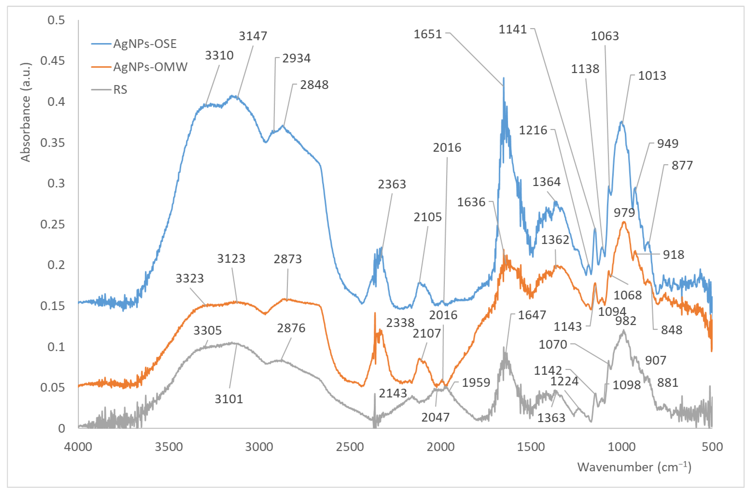
According to Figure 2, the bands at 3600–3300 cm−1 correspond to O-H stretching vibrations, while the wavenumbers between 2934 and 2848 depict C-H stretching vibrations of CH3 and CH2, respectively [74,75,76,77]. The sharp peaks at 2338–2363 cm−1 and 2016 cm−1 are characteristic bands in the formation of AgNPs from plant origin samples or O=C=O stretching vibrations from the air [69,78,79]. The band at around 1636–1651 cm−1 is due to the C-O bending associated with the OH group concerning starch and glycerol [74] or stretching vibration of the C=O group of agars [17]. In particular, the peak within the region 1420–1362 cm−1 is associated with O-H bending, while the peak within the region 1224–1063 cm−1 represents C-O stretching and C=C aromatic groups due to the presence of OS and OMW extracts [80,81]. The bands at 1140 cm−1 (stretching vibrations of C-O in the C-OH groups) and 1022 cm−1 (stretching vibrations of C-O in the C-OC groups) are characteristic functional groups of starch [74,82]. The bands at 1140 cm−1 may also depict P-OH stretching vibrations from extracts of OS or OMW [81]. However, the range from 1362 cm−1 to 1063 cm−1 also indicates the C-O stretching of primary to secondary alcohols, respectively [80,83]. The strong bands between 979 and 1013 cm−1 belong to C-OH groups, characteristic peaks of glycerol, while the weak bands at 881 to 949 cm−1 refer to C-C skeletal vibrations or C-H vibrations [76,77,80,81,82,83].
Similar results were presented in the aqueous assembly preparation of silver nanoparticles with phenols derived from olive oil industry by-products [69], where AgNPs appeared approximately at 2100 and 2300 cm−1, while the wavenumber at 1610 cm−1 corresponds to the stretching vibration of -C=O functional groups. A new study, concerning the production of agar–starch films with AgNP formation using enoki mushroom aqueous extract [17], has shown the presence of -OH stretching functional groups at approximately 3298 cm−1, the C-H stretching vibrations of alkane groups at 2931 and 2880 cm−1, the stretching vibration of the carbonyl group at 1643 cm−1, and the C-O stretching bond of starch at 1079 cm−1. A comparison of the spectrum of pure film without NPs with the films with NPs may reveal the disappearance, reduction in intensity, or shift of several characteristic peaks. Comparing the FT-IR spectra of starch/agar/AgNP film with starch/agar film, they present minor changes in peak intensity (1640, 1360, 1140, and 990 cm−1), corresponding probably to the van der Waals interactions and H-bonds between the biopolymers and the surface functional group of AgNPs [17,84].
The optical images of the three types of films can be associated with their morphology (Figure 3). According to Figure 3c, the starch–agar film with 30% w/w of glycerol appears to be transparent and colorless.
According to color analysis (Table 1), starch–agar films present high L values (>87%) and low chromaticity coordinates, a (2.39) and b (−9.84), where a values represent a color’s red–green axis and b values refer to a color’s yellow–blue axis, indicating that the film is transparent, while the introduction of AgNPs increases a (redness) and yellowness (b value) of the film [17]. Agar–starch films with AgNPs are typically odorless and tasteless, maintaining the desirable organoleptic qualities of the base film, which is a significant advantage for food packaging applications. However, the addition of AgNPs causes color changes, ranging from light yellow to dark brown depending on the AgNP concentration, the used raw materials, and their dispersion, as can be seen in Figure 3a,b [17]. The visual effect of AgNPs is influenced by the synthesis method, where reductions in silver can lead to browning reactions within the film matrix. In the present paper, the agar–starch AgNP coatings present an intense brown color, compared to the transparent, colorless agar–starch coating. According to Mahuwala et al. [20], the increase in AgNP concentration to agar–starch film from 0.5 to 2.0 mM changes the color of the films from light brown to dark brown, and the transparency is reduced owing to higher scattering of nanoparticles.
According to Figure 4, SEM images are included in two magnifications, i.e., 100× and 500×, for the three types of films. From the SEM images (Figure 4g,h), it is observed that the films are smooth with aggregate sizes ranging from approximately 1 to 10 μm due to the insolubility of starch in water. Moreover, the appearance of these microscopic aggregates indicates that starch and glycerol are partially miscible. According to the literature, the starch films without glycerol present more aggregates with higher sizes than films where glycerol is added [11]. Regular starch granules have an angular shape, like starch granules rich in amylopectin, while agar powders are irregular flakes with a wider size distribution than starch granules [85]. The addition of AgNPs derived from OSE and OMW in the film is presented in Figure 4a,b and Figure 4d,e, respectively. The appearance of Ag aggregates can be seen (the contrast between darker and lighter regions indicates the differences in atomic level), which are mentioned with the secondary electron images (lighter regions in the dark image indicate the presence of different atoms, i.e., Ag aggregates, Figure 4a,d). The inspection of the sample surface topography is best achieved using secondary electrons, as seen in Figure 4b,e. Comparing the surfaces of the films, it appears that Ag aggregates are distributed in the starch/agar/glycerol films. The evidence that these aggregates correspond to Ag aggregates can be ascertained by the EDS analysis. The latter is used to provide high-resolution chemical composition maps and give a clear understanding of processes occurring within a material.
According to EDS analysis (Figure 4i), it seems that the starch/agar/glycerol films contain only carbon (C) and oxygen (O) in a proportion of 56.6 and 43.4% w/w, respectively. The addition of Ag in the starch/agar/glycerol films is depicted in Figure 4c,f, where the lighter regions of Figure 4b,e are analyzed with EDS showing 39.6% C, 32.5% Ag, 23.2% O, and 4.8% Cl w/w for membrane with AgNPs derived from OSE and 58.0% C, 11.3% Ag, 28.1% O, 1.4% Na, 0.3% Cl, and 0.9% other elements w/w for AgNPs-OMW films. The appearance of Au in EDS images is the result of sample coating as it is mentioned in the Material and Methods section to prevent surface charging, to promote the secondary electrons’ emission so that the sample is uniformly conducted, and to provide a homogeneous surface for analysis and imaging. Similar studies have mentioned the deposition of AgNPs derived from leaf extract of the Mimusops elengi L. plant as a reducing agent, and their characterization through FT-IR (Fourier-transform infrared spectroscopy), XRD (X-ray diffraction), FESEM (Field Emission Scanning Electron Microscopy), HRTEM (High Resolution Transmission Electron Microscopy), and AFM (Atomic Force Microscopy) analyses on the surface of PES films [86]. Other studies [17] examined the microstructure of agar–starch–NP films with the produced AgNPs to come from silver nitrate with the water extract of the enoki mushroom as a reducing agent. According to the FESEM images, the starch/agar film presented a dense and smooth surface, while AgNP films have shown similar morphology with some spots spread over the aggregate-free polymer matrix.
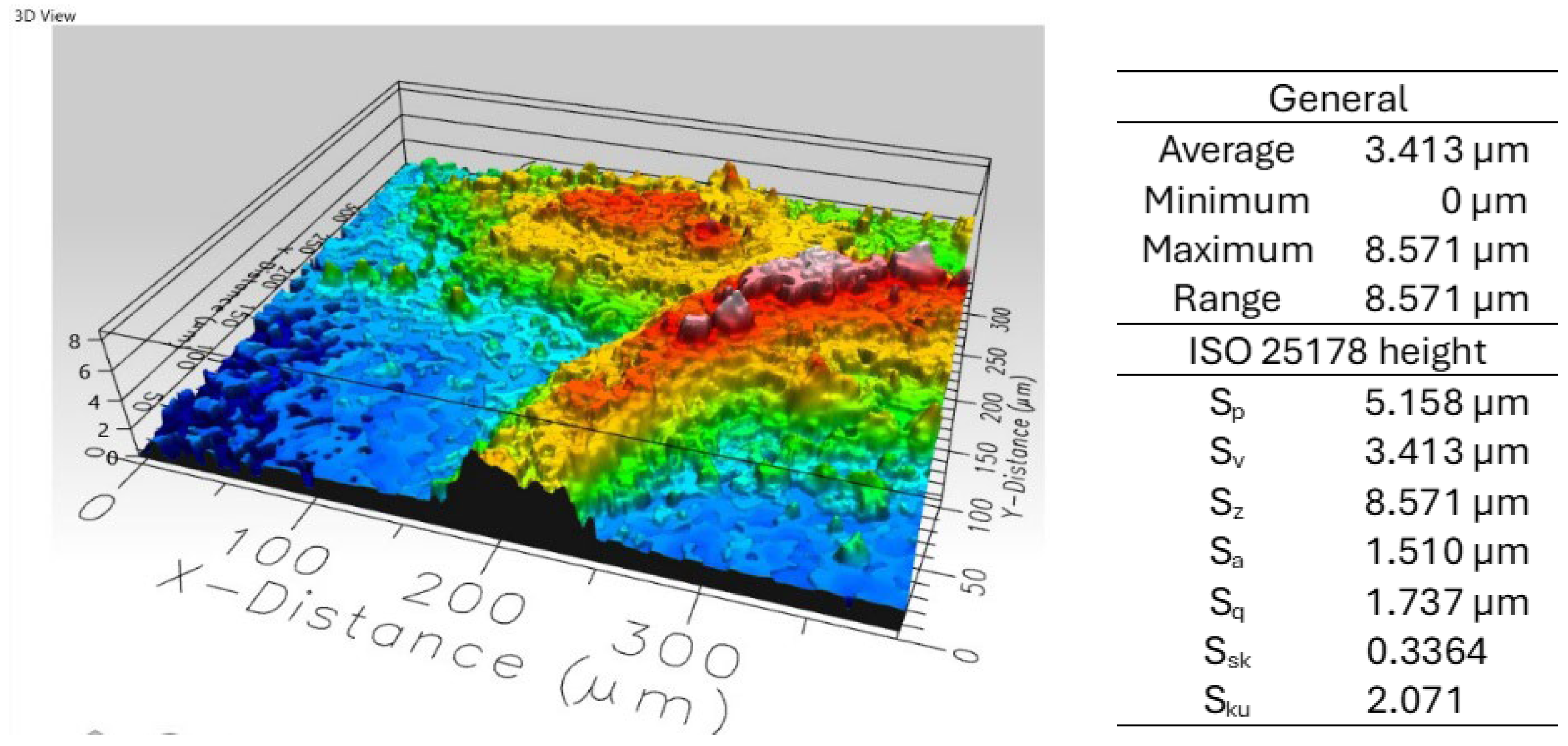
As a non-contact topography measurement technique, confocal profilometry is especially suitable for measuring the roughness of irregular, curved surfaces, such as those present in the starch/agar/glycerol specimens with or without AgNPs treated here (Figure 5).
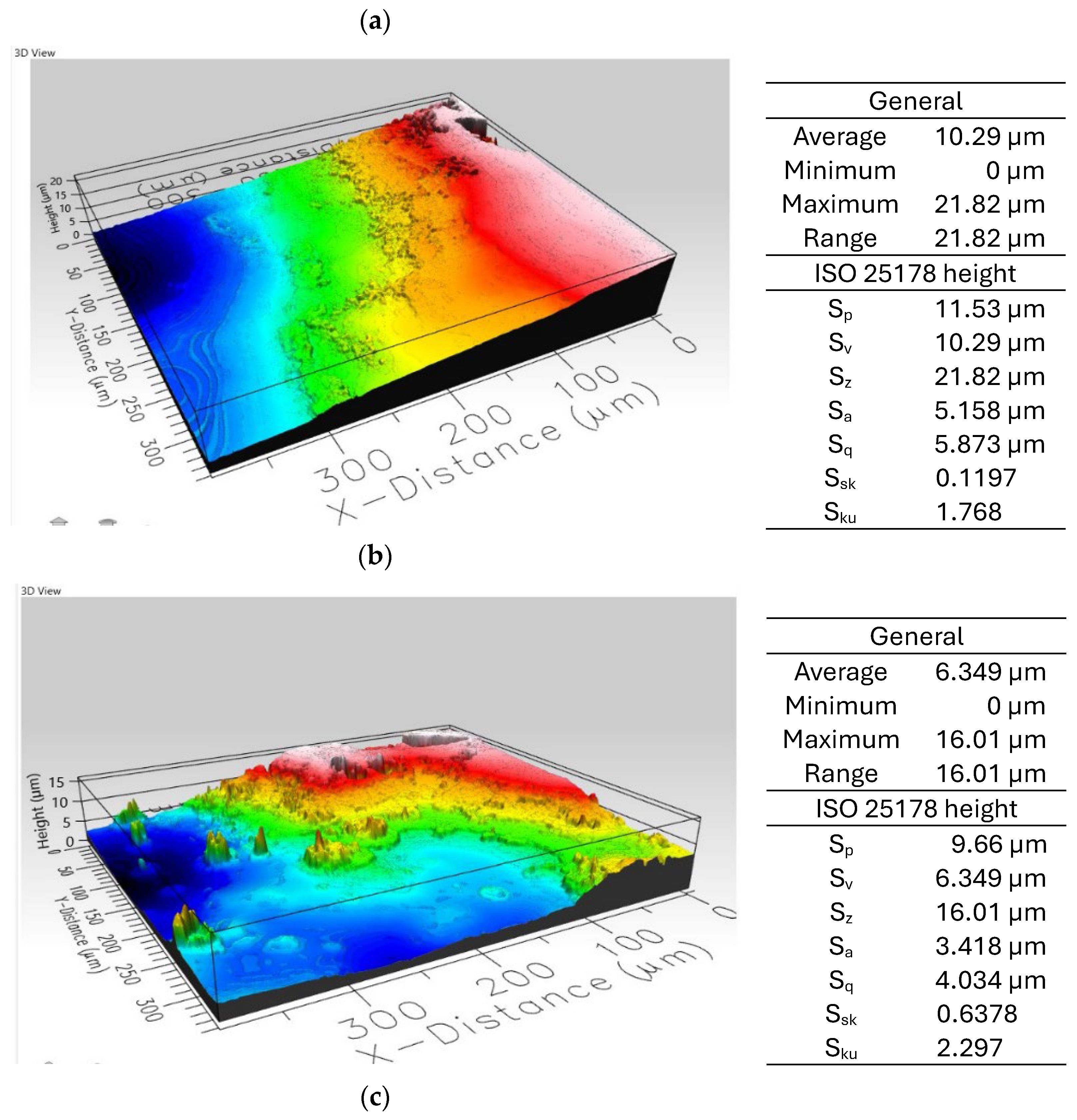
According to ISO 25178, the parameters Sq, Sa, Sp, Sv, and Sz characterize the surface amplitude, while Ssk and Sku describe the character of the height distribution and are defined in the caption of Figure 5. The Sp and Sv values represent the highest and lowest point on the surface within the measured area, respectively, showing that the AgNPs-OMW film presents the highest peak (Sp = 11.53) compared to AgNPs-OSE film (Sp = 5.16), while the AgNPs-OSE film presents the lowest peak (Sv = 3.41) compared to the AgNPs-OMW film (Sv = 10.29). Consequently, the total height difference between the highest peak and the lowest valley within the measured area (Sz) is equal to 21.82 for the AgNPs-OMW film and 8.57 for the AgNPs-OSE film. The above analysis shows that the AgNPs-OMW film presents a rougher surface than the AgNPs-OSE film. Moreover, Sq is a statistical measure of the roughness of the surface, calculated as the square root of the mean of the squared height differences from the mean line of the surface, showing that the AgNPs-OMW film presents higher roughness (Sq = 5.87) compared to the AgNPs-OSE film (Sq = 1.74).
A surface with a larger Sa (such as the AgNPs-OMW film) should be more difficult to wet than a surface with a smaller Sa (such as the AgNPs-OSE film), as the droplet would be less likely to contact with a starch/agar/glycerol surface at the bottom of a deeper valley or at the base between taller peaks than between shorter ones, maintaining a larger liquid–air interface as it was seen to Sheng et al. [87].
Moreover, surfaces with higher roughness featuring bumps or cavities can enhance droplet cohesion because the droplet is less likely to make contact with the surface at the bottom of valleys or between two peaks for surfaces with taller profiles than shorter ones [88].
Skewness can therefore be a helpful indicator of whether the surface is characterized by protruding particles (positive Ssk) or by voids (negative Ssk). The slightly higher than zero values of Ssk for all films indicate the presence of aggregates as a result of starch insolubility in water (more peaks with more material below the mean line), while the Sku values near two show that these aggregates present a platykurtic distribution, suggesting that the surface has few sharp peaks or deep indentations.

3.3. Films’ Antimicrobial Potential

Several metals with bactericidal properties have been proposed as antibacterial agents, including zinc (Zn), copper (Cu), and silver (Ag), each with a unique mechanism of action. For instance, the bacteria adhere to AgNPs, leading to local membrane perforation, and the AgNPs can then be internalized through the damaged membrane. The action of Ag+ ions significantly contributes to the antibacterial properties of AgNPs [89]. Moreover, Ag+ ions can cause cytoplasmic shrinkage, DNA condensation, and detachment of the cell-wall membrane. Furthermore, the toxic effects of Ag+ ions on bacteria have also been linked to the production of ROS [90]. The surface roughness and texture are also inherent parameters that can significantly impact the antibacterial properties of a given surface [91].
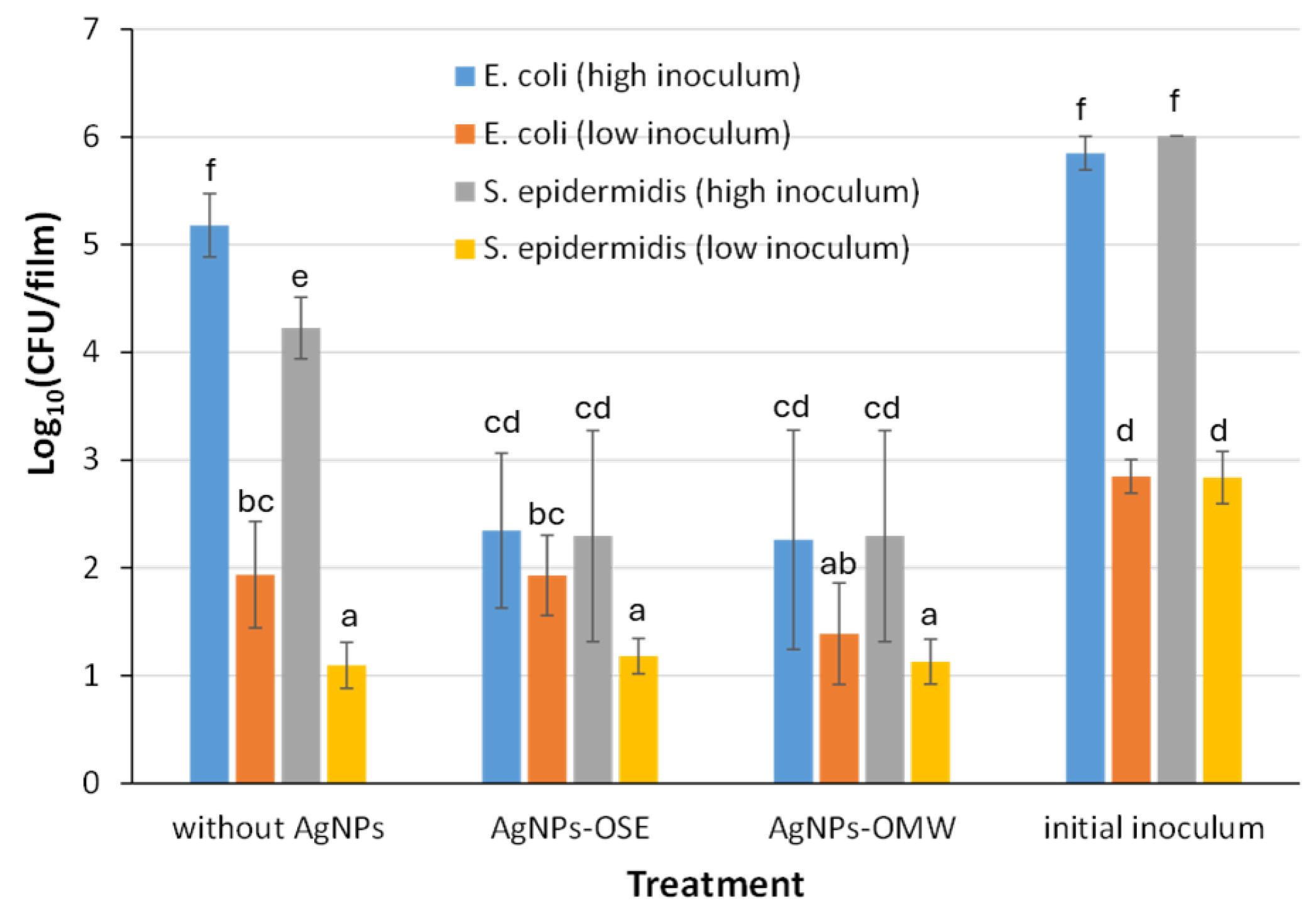
According to Figure 6, the two types of films with embedded AgNPs, presented a significant decrease in their bacterial load, when they had been initially inoculated with a high cellular concentration (106 CFU/film), for both bacterial species (E. coli and S. epidermidis). A slight decrease is also observed in the case of films that do not contain AgNPs (the reference sample) compared to the initial bacterial population. This occurred because agar–starch composite films do not provide the necessary nutritional ingredients and other environmental conditions for bacterial cells to grow and multiply or even survive. In contrast, these start to die. Under such conditions and between the two different bacterial species, and for both of their inoculation levels, S. epidermidis seems to be more vulnerable to stress, as its population noted a higher decrease, compared to that of E. coli. In the case of low inoculum (103 CFU/film), no significant differences were observed in the diminishment of the bacterial loads between films with no AgNPs and films with embedded AgNPs.
Nevertheless, all three types of films, initially inoculated with a low concentration of cells (103 CFU/film), both containing and not containing AgNPs, seem to be able to decrease their initial inoculated bacterial populations to less than 2 log10 (CFU/film). The incapability of total eradication of bacterial cells may be due to the existence of persisters, extremely resistant bacterial cells, among the total bacterial population [92,93]. Finally, no significant differences were observed in the antibacterial properties between AgNPs-OSE and AgNPs-OMW films, with the only exception being the films that were inoculated with the low inoculum of E. coli, although the difference was not significant (p > 0.05). In this case, AgNPs-OMW films presented a higher decrease in their bacterial load, i.e., the remaining viable and culturable population was 1.39 ± 0.47 log10 (CFU/film) compared to 1.93 ± 0.37 log10 (CFU/film) in the case of AgNPs-OSE. This slight difference between the two types of films may be due to the rougher surface of AgNPs-OMW films compared to that of AgNPs-OSE films, as shown in profilometry analysis (comparing Sp, Sv, Sz, and Sq parameters between the two types of films). It should thus be noted that at the nanoscale level, surfaces with roughness tend to show the best anti-adhesion properties (bacteria prefer smoother surfaces to grow and produce extracellular polymeric substances to survive), while at the micro-scale level, rougher surfaces tend to promote bacterial adhesion. This behavior is the result of the contact points to which the bacteria can adhere [94]. Confocal profilometry is applied for the first time to agar/starch/NP films, showing interesting results correlating the film surface with the antibacterial properties of the materials. In a previous study [30], the AgNP suspensions were also examined for their antimicrobial behavior, showing that both AgNPs exhibited the same antibacterial activity against S. epidermidis, while AgNPs derived from OMW presented a more effective inhibition of E. coli compared to AgNPs derived from OSE. The study of Qi et al. [69] reported on the antibacterial activities of AgNPs derived from olive mill wastewater phenols (OPs) and showed that 50 μg/mL of OP-AgNPs presented significant damage to the integrity of the E. coli membrane and inhibited the Ca2+-ATPase activity of E. coli, altering the cellular normal function. The antibacterial activity of AgNPs synthesized using trisodium citrate as a reducing and capping agent was tested against the bacteria S. taphylococcus aureus, P. seudomonas aeruginosa, and E. coli [68]. According to the results, a higher zone of inhibition was presented by S. aureus compared to P. aeruginosa and E. coli, showing a better susceptibility. Gram-negative bacteria like P. aeruginosa showed an increased resistance when exposed to AgNPs, compared to the most susceptible Gram-positive bacteria like S. aureus. The antimicrobial activity of the starch–agar–AgNP films, where nanoparticles were derived from the aqueous extract of the enoki mushroom, was studied elsewhere [17]. Their anti-microbial activity was measured against the bacteria L. monocytogenes, and E. coli. The results have shown higher antibacterial activity towards Gram-negative bacteria like E. coli than Gram-positive bacteria like Listeria monocytogenes.

4. Conclusions

The present study deals with the preparation of novel starch/agar/AgNP coatings for food packaging. The coatings, with NPs produced using Greek olive oil mill by-products as reducing agents, were successfully tested for the first time for antimicrobial properties.
TEM-EDS analysis has shown that AgNPs-OSE films present AgNPs that have different sizes starting from as small as a few nanometers, as was previously detected in AgNPs-OMW films.
FT-IR analysis revealed different functional groups, i.e., O-H stretching and bending, C-H stretching, C-O stretching, and bending vibrations associated with OH. The sharp peaks at 2338–2363 cm−1 and 2016 cm−1 are characteristic bands in the formation of AgNPs from plant origin samples or O=C=O stretching vibrations from the air.
The appearance of Ag aggregates is detected using secondary electron images at two different magnifications during SEM-EDS analysis. The addition of Ag in the starch/agar/glycerol films was analyzed with EDS, showing 39.6% C, 32.5% Ag, 23.2% O, and 4.8% ClL w/w for AgNPs-OSE films and 58.0% C, 11.3% Ag, 28.1% O, 1.4% Na, 0.3% Cl, and 0.9% other elements w/w for AgNPs-OMW films.
Profilometry analysis indicated that the AgNPs-OMW film presented a rougher surface compared to the AgNPs-OSE film, indicating that the roughest surface might also show the best anti-adhesion properties at the nanoscale level.
Agar–starch based films with embedded AgNPs (AgNPs-OSE or AgNPs-OMW) presented significant antibacterial activity against both Gram-negative (E. coli) and Gram-positive (S. epidermidis) model bacterial species, especially when they were initially inoculated with a high bacterial cell concentration (106 CFU/film). Comparing the AgNPs-OSE and AgNPs-OMW films, no significant differences were observed in the antibacterial properties, with the only exception of the films inoculated with the low inoculum of E. coli, although the difference was not significant (p > 0.05). In this case, AgNPs-OMW films presented a higher decrease in their bacterial load, indicating that they are ideal as packaging materials for the antimicrobial protection of foods.
To sum up, the synthesis of AgNPs by making good use of burdensome agro-industrial by-products, such as OS and OMW, and their subsequent incorporation into biopolymer films, was shown here to present promising potential. This approach can lead to the development of eco-friendly, biodegradable, and active antimicrobial packaging that can delay spoilage, reduce food loss, extend food shelf life, and promote sustainability and a green economy.
Future works may cover the examination of the antimicrobial action of agar–starch–AgNP coatings to other foodborne bacteria of interest such as Bacillus cereus, Pseudomonas fluorescens, and Salmonella enterica. Moreover, a basic aspect remains: the comprehensive toxicological assessments and regulatory scrutiny concerning the possible transportation of Ag ions from packages to food. Finally, future research should prioritize the combination of other physical polymers or nanocomposites with starch coatings to improve their properties and facilitate large-scale production.

Author Contributions

C.M.G.: synthesis of AgNPs and films, antimicrobial experiments, and initial draft; N.R.: synthesis of AgNPs and films; Z.I.: synthesis and characterization of films and writing the original draft; E.G.: antimicrobial analysis, writing the original draft; K.P.G.: SEM-EDS analysis; K.E.: 3D optical profilometer analysis. All authors have read and agreed to the published version of the manuscript.

Funding

This research received no external funding.

Institutional Review Board Statement

Not applicable.

Informed Consent Statement

Not applicable.

Data Availability Statement

The datasets used and analyzed during the current study are available within the article is available from the corresponding author upon reasonable request.

Acknowledgments

The authors express their deepest gratitude to Vafeiadis George, a student of the Department of Food science and Nutrition, University of the Aegean, for his precious contribution to the conduction of the antimicrobial experiments. Project SUB2: Universities of Excellence project code 5180665, National Recovery and Resilience Plan “Greece 2.0” NextGenerationEU is acknowledged for the purchase of the Optical Profilometer (Profilm 3D, Filmetrics).

Conflicts of Interest

The authors declare no competing interests.

References

  1. Macena, M.G.; Carvalho, R.; Cruz-Lopes, L.P.; Guiné, R.P.F. Perceptions and knowledge regarding quality and safety of plastic materials used for food packaging. Open Agric. 2022, 7, 132–146. [Google Scholar] [CrossRef]
  2. Ncube, L.K.; Ude, A.U.; Ogunmuyiwa, E.N.; Zulkifli, R.; Beas, I.N. Environmental Impact of Food Packaging Materials: A Review of Contemporary Development from Conventional Plastics to Polylactic Acid Based Materials. Materials 2020, 13, 4994. [Google Scholar] [CrossRef]
  3. Licciardello, F. Unexpected possible consequences of plastic packaging reuse. Curr. Opin. Food Sci. 2024, 56, 101131. [Google Scholar] [CrossRef]
  4. Roy, S.; Chawla, R.; Santhosh, R.; Thakur, R.; Sarkar, P.; Zhang, W. Agar-based edible films and food packaging application: A comprehensive review. Trends Food Sci. Technol. 2023, 141, 104198. [Google Scholar] [CrossRef]
  5. Jang, E.J.; Padhan, B.; Patel, M.; Pandey, J.K.; Xu, B.; Patel, R. Antibacterial and Biodegradable Food Packaging Film from Bacterial Cellulose. Food Control 2023, 153, 109902. [Google Scholar] [CrossRef]
  6. Rydz, J.; Musiol, M.; Zawidlak-Wegrzyńska, B.; Sikorska, W. Present and Future of Biodegradable Polymers for Food Packaging Applications. Biopolymers for Food Design. In Handbook of Food Bioengineering; Grumezescu, A.M., Holban, A.M., Eds.; Academic Press Inc.: Cambridge, MA, USA, 2018; pp. 431–467. [Google Scholar]
  7. Han, G.; Rui, G.; Yu, Z.; Chen, G. Progress on biodegradable films for antibacterial food packaging. E3S Web Conf. 2019, 145, 01036. [Google Scholar] [CrossRef]
  8. Motelica, L.; Ficai, D.; Ficai, A.; Oprea, O.C.; Kaya, D.A.; Andronescu, E. Biodegradable Antimicrobial Food Packaging. Foods 2020, 9, 1438. [Google Scholar] [CrossRef] [PubMed]
  9. Arruda, T.R.; Machado, G.d.O.; Marques, C.S.; de Souza, A.L.; Pelissari, F.M.; de Oliveira, T.V.; Silva, R.R.A. An Overview of Starch-Based Materials for Sustainable Food Packaging: Recent Advances, Limitations, and Perspectives. Macromol 2025, 5, 19. [Google Scholar] [CrossRef]
  10. Karnwal, A.; Rauf, A.; Jassim, A.Y.; Selvaraj, M.; Al-Tawaha, A.R.M.S.; Kashyap, P.; Kumar, D.; Malik, T. Advanced starch-based films for food packaging: Innovations in sustainability and functional properties. Food Chem. X 2025, 29, 102662. [Google Scholar] [CrossRef] [PubMed]
  11. Ramasamy, P. A dielectric relaxation study of starch-water and starch-glycerol films. Ionics 2012, 18, 413–423. [Google Scholar] [CrossRef]
  12. Wu, Y.; Geng, F.; Chang, P.R.; Yu, J.; Ma, X. Effect of agar on the microstructure and performance of potato starch film. Carbohydr. Polym. 2009, 76, 299–304. [Google Scholar] [CrossRef]
  13. Phan The, D.; Debeaufort, F.; Voilley, A.; Luu, D. Biopolymer interactions affect the functional properties of edible films based on agar, cassava starch and arabinoxylan blends. J. Food Eng. 2009, 90, 548–558. [Google Scholar] [CrossRef]
  14. Wongphan, P.; Harnkarnsujarit, N. Characterization of starch, agar and maltodextrin blends for controlled dissolution of edible films. Int. J. Biol. Macromol. 2020, 156, 80–93. [Google Scholar] [CrossRef] [PubMed]
  15. Hajipour, M.J.; Fromm, K.M.; Akbar Ashkarran, A.; Jimenez de Aberasturi, D.; Larramendi, I.R.D.; Rojo, T.; Serpooshan, V.; Parak, W.J.; Mahmoudi, M. Antibacterial properties of nanoparticles. Trends Biotechnol. 2012, 30, 499–511. [Google Scholar] [CrossRef]
  16. Rai, M.K.; Deshmukh, S.D.; Ingle, A.P.; Gade, A.K. Silver nanoparticles: The powerful nanoweapon against multidrug-resistant bacteria. J. Appl. Microbiol. 2012, 112, 841–852. [Google Scholar] [CrossRef]
  17. Roy, S.; Rhim, J.W. Starch/agar—Based functional films integrated with enoki mushroom- mediated silver nanoparticles for active packaging applications. Food Biosci. 2022, 49, 101867. [Google Scholar] [CrossRef]
  18. Ortega, F.; Giannuzzi, L.; Arce, V.B.; García, M.A. Active composite starch films containing green synthesized silver nanoparticles. Food Hydrocoll. 2017, 70, 152–162. [Google Scholar] [CrossRef]
  19. Roy, S.; Shankar, S.; Rhim, J.-W. Melanin-mediated synthesis of silver nanoparticles and its use for the preparation of carrageenan-based antibacterial films. Food Hydrocoll. 2019, 88, 237–246. [Google Scholar] [CrossRef]
  20. Mahuwala, A.A.; Hemant, V.; Meharwade, S.D.; Deb, A.; Chakravorty, A.; Grace, A.N.; Raghavan, V. Synthesis and characterisation of starch/agar nanocomposite films for food packaging application. IET Nanobiotechnol. 2020, 9, 809–814. [Google Scholar] [CrossRef]
  21. Sadeghi, B.; Jamali, M.; Kia, S.; Amininia, A.; Ghafari, S. Synthesis and characterization of silver nanoparticles for antibacterial activity. Int. J. Nano Dimens. 2010, 1, 119–124. [Google Scholar]
  22. Zulfiqar, Z.; Khan, R.R.M.; Summer, M.; Saeed, Z.; Pervaiz, M.; Rasheed, S.; Shehzad, B.; Kabir, F.; Ishaq, S. Plant-mediated green synthesis of silver nanoparticles: Synthesis, characterization, biological applications, and toxicological considerations: A review. Biocatal. Agric. Biotechnol. 2024, 57, 103121. [Google Scholar] [CrossRef]
  23. Wasiq, M.; Ahmed, F.; Saeed, F.; Raza, A.; Abdullah, H.M.; Saleem, S.; Shoukat, L.; Nuzhyma, N.; Afzaal, M. From waste to wellness: Green synthesis of nanoparticles from vegetable waste. Sustain. Environ. 2025, 11, 2491870. [Google Scholar] [CrossRef]
  24. Mathew, S.S.; Sunny, N.E.; Shanmugam, V. Green synthesis of anatase titanium dioxide nanoparticles using Cuminum cyminum seed extract; effect on Mung bean (Vigna radiata) seed germination. Inorg. Chem. Commun. 2021, 126, 108485. [Google Scholar] [CrossRef]
  25. Hamimed, S.; Chamekh, A.; Slimi, H.; Chatti, A. How olive mill wastewater could turn into valuable bionanoparticles in improving germination and soil bacteria. Ind. Crops Prod. 2022, 188, 115682. [Google Scholar] [CrossRef]
  26. Xu, J.; Yıldıztekin, M.; Han, D.; Keskin, C.; Baran, A.; Baran, M.F.; Eftekhari, A.; Aytuğ Ava, C.; İrtegün Kandemir, S.; Bariş Cebe, D.; et al. Biosynthesis, chara-cterization, and investigation of antimicrobial and cytotoxic activities of silver nanoparticles using Solanum tuberosum peel aqueous extract. Heliyon 2023, 9, e19061. [Google Scholar] [CrossRef]
  27. Daniel, S.; Gladis, L.R. Investigation of Antimicrobial Potential of Silver Nanoparticles Synthesised from Vegetable Peel Extracts. Nanochem. Res. 2024, 9, 242–247. [Google Scholar]
  28. Tongwanichniyom, S.; Phewrat, N.; Rangsarikorn, N.; Leasen, S.; Luangkamin, S.; Chumnanvej, N. Green synthesis of silver nanoparticles using mature-pseudostem extracts of Alpinia nigra and their bioactivities. Green Process. Synth. 2024, 13, 20230226. [Google Scholar] [CrossRef]
  29. Elshahat Abdallah, S.; Elmessery, W.M.; Elfallawi, F.E.; Shoueir, K.R. Utilizing agricultural biowaste for food safety: Integrating naturally synthesized silver nanoparticles as antibacterial coating. Inorg. Chem. Commun. 2024, 163, 112337. [Google Scholar] [CrossRef]
  30. Rigopoulos, N.; Gkaliouri, C.M.; Ioannou, Z.; Sakavitsi, V.; Gournis, D. Green synthesis of silver nanoparticles using olive mill wastewater and olive stones extract and testing their antimicrobial activities against Escherichia coli and Staphylococcus epidermidis. NanoExpress 2024, 5, 015026. [Google Scholar] [CrossRef]
  31. Rao, M.M.V.; Mohammad, N.; Banerjee, S.; Khanna, P.K. Synthesis and food packaging application of silver nanoparticles: A review. Hybrid Adv. 2024, 6, 100230. [Google Scholar] [CrossRef]
  32. Basumatary, K.; Daimary, P.; Das, S.K.; Thapa, M.; Singh, M.; Mukherjee, A.; Κumar, S. Lagerstroemia speciosa fruit-mediated synthesis of silver nanoparticles and its application as filler in agar-based nanocomposite films for antimicrobial food packaging. Food Packag. Shelf Life 2018, 17, 99–106. [Google Scholar] [CrossRef]
  33. Liu, J.; Ma, Z.; Liu, Y.; Zheng, X.; Pei, Y.; Tang, K. Soluble soybean polysaccharide films containing in-situ generated silver nanoparticles for antibacterial food packaging applications. Food Packag. Shelf Life 2022, 31, 100800. [Google Scholar] [CrossRef]
  34. Kwon, S.; Lee, W.; Choi, J.W.; Bumbudsanpharoke, N.; Ko, S. A Facile Green Fabrication and Characterization of Cellulose-Silver Nanoparticle Composite Sheets for an Antimicrobial Food Packaging. Front. Nutr. 2021, 8, 778310. [Google Scholar] [CrossRef] [PubMed]
  35. Pandian, H.; Senthilkumar, K.; Venkata Ratnam, M.; Naveenkumar, M.; Samraj, S. Azadirachta indica leaf extract mediated silver nanoparticles impregnated nano composite film (AgNP/MCC/starch/whey protein) for food packaging applications. Environ. Res. 2023, 216, 114641. [Google Scholar] [CrossRef] [PubMed]
  36. Zhong, K.; Yang, B.; Liu, C.; Xiong, Y.; Wen, Q.; Li, W.; Ren, J. Progress in the application of polysaccharide/nanomaterial composites in food safety. Food Chem. 2025, 495, 146317. [Google Scholar] [CrossRef]
  37. Akila, V.; Badwaik, L.S. Recent advancement in improvement of properties of polysaccharides and proteins-based packaging film with added nanoparticles: A review. Int. J. Biol. Macromol. 2022, 203, 515–525. [Google Scholar]
  38. Dejene, B.K. Eco-friendly synthesis of metallic nanoparticles from agri-food waste extracts: Applications in food packaging and healthcare–A critical review. Mater. Today Chem. 2025, 45, 102619. [Google Scholar] [CrossRef]
  39. Kyziol-Komosinska, J.; Dzieniszewska, A.; Czupioł, J. Behavior of silver species in soil: Ag nanoparticles versus ionic Ag. Molecules 2024, 29, 5531. [Google Scholar] [CrossRef]
  40. Benoit, R.; Wilkinson, K.J.; Sauvé, S. Partitioning of silver and chemical speciation of free Ag in soils amended with nanoparticles. Chem. Cent. J. 2013, 7, 75. [Google Scholar] [CrossRef]
  41. Saleeb, N.; Gooneratne, R.; Cavanagh, J.; Bunt, C.; Mofasser Hossain, A.K.M.; Gaw, S.; Robinson, B. The mobility of silver nanoparticles and silver ions in the soil-plant system. J. Environ. Qual. 2019, 48, 1835–1841. [Google Scholar] [CrossRef]
  42. Pradas del Real, A.E.; Castillo-Michel, H.; Kaegi, R.; Sinnet, B.; Magnin, V.; Findling, N.; Villanova, J.; Carrière, M.; Santaella, C.; Fernańdez-Martıńez, A.; et al. Fate of Ag-NPs in Sewage Sludge after Application on Agricultural Soils. Environ. Sci. Technol. 2016, 50, 1759–1768. [Google Scholar] [CrossRef]
  43. Maymuna, A.; Annisa, W.D.; Nandiyanto, A.B.D.; Ragadhita, R. Economic Perspective in the Production of Silver Nanoparticles on the Bacterial Cellulose Membrane as Antibacterial Material. Int. J. Energetica 2019, 4, 17–22. [Google Scholar] [CrossRef]
  44. Iravani, S.; Korbekandi, H.; Mirmohammadi, S.V.; Zolfaghari, B. Synthesis of silver nanoparticles: Chemical, physical and biological methods. Res. Pharm. Sci. 2014, 9, 385–406. [Google Scholar] [PubMed]
  45. Silver Nanoparticles Market. Available online: https://www.alliedmarketresearch.com/silver-nanoparticles-marketA06923#:~:text=The%20global%20silver%20nanoparticles%20market,11.7%25%20from%202025%20to%202034 (accessed on 25 September 2025).
  46. Silver Nitrate Price. Available online: https://www.metal.com/Silver/202203220001 (accessed on 25 September 2025).
  47. Silver Nitrate Price. Available online: https://www.alibaba.com/product-detail/Factory-Price-Silver-Nitrate-PowderAgno3_1600290497590.html?spm=a2700.7724857.0.0.1cb77187PiLhA9 (accessed on 25 September 2025).
  48. Global Cassava Starch Price. Available online: https://dir.tridge.com/prices/cassava-starch (accessed on 25 September 2025).
  49. Global Agar Price. Available online: https://dir.tridge.com/prices/agar (accessed on 25 September 2025).
  50. Sodium Hydroxide Price Index. Available online: https://businessanalytiq.com/procurementanalytics/index/sodium-hydroxide-price-index/ (accessed on 25 September 2025).
  51. Glycerol Price Index. Available online: https://businessanalytiq.com/procurementanalytics/index/glycerol-price-index/ (accessed on 25 September 2025).
  52. Puritan Springs. Available online: https://shop.puritansprings.com/55-gallon-drum-industrial-distilled-water/ (accessed on 25 September 2025).
  53. Cano, A.; Cháfer, M.; Chiralt, A.; González-Martínez, C. Development and characterization of active films based on starch-PVA, containing silver nanoparticles. Food Packag. Shelf Life 2016, 10, 16–24. [Google Scholar] [CrossRef]
  54. Krásniewska, K.; Galus, S.; Gniewosz, M. Biopolymers-Based Materials Containing Silver Nanoparticles as Active Packaging for Food Applications—A Review. Int. J. Mol. Sci. 2020, 21, 698. [Google Scholar] [CrossRef]
  55. Moulahoum, H.; Ghorbanizamani, F. Navigating the development of silver nanoparticles-based food analysis through the power of artificial intelligence. Food Chem. 2024, 445, 138800. [Google Scholar] [CrossRef]
  56. Paidari, S.; Tahergorabi, R.; Anari, E.S.; Nafchi, A.M.; Zamindar, N.; Goli, M. Migration of Various Nanoparticles into Food Samples: A Review. Foods 2021, 10, 2114. [Google Scholar] [CrossRef]
  57. Rybalchenko, N.P.; Demchenko, V.L.; Hnatiuk, T.T.; Vasyliuk, O.M.; Iurzhenko, T.V.; Rybalchenko, T.V.; Sytnyk, I.O.; Shtepa, D.V.; Marynin, A.I. Antimicrobial effect of biopolymer packaging materials with silver nanoparticles for food storage. Microbiol. J. 2024, 86, 30–41. [Google Scholar] [CrossRef]
  58. Suvarna, V.; Nair, A.; Mallya, R.; Khan, T.; Omri, A. Antimicrobial Nanomaterials for Food Packaging. Antibiotics 2022, 11, 729. [Google Scholar] [CrossRef]
  59. Trotta, F.; Da Silva, S.; Massironi, A.; Mirpoor, S.F.; Lignou, S.; Ghawi, S.K.; Charalampopoulos, D. Silver bionanocomposites as active food packaging: Recent advances & future trends tackling the food waste crisis. Polymers 2023, 15, 4243. [Google Scholar] [CrossRef]
  60. WHO. Guidelines for Drinking-water Quality, Recommendations, 3rd ed. 2004, Volume 1. Available online: https://www.who.int/publications/i/item/9789241547611 (accessed on 6 October 2025).
  61. Carbone, M.; Donia, D.T.; Sabbatella, G.; Antiochia, R. Silver nanoparticles in polymeric matrices for fresh food packaging. J. King Saud Univ. Sci. 2016, 28, 273–279. [Google Scholar] [CrossRef]
  62. Lambre, C.; Baviera, J.M.B.; Bolognesi, C.; Chesson, A.; Cocconcelli, P.S.; Crebelli, R.; Gott, D.M.; Grob, K.; Lampi, Ε.; Mengelers, M.; et al. Safety assessment of the substance silver nanoparticles for use in food contact materials, Enzymes and Processing Aids (CEP), Scientific opinion. EJ EFSA J. 2021, 19, 6790. [Google Scholar]
  63. Chadha, U.; Bhardwaj, P.; Selvaraj, S.; Arasu, K.; Praveena, S.; Pavan, A.; Khanna, M.; Singh, P.; Singh, S.; Chakravorty, A.; et al. Current trends and future perspectives of nanomaterials in food packaging application. J. Nanomater. 2022, 2022, 2745416. [Google Scholar] [CrossRef]
  64. Marchiore, N.G.; Manso, I.J.; Kaufmann, K.C.; Lemes, G.F.; de Oliveira Pizolli, A.P.; Droval, A.A.; Bracht, L.; Gonçalves, O.H.; Leimann, F.V. Migration evaluation of silver nanoparticles from antimicrobial edible coating to sausages. LWT-Food Sci. Technol. 2017, 76, 203–208. [Google Scholar] [CrossRef]
  65. Thapliyal, D.; Karale, M.; Diwan, V.; Kumra, S.; Arya, R.K.; Verros, G.D. Current Status of Sustainable Food Packaging Regulations: Global Perspective. Sustainability 2024, 16, 5554. [Google Scholar] [CrossRef]
  66. Rigopoulos, N.; Gkaliouri, C.M.; Sakavitsi, V.; Gournis, D. Full Factorial Design Synthesis of Silver Nanoparticles Using Origanum vulgare. Reactions 2023, 4, 505–517. [Google Scholar] [CrossRef]
  67. ImageJ. Image Processing and Analysis in Java. Available online: https://imagej.net/ij/ (accessed on 28 September 2025).
  68. Iwuji, C.; Saha, H.; Ghann, W.; Dotson, D.; Bhuiya, M.A.K.; Parvez, M.S.; Jahangir, Z.M.G.S.; Rahman, M.M.; Chowdhury, F.I.; Uddin, J. Synthesis and characterization of silver nanoparticles and their promising antimicrobial effects. Chem. Phys. Impact 2024, 9, 100758. [Google Scholar] [CrossRef]
  69. Qi, Z.; Xue, X.; Zhou, H.; Yuan, H.; Li, W.; Yang, G.; Xie, P.; Whang, C. The aqueous assembly preparation of OPs-AgNPs with phenols from olive mill wastewater and its mechanism on antimicrobial function study. Food Chem. 2022, 376, 131924. [Google Scholar] [CrossRef]
  70. Stozhko, N.; Tarasov, A.; Tamoshenko, V.; Bukharinova, M.; Khamzina, E.; Kolotygina, V. Green Silver Nanoparticles: Plant-Extract-Mediated Synthesis, Optical and Electrochemical Properties. Physchem 2024, 4, 402–419. [Google Scholar] [CrossRef]
  71. Scroccarello, A.; Junior, B.M.H.; Della Pelle, F.; Ciancetta, J.; Ferraro, G.; Fratini, E.; Valbonetti, L.; Copez, C.C.; Compagnone, D. Effect of phenolic compounds-capped AgNPs on growth inhibition of Aspergillus niger. Colloids Surf. B Biointerfaces 2021, 199, 111533. [Google Scholar] [CrossRef]
  72. Sudha, P.N.; Gomathi, T.; Vinodhini, P.A.; Nasreen, K. Marine Carbohydrates of Wastewater Treatment. In Advances in Food and Nutrition Research; Se-Kwon, K., Ed.; Academic Press Inc.: Cambridge, MA, USA, 2014; Volume 73, pp. 103–143. [Google Scholar] [CrossRef]
  73. Robyt, J.F. General Principles, General Properties, Occurrence and Preparation of Carbohydrates. In Glycoscience, Chemistry and Chemical Biology, 2nd ed.; Fraser-Reid, B.O., Tatsuta, K., Thiem, J., Eds.; Springer: Heidelberg, Germany, 2008; Volume 1, p. 72. [Google Scholar]
  74. Abdullah, A.H.D.; Chalimah, S.; Primadona, I.; Hanantyo, M.H.G. Physical and chemical properties of corn, cassava, and potato starchs. IOP Conf. Ser. Earth Environ. Sci. 2018, 160, 012003. [Google Scholar] [CrossRef]
  75. Karthik, L.; Kumar, G.; Kirthi, A.V.; Rahuman, A.A.; Bhaskara Rao, K.V. Streptomyces sp. LK3 mediated synthesis of silver nanoparticles and its biomedical application. Bioprocess Biosyst. Eng. 2014, 37, 261–267. [Google Scholar] [CrossRef]
  76. 79 Niraimathi, K.L.; Sudha, V.; Lavanya, R.; Brindha, P. Biosynthesis of silver nanoparticles using Alternanthera sessilis (Linn.) extract and their antimicrobial, antioxidant activities. Colloids Surf. B Biointerfaces 2013, 102, 288–291. [Google Scholar] [CrossRef]
  77. 80 Prakash, P.; Gnanaprakasam, P.; Emmanuel, R.; Arokiyaraj, S.; Saravanan, M. Green synthesis of silver nanoparticles from leaf extract of Mimusops elengi, Linn. for enhanced antibacterial activity against multi drug resistant clinical isolates. Colloids Surf. B Biointerfaces 2013, 108, 255–259. [Google Scholar] [CrossRef]
  78. Hemavathy, R.V.; Kattupalli, R.; Sridevi, V.; Talib Hamzah, H.; Prasad, K.S.N.V.; Yaashikaa, P.R. Green synthesis of Pelargonium radens derived silver nanoparticles: Characterization studies and anticancer activity. Mater. Lett. 2024, 372, 137075. [Google Scholar] [CrossRef]
  79. Chowdhury, M.A.; Iqbal, M.Z.; Rana, M.M.; Hossain, N.; Shahin, M.; Islam, M.A.; Rahman, M.M. Green synthesis of novel green ceramic-based nanoparticles prepared by sol-gel technique for diverse industrial application. Results Surf. Interfaces 2024, 14, 100178. [Google Scholar] [CrossRef]
  80. Joshi, R.; Joshi, R.; Amanah, H.Z.; Faqeerzada, M.A.; Jayapal, P.K.; Kim, G.; Baek, I.; Park, E.S.; Masithoh, R.E.; Cho, B.K. Quantitative analysis of glycerol concentration in red wine using Fourier transform infrared spectroscopy and chemometrics analysis. Korean J. Agric. Sci. 2021, 48, 299–310. [Google Scholar] [CrossRef]
  81. Alardhi, S.M.; Salih, H.G.; Ali, N.S.; Khalbas, A.H.; Salih, I.K.; Saady, N.M.C.; Zendehboudi, S.; Albayati, T.M.; Harharah, H.N. Olive stone as an eco-friendly bio-adsorbent for elimination of methylene blue dye from industrial wastewater. Sci. Rep. 2023, 13, 21063. [Google Scholar] [CrossRef]
  82. Ponsanti, K.; Tangnorawich, B.; Ngernyuang, N.; Pechyen, C. A flower shape-green synthesis and characterization of silver nanoparticles (AgNPs) with different starch as a reducing agent. J. Mater. Res. Technol. 2020, 9, 11003–11012. [Google Scholar] [CrossRef]
  83. Hanry, E.L.; Redzwan, N.F.M.; Badeges, N.F.A.K.; Surugau, N. Characterization of biofilms developed from alginate extracted from Padina sp. incorporated with calcium chloride (CaCL2). J. Phys. Conf. Ser. 2022, 2314, 012022. [Google Scholar] [CrossRef]
  84. Agarwal, S.; Hoque, M.; Bandara, N.; Pal, K.; Sarkar, P. Synthesis and characterization of tamarind kernel powder-based antimicrobial edible films loaded with geraniol. Food Packag. Shelf Life 2020, 26, 100562. [Google Scholar] [CrossRef]
  85. Guo, Y.; Zhang, B.; Zhao, S.; Qiao, D.; Xie, F. Plasticized starch/agar composite films: Processing, morphology, structure, mechanical properties and surface hydrophilicity. Coatings 2021, 11, 311. [Google Scholar] [CrossRef]
  86. Tiwari, H.; Samal, K.; Geed, S.R.; Bera, S.; Das, C.; Mohanty, K. Green synthesis of silver nanoparticles for ultrafiltration membrane surface modification and antimicrobial activity. Sustain. Chem. Clim. Action 2023, 3, 100031. [Google Scholar] [CrossRef]
  87. Sheng, Y.-J.; Jiang, S.; Tsao, H.-K. Effects of geometrical characteristics of surface roughness on droplet wetting. J. Chem. Phys. 2007, 127, 234704. [Google Scholar] [CrossRef]
  88. Abbott, J.P.R.; Zhu, H. 3D optical surface profiler for quantifying leaf surface roughness. Surf. Topogr. Metrol. Prop. 2019, 7, 045016. [Google Scholar] [CrossRef]
  89. Ahmad, S.A.; Das, S.S.; Khatoon, A.; Ansari, M.T.; Afzal, M.; Hasnain, M.S.; Nayak, A.K. Bactericidal activity of silver nanoparticles: A mechanistic review. Mater. Sci. Energy Technol. 2020, 3, 756–769. [Google Scholar] [CrossRef]
  90. Yin, I.X.; Zhang, J.; Zhao, I.S.; Mei, M.L.; Li, Q.; Chu, C.H. The Antibacterial Mechanism of Silver Nanoparticles and Its Application in Dentistry. Int. J. Nanomed. 2020, 15, 2555–2562. [Google Scholar] [CrossRef]
  91. Georgakopoulos-Soares, I.; Papazoglou, E.L.; Karmiris-Obratański, P.; Karkalos, N.E.; Markopoulos, A.P. Surface antibacterial properties enhanced through engineered textures and surface roughness: A review. Colloids Surf. B Biointerfaces 2023, 231, 113584. [Google Scholar] [CrossRef]
  92. Niu, H.; Gu, J.; Zhang, Y. Bacterial persisters: Molecular mechanisms and therapeutic development. Signal Transduct. Target. Ther. 2024, 9, 174. [Google Scholar] [CrossRef]
  93. Serrano, S.; Grujović, M.Z.; Marković, K.G.; Barreto-Crespo, M.T.; Semedo-Lemsaddek, T. From dormancy to eradication: Strategies for controlling bacterial persisters in food settings. Foods 2025, 14, 1075. [Google Scholar] [CrossRef]
  94. Mahmoudi-Qashqay, S.; Zamani-Meymian, M.R.; Sadati, S.J. Improving antibacterial ability of Ti-Cu thin films with co-sputtering method. Sci. Rep. 2023, 13, 16593. [Google Scholar] [CrossRef]
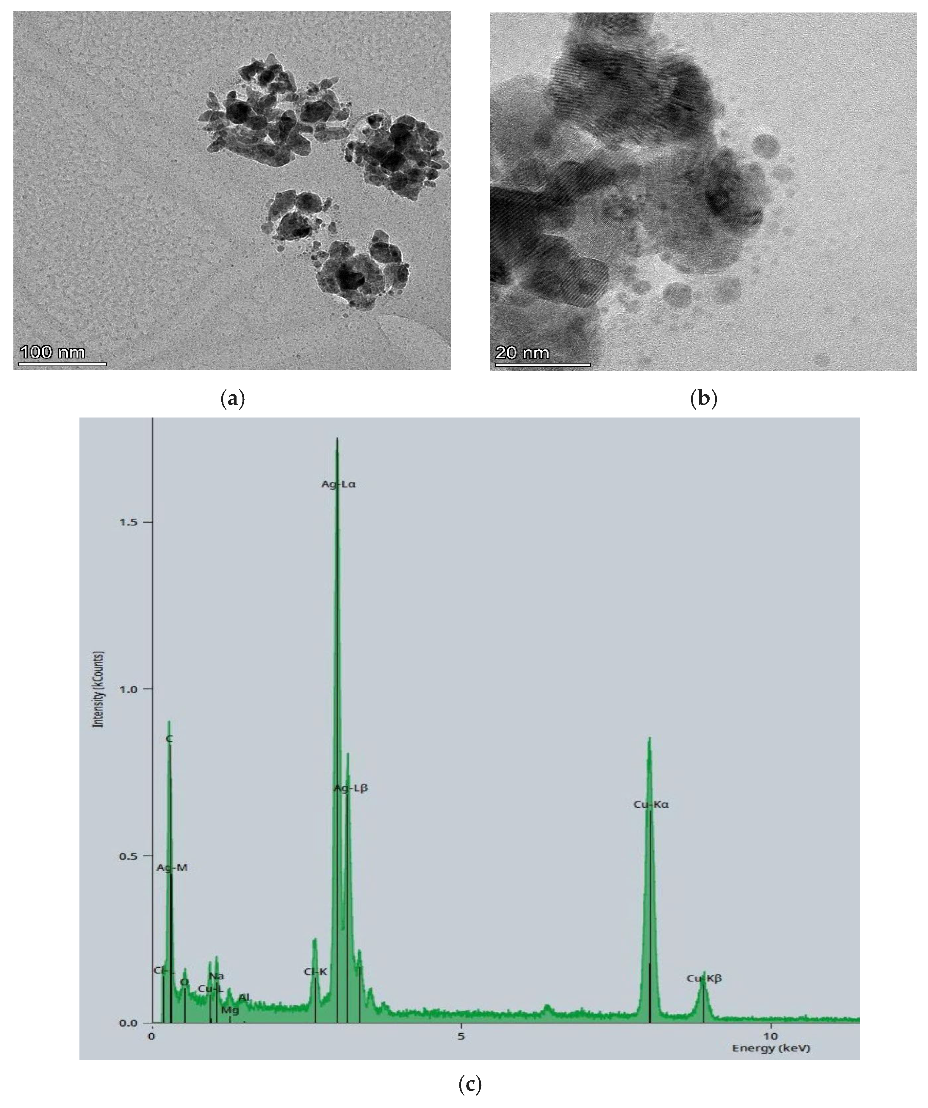
Figure 1. TEM images of the AgNPs-OSE samples at different resolutions, (a) ×100 and (b) ×20 nm, and (c) EDS analysis.
Sustainability 17 08916 g001
Figure 2. FT-IR spectra of starch–agar films with AgNPs, i.e., AgNPs-OSE or AgNPs-OMW, and without nanoparticles, i.e., RS films.
Sustainability 17 08916 g002
Figure 3. Pictures of the three different types of produced films: (a) with AgNPs-OSE, (b) with AgNPs-OMW, and (c) without AgNPs (RS).
Sustainability 17 08916 g003
Figure 4. SEM-EDS images of different films: 1st row (ac): AgNPs-OSE films; 2nd row (df): AgNPs-OMW films; and 3rd row (gi): films without AgNPs. SEM images are presented in two different magnifications, 100× (a,d,g) and 500× (b,c,h), where the arrows indicate the SEM points where EDS analysis was performed.
Sustainability 17 08916 g004aSustainability 17 08916 g004b
Figure 5. Three-dimensional topographical images and the corresponding parameters obtained by confocal profilometry for the different films (a) with AgNPs-OSE, (b) with AgNPs-OMW, and (c) without AgNPs, where Sp is the maximum peak height, Sv is the maximum pit height, Sz is the maximum height, Sa is the arithmetic mean height, Sq is the root mean square height, Ssk is the skewness, and Sku is kurtosis.
Sustainability 17 08916 g005aSustainability 17 08916 g005b
Figure 6. Logarithmic populations (log10 log10CFU/film) of surviving cells for each bacterial species (E. coli and S. epidermidis) on each type of film (AgNPs-OSE film, AgNPs-OMW film, and RS film without AgNPs) after 72 h of incubation at 4 °C. The initial populations of bacteria on the films after inoculation at two levels (high and low inoculum) are also shown on the right. Each bar represents the mean values ± standard deviations. The mean values followed by different superscript letters (a–f) differ significantly (p < 0.05).
Sustainability 17 08916 g006
Table 1. Color analysis of the three agar–starch films with and without AgNPs.
SamplesLab
AgNPs-OSE27.83 ± 0.6411.13 ± 0.64,28.03 ± 1.21
AgNPs-OMW32.10 ± 0.457.77 ± 0.3422.27 ± 2.02
RS87.46 ± 0.172.39 ± 1.25−9.84 ± 2.34
Disclaimer/Publisher’s Note: The statements, opinions and data contained in all publications are solely those of the individual author(s) and contributor(s) and not of MDPI and/or the editor(s). MDPI and/or the editor(s) disclaim responsibility for any injury to people or property resulting from any ideas, methods, instructions or products referred to in the content.