Article

Understanding the Inhibition Mechanism of Lignin Adsorption to Cellulase in Terms of Changes in Composition and Conformation of Free Enzymes

1
State Key Laboratory Base of Eco-Chemical Engineering, College of Chemical Engineering, Qingdao University of Science and Technology, 53# Zhengzhou Road, Qingdao 266042, China
2
Qingdao High-Tech Industry Promotion Center, Qingdao 266540, China
3
Guangxi Key Laboratory of Clean Pulp and Papermaking and Pollution Control, College of Light Industry and Food Engineering, Guangxi University, 100# Daxue Road, Nanning 530004, China
*
Author to whom correspondence should be addressed.
Sustainability 2023, 15(7), 6057; https://doi.org/10.3390/su15076057
Submission received: 6 March 2023 / Revised: 28 March 2023 / Accepted: 30 March 2023 / Published: 31 March 2023

Abstract

:
The adsorption of lignin to cellulase is the major obstacle in the sugar-platform conversion of lignocellulosic bioresources. In this study, the adsorption behavior of untreated and pretreated lignin samples from corn stover to cellulase was investigated, in particular the effects of lignin adsorption on the composition and spatial conformation of free enzymes were explored. The results showed that pretreatments decreased the hydrophobic groups contents of lignin, i.e., aromatic ring, ether and carbonyl, as well as the content of ionizable group, i.e., carboxyl, which reduced its hydrophobicity and negative charge density, thus weakening the adsorption ability of lignin to cellulase. The lignin samples mainly adsorbed the CBHII component of cellulase to inhibit the synergistic effect of free enzymes. Lignin adsorption altered the spatial position of tryptophan residues in free enzymes, exposing them to the protein surface. In addition, the secondary structure of free enzymes was altered, with a decrease in the alpha-helix content and an increase in the random coil content, thus loosening the spatial conformation of free enzymes. The change degree in the spatial structure of free enzymes correlated with the adsorption capacity of the lignin, i.e., lignin with low adsorption capacity caused the least damage to free enzyme, with NaOH pretreated lignin being the best. It appears that appropriate pretreatment and chemical modification of enzymes to resist lignin adsorption is a promising long-term pathway to overcome the lignin inhibition during sugar-platform conversion of lignocellulosic bioresources.

1. Introduction

Lignocellulosic biomass is the most abundant renewable resource on the earth, which can be converted into various fuels and chemicals by using sugar-platform conversion technology [1,2,3]. The sugar-platform conversion of lignocellulosic bio-resources is a complex process that involves several steps, including pretreatment, enzymatic hydrolysis and fermentation [4,5]. Pretreatment is a process involving physical and/or chemical alterations of lignocellulosic biomass to remove lignin and hemicellulose, making it more suitable for enzymatic hydrolysis. The enzymatic hydrolysis is a process that involves the use of enzymes to break down the remaining cellulose into simple sugars. The fermentation is a process that involves the use of microorganisms, such as yeast and bacteria, to convert the simple sugars produced by enzymatic hydrolysis into a variety of products, such as biofuels and bio-chemicals. The enzyme hydrolysis step has been identified as a major techno-economical bottleneck in the entire sugar-platform conversion process due to the inhibition of lignin adsorption.
The mechanisms of lignin inhibition during enzymatic hydrolysis can be divided into two categories [6,7,8]: physical and chemical. Physical inhibition occurs when lignin binds to the surface of the enzyme, preventing it from accessing the cellulose. Chemical inhibition occurs when lignin reacts with the enzyme, disrupting the spatial structure of enzymes that inactivate the cellulase. The pretreatment is the most effective way to overcome the lignin inhibition by reducing the lignin content in substrate [4,9]. However, lignin is a complex polymer composed of aromatic rings linked together by ether and carbon–carbon bonds that are difficult to break down. In industrial production, the intensity of pretreatment is relatively low in order to minimize the amount of chemicals used and their negative impact on the environment. Under low intensity pretreatment conditions, lignin is not completely removed, and a significant portion remains in the substrate. After pretreatment, the residual lignin generally has a strong inhibition to cellulase by unproductive adsorption and reducing the activity of enzymes [10].
The physical and chemical properties of lignin, such as hydrophobicity and charge, affect its binding ability to cellulase [11,12,13]. Hydrophobic interaction is generally considered to be the most important reason for lignin adsorption. Cellulase contains several non-polar regions, including the catalytic domain, which is composed of hydrophobic amino acids. These amino acids interact with the non-polar regions of lignin, forming a strong bond between the two molecules. Siqueira et al. [14] showed that increasing the carboxyl content can increase the hydrophilicity of lignin, thus making the interaction force between lignin and cellulase weaker. In addition, reducing the hydrophobic groups such as C-O-C bonds, carbonyl groups, and ester groups in lignin can also improve the efficiency of cellulase hydrolysis. Kim et al. [15] found that phenolic compounds can cause enzyme inactivation through reversible or irreversible adsorption, and the interactions between the cellulase and hydrophobic ends were 13% and 43% higher than those with -COOH and -OH groups, respectively [16].
Lignin contains carboxyl and sulfonic functional groups, while cellulase contains carboxyl and amino groups. These functional groups can become ionized in water, leading to a solid surface with a net charge. The charges on these functional groups follow the principle of like charges repelling each other and opposite charges attracting each other. Sun et al. [17] showed that the carboxyl group in lignin can reduce the interaction force by charge repulsion with cellulase. Cellulase contains a variety of components that carry carboxyl and amino amphiphilic groups. Cellulases bear carboxyl and amino amphiphilic groups. The charge properties of the enzyme components are different at different pH conditions. When the pH of the solution is greater than the isoelectric point (PI) of the protein, it is negatively charged and vice versa, positively charged. Under hydrolysis conditions (pH 4.8–5.0), CBHII, EGIII, and cellulases are positively charged and more readily adsorbed by negatively charged lignin, whereas CBHI, EGI, and EGII are negatively charged under hydrolysis conditions and their adsorption on lignin is attenuated by charge mutual repulsion with negatively charged lignin [18].
Hydrogen bonding is an important factor in the adsorption of lignin to cellulase. The hydroxyl groups (alcohol and phenolic hydroxyl groups) in lignin can form hydrogen bonds with the free amino or carboxyl groups in cellulase, potentially disrupting the protein’s original secondary structure [19]. Hydrogen bonding between the carbonyl and amide groups is responsible for maintaining the stability of the protein’s secondary structure. Rahikainen et al. [20] also showed that the interaction force with cellulase is stronger when lignin contains more phenolic hydroxyl groups; this is due to the fact that hydrogen bonding is an important way for lignin to interact with cellulase.
The reaction of lignin with enzymes can change the conformation of proteins, leading to protein denaturation and reducing the activity of the enzyme, which is also an important inhibitor during enzymatic hydrolysis. There is a lack of detailed studies on the effect of lignin adsorption on the spatial structure of free enzymes after lignin adsorption. In this study, five enzymatic lignin samples from corn stover, i.e., untreated lignin, p-toluenesulfonic acid (p-TsOH) pretreated lignin, ethanol pretreated lignin, sodium hydroxide (NaOH) pretreated lignin and hot water pretreated lignin, were prepared and subjected to investigate the effect of structure and properties of lignin on adsorption behavior to cellulase. Moreover, the changes in fraction and the spatial structure of free enzymes after lignin adsorption were investigated using SDS-PAGE, fluorescence spectroscopy and circular dichroism. This study provides a better understanding of the inhibiting mechanism of lignin adsorption to cellulase.

2. Experimental Section

2.1. Materials

Corn stover was harvested from Shandong Province, China, and crushed into powders (<40 meh). Cellulase 1.5 L and cellobiase were purchased from Sigma Chemical Reagent Co., Ltd. (St Saint Louis, MO, USA). para-Toluenesulfonic (p-TsOH), ethanol, sodium hydroxide (NaOH) and Coomassie brilliant blue were supported by Sinopharm Group Chemical Reagent Co., Ltd., Shanghai, China.

2.2. Preparation of Enzymatic Lignin Samples

The preparation of enzymatic lignin samples from untreated and pretreated corn stover was performed according to the method described by Nakagame [12], and the lignin content was analyzed following the National Renewable Energy Laboratory (NREL) method [13,21]. Four pretreatment methods including p-TsOH, NaOH, ethanol, and hot water were carried out to prepare pretreated corn stover. An amount of 10 g of corn stover powder was put into a 200 mL reactor made of poly(tetrafluoroethylene) (PTFE) and mixed with 100 mL 30% (v/v) p-TsOH, 2% (w/v) NaOH, ethanol (100%), and distilled water, respectively. After sealing, the system (PTFE reactor + stainless steel tank) was mounted onto a stainless-steel shaft and incubated for 2 h at the set temperature. The pretreatment temperature was 100 °C for p-TsOH pretreatment, 80 °C for NaOH pretreatment, and 180 °C for ethanol and hot water pretreatments. After pretreatment, the pretreated substrate was prepared following the process of filtration, rinsing with the corresponding pretreatment solvent, and washing with water until the neutral pH.

2.3. Lignin Adsorption

The dried lignin sample (1 g) was added into a conical flask containing 100 mL of acetic acid-sodium acetate solution (pH 4.8, 0.01 M). The amount of cellulase used in lignin adsorption was 23 FPU/g-lignin. The lignin adsorption was carried out in a thermostatic shaker at 50 °C and 150 rpm for 96 h. The samples taken at 0, 3, 6, 9, 12, 24, 36, 48, 72 and 96 h were centrifuged at 5000 rpm for 5 min and filtered through 0.22 μm needle filter. The amount of enzymes in the solution was detected using Coomassie brilliant blue method.

2.4. Characterization of Enzymatic Lignin

Fourier transform infrared (FT-IR) analysis was performed on a Nicolet 5700 FT-IR spectrophotometer using the KBr press method. The lignin samples were pressed into pellets at 15 Mpa and then placed on a contact angle measuring instrument (JY-82B Kruss DSA). The contact angle of sodium acetate buffer drops on its surface was measured within 10 s. The lignin samples were dispersed in acetate-sodium acetate buffer (pH 4.8) at a substrate concentration of 0.02% (w/v) and their surface charge density was measured using a Malvern Zetasier Nano ZS90.
The X-ray photoelectron spectroscopy (XPS) was used to analysis the surface characterization of lignin on an Axis Ultra spectrometer (Kratos Analytical Ltd., Stretford, UK) with monochromated Al Ka radiation at ca. The binding energy was calibrated with C1s (284.8 eV) as a standard. The high-resolution spectra were peak-sorted with XPSPEAK 4.1 software and Lorentz-Gaussian fit was performed. The elemental content was calculated based on the intensity of the peaks, and the lignin content of the sample surface could be calculated based on the elemental content.

2.5. Characterization of Free Enzyme

The free enzyme after 96 h of lignin adsorption was separated using centrifugation and they are employed to the characterization detection using fluorescence spectroscopy (FS), circular dichroism (CD) and SDS-PAGE. The UV-2D detection was carried out using a Hitachi F-4600 fluorescence spectrometer with a 1 cm quartz cell, a slit width of 2.5 nm, an excitation wavelength of 280 nm, a wavelength scan rate of 1200 nm/min and a scan wavelength of 300–540 nm. The FS detection was performed using a Pu-Analysis TU-1810 UV-visible spectrophotometer with a 1 cm quartz cell, a scan interval of 1 nm and a scan wavelength of 200–400 nm. The CD spectrum was measured using an Applied Light Physics ChirascanPlus instrument (British) with a slit width of 1 nm, a scanning speed of 50 nm/min, a scanning optical range of 0.2 cm and a scanning wavelength of 190–250 nm in the near ultraviolet region. The components of cellulase were determined before and after lignin adsorption using SDS-PAGE.

3. Results and Discussion

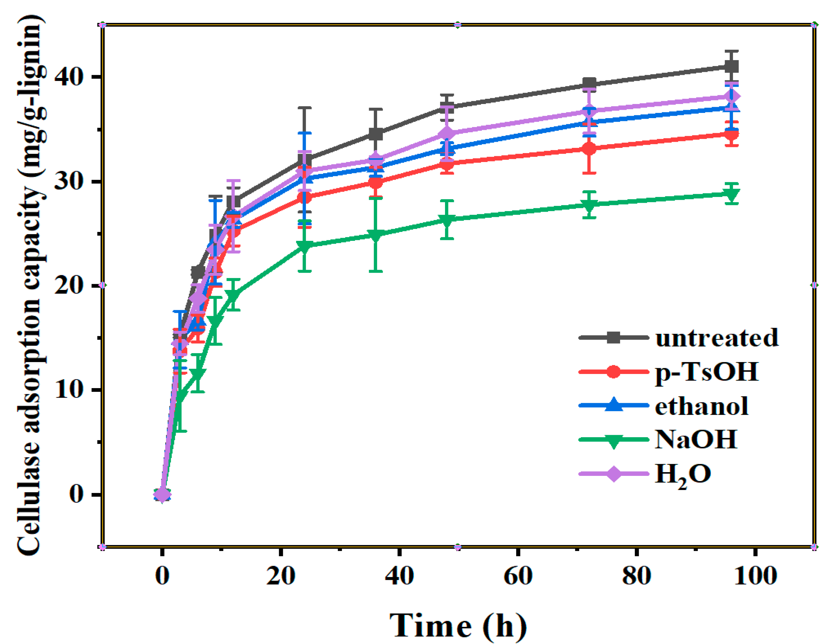
3.1. The Adsorption Curves of Lignin to Cellulase

3.1.1. Adsorption Curves of Cellulase on Lignin

Pretreatment is the primary procedure during sugar-platform conversion of lignocellulosic bioresource, aiming to remove lignin and hemicellulose to obtain the cellulose-rich substrate. However, the lignin remaining in the substrate, which has a stubborn structure, could have a strong inhibiting effect on enzymatic hydrolysis via the unproductive adsorption. The structure of residual lignin varies depending on the pretreatment method, and its adsorption capacity to cellulase is also different. Four pretreated residual lignin samples as well as untreated lignin were prepared using an enzymatic method to further investigate the adsorption capacity of lignin with different characteristics to cellulase, as shown in Figure 1. There were two stages during lignin adsorption, a period of rapid growth and a period of levelling off according to Figure 1. The adsorption time before 20 h was the rapid growth period, during which the adsorption amount of cellulase increased sharply and more than 60% of the adsorbed cellulases were bonded onto lignin at this stage. The time after 20 h was the levelling off period, during which the adsorption amount of cellulase increased slowly until adsorption/reabsorption equilibrium reached. The adsorption capacity of pretreated lignin to cellulase obtained from different pretreatments was ordered as: untreated lignin > hot water pretreated lignin > ethanol pretreated lignin > p-TsOH pretreated lignin > NaOH pretreated lignin, and the maximum adsorption amounts of cellulase obtained at 96 h were 41.1 mg/g-lignin, 38.2 mg/g-lignin, 37.1 mg/g-lignin, 34.6 mg/g-lignin and 28.8 mg/g-lignin, respectively. Notably, the NaOH pretreated lignin has the lowest adsorption capacity to cellulase.

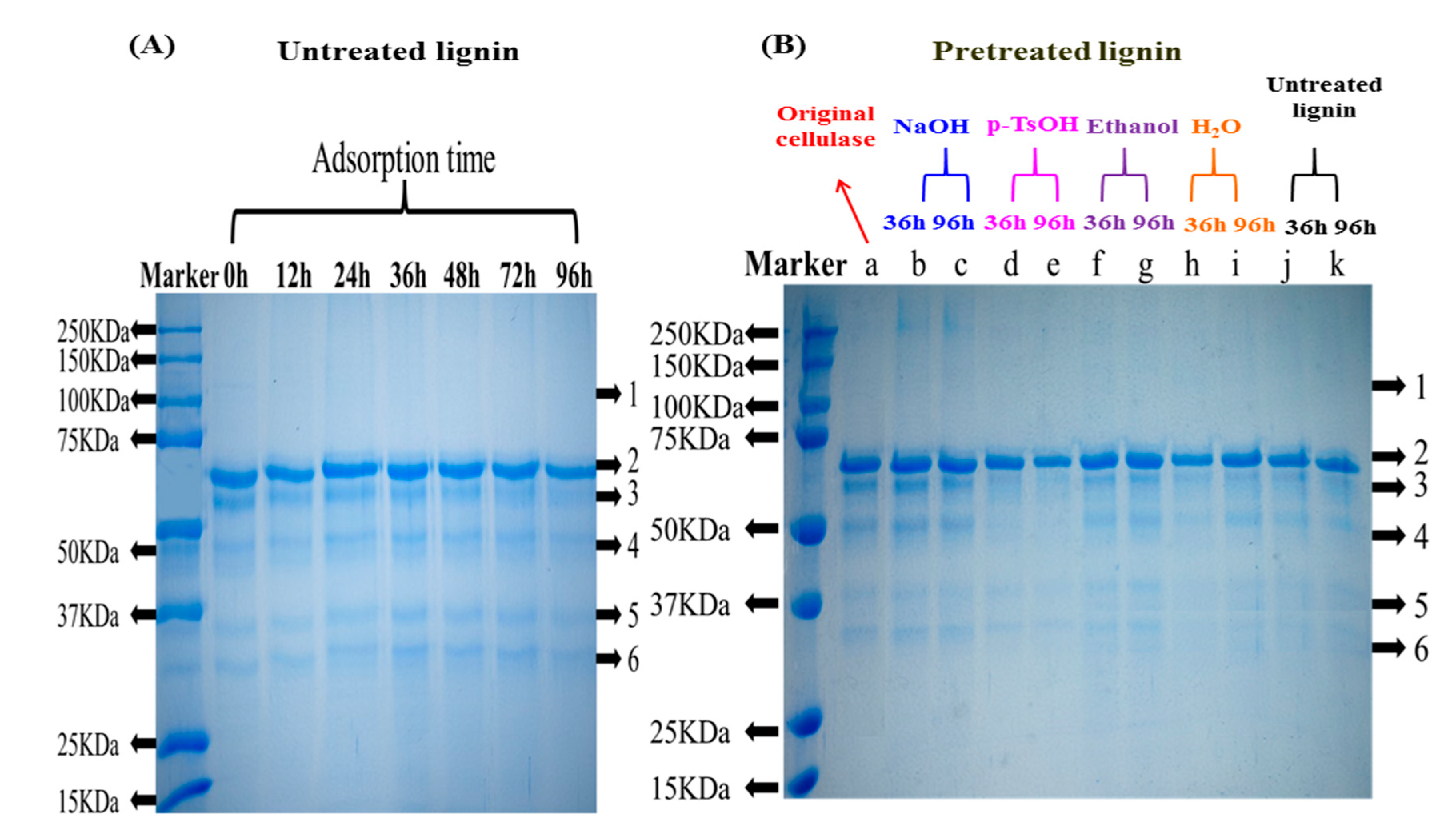
3.1.2. Composition Analysis of Free Enzymes

Cellulase is a hybrid enzyme consisting of three main components [22]: (1) endoglucanases (EG), which can randomly break β-1,4-glycosidic bonds within cellulose molecules, and which not only generate two chain ends, but also temporarily loosen the tight binding between these two chain segments and the cellulose molecule; (2) exoglucanase (CBH), which cleaves the reducing and non-reducing ends of the cellulose molecular chain to release soluble cellobiose, and which is divided into CBHI and CBHII, with CBHII degrading microcrystalline to produce reducing sugars about twice as much as CBHI; and (3) β-glucosidase (βG), which hydrolyses the resulting cellobiose to produce glucose. The three components convert cellulose to glucose by synergistic action. The composition of free cellulase after untreated lignin adsorption was analyzed by SDS-PAGE, as shown in Figure 2A. There were five relatively clear bands in the lane before adsorption (Figure 2A), indicating that the cellulase mainly contains five different fractions. The band 1 around 68 KDa is CBHI, band 2 around 60 KDa is CBHII, band 3 around 48 KDa is EGI, band 4 around 35 KDa is EGII and band 5 around 30 KDa is EGIII. From Figure 2A, after untreated lignin adsorption, the color of band 2 gradually becomes lighter as the time increases, and basically disappears after 48 h. Shown in Figure 2B is the composition of free cellulase after pretreated lignin adsorption. Similarly, the color of band 2 was lighter after all pretreated lignin samples adsorption. Although the structures of lignin samples are different, they adsorb the same components of cellulase, mainly adsorbing the CBHII component. Although the concentration of CBHII component in cellulsase is low (seeing from the light color of band 2), it has a great role in enzymatic hydrolysis due to its double ability to release reducing sugars than that of CBHI, which has a darker color in band 1.

3.2. The Characterization of Lignin

3.2.1. Composition Analysis of Lignin

The composition of pretreated lignin samples is shown in Table 1. The content of Klason lignin in five lignin samples was higher than 80% and the content of acid soluble lignin was lower than 1.1%. This avoids the influence of soluble lignin on the results and ensures that the structural changes of cellulase are due to the adsorption effect of lignin. The untreated lignin contained 5.3% glucan resulting from the lignin carbohydrate complex (LCC). The glucan content in the pretreated lignin samples was higher than that in the untreated lignin, which is an indication that pretreatments could remove the LCC. Interestingly, NaOH pretreated lignin contained the highest Klason lignin of 93.4%. However, the lowest adsorption capacity of NaOH pretreated residual lignin for cellulase was observed (Figure 1), indicating that the structural characteristics of lignin have a major effect on the cellulase adsorption.

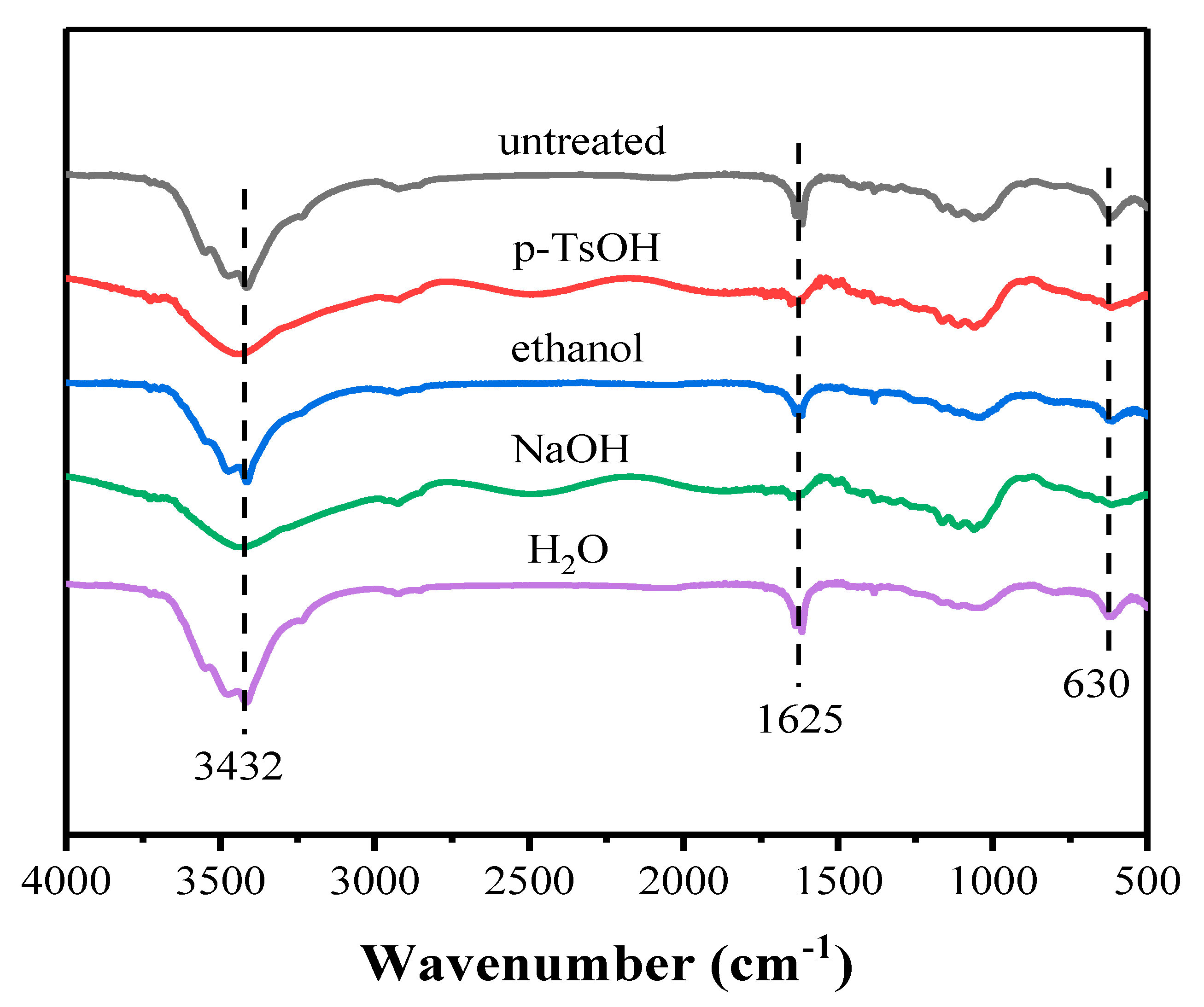
3.2.2. FT-IR Analysis of Lignin

The FT-IR spectra of lignin samples are shown in Figure 3. The broad adsorption peak at 3200–3700 cm−1 is attributed to the stretching vibration of -OH. The lignin has three -OH types, i.e., alcoholic -OH, phenolic -OH and carboxyl -OH, and they are generally conjoined to form a broad peak. In samples of untreated lignin, ethanol pretreated lignin and hot water pretreated lignin, three split peaks at 3420 cm−1, 3480 cm−1 and 3550 cm−1 were observed, indicating that they have all the three -OH forms. However, a broad peak at 3450 cm−1 was obtained in p-TsOH and NaOH pretreated lignin samples, indicating that the alcoholic -OH is dominant. Two peaks at 1620 cm−1 and 1650 cm−1, attributing to the aromatic skeletal vibration and -C=O stretching, were obtained in samples of untreated lignin, ethanol pretreated lignin and hot water pretreated lignin. In p-TsOH and NaOH pretreated lignin samples, only one peak was obtained at 1640 cm−1 assigning to the aromatic skeletal vibration. It can be seen that the p-TsOH and NaOH pretreated lignin samples had lower phenolic and carboxyl -OH contents than those in samples of untreated lignin, ethanol pretreated lignin and hot water pretreated lignin. The peak at 630 cm−1 was assigned to the bending vibration of the benzene ring. The pretreatments (except for hot water pretreatment) significantly decreased the intensity at 630 cm−1, indicating the lignin structure was destroyed.

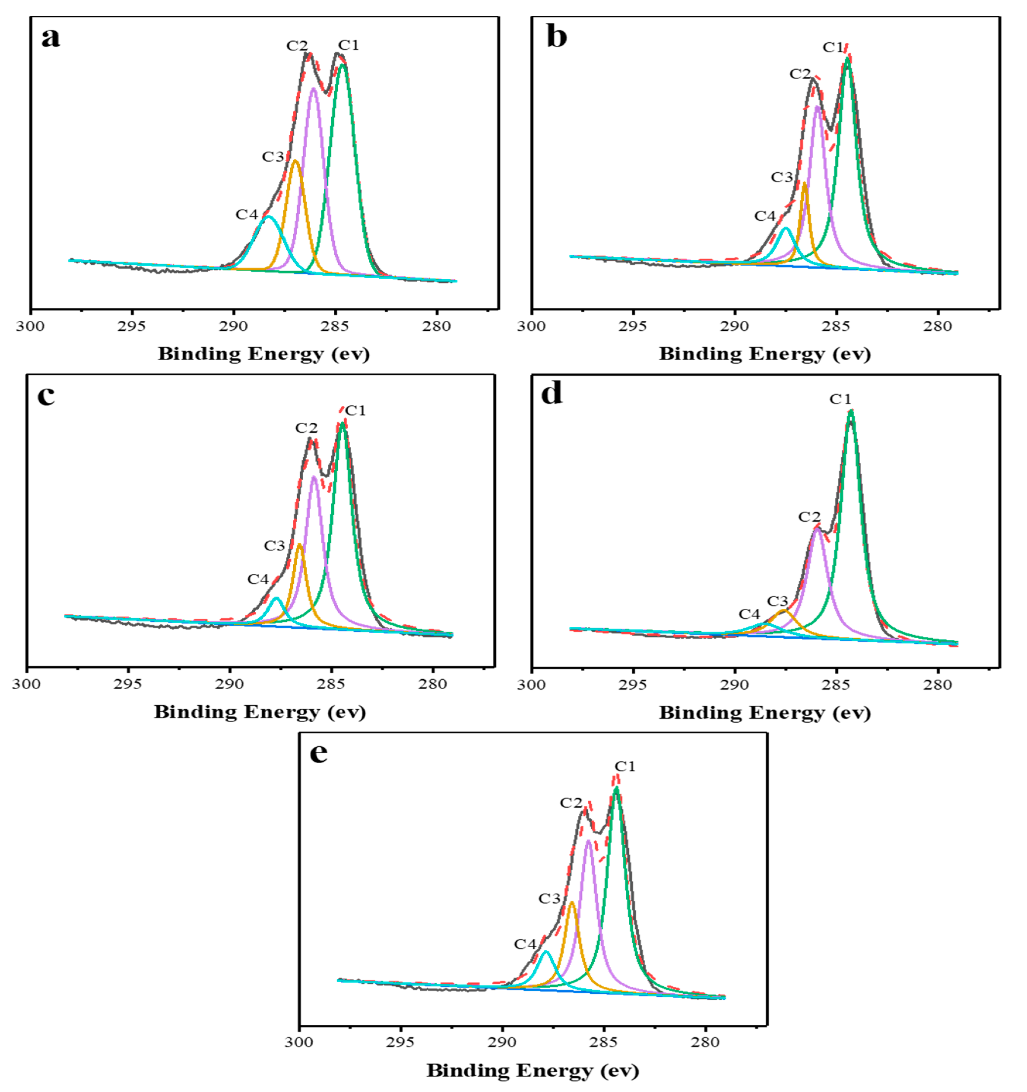
3.2.3. XPS Analysis of Lignin

XPS analysis was used to provide the surface chemical information of lignin. The carbon in lignin can be classified into four types, C1, C2, C3 and C4, as shown in Figure 4. C1 is a carbon attached to carbon or hydrogen (C-C or C-H); C2 is a carbon attached to an oxygen (-C-O-); C3 is a carbon attached to two oxygens or a carbonyl group (O-C-O or C=O), resulting from the carbonyl structure in lignin; and C4 is a carbon attached to both a carbonyl and a non-carbonyl oxygen (O-C=O), resulting from the carboxyl group in lignin [23]. Based on the results of the peak fitting, the relative content of C1–C4 for each lignin sample was calculated and is shown in Table 2. The pretreatments increased the C1 contents of all pretreated lignin samples and correspondingly decreased the C3 and C4 content. This indicated that the pretreatment decreased the contents of carbonyl and carboxyl groups in lignin. Interestingly, the order of total concentration of C3 and C4 was consistent with the order of cellulase adsorption, i.e., untreated lignin > hot water pretreated lignin > ethanol pretreated lignin > p-TsOH pretreated lignin > NaOH pretreated lignin. Therefore, the carbonyl and carboxyl groups of lignin play crucial role in cellulase adsorption.

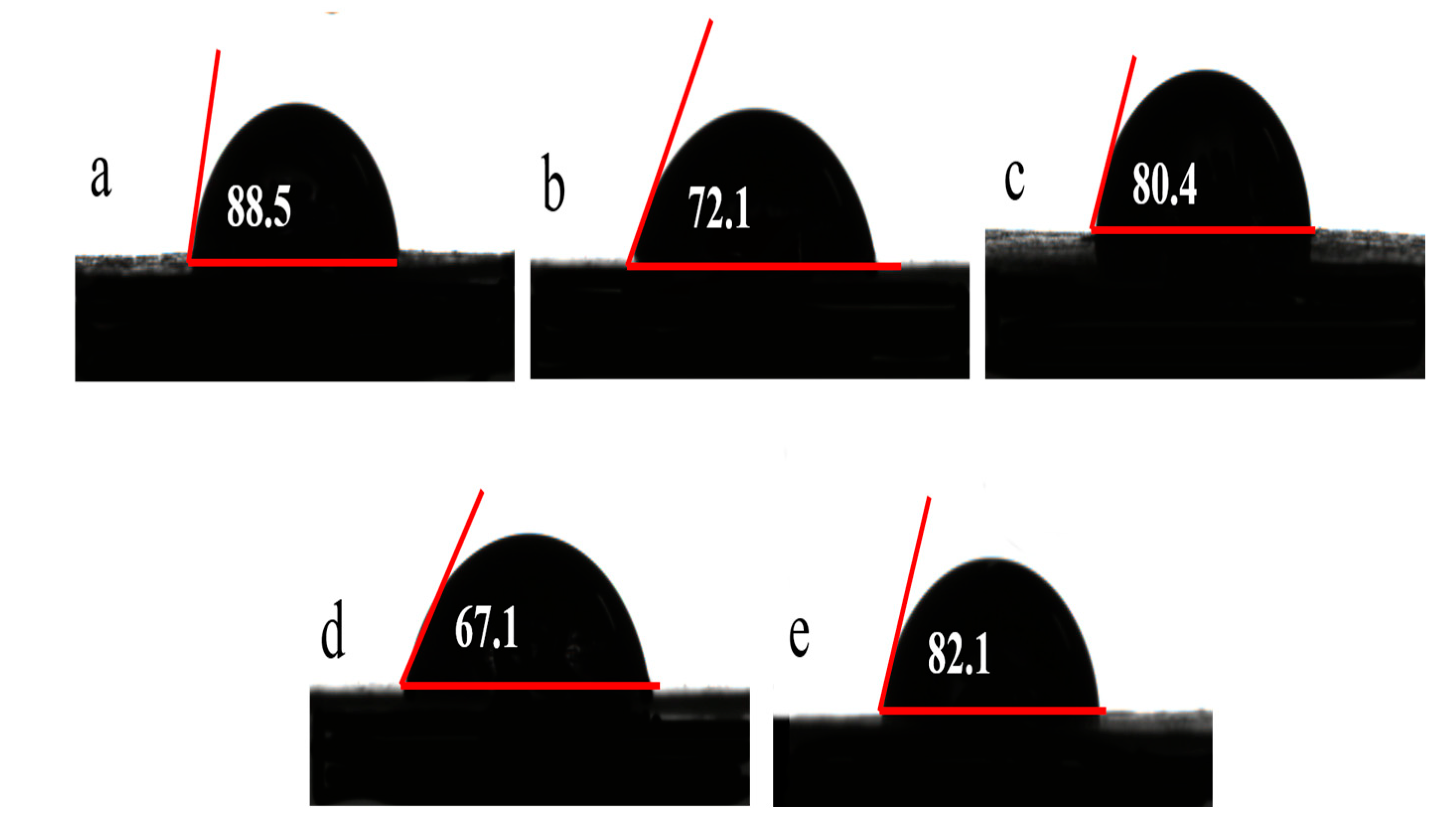
3.2.4. Contact Angle and Zeta Potential of Lignin

The hydrophilicity of lignin samples was investigated by measuring their contact angles, as shown in Figure 5. The untreated lignin had the highest contact angle of 88.5°, indicating it has the maximum hydrophobicity. The pretreatments reduced the contact angle of lignin, implying an increase in hydrophilicity. According to Figure 5, the order of hydrophilicity of the lignin samples is as follows: NaOH pretreated lignin (67.1°) > p-TsOH pretreated lignin (72.1°) > ethanol pretreated lignin (80.4°) > hot water pretreated lignin (82.1°) > untreated lignin (88.5°). The surface charge density of lignin was analyzed by detecting their zeta potentials. The untreated lignin had the highest negative charge density, probably due to its high carboxyl groups (see Table 2) which are more easily ionized in aqueous solutions with a negative charge. The pretreatment decreased the concentration of carboxyl group, thereby reducing the negative charge density of pretreated lignin. As shown in Table 3, the order of negative charge density of lignin samples is as follows: untreated lignin > hot water pretreated lignin > ethanol pretreated lignin > p-TsOH pretreated ligni > NaOH pretreated lignin.
The characterization of lignin, i.e., the hydrophobicity and charge, play an essential role in cellulase adsorption. Increasing the hydrophilicity of lignin reduces the binding ability of lignin to cellulase. In terms of molecular level, the hydrophobic groups, i.e., aryl, ether and carbonyl, are responsible for the hydrophobicity of lignin. The pretreatments destroyed the aromatic structure and removed the etheric and carbonyl groups of lignin, thereby decreasing the adsorption ability to cellulase. The CBHII has PI value of 5.2–5.9 and are positively charged under the hydrolysis pH (pH 4.8–5.0). For the negative charged lignin, it is easily to bind the positive charged CBHII. In terms of molecular level, the carboxyl is mainly responsible for the negative charge of lignin. Therefore, the decrease in negative charge density of lignin after pretreatments could reduce its adsorption capacity to CBHII of cellulase. In a word, a pretreatment method that can increase the hydrophilicity and reduce the negative charge density of lignin is a promising candidate for pretreatment of lignocellulosic materials, i.e., the NaOH pretreatment method showing promising results.

3.3. The Characterization of free Enzymes after Lignin Adsorption

3.3.1. Fluorescence Spectroscopy of Free Enzymes

The fluorescence spectroscopy (FS) was used to analyze the spatial structure of free enzymes, as shown in Figure 6. A broad peak at ~350 nm of original cellulase was obtained resulting from the aromatic amino acid residues in proteins including tryptophan (Trp), tyrosine (Tyr) and phenylalanine (Phe) [24,25]. The relative fluorescence intensity of Trp: Tyr: Phe is 100: 9: 0.5. Therefore, the fluorescence peak at ~350 nm is mainly from the Trp residues. As shown in Figure 6, a significant fluorescence quenching of free enzymes was obtained after lignin adsorption. The binding of lignin to cellulase altered the spatial structure of enzymes, which changed the microenvironment of fluorescent groups and thus reduced the fluorescence intensity of Trp-residues. Generally, the aromatic amino acid residues are located inside the enzyme and are surrounded by a variety of non-polar amino acid residues, so that the local environment they are in is less polar than the external aqueous solution. When the aromatic amino acid residues are gradually exposed to the aqueous solution, the polarity of the microenvironment gradually increases, causing a red shift in the maximum peak. The lignin samples, i.e., untreated lignin, hot water pretreated lignin and ethanol pretreated lignin, which had a strong adsorption capacity to cellulase, not only caused a great reduction in the fluorescence intensity of enzymes, but also caused a significant red shift in the maximum emission wavelength, indicating that the spatial structure of enzymes was changed more with the transfer of Trp-residues to the surface of enzymes. Interestingly, the lignin samples with low adsorption capacity, i.e., NaOH pretreated lignin and p-TsOH pretreated lignin, had a less pronounced effect on the spatial structure of enzymes due to the minor red shift in the maximum emission wavelength. It is probable that the adsorption of NaOH and p-TsOH pretreated lignins merely loosened the spatial structure, allowing polar aqueous solutions to enter the interior of enzymes, resulting in the fluorescence quenching, while the location of Trp-residues in enzymes did not change significantly. Clearly, the NaOH pretreated lignin had the minimal effect on the spatial structure of enzymes. It has been suggested that the spatial location of Trp at the entrance and exit of the catalytic tunnel of Trichoderma reesei family 6 cellulase (Cel6A) has a dramatic impact on its binding affinity with carbohydrates, as it is involved in recruiting individual substrate chains into the active site tunnel to initiate processive hydrolysis [26,27]. Thus, the change in the spatial position of Trp could influence the activity of enzymes.

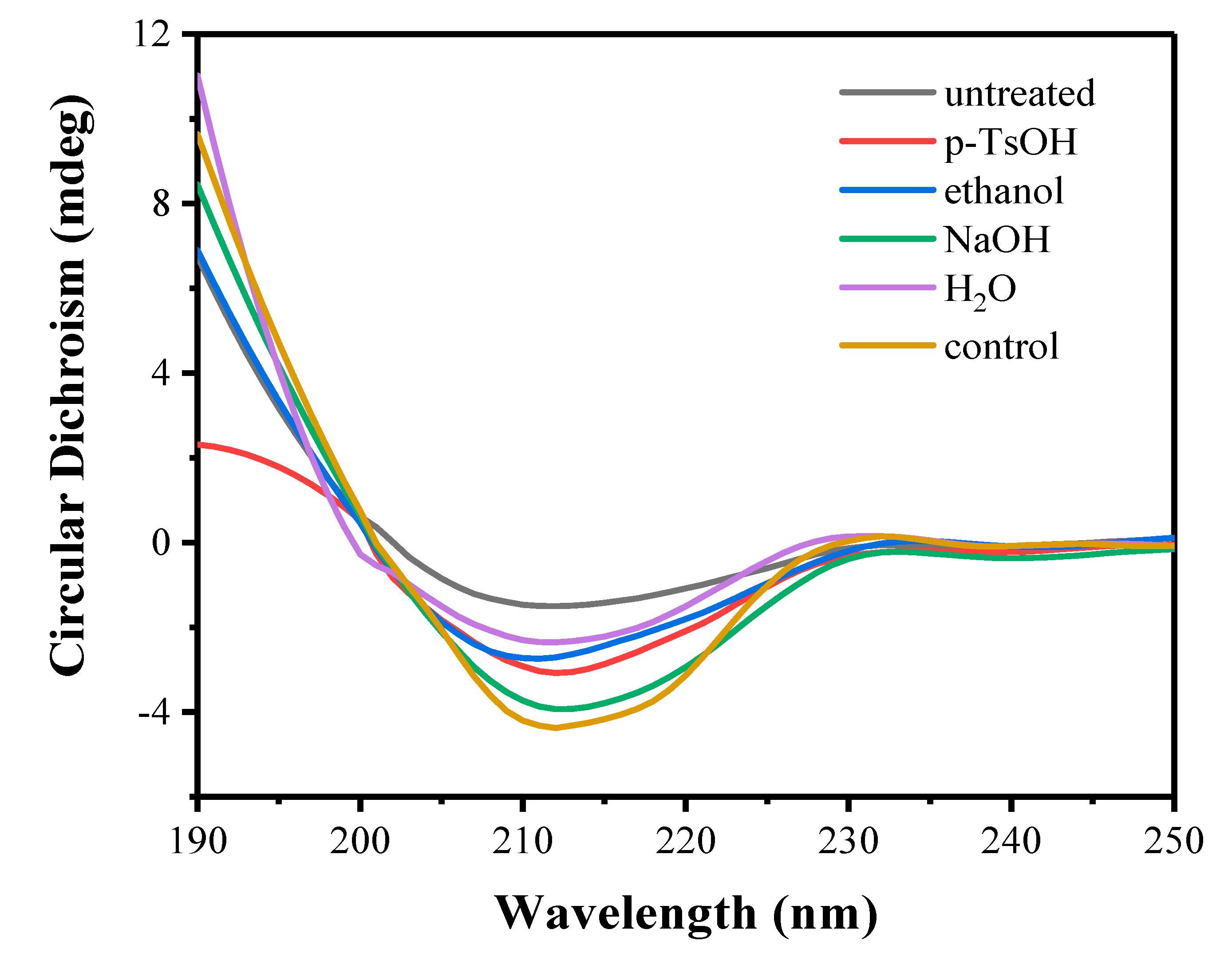
3.3.2. Circular Dichroism of Free Enzymes

Circular dichroism (CD) is a widely used technique to analyze the secondary structure of enzymes [25], and the CD spectra of free enzymes after lignin adsorption is shown in Figure 7. A broad peak at around 210 nm was observed, indicating the ‘α + β’ type (separate α-helix- and β-sheet-rich regions) of cellulase. After lignin adsorption, the intensity of peak at around 210 nm was decreased, implying a change in the secondary structure of free enzymes. The relative concentration of secondary structure of free enzymes after lignin adsorption is presented in Table 4. The concentrations of α-helix and β-turn were reduced and the concentrations of β-sheet and random coil were increased after lignin adsorption. The formation of α-helix structure requires strong hydrogen bonding, which is an important factor for the stability of protein. The decrease in concentration of α-helix and the increase in concentration of random coil indicated that the tight spatial structure of enzymes becomes relatively loose. It is well known that enzymes are active due to their relatively tight three-dimensional spatial structure, and that a loose spatial structure of enzymes could reduce their activities. Notably, the alteration in secondary structure of enzymes is dependent on the adsorption capacity of lignin, i.e., the higher the adsorption capacity of lignin the greater the disruption of the secondary structure of enzymes. The NaOH pretreated lignin caused the lowest degree of disruption to the secondary structure of free enzymes.

4. Conclusions

The residual lignin from different pretreatment methods had different adsorption capacities to cellulase. The adsorption capacity of lignin is strongly correlated with its hydrophilicity/hydrophobicity and surface charge density. The lower hydrophobicity and the surface negative charge density of lignin, the lower its adsorption capacity for cellulase. Pretreatments destroyed the aromatic structure and reduced the carbonyl and carboxyl contents on the surface of lignin, thus weakening the adsorption capacity of lignin for cellulase. Lignin has a strong adsorption capacity for the CBHII component of cellulase. Moreover, the lignin adsorption disrupted the spatial structure of cellulase, changing the spatial distribution of aromatic amino acid residues and the relative contents of secondary structures with reducing the α-helix content and increasing the random coil content. Notably, NaOH pretreated lignin had the highest hydrophilicity and the lowest surface negative charge density, and its adsorption capacity and distribution degree for cellulase was the lowest. Improving the catalytic activity and stability of cellulases, as well as addressing the challenge of enzyme recovery, are crucial to overcoming lignin inhibition.

Author Contributions

Conceptualization, H.Y.; Methodology, C.C. (Can Cui); Software, C.C. (Can Cui) and C.Y.; Validation, C.C. (Can Cui), C.Y. and A.W.; Formal analysis, C.C. (Can Cui), A.W. and C.C. (Cui Chen); Investigation, C.C. (Cui Chen) and D.C.; Data curation, D.C.; Writing—original draft, C.C. (Can Cui); Writing—review & editing, Y.L. (Yue Liu), Y.L. (Yuxiang Liu), G.N., X.J. and H.Y.; Visualization, Q.W.; Supervision, S.L., L.L., X.J. and H.Y.; Project administration, H.Y.; Funding acquisition, S.N., S.Y. and H.Y. All authors have read and agreed to the published version of the manuscript.

Funding

This work was financially supported by the National Natural Science Foundation of China (32171735) and the Open Project of Guangxi Key Laboratory of Clean Pulp and Papermaking and Pollution Control (2021KF04).

Institutional Review Board Statement

Not applicable.

Informed Consent Statement

Not applicable.

Data Availability Statement

Not applicable.

Conflicts of Interest

The authors declare no conflict of interest.

References

  1. Raj, T.; Chandrasekhar, K.; Naresh Kumar, A.; Kim, S.-H. Lignocellulosic biomass as renewable feedstock for biodegradable and recyclable plastics production: A sustainable approach. Renew. Sustain. Energy Rev. 2022, 158, 112130. [Google Scholar] [CrossRef]
  2. Awasthi, M.K.; Sindhu, R.; Sirohi, R.; Kumar, V.; Ahluwalia, V.; Binod, P.; Juneja, A.; Kumar, D.; Yan, B.; Sarsaiya, S.; et al. Agricultural waste biorefinery development towards circular bioeconomy. Renew. Sustain. Energy Rev. 2022, 158, 112122. [Google Scholar] [CrossRef]
  3. Yu, H.; Xu, Y.; Hou, J.; Ni, Y.; Liu, S.; Liu, Y.; Yu, S.; Nie, S.; Wu, Q.; Wu, C. Efficient Fractionation of Corn Stover for Biorefinery Using a Sustainable Pathway. ACS Sustain. Chem. Eng. 2020, 8, 3454–3464. [Google Scholar] [CrossRef]
  4. Su, Y.; Huang, C.; Lai, C.; Yong, Q. Green solvent pretreatment for enhanced production of sugars and antioxidative lignin from poplar. Bioresour. Technol. 2021, 321, 124471. [Google Scholar] [CrossRef] [PubMed]
  5. Rajak, R.C.; Saha, P.; Singhvi, M.; Kwak, D.; Kim, D.; Lee, H.; Deshmukh, A.R.; Bu, Y.; Kim, B.S. An eco-friendly biomass pretreatment strategy utilizing reusable enzyme mimicking nanoparticles for lignin depolymerization and biofuel production. Green Chem. 2021, 23, 5584–5599. [Google Scholar] [CrossRef]
  6. Saini, J.K.; Patel, A.K.; Adsul, M.; Singhania, R.R. Cellulase adsorption on lignin: A roadblock for economic hydrolysis of biomass. Renew. Energy 2016, 98, 29–42. [Google Scholar] [CrossRef]
  7. Rahikainen, J.L.; Martin-Sampedro, R.; Heikkinen, H.; Rovio, S.; Marjamaa, K.; Tamminen, T.; Rojas, O.J.; Kruus, K. Inhibitory effect of lignin during cellulose bioconversion: The effect of lignin chemistry on non-productive enzyme adsorption. Bioresour. Technol. 2013, 133, 270–278. [Google Scholar] [CrossRef]
  8. Chu, Q.; Wang, R.; Tong, W.; Jin, Y.; Hu, J.; Song, K. Improving Enzymatic Saccharification and Ethanol Production from Hardwood by Deacetylation and Steam Pretreatment: Insight into Mitigating Lignin Inhibition. ACS Sustain. Chem. Eng. 2020, 8, 17967–17978. [Google Scholar] [CrossRef]
  9. Yoo, C.G.; Meng, X.; Pu, Y.; Ragauskas, A.J. The critical role of lignin in lignocellulosic biomass conversion and recent pretreatment strategies: A comprehensive review. Bioresour. Technol. 2020, 301, 122784. [Google Scholar] [CrossRef]
  10. Wang, J.; Wang, J.; Lu, Z.; Zhang, J. Adsorption and desorption of cellulase on/from enzymatic residual lignin after alkali pretreatment. Ind. Crop Prod. 2020, 155, 112811. [Google Scholar] [CrossRef]
  11. Yu, H.; Hou, J.; Yu, S.; Liu, S.; Li, L.; Wu, Q.; Liu, Y.; Jiang, J.; Nie, S. Comprehensive understanding of the non-productive adsorption of cellulolytic enzymes onto lignins isolated from furfural residues. Cellulose 2019, 26, 3111–3125. [Google Scholar] [CrossRef]
  12. Nakagame, S.; Chandra, R.P.; Saddler, J.N. The effect of isolated lignins, obtained from a range of pretreated lignocellulosic substrates, on enzymatic hydrolysis. Biotechnol. Bioeng 2010, 105, 871–879. [Google Scholar] [CrossRef]
  13. Kellock, M.; Rahikainen, J.; Marjamaa, K.; Kruus, K. Lignin-derived inhibition of monocomponent cellulases and a xylanase in the hydrolysis of lignocellulosics. Bioresour. Technol. 2017, 232, 183–191. [Google Scholar] [CrossRef]
  14. Siqueira, G.; Arantes, V.; Saddler, J.N.; Ferraz, A.; Milagres, A.M.F. Limitation of cellulose accessibility and unproductive binding of cellulases by pretreated sugarcane bagasse lignin. Biotechnol. Biofuels 2017, 10, 176. [Google Scholar] [CrossRef] [PubMed]
  15. Kim, Y.; Ximenes, E.; Mosier, N.S.; Ladisch, M.R. Soluble inhibitors/deactivators of cellulase enzymes from lignocellulosic biomass. Enzym. Microb Technol. 2011, 48, 408–415. [Google Scholar] [CrossRef]
  16. Qin, C.; Clarke, K.; Li, K. Interactive forces between lignin and cellulase as determined by atomic force microscopy. Biotechnol. Biofuels 2014, 7, 65. [Google Scholar] [CrossRef] [Green Version]
  17. Sun, S.; Huang, Y.; Sun, R.; Tu, M. The strong association of condensed phenolic moieties in isolated lignins with their inhibition of enzymatic hydrolysis. Green Chem. 2016, 18, 4276–4286. [Google Scholar] [CrossRef]
  18. Rahikainen, J.L.; Evans, J.D.; Mikander, S.; Kalliola, A.; Puranen, T.; Tamminen, T.; Marjamaa, K.; Kruus, K. Cellulase-lignin interactions-the role of carbohydrate-binding module and pH in non-productive binding. Enzym. Microb Technol. 2013, 53, 315–321. [Google Scholar] [CrossRef] [PubMed]
  19. Pace, C.N.; Fu, H.; Lee Fryar, K.; Landua, J.; Trevino, S.R.; Schell, D.; Thurlkill, R.L.; Imura, S.; Scholtz, J.M.; Gajiwala, K.; et al. Contribution of hydrogen bonds to protein stability. Protein Sci. 2014, 23, 652–661. [Google Scholar] [CrossRef]
  20. Rahikainen, J.; Mikander, S.; Marjamaa, K.; Tamminen, T.; Lappas, A.; Viikari, L.; Kruus, K. Inhibition of enzymatic hydrolysis by residual lignins from softwood--study of enzyme binding and inactivation on lignin-rich surface. Biotechnol. Bioeng 2011, 108, 2823–2834. [Google Scholar] [CrossRef]
  21. Wang, J.; Hao, X.; Wen, P.; Zhang, T.; Zhang, J. Adsorption and desorption of cellulase on/from lignin pretreated by dilute acid with different severities. Ind. Crop Prod. 2020, 148, 112309. [Google Scholar] [CrossRef]
  22. Yoo, C.G.; Li, M.; Meng, X.; Pu, Y.; Ragauskas, A.J. Effects of organosolv and ammonia pretreatments on lignin properties and its inhibition for enzymatic hydrolysis. Green Chem. 2017, 19, 2006–2016. [Google Scholar] [CrossRef]
  23. Pang, C.; Xie, T.; Lin, L.; Zhuang, J.; Liu, Y.; Shi, J.; Yang, Q. Changes of the surface structure of corn stalk in the cooking process with active oxygen and MgO-based solid alkali as a pretreatment of its biomass conversion. Bioresour. Technol. 2012, 103, 432–439. [Google Scholar] [CrossRef] [PubMed]
  24. van de Weert, M. Fluorescence quenching to study protein-ligand binding: Common errors. J. Fluoresc. 2010, 20, 625–629. [Google Scholar] [CrossRef]
  25. Anand, U.; Jash, C.; Boddepalli, R.K.; Shrivastava, A.; Mukherjee, S. Exploring the mechanism of fluorescence quenching in proteins induced by tetracycline. J. Phys. Chem. B 2011, 115, 6312–6320. [Google Scholar] [CrossRef]
  26. Payne, C.M.; Bomble, Y.J.; Taylor, C.B.; McCabe, C.; Himmel, M.E.; Crowley, M.F.; Beckham, G.T. Multiple functions of aromatic-carbohydrate interactions in a processive cellulase examined with molecular simulation. J. Biol. Chem. 2011, 286, 41028–41035. [Google Scholar] [CrossRef] [Green Version]
  27. Nakamura, A.; Tsukada, T.; Auer, S.; Furuta, T.; Wada, M.; Koivula, A.; Igarashi, K.; Samejima, M. The tryptophan residue at the active site tunnel entrance of Trichoderma reesei cellobiohydrolase Cel7A is important for initiation of degradation of crystalline cellulose. J. Biol. Chem. 2013, 288, 13503–13510. [Google Scholar] [CrossRef] [Green Version]
Figure 1. Adsorption curves of cellulase on different lignin samples.
Sustainability 15 06057 g001
Figure 2. SDS-PAGE analysis of free enzymes after adsorption of untreated lignin (A) and pretreated lignin samples (B).
Sustainability 15 06057 g002
Figure 3. FT-IR spectra of lignin samples.
Sustainability 15 06057 g003
Figure 4. Deconvolution of high-resolution C1s spectra of lignin samples. (a) Untreated lignin; (b) p-TsOH pretreated lignin; (c) ethanol pretreated lignin; (d) NaOH pretreated lignin; (e) hot water pretreated lignin.
Sustainability 15 06057 g004
Figure 5. Contact angles of lignin samples. (a) Udntreated lignin; (b) p-TsOH pretreated lignin; (c) ethanol pretreated lignin; (d) NaOH pretreated lignin; (e) hot water pretreated lignin.
Sustainability 15 06057 g005
Figure 6. Fluorescence spectra of free enzymes after 96 h of lignin adsorption.
Sustainability 15 06057 g006
Figure 7. Circular dichroism of free enzymes after 96 h of lignin adsorption.
Sustainability 15 06057 g007
Table 1. Composition analysis of untreated lignin and pretreated lignin samples.
MethodsTemperature (°C)Concentration (%)Glucan (%)Xylan (%)Klason Lignin (%)Acid-Soluble Lignin (%)Ash (%)
Untreated--5.363.9180.721.102.86
p-TsOH100301.513.2486.670.414.02
Ethanol180503.993.6684.641.033.00
NaOH8020.362.8293.400.870.97
H2O180-4.433.7181.390.801.96
Table 2. The concentration of functionality by percentage carbon, determined by curve fitting the C1s peak from the X-ray photoelectron spectra.
Lignin SamplesC1 (%)C2 (%)C3 (%)C4 (%)
Untreated39.5630.9217.3312.19
p-TsOH48.3133.419.338.95
Ethanol48.2532.2513.685.82
NaOH57.3328.598.725.35
Hot water45.0730.4416.088.42
Table 3. Zeta potentials of lignin samples at pH 4.8.
Lignin SamplesZeta (mV)
Untreated−14.50 ± 1.42
p-TsOH−9.21 ± 3.13
Ethanol−10.20 ± 1.77
NaOH−6.71 ± 2.50
H2O−13.50 ± 1.87
Table 4. The content of secondary structure of free enzymes after 96 h of lignin adsorption.
Lignin SamplesEnzymeα-Helix (%)β-Sheet (%)β-Turns (%)Unordered (%)
untreatedCell15.8035.4016.8033.10
p-TsOHCell20.3033.3017.8030.50
ethanolCell19.5032.4016.3033.70
NaOHCell24.5027.8017.7029.60
H2OCell21.1028.0017.6033.40
controlCell26.0027.3018.4029.00
Note: Cell is original cellulase.
Disclaimer/Publisher’s Note: The statements, opinions and data contained in all publications are solely those of the individual author(s) and contributor(s) and not of MDPI and/or the editor(s). MDPI and/or the editor(s) disclaim responsibility for any injury to people or property resulting from any ideas, methods, instructions or products referred to in the content.

