Article

The Biosynthesis of Nickel Oxide Nanoparticles: An Eco-Friendly Approach for Azo Dye Decolorization and Industrial Wastewater Treatment

1
Department of Environmental Sciences, Government College University, Faisalabad 38000, Pakistan
2
Department of Bioinformatics and Biotechnology, Government College University, Faisalabad 38000, Pakistan
3
Biology Department, Faculty of Sciences and Arts, King Khalid University, Abha 64353, Saudi Arabia
4
Department of Biology, College of Science, King Khalid University, Abha 61413, Saudi Arabia
*
Author to whom correspondence should be addressed.
Sustainability 2023, 15(20), 14965; https://doi.org/10.3390/su152014965
Submission received: 12 September 2023 / Revised: 5 October 2023 / Accepted: 11 October 2023 / Published: 17 October 2023

Abstract

:
Wastewater is one of the major concerns for agriculture, and the composition of wastewater depends on its origin. Generally, industrial wastewater consists of azo dyes and heavy metals that contaminate the food chain. In this study, nickel oxide nanoparticles (NiO-NPs) were biosynthesized from Shewnella spp. and characterized by UV–visible spectroscopy (UV–vis), Scanning Electron Microscopy (SEM), X-ray Diffraction (XRD) and Fourier Transform Infrared Spectroscopy (FTIR). Azo dye decolorization indicated that NiO-NPs decolorize methylene blue (MB) (82.36%), Congo red (CR) (93.57%), malachite green (MCG) (91.05%), reactive black 5(RB5) (55.17%), reactive red-II(RR-II) (55.45%), and direct blue-I(DB-I) (59.94%) at a dye concentration of 25 mg L−1 after 4 h of sunlight exposure. Additionally, the rate of decolorization was also examined for a 50 mg L−1 concentration of dye. In order to investigate the photocatalytic potential of NiO-NPs, different dyes were also subjected to static and shaking conditions for dye decolorization. The treatment of industrial wastewater with NiO-NPs showed a significant reduction in pH from 8.5 to 6.1, EC (48.38%), chemical oxygen demand (49.24%), total dissolved solids (67.05%), sulfates (52.5%), and phosphates (49.49%). The results of this study indicated that biosynthesized NiO-NPs are an attractive choice for azo dye degradation and industrial wastewater treatment, and they can help save the depleted natural resources of water for agricultural purposes.

1. Introduction

In spite of rapid development in water restoration technologies, water pollution continues to be one of the dominating global problems [1]. Due to rapid urbanization and industrialization, securing the availability of clean water has become a challenge. The tendency to add pollutants into water bodies has become common practice, but removing these pollutants from them is a difficult task. The situation becomes more difficult when these pollutants become part of biological systems. Mostly in developing countries like Pakistan, industrial wastewater is often used for agricultural purposes. Unfortunately, this wastewater often contains azo dyes, which may have detrimental effects on the nutrient and biological properties of soil, consequently affecting crop production. Synthetic dyes are broadly used in paper, leather, soil, medicine, and food industries as coloring agents [2]. The challenging molecular structure of non-biodegradable dyes, like azo dyes, adversely affects the aquatic environment when these color products and their byproducts are directly discharged into water bodies [3].
Underdeveloped countries like Pakistan face a shortage of water resources; these are insufficient to meet the needs of human beings, agriculture, and industries. As a result of this scarcity of clean water, underdeveloped countries are compelled to utilize contaminated and polluted water for the irrigation of agricultural crops [4]. To overcome this problem, different physicochemical methods, including adsorption and ozonation, are used for treating textile wastewater [5]. These methods help to remove or minimize contaminants and pollutants from wastewater, making it safe for the environment and aquatic ecosystems [6]. A study by Danouche et al. (2022) [7] addressed a batch experiment in which the biomass of a Wickerhamomyces anomalus yeast stain was assessed as a natural biosorbent for the removal of an Acid Red 14 dye (AR14). The results showed that 71.37 mg g−1 was the most significant biosorption capability. In addition to adsorption, many other methods are also used for the treatment of industrial wastewater, like nanotechnology. However, these methods have their own advantages and disadvantages for wastewater treatment [4,8,9].
Nanotechnology includes atomic, molecular, and macromolecular level research, study, and technology development. The NP range is almost 1–100 nanometers, to give a fundamental understanding of the nano-scale of phenomena and materials [10]. Nanotechnology is an integrative field at the junction of applied disciplines and basic sciences such as bioengineering, biophysics, and molecular biology [11,12]. Nanoparticles have obtained fame in technological advancements because of their tunable physicochemical characteristics like wettability, thermal or electrical conductivity, melting points, scattering, and light absorption. These may result in enhanced performance over the majority of their counterparts [13]. Some important applications for nanoparticles include functional nanostructures, environment and biological systems, and consolidated materials [14]. Because nanotechnology is new and is enlarging technology, its important applications may include the development of innovative methods for the formation of new products to form new chemicals and materials or to replace the present generation of equipment with new and improved performance equipment. This would result in less consumption of energy and materials, lower the harm to the environment, and also offer environmental remediation [15]. Previous studies showed that researchers used different absorbents for the adsorption of azo dyes, i.e., molecularly imprinted polymers (MIP), monometallic NPs and bimetallic NPs, fly ash, kaolinite, activated clay, hydrogels, metal oxide nanoparticles, activated carbon, modified alumina, and mesoporous zeolite [16,17,18,19].
Various techniques including physical, biological, chemical, or hybrid methods are currently being used for the synthesis of nanoparticles [20,21]. Mostly physiochemical methods are the ones used for the synthesis of nanoparticles, but due to the adverse environmental impacts of the chemical and physical synthesis of nanoparticles, biosynthesized nanoparticles are considered environmentally friendly methods [22]. Nowadays, the biosynthesis of nanoparticles has received considerable attention from the scientific community due to its eco-friendly approach, allowing for the production of nanoparticles with diverse sizes and shapes while minimizing or eliminating the use of damaging solvents [23,24]. Utilizing biological techniques for nanoparticle fabrication is an economical choice because they require less energy [25]. For example, bacteria with the unique property of metal reduction are taken as a potential bioresource for the synthesis of nanoparticles [26,27]. The resistance of bacteria to extreme environmental conditions makes them satisfactory candidates for the synthesis of nanoparticles [28,29]. Many studies have reported on the biosynthesis of NiO-NPs through a microwave-assisted route, which is more eco-friendly and cost-effective as compared to physical and chemical methods [30].
The green synthesis of nanoparticles is consistent with the United Nations’ Sustainable Development Goal (SDG) 6: Clean Water and Sanitation, which emphasized the cleaning of wastewater. Green nanoparticle synthesis for azo dye decolorization and industrial wastewater treatment is a sustainable approach, which ensures the availability and sustainable management of water.
In the field of environmental remediation, metal and metal oxide materials have gained significant recognition as advanced adsorbents. Scientists have been actively exploring readily available metal and metal oxide nano-adsorbents for the efficient removal of dyes, which has become a topic of great interest in recent times. Certain metals like silver (Ag) and metal oxides such as iron (Fe), cobalt (Co), and nickel (Ni) oxides can be synthesized using simple processes. These nanomaterials hold promise for their potential application in dye removal and offer a cost-effective and accessible solution for environmental remediation including textile dye degradation; however, NiO-NPs are reported to have promising photocatalytic potential [31,32]. Currently, several reports have addressed the synthesis of NiO nanoparticles with different bacteria. For example, from the nickel electroplating industry, a bacterium—MRS-1—was isolated, which was nickel resistant and was capable of synthesizing NiO nanoparticles from NiSO4 [33]. Another study reported the biosynthesis of Li, Ru, Ni, Fe, Rh, Pd, Ag, Pt, and Co nanoparticles at room temperature by using Pseudomonas aeruginosa SM1 [34]. However, many research articles have been published on using ZnO nanoparticles as adsorbents for the removal of azo dyes [35].
Treated wastewater should meet some important requirements in order to be deemed suitable for agriculture use, since plants require optimum pH, EC, COD, and nutrients for better growth. pH and EC directly affect the nutrient uptake of plants. A higher EC reduces nutrient uptake. Similarly, a high or low pH also affects nutrient cycling. Dyes reduce the photosynthetic spectra of plants and ultimately reduce plant growth and biomass. Treated wastewater should also have a low COD level, because a high COD can indicate the presence of organic pollutants, which may harm the plants or soil. To prevent overfertilization and eutrophication in receiving bodies of water, low phosphate levels are suggested.
This study focused on an eco-friendly and proficient techniques for the bacterial synthesis of NiO-NPs by Shewanella sp. SM33 with the characterization of NiO-NPs through different characterization techniques like ultraviolet–visible spectroscopy (UV–vis), scanning electron microscopy (SEM), Fourier-transform infrared spectroscopy (FTIR), and X-ray diffraction (XRD). The ultimate objective of this study was to check the photocatalytic potential of NiO-NPs for the decolorization of azo dyes and the treatment of industrial wastewater. Perhaps there is not even a single report that has addressed the biosynthesis of nanoparticles from the bacterial strain that belongs to the Shewanella species. Hence, the Shewanella species may be a new potential bioresource for biogenic nanoparticles.

2. Materials and Methods

2.1. Sample Collection and the Isolation of Bacterial Strain

The strain SM33 was isolated from wastewater, which was taken in sterilized bottles from the Paharrang drain Faisalabad, Pakistan (31°40′0″ N, 73°17′30″ E), and transferred to the Environmental microbiology and biotechnology laboratory, department of Environmental Sciences in the Government College University Faisalabad for further processing. To synthesis the nanoparticles, isolated bacterial strain samples were serially diluted (10−1–10−6). The last dilutions (10−3–10−6) were spread on nutrient agar plates amended with 1 mM NiCl and kept in an incubator at 30 °C for 48 h. The bacterial colonies capable of growing in the presence of nickel were purified by repeated streaking on fresh media plates and selected for further investigations, including the determination of minimum inhibitory concentration (MIC) for NiCl. Due to a maximum MIC value for NiCl (5 mM), the isolate SM33 was selected and stored at −80 °C in glycerol (20% v/v) stocks.
All the chemicals used in this experiment were purchased from Faisalabad Scientific store, and these were applied for experimental analysis without any further purifying techniques. For the isolation of bacterial strains, a nutrient agar (NA) medium was used. Congo-red, methylene blue, direct blue, reactive red II (RRII), and reactive black 5 (RB5) dyes that were used for decolorization were purchased from Sigma Aldrich.

2.2. The Biosynthesis of NiO-NPs by the Shewanella Species

For the synthesis of NiO nanoparticles, nickel chloride (NiCl · 7H2O) (Sigma-Aldrich CAS number: 13446-34-9) was used as a precursor salt. The highly Ni-resistant bacterial strain Shewanella species (SM33) was selected by optimization for the microbial synthesis of nanoparticles. Nutrient broth (NB) media was prepared in conical flasks, and the bacterial culture of screened bacteria was cultured into this NB media and kept on an incubating shaker for 24 h at 150 rpm (30 °C). After 24 h, NiCl · 7H2O salt solutions of 10 mM concentrations (selected by optimizations) were separately added into the culture medium to investigate the synthesis of nanoparticles and again incubated under same conditions previously mentioned. The color of the mixture turned dense green from pale green, and the color was strengthened with time. After 24 h, the reaction mixture was centrifuged for 10 min (4 °C) at 7000 rpm to obtain the nanoparticles. The supernatant was wasted, and the palate was washed with double-distilled water again and again to obtain purified NiO nanoparticles. The resulting palate was collected and kept in the oven at 85 °C for 12 h; the dried product was collected and ground into fine powder using a mortar and pestle.

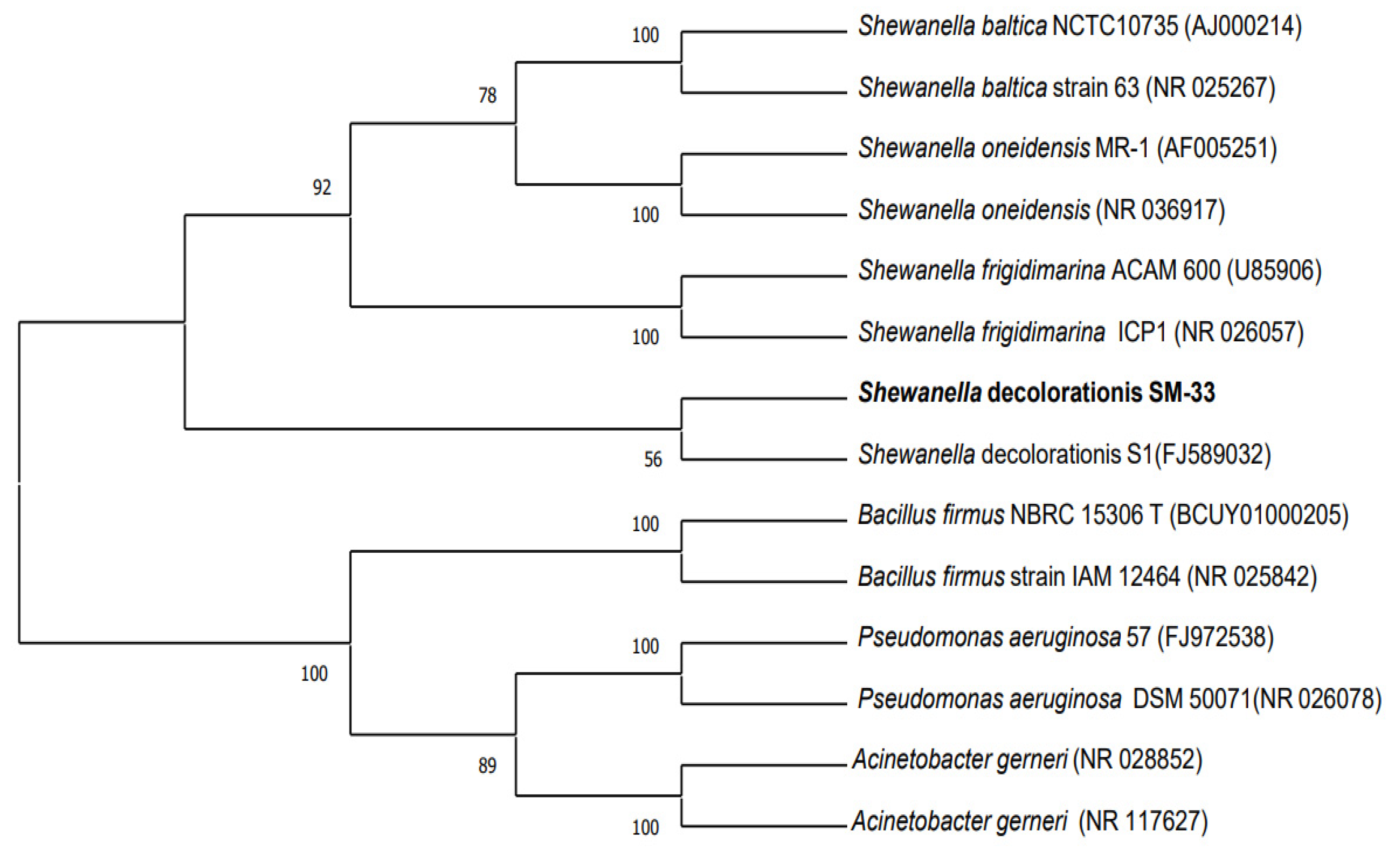
2.3. Molecular Characterization and Phylogenetic Analysis

Bacterial cultures that had specific potential for NiO-NP synthesis were identified on a molecular basis. Amplification of the 16S rRNA gene was carried out with the help of the rD1 (5′-AAGGAGGTGATCCAGCC-3′) and fD1 (5′-AGAGTTTGATCCTGGCTCAG-3′) primer pair [36]. The recipe for the 1X PCR reaction mixture and the PCR protocol for 16S rRNA gene amplification are described below in Table 1 and Table 2.

2.4. The Characterization of NiO-NPs

A UV–visible spectrophotometer was used for the confirmation of the production of biologically synthesized NiO nanoparticles (Shimadzu, Kyoto, Japan). UV–visible absorption spectra having a wavelength range of 250 to 600 nm were measured for confirmation of the presence of nickel nanoparticles. To investigate surface morphology and the shape of NiO-NPs, a scanning electron microscopy technique was used (TM-1000, Hitachi, Japan). An SEM technique was also utilized to analyze the surface or near-surface structure of the specimens. It was performed according to methodology described by Tiwari et al., 2016 [37]. For performing an analysis of SEM (SEM LEO 1530, Germany), 10 µL of the reaction mixture was put on a glass coverslip and dried on a hot plate at 100 °C. Then, the cover slip was cooled down and fixed on aluminum stub via double-sided adhesive carbon tape. The characteristic functional groups and associated proteins present on the surfaces of the biologically synthesized nanoparticles were analyzed with FTIR spectroscopy. This was performed using a PerkinElmer Spectrum-100 FTIR spectrometer (FTIR-Bruker TENSOR-27). Developing an infrared absorption spectrum that acts as a molecular “fingerprint” is an effective method to detect the different types of chemical bonds present in a molecule. FTIR investigation was carried out on the fine dried powder of NiO-NPs, and spectra was noted [38] (Griffiths and De Haseth, 2007). FTIR was performed in the spectral range of 2000 to 500 cm−1. A 1 g sample of dried powder was placed on the small crystal area in front of a high refractive index prism. An IR (infrared radiations) of 10,000 to 100 cm−1 was passed toward the sample, which absorbed them and converted them into vibrational energy. The resultant molecular fingerprints were presented as a signal on a detector range from 2000 to 500 cm−1. XRD is a commonly used analytical technique to detect the crystalline nature of biologically synthesized NiO-NPs. XRD (PANalytical X’PERT PRO, manufactured by USA and sourced in Pakistan) was carried out according to the methodology addressed by Varshney et al., 2010 [39]. The crystalline nature of NiO-NPs and the development of crystalline sizes were revealed by a strong and high intensity peak in the XRD analysis. Structural crystallinity was manually analyzed, while average particle size was calculated using Debye-Scherrer’s formula in the following way:
D = Kλ/β·cosθ
where D = crystalline size (nm); λ = the wavelength of the X-ray source; K = Sharrer’s constant (0.9); θ = peaks’ position (radians); β = full width at the half maximum of the reflection peaks (radians)

2.5. The Photocatalytic Degradation of Azo Dyes

The potential of synthesized NiO-NPs was investigated against a 50 mg L−1 concentration of Congo red dye using different concentrations of nanoparticles (i.e., 0.05, 0.1, 0.2, 0.5, 1, 3, 5, mg/mL) for optimization. We obtained the best results at 1 mg/mL and 5 mg/mL concentrations, but we took a 1 mg/mL concentration for further processing. Biologically synthesized NiO-NPs were investigated to check their photocatalytic potential at various concentrations (i.e., 25, 50 mg L−1) of different commonly used dyes, i.e., methylene blue, malachite green, Congo red, reactive black-5, reactive red II, and direct blue at 561, 624, 478, 597, 540, and 592 wavelengths, respectively. For this purpose, 50 mg of NPs (an optimized concentration, i.e., 1 mg/mL) was added into a 50 mL solution of the azo dyes. Then, the reaction mixture was placed under sunlight for 4 h. After every hour, a little quantity of the reaction mixture was taken out and centrifuged to settle suspended particles. After this, the absorbance of the cleared solution was measured with the use of a UV–visible spectrophotometer (STA- 8200V STALWART), according to Roy et al., 2017 [40].
The same procedure was also repeated to check the potential of NPs at shaking and static conditions by putting the reaction mixture under sunlight for 7 h.

2.6. The Application of NiO-NPs for Industrial Wastewater Treatment

To assess their potential, biologically synthesized nanoparticles were tested for their ability to degrade dyes and to treat some other parameters in actual industrial wastewater. For this purpose, wastewater was collected from the Paharang drain, Sargodha Road, Faisalabad. The wastewater was collected at the end of processing, resulting in water that was nearly colorless. Firstly, the wastewater sample was centrifuged to remove particulate matter, and then it was distributed into two portions, which were spiked with Congo red dye. The wastewater sample was collected from the end of the drain, resulting in a minimal initial dye concentration. Therefore, a supplementary quantity of 50 mg L−1 Congo red dye was introduced into the sample to enable the assessment of the impact of nanoparticles on dye removal alongside the evaluation of other wastewater parameters. Therefore, we added nanoparticles in one portion, while the other portion remained nanoparticle-free. To assess the potential of NiO-NPs, 50 mg of the nanoparticles were added to 50 mL of wastewater. Three replicates of the sample were vortexed and incubated under sunlight with their respective controls. After a 7 h incubation period, the samples were centrifuged at 9000 rpm for 10 min for the removal of suspended nanoparticles, and then both the samples with and without nanoparticles were analyzed for pH, electrical conductivity, chemical oxygen demand (COD), color intensity, phosphates, and sulphates by following the procedures [41] (APHA, 2005).

2.6.1. pH Analysis Method

The pH meter was standardized via buffer solution to check the pH of the wastewater. With the help of distilled water, the pH electrode was rinsed after buffering, and the electrode was dried with filter paper. The dried electrode was dipped into the beaker containing a homogenous wastewater sample to check its pH, and the reading was noted. To obtain an average reading, this step was repeated two to three times.

2.6.2. Electrical Conductivity Analysis Method

The conductivity cell was calibrated with a standard solution of KCl to check the electrical conductivity of the wastewater. The conductivity cell was washed with distilled water to avoid error in final readings, and before every measurement it was cleaned with filter paper. The conductivity cell was dipped into the beaker containing the homogeneous wastewater sample and the exact reading was obtained after waiting a while. After one or two minutes, the value of the sample was constant, which was noted. The step was repeated again and again to obtain the average value of the sample’s electrical conductivity.

2.6.3. Chemical Oxygen Demand (COD) Analysis Method

To perform COD analysis, a sample of 25 mL of wastewater was taken into a 250 mL beaker and one gram of HgSO4 was also added into it. Then, the solution was shifted to a refluxing flask of 250 mL, and 33 mL of concentrated H2SO4 was also added into it. Furthermore, for 2 h, the flask was attached to a condenser by adding a 10 mL potassium dichromate solution. After 2 h, the mixture was cooled at room temperature by removing the flask from the condenser. Excess potassium dichromate was titrated against FAS (ferroin indicator). Lastly, the color of the sample changed to reddish brown from blue green.

2.6.4. Phosphorus

Phosphorus was checked using colorimetric methods. Samples of 25 mL were placed into Erlenmeyer flasks. The pH of the samples was set at 5 with the help of 5N H2SO4, and 8 mL reagent B (1.056 g L-Ascorbic acid dissolved in 200 mL reagent A) was added into the sample; then, the sample was diluted to 50 mL with distilled water. A standard solution was set from 1 to 5 ppm by using KH2PO4, and two blank samples were synthesized by using distilled water. From each sample, 2 mL were placed one by one in a cuvette, and the absorbance was checked with a spectrophotometer at 882 nm. Textile wastewater samples and post treated wastewater proceeded as samples. A standard curve was drawn by using the absorbance of the standard solution; the absorbance was given on a Y-axis, and concentration was given on an X-axis. The initial concentration of phosphorus was estimated by using the standard curve. The formula to measure the phosphate of industrial wastewater is given below:
P(ppm) = ppm P (from calibration curve) × V1/V
where V = volume of wastewater sample; V1 = the final volume of the flask used for measurement.

2.6.5. Sulfates

The sulfate was estimated using gravimetric methods. Samples of 25 mL were placed in 150 mL Erlenmeyer flasks. A 1:1 HCl solution was prepared, and 1 mL was placed into each sample. A 5 mL BaCl2 solution was added, and it was shaken well for 1–2 min. Standard solutions of 10 to 200 ppm were prepared by using K2SO4, and two blank samples were also prepared by using distilled water. The standard solution was placed in a quartz cuvette, and light absorbance was measured at 420 nm using a spectrophotometer. The standard curve was drawn by using the absorbance of the standard solution, the absorbance given on a Y-axis, and the concentration on an X-axis. The starting concentration of the sulfate was estimated via standard curve. The sulfate of industrial wastewater was measured using the following formula:
Sulfate as SO4−2 (ppm) = (mg SO4−2 × 1000)/mL of sample

3. Results

3.1. Molecular Characterization and Phylogenetic Analysis

The SM33 strain was taken from wastewater and grown on nutrient agar media plates. BLASTn and phylogenetic analysis of the 16s rRNA gene of the SM-33 isolate revealed that the selected isolate (SM-33) belongs to the genus Shewanella. In the phylogenetic tree, the selected strain clustered with the Shewanella decolorationis species (Figure 1). The 16s rRNA gene sequence of the selected strain was investigated in a database using the BLASTn program of NCBI (National Center for Biotechnology Information), and the sequence showed 99.70% similarity with the Shewanella decolorationis strain (Gene bank accession no. FJ589032.1). Different sequences were considered for the phylogenetic analysis, and we constructed a phylogenetic tree using software named mega X version 10.2.4.

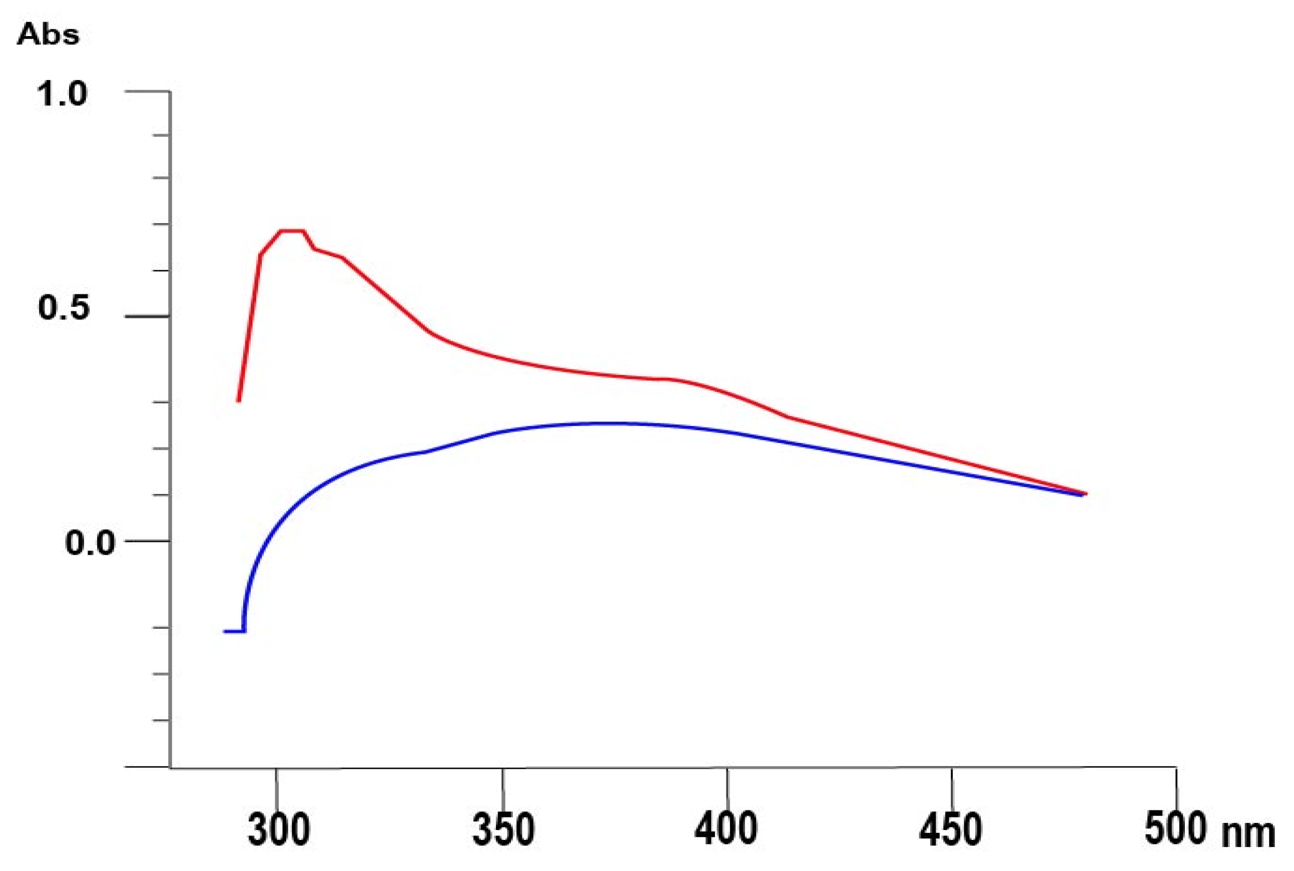
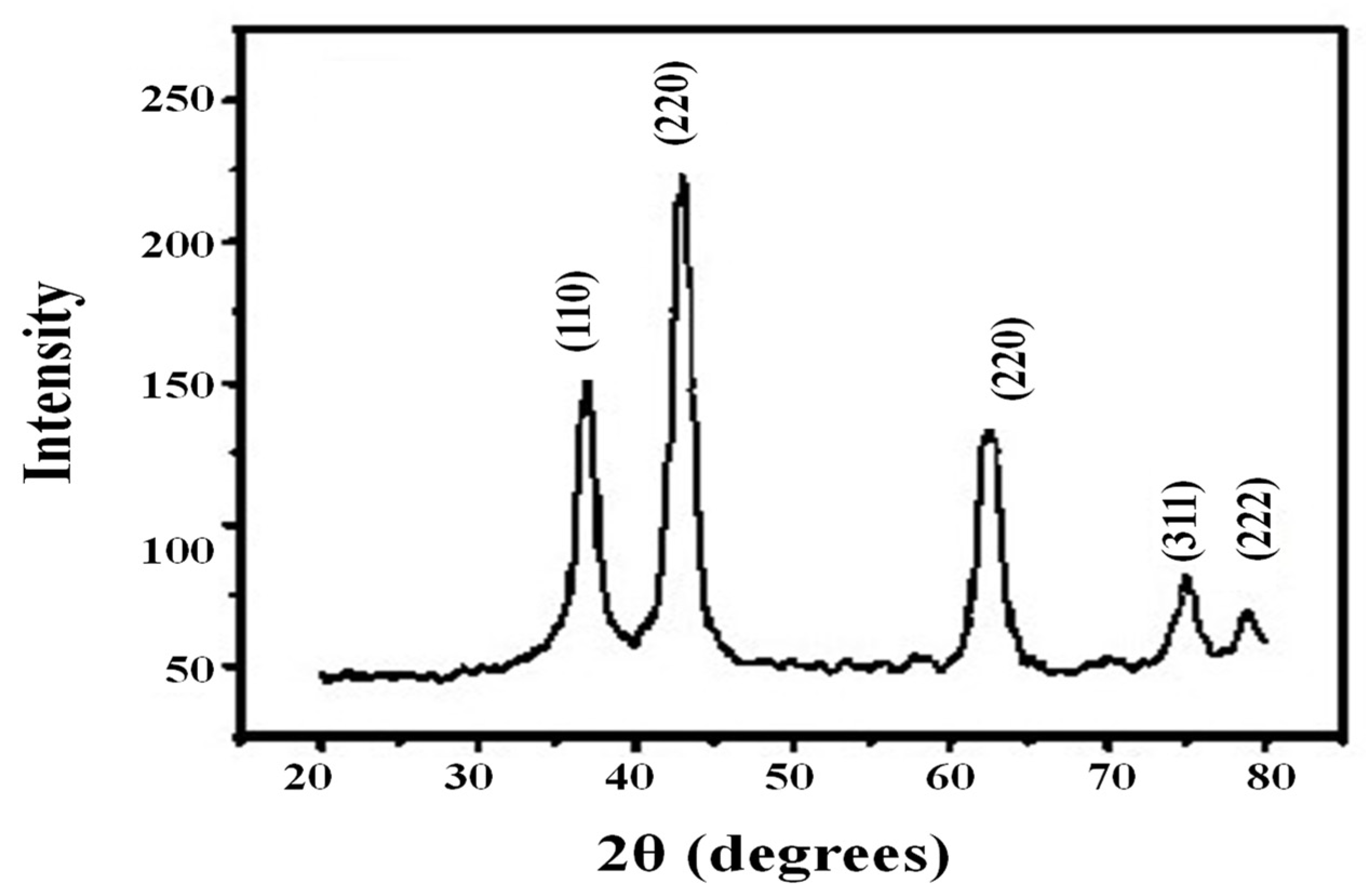
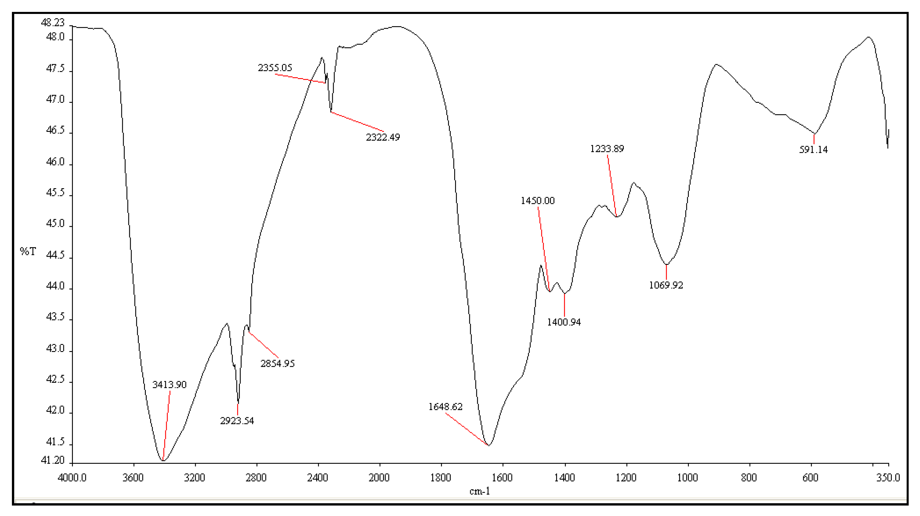
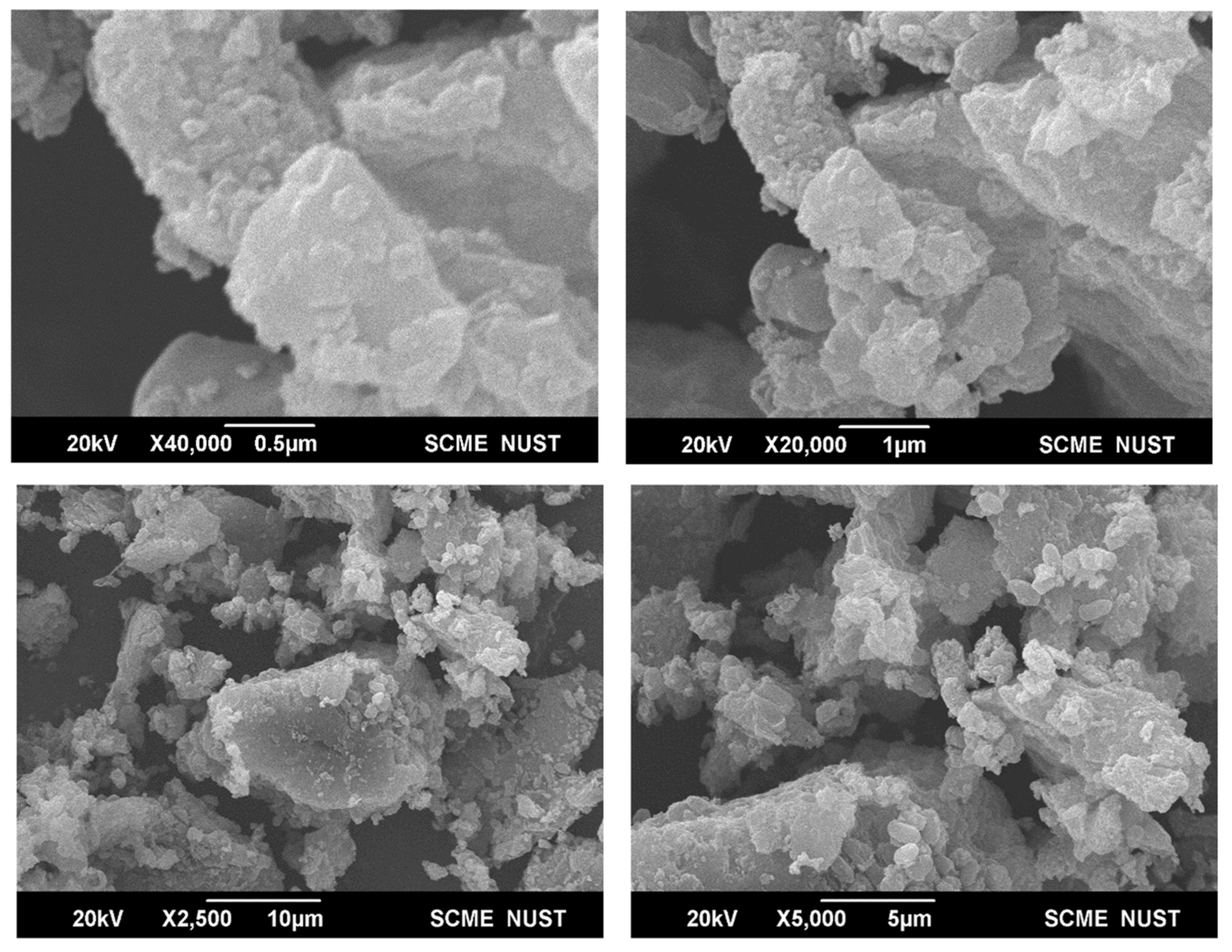
3.2. Characterization of NiO Nanoparticles

The culture of Shewanella sp. SM33 was incubated at optimum conditions, i.e., at 28 °C for 24 h after the addition of a 10 mM NiCl salt solution. After 24 h of incubation, it was observed that the color of the reaction mixture changed from pale green to dense green. Then, a trace amount of the reaction mixture was subjected to a UV–vis spectrophotometer analysis. The Shewanella sp. SM33 showed a peak at 310.23 nm, which was the first indication of the biosynthesis of NiO-NPs (Figure 2).
The XRD pattern indicated the crystalline nature of the nanoparticles. The diffraction data of NiO-NPs produced from Shewanella sp. SM33 showed characteristic diffraction peaks at 37.5°, 42.5°, 75.25°, and 80°, as shown in Figure 3. These peak positions at 2ϴ degrees corresponded to 110, 220, 311, and 222 places of Ni, which were according to the standard diffraction data of NiO-NPs. There were a few extra peaks, which may be due to the small amount of nickel oxides present there. It was observed that the average particle size of biogenic NiO-NPs was less than 20 nm, which was calculated with the use of the Debye–Scherrer equation.
FTIR analysis was used to investigate the surface characteristics of biosynthesized NiO-NPs in a wave number range of 350–4000 cm−1. While the FTIR spectra of NiO-NPs produced from Shewanella sp. SM33 showed different absorption peaks at 3413.90, 2923.54, 2854.95, 2355.05, 2322.49, 1648.62, 1450.00, 1233.89, and 591.14 cm−1 (Figure 4), the peak at 3413.90 was because of the broad absorption of the O-H group of alcohol and C-H stretching of amines and amides. The peaks at 2923.54 and 2854.95 were due to -C-H (CH2) stretching. While the peaks at 2353, 2322.49, and 1648.62 were due to C-O bond, C-N bond, and -C=C- (cis) stretching, respectively, the absorption at 1450.00, 1233.89 and 591.14 cm−1 were because of nitrosamine, alkyl ketone, and halogen compound (C-Cl) bonds, respectively. The FTIR spectra of the biosynthesized NiO-NPs confirmed the presence of proteins and alcoholic groups in NiO-NPs.
SEM images at various magnitude scales ranging from 5000× to 40,000× were collected and studied. The image of the sample indicated that sample consisted of Ni nanoclusters with different morphological forms. Moreover, an agglomeration was found between particles of larger sizes. SEM images of biogenic NiO-NPs that were prepared from Strain SM33 are shown in Figure 5.

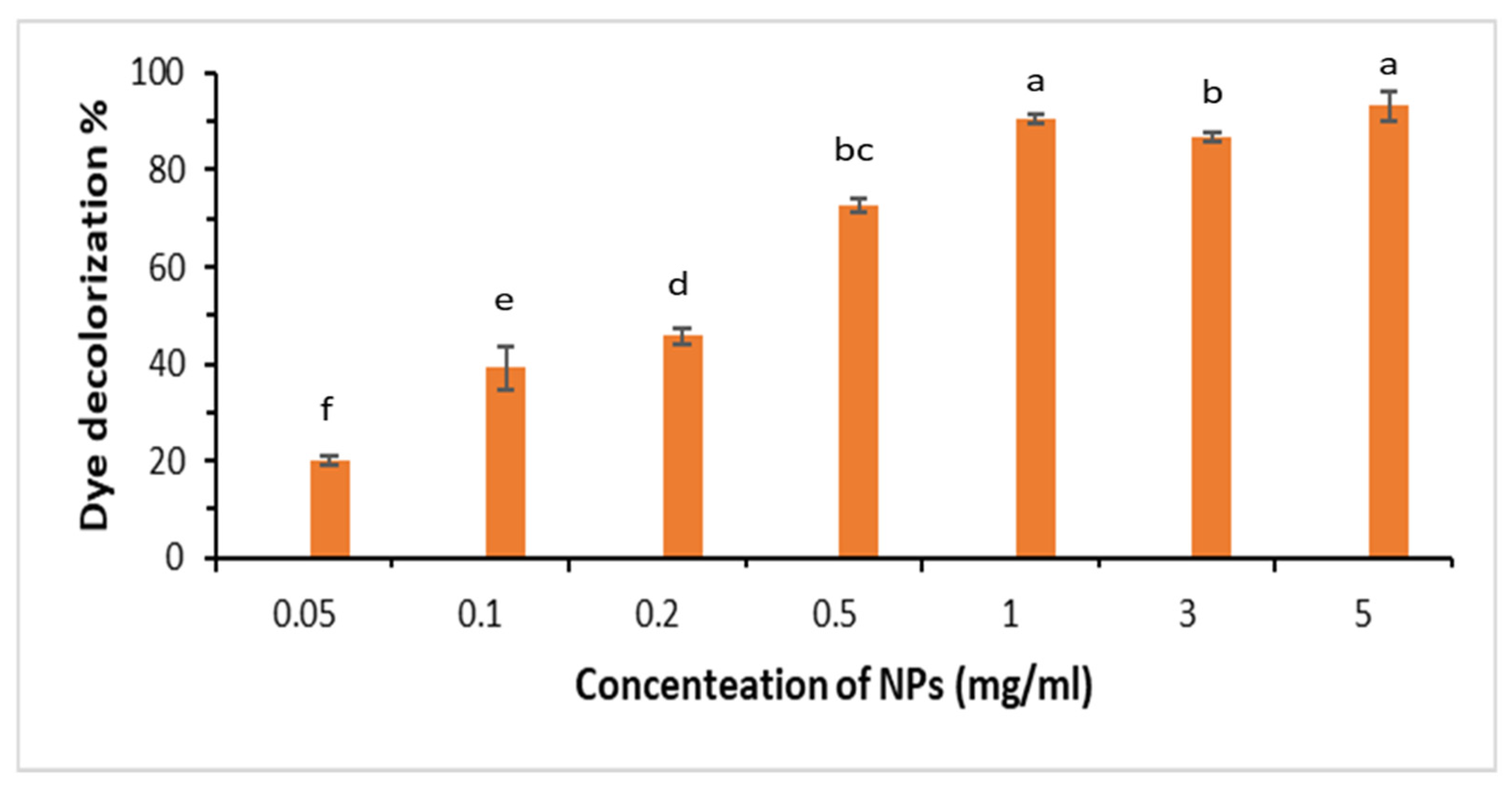
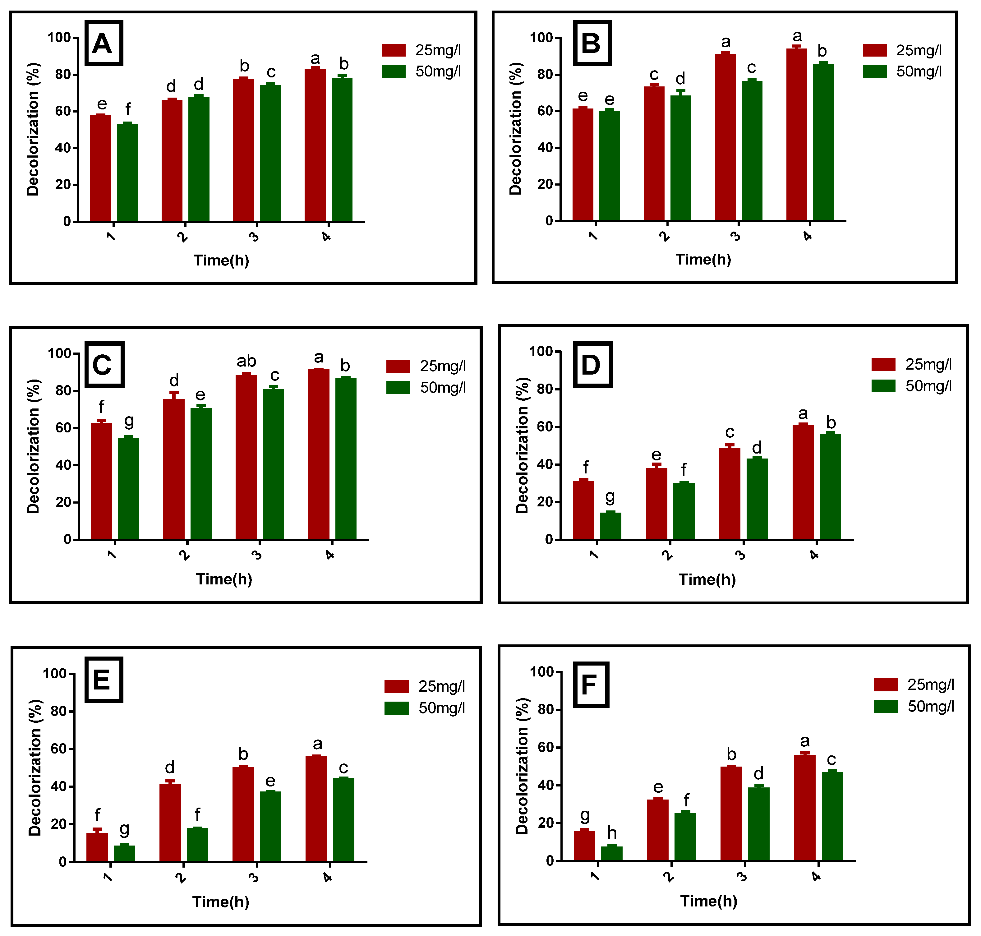
3.3. Photocatalytic Degradation of Organic Azo Dyes by Using Biogenic NiO-NPs

The addition of NiO-NPs to the solution resulted in the decolorization of the dyes, which was observed through a decrease in absorption values at the corresponding λmax wavelengths of dyes. For the purpose of decolorizing the dyes, a 1 mg/mL (an optimized) concentration of NiO-NPs was used because it was the lowest concentration of nanoparticles, which showed the best results after 4 h of incubation, as shown in Figure 6.
In accordance with Figure 7, which shows the dye decolorization data, the decolorization rate observed at a 25 mg L−1 dye concentration was 82.36 ± 1.48%, 93.57 ± 2.11%, 91.05 ± 0.45%, 59.94 ± 1.51%, 55.45 ± 0.91%, and 55.17 ± 2.11% for MB, CR, MCG, DB-I, RR-II, and RB-5 after 4 h under sunlight, while at a 50 mg L−1 concentration of the dye, the decolorization of MB, CR, MCG, DB-I, RR-II, and RB-5 was 77.52 ± 2.11%, 85.19 ± 1.40%, 86.09 ± 0.95%, 55.15 ± 1.64%, 43.81 ± 0.68%, and 46.11 ± 1.67%, respectively, by the addition of NiO-NPs under 4 h of sunlight. The results show that biosynthesized NiO-NPs show an excellent photocatalytic potential to degrade each dye at a 25 mg L−1 concentration. After sunlight exposure of 1 h, the photocatalytic degradation rate of a 25 mg L−1 concentration of Congo red dye is 1.41% higher than the decolorization of a 50 mg L−1 dye concentration. Similarly, after 4 h of contact with sunlight, the trend of decolorization remains the same as at a 25 mg L−1 dye concentration, and the decolorization rate is 8.38% higher than decolorization at a 50 mg L−1. Likewise, the photocatalytic decolorization of a 25 mg L−1 dye concentration of MB, DB, MCG, RB-5, and RR-II after 1 h of sunlight exposure is 4.88%, 16.87%, 8.28%, 8.0%, and 6.7% higher, respectively, than the decolorization of a 50 mg L−1 dye concentration. Similarly, after 4 h of sunlight exposure, the decolorization rate of a 25 mg L−1 dye concentration of MB, DB, MCG, RB-5, and RR-II is 4.84%, 4.79%, 4.96%, 9.06%, and 11.64% higher, respectively, than the decolorization of a 50 mg L−1 dye concentration. This shows that NiO-NPs were found capable of decolorizing a 25 mg L−1 dye concentration more accurately then a 50 mg L−1 dye concentration.
According to Figure 8, which shows the dye decolorization data at shaking and static conditions, the decolorization rate at shaking conditions was observed to be 72.16 ± 1.29%, 90.76 ± 2.18%, 86.09 ± 0.79%, 64.50 ± 0.44%, 34.14 ± 0.28%, and 33.52 ± 0.48% for MB, CR, MCG, DB-1, RR-II, and RB-5, respectively, while at static conditions, the decolorization rate of MB, CR, MG, DB-1, RR-II, and RB-5 was 88.66 ± 1.53%, 95.35 ± 1.71%, 93.52 ± 1.02%, 72.17 ± 0.28%, 69.52 ± 0.31%, and 52.05 ± 0.57%, respectively, after 6 h of sunlight exposure. Due to the fact that dye decolorization tended to change after the 7th hour of incubation, as is shown in Figure 8, the rate of decolorization tended to decrease instead of increase. In Figure 8, it is clearly shown that the decolorization rate at static conditions is higher than at shaking conditions.

3.4. Treatment of Industrial Wastewater by Using Biosynthesized Nickel Oxide Nanoparticles

Industrial wastewater containing Congo-red dye was treated using biosynthesized nickel oxide nanoparticles. Table 1 shows the data of different parameters like color removal, pH, EC, COD, TDS, sulfates, and phosphates, which measured untreated wastewater and treated wastewater with NiO-NPs. In untreated wastewater, the values of the different parameters were beyond the permissible range given by the Environmental Protection Agency, while wastewater that was treated with NiO-NPs demonstrated significantly decreased values of these parameters. In this study, NiO-NPs showed 86.79% reduction in color removal, while the pH value decreased from 8.5 to 6.1, and EC decreased from 9.3 to 4.5. Other factors like COD, TDS, sulfates, and phosphates show 86.79%, 67.05%, 52.5, and 49.49% reduction, respectively, after the treatment of the wastewater sample. Some other studies also reported the treatment of wastewater samples; Soltani and Safari (2016) [42] reported the treatment of different parameters of wastewater with the use of MgO NPs, and these showed an 85% and 63.34% reduction in COD and total organic carbon, respectively, in wastewater.

4. Discussion

This study mainly focuses on the biosynthesis of NiO-NPs from Shewanella sp. SM33 and their application on industrial wastewater in order to check their decolorizing potential. The strain Shewanella sp. SM33 was isolated from the wastewater that was collected from the Paharrang drain in Faisalabad, Pakistan. The isolated strain was taxonomically identified by the 16S rRNA gene sequence, which is a familiar bacterial taxonomic marker and has already been used for the taxonomic identification of gene sequences by Noman et al., 2020 [26]. BLASTn and phylogenetic analysis of the 16s rRNA gene of isolate SM-33 revealed that the selected isolate (SM-33) belongs to the Shewanella genus. The BLASTn and phylogenetic analysis of 16s rRNA gene of isolate SM-33 revealed that the selected isolate (SM-33) belongs to genus Shewanella. Besides this, a number of other bacterial strains that belong to different types of genera have been reported for the biosynthesis of different nanoparticles [37,43,44,45].
The synthesized nanoparticles from the strain SM33 were initially confirmed by the synthesis of NiO-NPs at 310.23 nm via UV–vis spectral analysis. These results are quite similar to Bashir et al. (2019) [46], who confirmed the synthesis of NiO-NPs by the formation of a peak at 338.9 nm. The XRD pattern of NiO-NPs showed a sharp peak at 110, 220, 311, and 222 ranges at 2ϴ degrees. The peaks confirmed the crystalline nature of NiO-NPs with an average size of 20 nm. The result are inconsistent with the findings of Zorkipli et al. (2016) [47], who synthesized the NiO-NPs with a sol–gel method. An FTIR spectra of NiO-NPs produced from Shewanella sp. SM33 showed different absorption peaks at 3413.90, 2923.54, 2854.95, 2355.05, 2322.49, 1648.62, 1450.00, 1233.89, and 591.14 cm−1 (Figure 4). The FTIR spectra of biosynthesized NiO-NPs confirmed the presence of proteins and alcoholic groups in NiO-NPs.
In this study, after the optimization of the concentration of NPs, we obtained the best results at 1 mg/mL and 5 mg/mL concentrations, but we selected a 1 mg/mL concentration of NiO-NPs because this was the lowest concentration of NPs at which we obtained best results and in which the lowest amount of metal salt was used, rendering this less toxic.
This study also addressed the fact that biosynthesized NiO-NPs were found to be efficient for the decolorization of various types of azo dyes (i.e., MB, CR, DB, RRII, RB5 and MG) at different concentrations (i.e., 25 and 50 mg L−1) under 4 to 7 h of incubation in sunlight. These results are also in accordance with those of Sabouri et al. (2019) [48], in which they synthesized nickel oxide nanoparticles by using the sol–gel method employed for the photocatalytic degradation of methylene blue. According to the results of this study, it was clearly shown that the photocatalytic degradation of selected azo dyes was significantly higher at a 25 mg L−1 concentration of biogenic nickel nanoparticles in static conditions. The results showed that the decolorization rate of MB dye at a concentration of 25 mg L−1, degraded by NiO-NPs under static conditions, was observed to be 82.36 ± 1.48%. These obtained results were inconsistent with those of Rong et al. (2015) [49], where they synthesized nickel oxide/graphene oxide (NiO/GO) nanocomposite using a hydrothermal process and applied it to the process of removing aromatic heterocyclic dye methylene blue (MB) from aqueous solutions. Their findings demonstrated that under visible light irradiation, a NiO/GO nanocomposite displayed significant removal efficiency, in contrast to conditions in the absence of light. In another study, Khairnar and Shrivastava (2019) [50] synthesized NiO nanoparticles using a chemical method, employing NiCl2 and NaHCO3 as precursors. They assessed the photocatalytic proficiency of these NiO nanoparticles in degrading methylene blue and rhodamine B dyes under visible light irradiation. Their results showed that at a pH of 2, there was a 98.7% degradation of methylene blue, while at pH 10, the degradation efficiency for rhodamine B was 80.33%. The same results were also noticed in the study of Noman et al. (2020) [26], according to which nanoparticles showed a high photocatalytic degradation at 25 mg L−1 compared to higher concentrations. The higher result at 25 mg L−1 might be due to molecules being more dispersed and having better contact with nanoparticles compared to higher concentrations. At higher concentrations, the solution was saturated, and it became overwhelmed with dye molecules, resulting in less interaction between the dye and nanoparticles [51].
Figure 7 shows that the reactive black 5 dye was degraded by 93.57 ± 2.11% by a nickel oxide nanoparticle in the presence of sunlight. The degradation of the reactive black dye yielded similar results, as demonstrated by Prabhu et al., 2022 [52]. They synthesized NiO-NPs using an extract of Clitoria ternatea. The photocatalytic activity of the NiO-NPs was investigated for the degradation of fast green (FG) and rose bengal (RB) dye molecules under sunlight. The results revealed that the NiO-NPs were effective in degrading these dye molecules, with a degradation efficiency of 89% for FG and 77% for RB. Ahsan et al. (2022) [53] synthesized nickel oxide nanoparticles (NiO-NPs), copper oxide nanoparticles (CuO-NPs), and their nanocomposite (NiO/CuO-NC) via precipitation method. Results revealed that at an optimum concentration (60 mg L−1) of RR-2, RB-5, and OII, the dyes were degraded 90, 82, and 83% by NiO-NPs; 49, 34, and 44% degraded by CuO-NPs; whereas the nanocomposite NiO/CuO-NC was degraded by 92, 93, and 96%, respectively.
Results showed that the NiO-NPs degraded Congo red dye up to 91.05 ± 0.45% at a concentration of 25 mg/L under sunlight. A study conducted by Rafique et al. (2021) [54] showed consistent results for the degradation of Congo red dye by using nickel oxide nanoparticles. They synthesized nickel oxide nanoparticles via use of aqueous extracts from Allium cepa peels. Their results showed that under optimized conditions (Congo Red Direct dye concentration of 0.02%, a catalyst dose of 0.003 g·L−1, pH of 6, and a temperature of 50 °C), a decolorization efficiency of up to 90% was attained.
MG dye was degraded by 46.81 ± 0.836, and a similar result for MG was shown by Ghazal et al. (2021) [55]. They synthesized carbon quantum dots/nickel oxide (CQDs/NiO) composites from the seeds of Nigella sativa via hydrothermal method. The photocatalytic degradation of the composites was determined using malachite green as the pollutant in the presence of sunlight. The results showed that MG was degraded approximately by 98 % in the solution after 60 min in the presence of sunlight. Figure 7 showed the photocatalytic degradation of six different dyes in the order of MG > MB > CR > DB > RB5 > RR-II after 4 h of incubation in sunlight. Malachite green showed a maximum decolorization of 89.11 ± 1.018, while reactive red II showed a minimum decolorization of 46.81 ± 0.836, respectively. The lower decolorization of reactive red II might be due to its complex aromatic structure. Dyes that have more complex chemical structures were difficult for nanoparticles to decolorize due to their high stability against oxidation or reduction [56].
The decolorization rate of dyes were checked at both shaking and static conditions in order to find the best result. Results demonstrated that at static conditions, MG, CR, MB, RR-II, RB5, and DB showed higher decolorization rates as compared to shaking conditions (Figure 8).
Nanoparticles used for decolorization may tend to aggregate or settle under shaking conditions. Aggregation can reduce the available surface area of nanoparticles for dye adsorption or reaction, leading to a slower decolorization rate. By contrast, in static conditions, nanoparticles may have better dispersion, allowing for more efficient interaction with dye molecules. The results of the study were inconsistent with those of Kannan et al. (2020) [30], where they used biogenic nanoparticles for the decolorization of azo dyes. The decolorization of organic azo dyes by NiO-NPs might have generated electron–hole pairs when the dyes were exposed to sunlight. These electron–hole pairs might have participated in a subsequent redox reaction, leading to the degradation of the dyes [57].
It was also noted that NiO-NPs are not only efficient for the treatment of azo dyes, but they can also reduce values of other effluents in industrial wastewater like COD (chemical oxygen demand), TDS (total dissolved solids), TSS (total suspended solids), phosphates, sulphates, pH, and EC (electrical conductivity), assuring their percentage degradation. The gradual reduction in pH and EC in treated wastewater might be due to breakdown of the organic matter present in municipal wastewater through oxidation or other chemical processes. Nanoparticles with a high surface area and specific surface chemistry can adsorb ions, pollutants, or organic compounds present in wastewater. This adsorption process helps in the removal of ions and contaminants that contribute to high EC and pH levels. For example, activated carbon nanoparticles have a high adsorption capacity for organic compounds [58]. Similar results for the treatment of wastewater were also documented by Mehwish et al. 2021 [59] using MOS-AgNPs, which significantly reduced pH (10.1–7.9) and EC values (11.8–3.7 dS m−1).
Table 3 illustrates the reduction in color intensity and COD by 86.79% and 49.24% in treated wastewater as compared to untreated wastewater. These results were consistent with [60,61]. The decrease in the concentration of the chemical oxygen demand might be the process of oxidation and catalysis. The process of oxidation and reduction were initiated by ultraviolet (UV) light, which created electron–hole pairs in the presence of nanoparticles. Organic compounds were oxidized and degraded by generated ROS, leading to a decrease in COD [62]. A similar result for the treatment of wastewater, along with the degradation of dye, was shown by [54]. The actual effluent from the textile industry revealed a 70% reduction in color at optimized conditions. Reduction percentages for total organic carbon (TOC) and chemical oxygen demand (COD) were determined to be 73.24% and 74.56%, respectively. For the catalytic treatment of Congo red dye, the reduction percentages in TOC and COD were measured at 62.47% and 60.23%, respectively. In the context of treating textile effluents, the utilization of nickel oxide nanoparticles as a catalyst yielded these results. Similar results were stated by [6], where COD removal efficiency was achieved at 47% after 8.5 min. Table 2 illustrated that the activity of phosphates and sulphates in non-treated wastewater decreased compared to treated wastewater. The reduction in the concentration of phosphates and sulphates were due to the precipitation of these molecules by nanoparticles in the presence of sunlight [63]. Similarly, the treatment of wastewater by Cu-NPs was reported by Noman et al. (2020) [45], where a 20.71%, 79.79%, 50%, and 78.57% decrease was reported in pH, EC, turbidity, and TSS, respectively. In the present study, Ni-ONPs showed a high efficiency of removal and a degradation of 67.45% against TDS compared to other parameters. This might be due to the degradation of total dissolved solids, as generated reactive oxygen species oxidize TDS ions and convert them into less harmful substances [64].
Therefore, according to our observations and knowledge, many of the studies mentioned before synthesized nickel nanoparticles from bacteria and checked their potential for azo dye decolorization and for wastewater treatment, but not a single study on the production of nickel nanoparticles from Shewanella sp. exist. As a result, Shewanella sp. might be a new biosource for the production of nickel nanoparticles because of its catalytic activity and role in maintaining pH.
The microbial synthesis of nickel oxide nanoparticles provides a sustainable approach to implement azo dye decolorization and industrial wastewater remediation. This ecologically beneficial technology reduces the need for renewable energy and environmentally hazardous chemical processes through the benefit of microorganisms’ natural capacity to convert nickel ions into nanoparticles. Furthermore, in efficiently decolorizing azo dyes, it assists in the removal of heavy metals from industrial effluents, resulting in cleaner water resources. This sustainable methodology reduces the carbon footprint and chemical waste overall, coordinating with eco-friendly practices and promoting environmental conservation in industrial processes.

5. Conclusions

Metallic nanoparticles have emerged as a valuable tool for the degradation of azo dyes due to their different biological and physicochemical properties. Currently, the scientific community predominantly prefers biologically synthesized nanoparticles over chemical or physical methods, given their eco-friendly and cost-effective attributes. In this study, we investigated the nickel-resistant bacterium SM33 for the synthesis of NiO-NPs at a concentration of 10 mM NiCl salt. Phylogenetic analysis of the 16S rRNA sequences identified strain SM33 as belonging to the Shewanella genus. The biogenically synthesized NiO-NPs were examined for their photocatalytic activity under sunlight using various concentrations of azo dyes and also under different static and shaking conditions. Furthermore, the photocatalytic activity of these biologically synthesized NiO-NPs was checked for the treatment of industrial wastewater, considering various parameters such as pH, EC, COD, TDS, sulfates, and phosphates before and after treatment. Our findings demonstrated that approximately 60% of the wastewater was effectively treated in this study. These results demonstrate the potential of biogenically synthesized NiO-NPs as an attractive option for azo dye degradation and industrial wastewater treatment; they may contribute to the preservation of dwindling water resources for agricultural purposes, in power plants for cooling, watering golf courses, building construction, firefighting, and car washing.

Author Contributions

Formal analysis, S.M., F.M., U.S. and R.O.E.; Funding acquisition, R.O.E., M.H. and T.A.Y.A.; Investigation, S.M., U.S. and F.B.; Methodology, F.M., M.S. and F.B.; Supervision, F.M., S.H., M.S. and T.S.; Writing—original draft, S.M., U.S. and F.B.; Writing—review and editing, F.M., U.S., S.H., M.S., F.B., R.O.E., M.H., T.A.Y.A. and T.S. All authors have read and agreed to the published version of the manuscript.

Funding

This research was funded by the Deanship of Scientific Research, King Khalid University through research groups program under grant number R.G.P. 2/75/44.

Institutional Review Board Statement

Not applicable.

Informed Consent Statement

Not applicable.

Data Availability Statement

Other data could be made available upon request to the corresponding author.

Acknowledgments

The authors extend their appreciation to the Deanship of Scientific Research, King Khalid University for funding this work through research groups program under grant number R.G.P. 2/75/44.

Conflicts of Interest

The authors declare that they have no competing interest.

References

  1. Anastopoulos, I.; Hosseini-Bandegharaei, A.; Fu, J.; Mitropoulos, A.C.; Kyzas, G.Z. Use of Nanoparticles for Dye Adsorption: Review. J. Dispers. Sci. Technol. 2017, 39, 836–847. [Google Scholar] [CrossRef]
  2. Gupta, V.K.; Kumar, R.; Nayak, A.; Saleh, T.A.; Barakat, M.A. Adsorptive Removal of Dyes from Aqueous Solution onto Carbon Nanotubes: A Review. Adv. Colloid Interface Sci. 2013, 193–194, 24–34. [Google Scholar] [CrossRef]
  3. Routoula, E.; Patwardhan, S.V. Degradation of Anthraquinone Dyes from Effluents: A Review Focusing on Enzymatic Dye Degradation with Industrial Potential. Environ. Sci. Technol. 2020, 54, 647–664. [Google Scholar] [CrossRef] [PubMed]
  4. Deniz, F. Adsorption Properties of Low-Cost Biomaterial Derived from Prunus amygdalus L. for Dye Removal from Water. Sci. World J. 2013, 2013, 961671. [Google Scholar] [CrossRef] [PubMed]
  5. Katheresan, V.; Kansedo, J.; Lau, S.Y. Efficiency of Various Recent Wastewater Dye Removal Methods: A Review. J. Environ. Chem. Eng. 2018, 6, 4676–4697. [Google Scholar]
  6. Keramati, M.; Ayati, B. Petroleum Wastewater Treatment Using a Combination of Electrocoagulation and Photocatalytic Process with Immobilized ZnO Nanoparticles on Concrete Surface. Process Saf. Environ. Prot. 2019, 126, 356–365. [Google Scholar] [CrossRef]
  7. Danouche, M.; El Arroussi, H.; El Ghachtouli, N. Bioremoval of Acid Red 14 Dye by Wickerhamomyces Anomalus Biomass: Kinetic and Thermodynamic Study, Characterization of Physicochemical Interactions, and Statistical Optimization of the Biosorption Process. Biomass Convers. Biorefinery 2022, 1–20. [Google Scholar] [CrossRef]
  8. Imran, M.; Arshad, M.; Negm, F.; Khalid, A.; Shaharoona, B.; Hussain, S.; Nadeem, S.M.; Crowley, D.E. Yeast Extract Promotes Decolorization of Azo Dyes by Stimulating Azoreductase Activity in Shewanella Sp. Strain IFN4. Ecotoxicol. Environ. Saf. 2016, 124, 42–49. [Google Scholar] [CrossRef]
  9. Rafatullah, M.; Sulaiman, O.; Hashim, R.; Ahmad, A. Adsorption of Methylene Blue on Low-Cost Adsorbents: A Review. J. Hazard. Mater. 2010, 177, 70–80. [Google Scholar] [CrossRef]
  10. Subedi, S.K. An Introduction to Nanotechnology and Its Implications. Himal. Phys. 2015, 5, 78–81. [Google Scholar] [CrossRef]
  11. Rangasamy, M. Nano Technology: A Review. J. Appl. Pharm. Sci. 2011, 1, 8–16. [Google Scholar]
  12. Haidri, I.; Shahid, M.; Hussain, S.; Shahzad, T.; Mahmood, F.; Hassan, M.U.; Al-Khayri, J.M.; Aldaej, M.I.; Sattar, M.N.; Rezk, A.A.-S. Efficacy of Biogenic Zinc Oxide Nanoparticles in Treating Wastewater for Sustainable Wheat Cultivation. Plants 2023, 12, 3058. [Google Scholar] [PubMed]
  13. Jeevanandam, J.; Barhoum, A.; Chan, Y.S.; Dufresne, A.; Danquah, M.K. Review on Nanoparticles and Nanostructured Materials: History, Sources, Toxicity and Regulations. Beilstein J. Nanotechnol. 2018, 9, 1050–1074. [Google Scholar] [CrossRef] [PubMed]
  14. Roco, M.C. Nanoparticles and Nanotechnology Research. J. Nanoparticle Res. 1999, 1, 1–6. [Google Scholar]
  15. Nasrollahzadeh, M.; Sajadi, S.M.; Sajjadi, M.; Issaabadi, Z. Applications of Nanotechnology in Daily Life. In Interface Science and Technology; Elsevier: Amsterdam, The Netherlands, 2019; pp. 113–143. [Google Scholar]
  16. Mouni, L.; Belkhiri, L.; Bollinger, J.-C.; Bouzaza, A.; Assadi, A.; Tirri, A.; Dahmoune, F.; Madani, K.; Remini, H. Removal of Methylene Blue from Aqueous Solutions by Adsorption on Kaolin: Kinetic and Equilibrium Studies. Appl. Clay Sci. 2018, 153, 38–45. [Google Scholar] [CrossRef]
  17. Adak, A.; Bandyopadhyay, M.; Pal, A. Adsorption of Anionic Surfactant on Alumina and Reuse of the Surfactant-Modified Alumina for the Removal of Crystal Violet from Aquatic Environment. J. Environ. Sci. Health Part A 2005, 40, 167–182. [Google Scholar] [CrossRef]
  18. Kumar, P.S.; Joshiba, G.J.; Femina, C.C.; Varshini, P.; Priyadharshini, S.; Karthick, M.S.A.; Jothirani, R. A Critical Review on Recent Developments in the Low-Cost Adsorption of Dyes from Wastewater. Desalination Water Treat. 2019, 172, 395–416. [Google Scholar] [CrossRef]
  19. Kausar, A.; Iqbal, M.; Javed, A.; Aftab, K.; Nazli, Z.-H.; Bhatti, H.N.; Nouren, S. Dyes Adsorption Using Clay and Modified Clay: A Review. J. Mol. Liq. 2018, 256, 395–407. [Google Scholar] [CrossRef]
  20. Liu, J.; Qiao, S.Z.; Hu, Q.H.; Lu, G.Q.M. Magnetic Nanocomposites with Mesoporous Structures: Synthesis and Applications. Small 2011, 7, 425–443. [Google Scholar] [CrossRef]
  21. Mohanpuria, P.; Rana, N.K.; Yadav, S.K. Biosynthesis of Nanoparticles: Technological Concepts and Future Applications. J. Nanoparticle Res. 2007, 10, 507–517. [Google Scholar] [CrossRef]
  22. Shafqat, U.; Hussain, S.; Shahzad, T.; Shahid, M.; Mahmood, F. Elucidating the Phytotoxicity Thresholds of Various Biosynthesized Nanoparticles on Physical and Biochemical Attributes of Cotton. Chem. Biol. Technol. Agric. 2023, 10, 30. [Google Scholar]
  23. Maurer-Jones, M.A.; Bantz, K.C.; Love, S.A.; Marquis, B.J.; Haynes, C.L. Toxicity of Therapeutic Nanoparticles. Nanomedicine 2009, 4, 219–241. [Google Scholar] [CrossRef] [PubMed]
  24. Sharifi, S.; Behzadi, S.; Laurent, S.; Forrest, M.L.; Stroeve, P.; Mahmoudi, M. Toxicity of Nanomaterials. Chem. Soc. Rev. 2012, 41, 2323–2343. [Google Scholar] [CrossRef] [PubMed]
  25. Pearce, C.I.; Coker, V.S.; Charnock, J.M.; Pattrick, R.A.D.; Mosselmans, J.F.W.; Law, N.; Beveridge, T.J.; Lloyd, J.R. Microbial Manufacture of Chalcogenide-Based Nanoparticles via the Reduction of Selenite Using Veillonella atypica: An in Situ EXAFS Study. Nanotechnology 2008, 19, 155603. [Google Scholar] [CrossRef]
  26. Noman, M.; Shahid, M.; Ahmed, T.; Niazi, M.B.K.; Hussain, S.; Song, F.; Manzoor, I. Use of Biogenic Copper Nanoparticles Synthesized from a Native Escherichia Sp. as Photocatalysts for Azo Dye Degradation and Treatment of Textile Effluents. Environ. Pollut. 2020, 257, 113514. [Google Scholar]
  27. Jamkhande, P.G.; Ghule, N.W.; Bamer, A.H.; Kalaskar, M.G. Metal Nanoparticles Synthesis: An Overview on Methods of Preparation, Advantages and Disadvantages, and Applications. J. Drug Deliv. Sci. Technol. 2019, 53, 101174. [Google Scholar] [CrossRef]
  28. Rouch, D.A.; Lee, B.T.O.; Morby, A.P. Understanding Cellular Responses to Toxic Agents: A Model for Mechanism-Choice in Bacterial Metal Resistance. J. Ind. Microbiol. 1995, 14, 132–141. [Google Scholar] [CrossRef]
  29. Lee, N.-Y.; Ko, W.-C.; Hsueh, P.-R. Nanoparticles in the Treatment of Infections Caused by Multidrug-Resistant Organisms. Front. Pharmacol. 2019, 10, 1153. [Google Scholar] [CrossRef]
  30. Kannan, K.; Radhika, D.; Nikolova, M.P.; Sadasivuni, K.K.; Mahdizadeh, H.; Verma, U. Structural Studies of Bio-Mediated NiO Nanoparticles for Photocatalytic and Antibacterial Activities. Inorg. Chem. Commun. 2020, 113, 107755. [Google Scholar] [CrossRef]
  31. Li, L.; Nawaz, T.; Ferryman, J. PETS 2015: Datasets and Challenge. In Proceedings of the 2015 12th IEEE International Conference on Advanced Video and Signal Based Surveillance (AVSS), Karlsruhe, Germany, 25–28 August 2015. [Google Scholar]
  32. Fei, J.; Cui, Y.; Wang, A.; Zhu, P.; Li, J. Noble Metal Nanochains through Helical Self-Assembly. Chem. Commun. 2010, 46, 2310–2312. [Google Scholar] [CrossRef]
  33. Sathyavathi, S.; Manjula, A.; Rajendhran, J.; Gunasekaran, P. Extracellular Synthesis and Characterization of Nickel Oxide Nanoparticles from Microbacterium Sp. MRS-1 towards Bioremediation of Nickel Electroplating Industrial Effluent. Bioresour. Technol. 2014, 165, 270–273. [Google Scholar] [CrossRef] [PubMed]
  34. Srivastava, S.K.; Constanti, M. Room Temperature Biogenic Synthesis of Multiple Nanoparticles (Ag, Pd, Fe, Rh, Ni, Ru, Pt, Co, and Li) by Pseudomonas Aeruginosa SM1. J. Nanoparticle Res. 2012, 14, 831. [Google Scholar] [CrossRef]
  35. Khoshhesab, Z.M.; Sarfaraz, M.; Asadabad, M.A. Preparation of ZnO Nanostructures by Chemical Precipitation Method. Synth. React. Inorg. Met. Nano-Metal Chem. 2011, 41, 814–819. [Google Scholar] [CrossRef]
  36. Weisburg, W.G.; Barns, S.M.; Pelletier, D.A.; Lane, D.J. 16S Ribosomal DNA Amplification for Phylogenetic Study. J. Bacteriol. 1991, 173, 697–703. [Google Scholar] [PubMed]
  37. Tiwari, M.; Jain, P.; Hariharapura, R.C.; Narayanan, K.; Bhat, U.; Udupa, N.; Rao, J.V. Biosynthesis of Copper Nanoparticles Using Copper-Resistant Bacillus Cereus, a Soil Isolate. Process Biochem. 2016, 51, 1348–1356. [Google Scholar]
  38. Griffiths, P.R.; de Haseth, J.A. Fourier Transform Infrared Spectrometry, 2nd ed; John Wiley & Sons: Hoboken, NJ, USA, 2007. [Google Scholar]
  39. Varshney, R.; Bhadauria, S.; Gaur, M.S.; Pasricha, R. Characterization of Copper Nanoparticles Synthesized by a Novel Microbiological Method. JOM 2010, 62, 102–104. [Google Scholar]
  40. Roy, K.; Ghosh, C.K.; Sarkar, C.K. Degradation of Toxic Textile Dyes and Detection of Hazardous Hg2+ by Low-Cost Bioengineered Copper Nanoparticles Synthesized Using Impatiens Balsamina Leaf Extract. Mater. Res. Bull. 2017, 94, 257–262. [Google Scholar]
  41. Allen, S.J.; Koumanova, B. Decolourisation of water/wastewater using adsorption. J. Univ. Chem. Technol. Metal. 2005, 40, 175–192. [Google Scholar]
  42. Soltani, R.D.C.; Safari, M. Periodate-Assisted Pulsed Sonocatalysis of Real Textile Wastewater in the Presence of MgO Nanoparticles: Response Surface Methodological Optimization. Ultrason. Sonochem. 2016, 32, 181–190. [Google Scholar]
  43. Lingaraju, K.; Raja Naika, H.; Nagabhushana, H.; Jayanna, K.; Devaraja, S.; Nagaraju, G. Biosynthesis of Nickel Oxide Nanoparticles from Euphorbia heterophylla (L.) and Their Biological Application. Arab. J. Chem. 2020, 13, 4712–4719. [Google Scholar] [CrossRef]
  44. Zaki, S.; El Kady, M.F.; Abd-El-Haleem, D. Biosynthesis and Structural Characterization of Silver Nanoparticles from Bacterial Isolates. Mater. Res. Bull. 2011, 46, 1571–1576. [Google Scholar] [CrossRef]
  45. Noman, M.; Ahmed, T.; Hussain, S.; Niazi, M.B.K.; Shahid, M.; Song, F. Biogenic Copper Nanoparticles Synthesized by Using a Copper-Resistant Strain Shigella Flexneri SNT22 Reduced the Translocation of Cadmium from Soil to Wheat Plants. J. Hazard. Mater. 2020, 398, 123175. [Google Scholar] [PubMed]
  46. Bashir, A.K.H.; Razanamahandry, L.C.; Nwanya, A.C.; Kaviyarasu, K.; Saban, W.; Mohamed, H.E.A.; Ntwampe, S.K.O.; Ezema, F.I.; Maaza, M. Biosynthesis of NiO Nanoparticles for Photodegradation of Free Cyanide Solutions under Ultraviolet Light. J. Phys. Chem. Solids 2019, 134, 133–140. [Google Scholar] [CrossRef]
  47. Zorkipli, N.N.M.; Kaus, N.H.M.; Mohamad, A.A. Synthesis of NiO Nanoparticles through Sol-Gel Method. Procedia Chem. 2016, 19, 626–631. [Google Scholar] [CrossRef]
  48. Sabouri, Z.; Akbari, A.; Hosseini, H.A.; Hashemzadeh, A.; Darroudi, M. Bio-Based Synthesized NiO Nanoparticles and Evaluation of Their Cellular Toxicity and Wastewater Treatment Effects. J. Mol. Struct. 2019, 1191, 101–109. [Google Scholar]
  49. Rong, X.; Qiu, F.; Zhang, C.; Fu, L.; Wang, Y.; Yang, D. Adsorption–Photodegradation Synergetic Removal of Methylene Blue from Aqueous Solution by NiO/Graphene Oxide Nanocomposite. Powder Technol. 2015, 275, 322–328. [Google Scholar]
  50. Khairnar, S.D.; Shrivastava, V.S. Facile Synthesis of Nickel Oxide Nanoparticles for the Degradation of Methylene Blue and Rhodamine B Dye: A Comparative Study. J. Taibah Univ. Sci. 2019, 13, 1108–1118. [Google Scholar]
  51. Saifuddin, N.; Nian, C.Y.; Zhan, L.W.; Ning, K.X. Chitosan-Silver Nanoparticles Composite as Point-of-Use Drinking Water Filtration System for Household to Remove Pesticides in Water. Asian J. Biochem. 2011, 6, 142–159. [Google Scholar]
  52. Prabhu, S.; Thangadurai, T.D.; Bharathy, P.V.; Kalugasalam, P. Synthesis and Characterization of Nickel Oxide Nanoparticles Using Clitoria Ternatea Flower Extract: Photocatalytic Dye Degradation under Sunlight and Antibacterial Activity Applications. Results Chem. 2022, 4, 100285. [Google Scholar]
  53. Ahsan, H.; Shahid, M.; Imran, M.; Mahmood, F.; Siddique, M.H.; Ali, H.M.; Niazi, M.B.K.; Hussain, S.; Shahbaz, M.; Ayyub, M. Photocatalysis and Adsorption Kinetics of Azo Dyes by Nanoparticles of Nickel Oxide and Copper Oxide and Their Nanocomposite in an Aqueous Medium. PeerJ 2022, 10, e14358. [Google Scholar]
  54. Rafique, M.A.; Kiran, S.; Javed, S.; Ahmad, I.; Yousaf, S.; Iqbal, N.; Afzal, G.; Rani, F. Green Synthesis of Nickel Oxide Nanoparticles Using Allium Cepa Peels for Degradation of Congo Red Direct Dye: An Environmental Remedial Approach. Water Sci. Technol. 2021, 84, 2793–2804. [Google Scholar]
  55. Ghazal, S.; Khandannasab, N.; Hosseini, H.A.; Sabouri, Z.; Rangrazi, A.; Darroudi, M. Green Synthesis of Copper-Doped Nickel Oxide Nanoparticles Using Okra Plant Extract for the Evaluation of Their Cytotoxicity and Photocatalytic Properties. Ceram. Int. 2021, 47, 27165–27176. [Google Scholar]
  56. Naseem, S.; Rawal, R.S.; Pandey, D.; Suman, S.K. Immobilized Laccase: An Effective Biocatalyst for Industrial Dye Degradation from Wastewater. Environ. Sci. Pollut. Res. 2023, 30, 84898–84917. [Google Scholar]
  57. Mehta, A.; Mishra, A.; Basu, S.; Shetti, N.P.; Reddy, K.R.; Saleh, T.A.; Aminabhavi, T.M. Band Gap Tuning and Surface Modification of Carbon Dots for Sustainable Environmental Remediation and Photocatalytic Hydrogen Production—A Review. J. Environ. Manag. 2019, 250, 109486. [Google Scholar] [CrossRef]
  58. Wadhawan, S.; Jain, A.; Nayyar, J.; Mehta, S.K. Role of Nanomaterials as Adsorbents in Heavy Metal Ion Removal from Waste Water: A Review. J. Water Process Eng. 2020, 33, 101038. [Google Scholar] [CrossRef]
  59. Mehwish, H.M.; Rajoka, M.S.R.; Xiong, Y.; Cai, H.; Aadil, R.M.; Mahmood, Q.; He, Z.; Zhu, Q. Green Synthesis of a Silver Nanoparticle Using Moringa Oleifera Seed and Its Applications for Antimicrobial and Sun-Light Mediated Photocatalytic Water Detoxification. J. Environ. Chem. Eng. 2021, 9, 105290. [Google Scholar] [CrossRef]
  60. Fouda, A.; Eid, A.M.; Abdelkareem, A.; Said, H.A.; El-Belely, E.F.; Alkhalifah, D.H.M.; Alshallash, K.S.; Hassan, S.E.D. Phyco-Synthesized Zinc Oxide Nanoparticles Using Marine Macroalgae, Ulva Fasciata Delile, Characterization, Antibacterial Activity, Photocatalysis, and Tanning Wastewater Treatment. Catalysts 2022, 12, 756. [Google Scholar] [CrossRef]
  61. Puasa, N.A.; Hairom, N.H.H.; Dzinun, H.; Madon, R.H.; Ahmad, N.S.; Sidik, D.A.B.; Azmi, A.A.A.R. Photocatalytic Degradation of Palm Oil Mill Secondary Effluent in Presence of Zinc Oxide Nanoparticles. Environ. Nanotechnol. Monit. Manag. 2021, 15, 100413. [Google Scholar]
  62. Som, I.; Roy, M.; Saha, R. Advances in Nanomaterial-Based Water Treatment Approaches for Photocatalytic Degradation of Water Pollutants. ChemCatChem 2020, 12, 3409–3433. [Google Scholar] [CrossRef]
  63. Giles, D.E.; Mohapatra, M.; Issa, T.B.; Anand, S.; Singh, P. Iron and Aluminium Based Adsorption Strategies for Removing Arsenic from Water. J. Environ. Manag. 2011, 92, 3011–3022. [Google Scholar] [CrossRef]
  64. Dihom, H.R.; Al-Shaibani, M.M.; Mohamed, R.M.S.R.; Al-Gheethi, A.A.; Sharma, A.; Khamidun, M.H. Bin Photocatalytic Degradation of Disperse Azo Dyes in Textile Wastewater Using Green Zinc Oxide Nanoparticles Synthesized in Plant Extract: A Critical Review. J. Water Process Eng. 2022, 47, 102705. [Google Scholar] [CrossRef]
Figure 1. BLASTn and phylogenetic analysis of the 16s rRNA gene of isolate SM-33 revealed that the selected isolate (SM-33) belongs to the Shewanella genus. In the phylogenetic tree, the selected strain clustered with the Shewanella decolorationis sp. The 16s rRNA gene sequence of the selected strain was searched in a database using the BLASTn program of NCBI (National Center for Biotechnology Information), and the sequence showed 99.70% similarity with the Shewanella decolorationis strain (Gene bank accession no. FJ589032.1). The phylogenetic tree was constructed by using a NJ (neighbor joining) method to describe the identity of the selected strain.
Sustainability 15 14965 g001
Figure 2. UV–vis spectrum of biosynthesized nickel nanoparticles from Shewanella sp. SM33. The red line represents the nanoparticle synthesis while blue line denotes the precursor salt.
Sustainability 15 14965 g002
Figure 3. XRD spectrum of nickel nanoparticles biologically synthesized from Shewanella sp. SM33.
Sustainability 15 14965 g003
Figure 4. FTIR spectra of biogenic nickel nanoparticles obtained from Shewanella sp. SM33.
Sustainability 15 14965 g004
Figure 5. SEM images of biosynthesized nickel nanoparticles from mg sp. SM33 captured at different magnifications.
Sustainability 15 14965 g005
Figure 6. Decolorization of a 50 mg L−1 concentration of Congo red dye at different concentrations in nanoparticles after 4 h of sunlight exposure. The given data is the mean of 3 replicates and different small letters from ‘a’ to ‘f’ showed the significance difference between the treatments. It represents that ‘a’ depicts the most significant treatment and ‘f’ depicts the least significant treatment.
Sustainability 15 14965 g006
Figure 7. Photocatalytic decolorization of (A) MB, (B) CR, (C) MG, (D) DB-I, (E) RR-II, and (F) RB-5 at different concentrations (25 mg L−1 and 50 mg L−1) of dye at different intervals of time in the presence of biosynthesized NiO-NPs. The given data is the mean of 3 replicates and different small letters showed the significance difference between the treatments.
Sustainability 15 14965 g007
Figure 8. Photocatalytic decolorization of (A) MB, (B) CR, (C) MG, (D) DB-I, (E) RR-II, and (F) RB-5 at different conditions (static and shaking) and at different intervals of time in the presence of biosynthesized NiO-NPs. The given data is the mean of 3 replicates and different small letters showed the significance difference between the treatments.
Sustainability 15 14965 g008
Table 1. Recipe for the 1X PCR reaction mixture.
IngredientsVolume (µL)
PCR mix12.5
Water9.00
Forward primer0.75
Reverse primer0.75
Template2.00
Total Volume25
Table 2. The conditions of Thermocycler for PCR.
ProcessCondition
Pre-denaturation94 °C for 5 min
Denaturation94 °C for 1 minSustainability 15 14965 i001
Annealing54 °C for 1 min(30 cycles)
Extension72 °C for 1 min
Final extension72 °C for 10 min
Table 3. Comparison of some parameters of treated and untreated industrial wastewater after an 8 h incubation under sunlight.
ParametersWastewaterTreated WastewaterNEQS Limits%Decrease
pH8.56.16–1028.23
EC (dS m−1)9.34.5Not given48.38
Color Removal (%)0.530.07Not given86.79
COD (mg L−1)152377315049.24
TDS (mg L−1)1891623350067.05
Sulfates (mg L−1)145.3769.0260052.5
Phosphates (mg L−1)27.6413.96Not given49.49
Disclaimer/Publisher’s Note: The statements, opinions and data contained in all publications are solely those of the individual author(s) and contributor(s) and not of MDPI and/or the editor(s). MDPI and/or the editor(s) disclaim responsibility for any injury to people or property resulting from any ideas, methods, instructions or products referred to in the content.

