Brief Report

Biofabricated Aluminium Oxide Nanoparticles Derived from Citrus aurantium L.: Antimicrobial, Anti-Proliferation, and Photocatalytic Efficiencies

by
Punitha Nagarajan
1,
Vijayakumar Subramaniyan
1,*,
Vidhya Elavarasan
1,
Nilavukkarasi Mohandoss
1,
Prathipkumar Subramaniyan
2 and
Sekar Vijayakumar
3,*
1
PG and Research Department of Botany, A.V.V.M. Sri Pushpam College, Bharathidasan University, Tiruchirappalli 613503, Tamil Nadu, India
2
National Institute of Technology, Tiruchirappalli 620015, Tamil Nadu, India
3
Marine College, Shandong University, Weihai 264209, China
*
Authors to whom correspondence should be addressed.
Sustainability 2023, 15(2), 1743; https://doi.org/10.3390/su15021743
Submission received: 3 December 2022 / Revised: 12 January 2023 / Accepted: 14 January 2023 / Published: 16 January 2023

Abstract

:
A current strategy in material science and nanotechnology is the creation of green metal oxide nanoparticles. Citrus aurantium peel extract was used to create aluminium oxide nanoparticles (Al2O3 NPs) in an efficient, affordable, environmentally friendly, and simple manner. Various characterisation methods such as UV-vis spectrophotometer (UV), X-ray diffraction (XRD), Fourier transform infrared spectroscopy (FT-IR), and field emission scanning electron microscopy (FE-SEM) were utilised to assess the morphology of Al2O3 NPs. The elemental composition was performed by EDX analysis. Using the well diffusion method, Al2O3 NPs’ antimicrobial properties were used against pathogenic organisms. The antiproliferation efficacy of a neuronal cell line was investigated using the MTT assay. The photocatalytic activities were studied against methylene blue dye. In this study, Al2O3 NPs were found to have an average crystallite size of 28 nm in the XRD, an absorption peak at 322 nm in the UV spectrum, and functional groups from 406 to 432 in the FT-IR spectrum, which were ascribed to the stretching of aluminium oxide. Antimicrobial efficiencies were observed against Pseudomonas aeruginosa [36 ± 2.12], Staphylococcus aureus [35 ± 1.23], Staphylococcus epidermis [27 ± 0.06], Klebsiella pneumonia [25 ± 1.65], Candida albicans [28 ± 1.06], and Aspergillus niger [27 ± 2.23], as well as the cell proliferation of a PC 12 cell line (54.09 at 31.2 μg/mL). Furthermore, photocatalytic degradation of methylene blue dye decreased up to 89.1 percent after 150 min. The current investigation concluded that biosynthesised Al2O3 NPs exhibit feasible antimicrobial, anti-proliferative, and photocatalytic behaviours.

1. Introduction

Aluminium oxide nanoparticles (Al2O3 NPs) are stable crystalline particles over a broad temperature range. They have a structure similar to that of crystals, with oxygen atoms arranged hexagonally near one another and ions of aluminium loading octahedral holes in two-thirds of the lattice [1]. Al2O3 nanopowders are made using a variety of traditional chemical and physical processes, such as sol-gel [2], sputtering [3], mechanical milling [4], and hydrothermal [5]. Though there are only a few environmentally friendly processes utilised to create alumina nanoparticles [6].
When compared to their chemically generated equivalents, bioactive components of plant extracts were shown to be harmless to humans, with superior biocompatibility and outstanding antibacterial activities [7]. Due to the possible aldehyde components of its own bioactive chemicals, the natural extract is expected to be beneficial as a chelating agent for bioengineering nanoscale oxides [8]. Citrus aurantium L. is a traditional medicinal plant with a wide range of therapeutic applications. It is a rich source of the alkaloid p-synephrine as well as many other bioactive compounds, including flavonoids. This plant is employed to treat a range of conditions, including anxiety, lung and prostate cancer, obesity, and digestive problems [9]. The lack of physical activity, cerebral worry, and unease today cause people to live disturbing lives. These factors are solidly linked to the progression of several illnesses, including neurodegenerative disorders [10]. Consequently, the current goal of the investigation is to fabricate the Al2O3 NPs via Citrus aurantium peel extract. In addition, this synthesised nanoparticle has proven amazing antimicrobial, anti-neuronal, and photodegradation activities.

2. Materials and Methods

2.1. Chemicals

Potassium aluminium sulphates (KAl(SO4)2·12H2O), zinc acetate (ZnC4H6O4), and sodium hydroxide (NaOH) were used in this experiment; all of the chemicals and solvents were provided by Sigma Aldrich Chemicals, India.

2.2. Collection and Preparation of Plant Materials

In May 2019, the peels of the fruit Citrus aurantium were purchased at Kamaraj market in Thanjavur, Tamil Nadu, India. The plant was identified and validated by John Brito of the Rapinot Herbarium in Tiruchirappalli, Tamil Nadu, India. Fruit peels were pulverised into a fine powder. Ten grammes of peel powder were extracted with 100 mL of distilled water. After filtering the extract via Whatman No. 1 filter paper, the resultant mixture was taken as a plant extract.

2.3. Synthesis of Aluminium Oxide Nanoparticles

A solution of 0.5 M potassium aluminium sulphate is prepared by using 100 mL of double-distilled water, which is then added to 5 mL of Citrus aurantium peel extract. To that, 2 g of sodium hydroxide followed by 2 g of zinc acetate solution were added. A constant pH of 12 was used for the solution after the addition of 4 g of sodium hydroxide, followed by 2 h of continuous stirring. After 2 hours, the solution was centrifuged at 6000 rpm for 30 min. After that, the aqueous solution was heated for 60 min until it transformed into a pale brown adhesive. After that, this adhesive was mixed and heated at 400 °C in a muffle furnace. A pale brown powder was the end product, which was carefully gathered and preserved for characterisation.

2.4. Characterisation of Aluminium Oxide Nanoparticles

Utilising UV-vis absorption spectroscopy, the optical properties of biofabricated Al2O3 NPs were diluted and investigated at various wavelengths between 250 and 700 nm (Hitachi U-2001). Al2O3 NPs’ crystallinity was assessed using XRD. The crystalline size and phase purity of the Al2O3 NPs were determined using the X-ray diffraction (XRD) technique (Model D8 Advance, BRUKER, Germany). Fourier transform infrared spectroscopy was used to study the functional groups of the nanoparticles (FTIR- Jascov-650 spectrophotometer). Field emission Scanning electron microscopy (FE-SEM, Hi-Tech model s-3400n). The composition of the Al2O3 NPs was examined using the Energy Dispersive X-ray (EDX) method.

2.5. Antimicrobial Activity

The antimicrobial activity of aluminium oxide nanoparticles was examined using the agar well diffusion method [11]. This study used Gram-positive bacteria Staphylococcus aureus (MTCC737), Staphylococcus epidermis (MTCC10656), Gram-negative bacteria Pseudomonas aeruginosa (MTCC429), Klebsiella pneumoniae (MTCC618), and fungal strains like Candida albicans (MTCC227) and Aspergillus niger (MTCC281) to investigate the antimicrobial efficiency of the plant extract and biofabricated Al2O3 NPs. These were purchased from the Eunice Analytical Lab and Research Institute, Tiruchirappalli, India, and also obtained from the microbial type culture collection (MTCC), Chandigarh. The fungal and bacterial cultures were subcultured in MHA (Mueller–Hinton Agar) medium at 35 °C and at 30 °C in Sabouraud dextrose agar media, respectively. Gentamycin (5 µg) and nystatin (50 µg) were used as positive controls for bacteria and fungus, respectively.

2.6. Anti-Proliferative Activity

The National Centre for Cell Sciences (NCCS), in Pune, India, provided PC-12 cell cultures. While all research was done on 96 microtiter plates, the stock cultures were made in 25 cm2 culture flasks. Trypsinisation of the cell culture and adjustment of the cell density to 1 × 10⁵ cells per well using 10% FBS. Cell viability was determined using MTT assays in accordance with Salameh et al. [12]. The absorbance at 540 nm was measured using a UV spectrophotometer. The 50% (IC50) values for the various sample concentrations used in the cell growth inhibition experiment were determined using the formula below.
% growth   inhibition = 100 M e a n   O D   o f   i n d i v i d u a l   g r o u p M e a n   O D   o f   t h e   c o n t r o l   g r o u p

2.7. Photocatalytic Activity

Methylene blue (MB) dye was photodegraded via an annular type of photoreaction with aluminium oxide in an aqueous solution, and UV light is produced by a 100 W halogen lamp. In the experiment, 150 mL of MB solution were used to fill the reactant with 50 mg of the substance. The mixture was then sonicated for 30 min in the dark to achieve an adsorption behaviour. The reactant-filled methylene blue emulsion is exposed to various durations of visible light irradiation.

2.8. Statistical Analysis

All the tests were done in triplicate. The outcomes were presented as mean ± standard errors, and the antimicrobial activity of the samples was compared to that of conventional antibiotics using one-way analysis of variations.

3. Results and Discussion

3.1. UV-vis Absorption

UV-vis spectroscopy and photoluminescence spectroscopy were used to demonstrate the optical properties of the biofabricated Al2O3 NPs at room temperature. By their 322 nm absorption wavelengths, Figure 1 demonstrates the excitonic nature of Al2O3 NPs. The current findings were in strong comparison to previous reports of an absorption peak for the biogenesis of Al2O3 NPs at 326 nm [11].

3.2. X-ray Diffraction Pattern

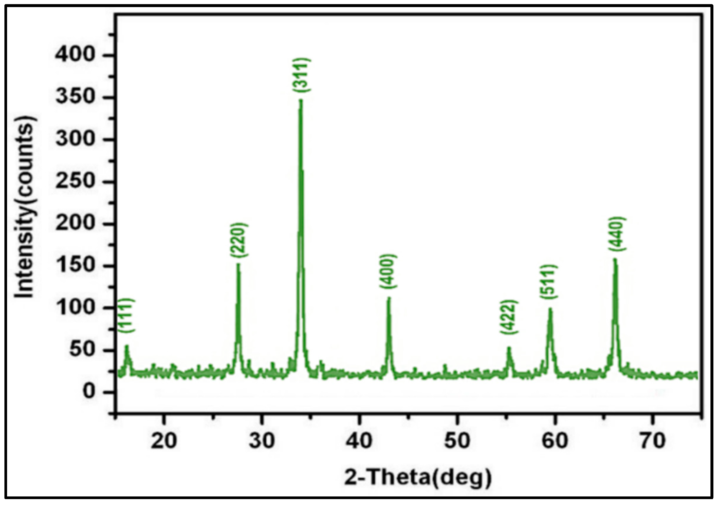
The X-ray diffraction image of the biofabricated Al2O3 NPs is shown in Figure 2. The indexes for the diffraction peaks at the corresponding planes 2θ = 27.2, 34.8, 44.7, 55.4, 59.9, and 66.3 are (220), (311), (400), (422), (511), and (440). The sample’s peak intensity indicates a higher rate of crystalline structure and a predictable structure, as seen in the illustration. As the peak’s breadth increased, the particle size decreased. This finding is in accordance with the literature, which has been published by Duraisamy [13]. The crystallite size of the nanoparticles was 28 nm, which is the common nanoparticle size. The Debye–Scherrers formula can be used to predict the crystal size of the derived NPs [10].

3.3. FT-IR Analysis

The FT-IR investigation of biofabricated Al2O3 NPs from fruit peel extract exposes numerous functional groups, which were observed in the range of 406−3412 cm−1 (Figure 3). The peaks at 406 cm−1, 1110 cm−1, and 1369 cm−1 are conspicuous peaks of Al2O3 NPs [11]. The peaks at 432 cm−1, 584 cm−1, and 830 cm−1 are because of Al-O-Al bonds [13]. An absorption peak at 1620 cm−1 and 3412 cm−1 is owing to the presence of C-O-C stretching [14].

3.4. FE-SEM with EDAX Analysis

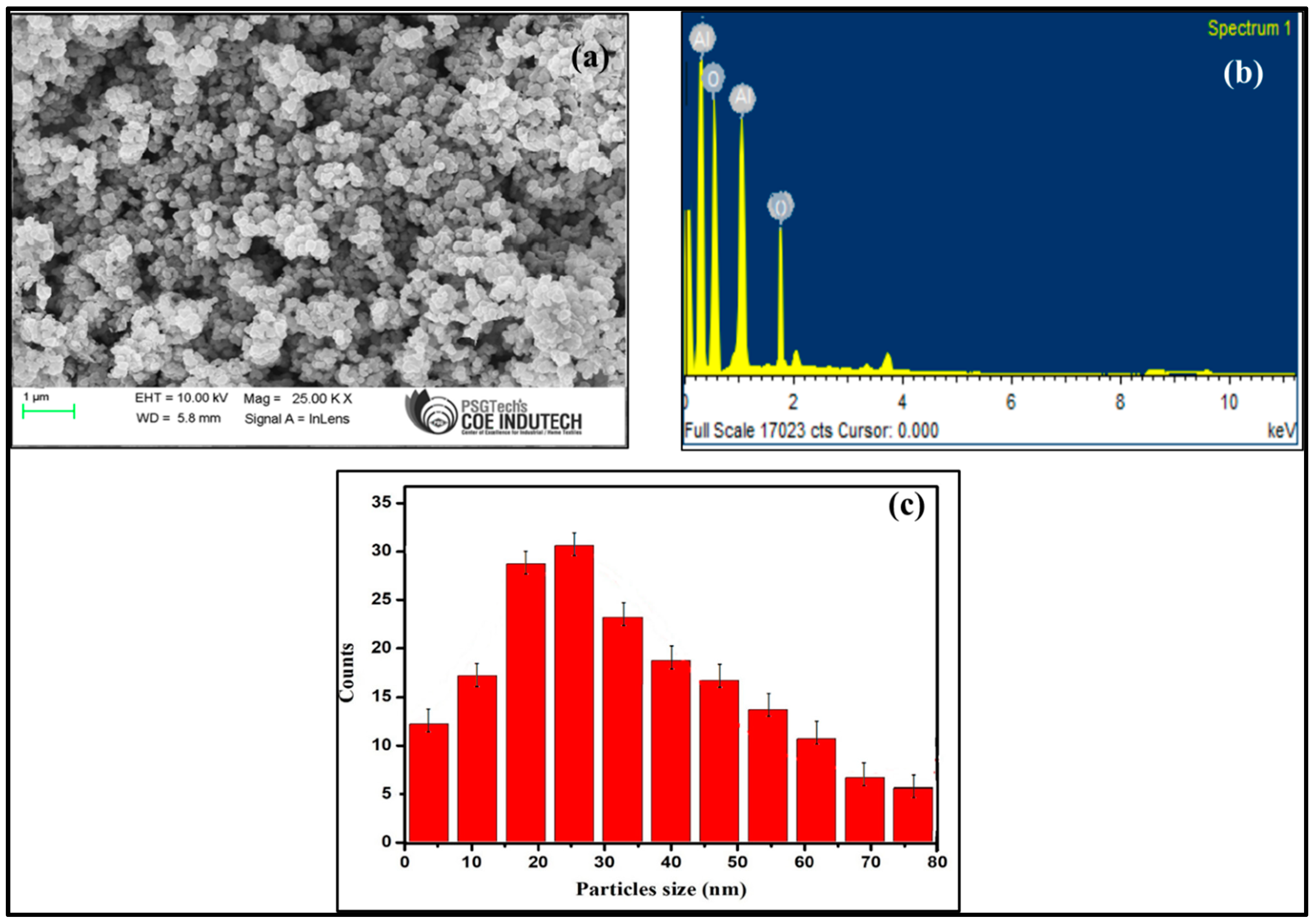
The usage of FE-SEM analysis was to analyse the structure of biofabricated Al2O3 NPs, and the results are displayed in Figure 4a with an agglomerated micrograph. It is evident that the structure of biofabricated Al2O3 NPs is spherical. To ascertain the elemental makeup and purity of Al2O3 NPs, EDAX analysis was performed. The sharp, lengthy peaks indicate the purity and atomic composition of the elements Al and O (Figure 4b). The histogram of the 28 nm particle size distribution of biofabricated Al2O3 NPs is shown in Figure 4c.

3.5. Antimicrobial Activity

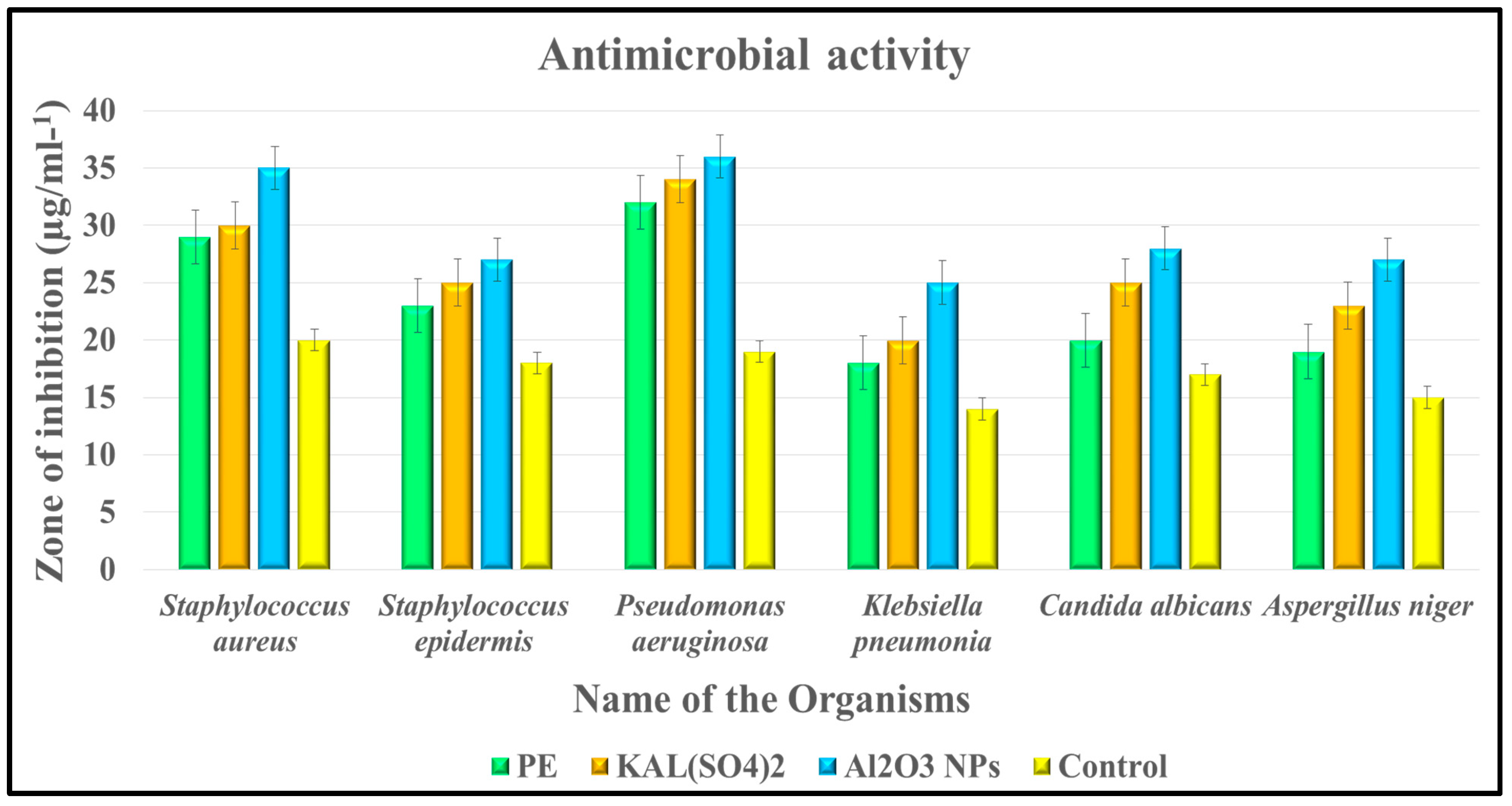
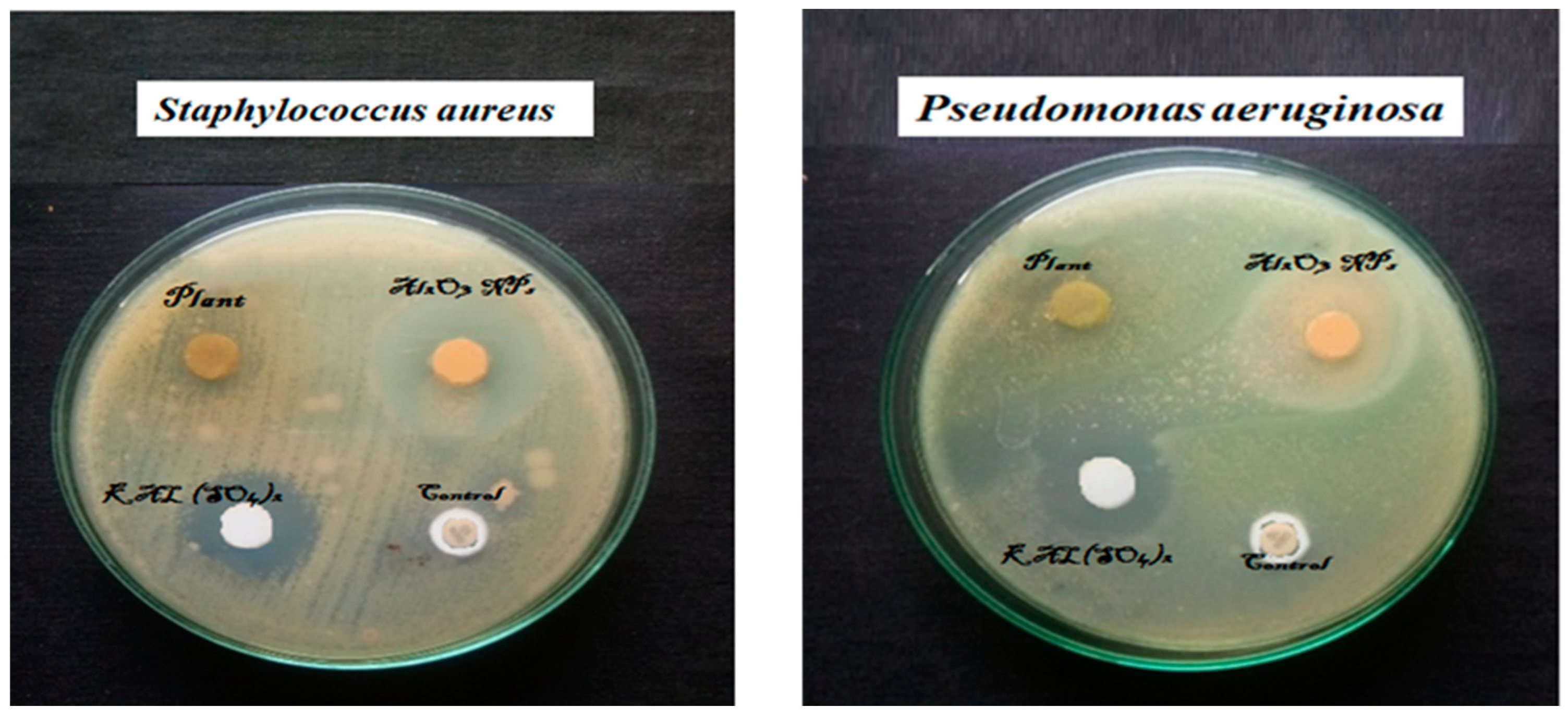
Al2O3 NPs’ antimicrobial properties were studied against pathogens like Staphylococcus aureus, Staphylococcus epidermis, Pseudomonas aeruginosa, Klebsiella pneumoniae, Candida albicans, and Aspergillus niger (Figure 5). Results showed that the Al2O3 NPs produced had strong antimicrobial action, with potent efficacy values for P. aeruginosa (36 ± 2.12), S. aureus (35 ± 1.23), S. epidermis (27 ± 0.06), K. pneumoniae (25 ± 1.65), C. albicans (28 ± 1.06), and A. niger (27 ± 2.23). Furthermore, a larger zone of inhibition was observed in S. aureus and P. aeruginosa when compared to control biofabricated Al2O3 NPs (Figure 6). A similar approach was noticed by Manogar et al. [11], who reported that Al2O3 NPs’ inhibitory impact increased when concentration was increased, but their activities other than Klebsiella pneumoniae and Candida albicans had a similar inhibiting effect (22 mm). The least inhibition activity was observed in Klebsiella pneumoniae using Citrus aurantium peel extract. Manyashree et al. [1] reported similar findings.

3.6. Antiproliferative Activity

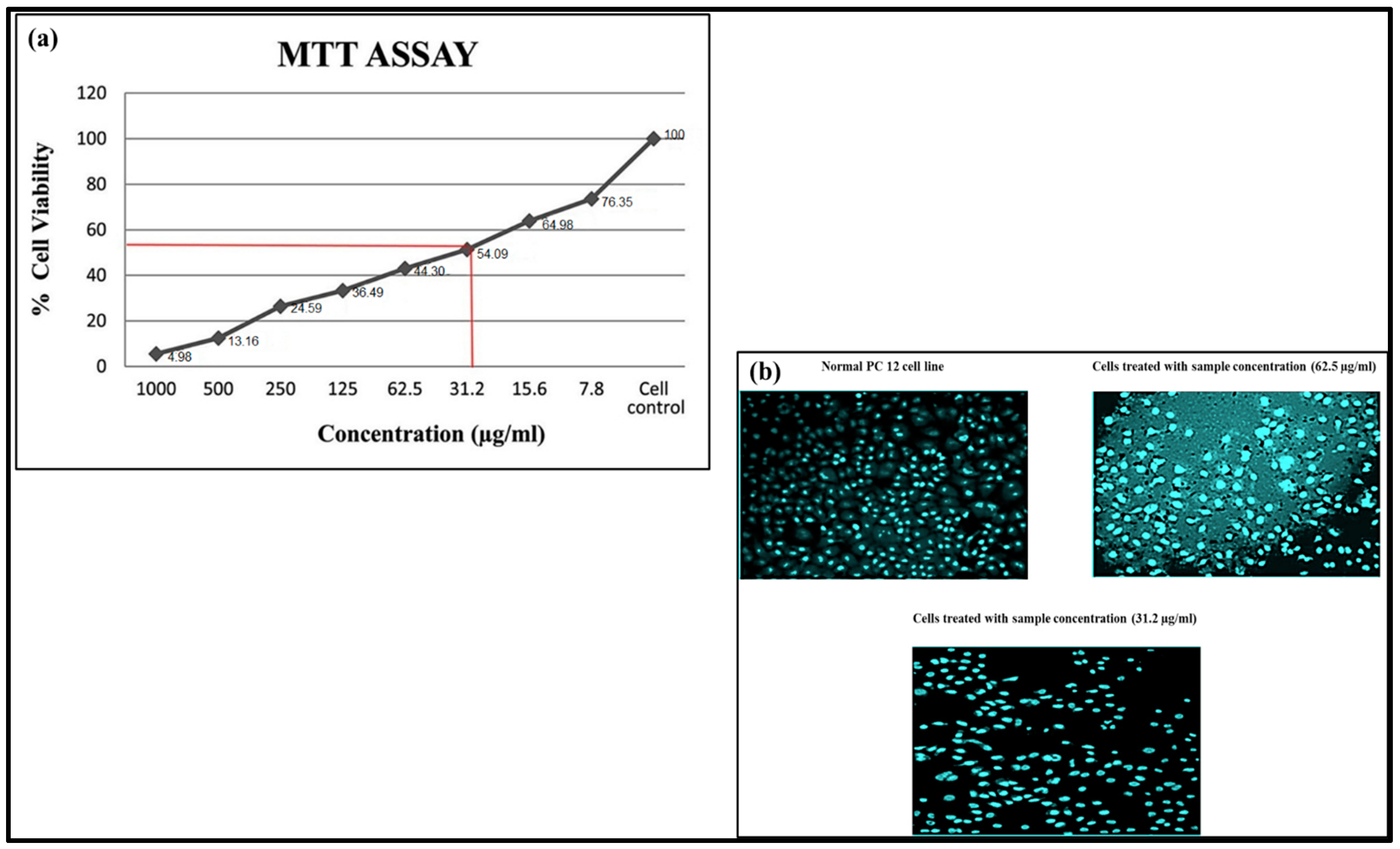
The results of cytotoxicity investigations using the PC12 cell line and MTT analysis of biofabricated Al2O3 NPs were reported in this work, and the results were 54.09% at 31.2 µg/mL. (Figure 7a). It indicates that the IC50 values for the biofabricated Al2O3 NPs are 31.2 and 62.5 µg/mL. During 24 h of treatment with various concentrations of biofabricated Al2O3 NPs, the PC12 cell line’s morphological analysis is displayed in Figure 7b.

3.7. Photocatalytic Activity

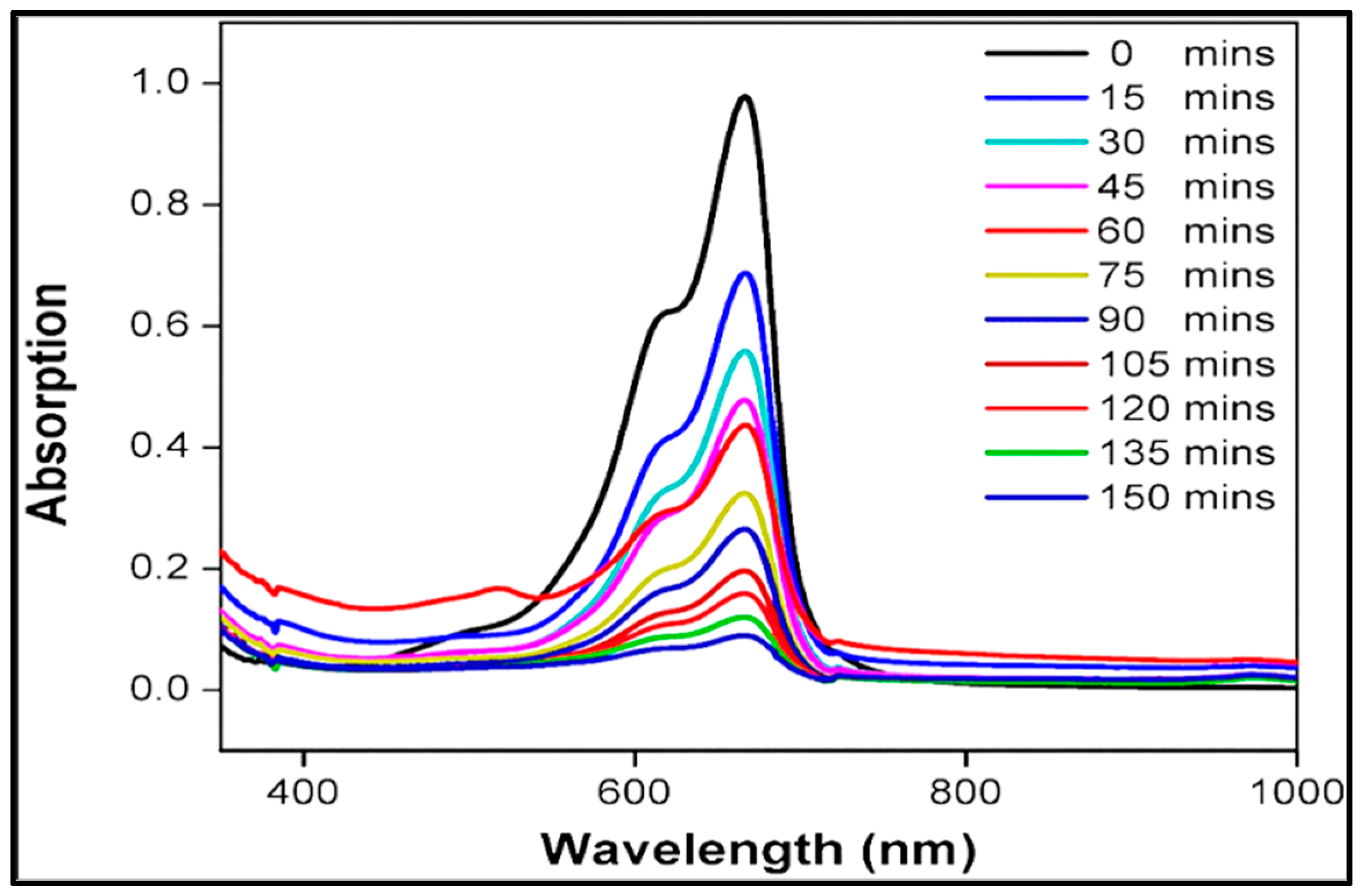
The photodegradation capability of biofabricated Al2O3 NPs and the absorption spectra for MB dye degradation are shown in Figure 8. Visible light does not exhibit an absorption spectrum peak, and the typical absorption maximum peak of MB dye appears at 665 nm. The band of MB’s absorption is reduced with respect to time by the addition of biofabricated Al2O3 NPs. Within 150 min, the MB dye’s absorption band fully deteriorated, with a maximum percentage of 89.1%. Accordingly, Kiran Kumar et al. [15] found that using 10 mg of γ-Al2O3 as a photocatalyst under sunlight resulted in 91.6% MB photodegradation in 240 min. Metal oxide nanoparticles such as ZnO, NiO, palladium oxide, and CuO have also been investigated for their electrochemical activity and have proven to be viable electrocatalysts. [16,17,18].

4. Conclusions

Based on the findings, it was determined that biofabricated Al2O3 NPs were successfully synthesised by Citrus aurantium fruit peel extract. The parameters of the generated Al2O3 NPs, including their structural, optical, elemental, and morphological characteristics, were examined using UV-vis spectroscopy, XRD, FE-SEM with EDX, and FTIR. The purity and composition of the biofabricated Al2O3 NPs can be observed by the sharp, lengthy peaks in the EDAX analysis. The study established amazing antimicrobial activity, specifically in S. aureus and P. aeruginosa, and anti-neuronal activity results show 54.09% at 31.2 µg/mL. Additionally, photodegradation efficiency is admirable with 89.1% dye degradation.

Author Contributions

Synthesised nanoparticles and investigated the antiproliferative activity, P.N.; conceptulisation and monitored the overall setup, manuscript—writing, S.V.; data analysis of characterisations of nanoparticles, V.S., V.E., N.M., P.S. and S.V. All authors have read and agreed to the published version of the manuscript. All authors have helped to finish the paper.

Funding

The authors are appreciative of the financial support provided by the DST-FIST (SR/FST/College-222/2014) and DBT-STAR (HRD-11011/18/2022-HRD-DBT) for this study.

Institutional Review Board Statement

Not Applicable.

Informed Consent Statement

Not Applicable.

Data Availability Statement

On behalf of all the authors, the corresponding author states that our data are available upon reasonable request.

Acknowledgments

The authors are appreciative of the financial support provided by the DST-FIST (SR/FST/College-222/2014) and DBT-STAR (HRD-11011/18/2022-HRD-DBT) for this study. We gratefully thank the administration of A.V.V.M. Sri Pushpam College (Autonomous), Poondi, for providing us with the resources and assistance we needed to complete this task.

Conflicts of Interest

The authors have declared no conflict of interest.

References

  1. Manyasree, D.; Kiranmayi, P.; Ravi Kumar, R.V.S.S.N. Synthesis, characterization and antibacterial activity of aluminium oxide nanoparticles. Int. J. Pharm. Pharm. Sci. 2018, 10, 32–35. [Google Scholar] [CrossRef] [Green Version]
  2. Yang, H.; Ouyang, J.; Tang, A.; Xiao, Y.; Li, X.; Dong, X.; Yu, Y. Electrochemical synthesis and photocatalytic property of cuprous oxide nanoparticles. Mater. Res. Bull. 2006, 41, 1310–1318. [Google Scholar] [CrossRef]
  3. Trinh, D.H.; Ottosson, M.; Beckers, M.; Collin, M.; Reineck, I.; Hultman, L.; Högberg, H. Structural and me-chanical characterisation of nanocomposite Al2O3-ZrO2 thin films grown by reactive dual radiofrequency magnetron sputtering. Thin Solid Film. 2008, 516, 4977–4982. [Google Scholar] [CrossRef]
  4. Reid, C.B.; Forrester, J.S.; Goodshaw, H.J.; Kisi, E.H.; Suaning, G.J. A study in the mechanical milling of alumina powder. Ceram. Int. 2008, 34, 1551–1556. [Google Scholar] [CrossRef]
  5. Qu, L.; He, C.; Yang, Y.; He, Y.; Liu, Z. Hydrothermal synthesis of alumina nanotubes templated by anionic surfactant. Mater. Lett. 2005, 59, 4034–4037. [Google Scholar] [CrossRef]
  6. Hasanpoor, M.; Nabavi, H.F.; Aliofkhazraei, M. Microwave-Assisted Synthesis of Alumina Nanoparticles Using Some Plants Extracts. J. Nanostruct. 2017, 7, 40–46. [Google Scholar] [CrossRef]
  7. Khalil, A.T.; Ovais, M.; Ullah, I.; Ali, M.; Shinwari, Z.K.; Maaza, M. Biosynthesis of iron oxide (Fe2O3) nanoparticles via aqueous extracts of Sageretia thea (Osbeck.) and their pharmacognostic properties. Green Chem. Lett. Rev. 2017, 10, 186–201. [Google Scholar] [CrossRef] [Green Version]
  8. Ditlopo, N.; Sintwa, N.; Khamlich, S.; Manikandan, E.; Gnanasekaran, K.; Henini, M.; Gibaud, A.; Krief, A.; Maaza, M. From Khoi-San indigenous knowledge to bioengineered CeO2 nanocrystals to exceptional UV-blocking green nanocosmetics. Sci. Rep. 2022, 12, 3468. [Google Scholar] [CrossRef] [PubMed]
  9. Vijayakumar, S.; Punitha, V.N.; Parameswari, N. Phytonanosynthesis of MgO Nanoparticles: Green Synthesis, Characterization and Antimicrobial Evaluation. Arab. J. Sci. Eng. 2021, 47, 6729–6734. [Google Scholar] [CrossRef]
  10. Punitha, V.N.; Vijayakumar, S.; Sakthivel, B.; Praseetha, P.K. Protection of neuronal cell lines, antimicrobial and photocatalytic behaviours of eco-friendly TiO2 nanoparticles. J. Environ. Chem. Eng. 2020, 8, 104343. [Google Scholar] [CrossRef]
  11. Manogar, P.; Morvinyabesh, J.E.; Ramesh, P.; Jeyaleela, G.D.; Amalan, V.; Ajarem, J.S.; Allam, A.A.; Khim, J.S.; Vijayakumar, N. Biosynthesis and antimicrobial activity of aluminium oxide nanoparticles using Lyngbya majuscula extract. Mater. Lett. 2022, 311, 131569. [Google Scholar] [CrossRef]
  12. Salameh, B.A.; Cumpstey, I.; Sundin, A.; Leffler, H.; Nilsson, U.J. 1H-1,2,3-Triazol-1-yl thiodigalactoside derivatives as high affinity galectin-3 inhibitors. Bioorg. Med. Chem. 2018, 18, 5367–5378. [Google Scholar] [CrossRef] [PubMed]
  13. Duraisamy, P. Green Synthesis of Aluminium Oxide Nanoparticles by using Aerva Lanta and Terminalia Chebula Extracts. IJRASET 2018, 6, 428–433. [Google Scholar] [CrossRef]
  14. Sutradhar, P.; Debnath, N.; Saha, M. Microwave-assisted rapid synthesis of alumina nanoparticles using tea, coffee and triphala extracts. Adv. Manuf. 2013, 1, 357–361. [Google Scholar] [CrossRef] [Green Version]
  15. Anna, K.K.; Bogireddy, N.K.R.; Agarwal, V.; Bon, R.R. Synthesis of α and γ phase of aluminium oxide nanoparticles for the photocatalytic degradation of methylene blue under sunlight: A comparative study. Mater. Lett. 2022, 317, 132085. [Google Scholar] [CrossRef]
  16. Matinise, N.; Fuku, X.; Kaviyarasu, K.; Mayedwa, N.; Maaza, M. ZnO nanoparticles via Moringa oleifera green synthesis: Physical properties & mechanism of formation. Appl. Surf. Sci. 2017, 406, 339–347. [Google Scholar] [CrossRef]
  17. Mayedwa, N.; Mongwaketsi, N.; Khamlich, S.; Kaviyarasu, K.; Matinise, N.; Maaza, M. Green synthesis of nickel oxide, palladium and palladium oxide synthesized via Aspalathus linearis natural extracts: Physical properties & mechanism of formation. Appl. Surf. Sci. 2010, 446, 266–272. [Google Scholar] [CrossRef]
  18. Rathnakumar, S.S.; Noluthando, K.; Kulandaiswamy, A.J.; Rayappan, J.B.B.; Kasinathan, K.; Kennedy, J.; Maaza, M. Stalling behaviour of chloride ions: A non-enzymatic electrochemical detection of α-Endosulfan using CuO interface. Sens. Actuators B Chem. 2019, 293, 100–106. [Google Scholar] [CrossRef]
Figure 1. UV-vis absorption of Al2O3 NPs.
Sustainability 15 01743 g001
Figure 2. XRD patterns of Al2O3 NPs.
Sustainability 15 01743 g002
Figure 3. FT-IR spectrum of Al2O3 NPs.
Sustainability 15 01743 g003
Figure 4. (a) FE-SEM image of Al2O3 NPs, (b) EDAX analysis of Al2O3 NPs, and (c) histogram distribution of Al2O3 NPs.
Sustainability 15 01743 g004
Figure 5. Antimicrobial analysis of biofabricated Al2O3 NPs against pathogens.
Sustainability 15 01743 g005
Figure 6. Antimicrobial evaluation of biofabricated Al2O3 NPs using the well method.
Sustainability 15 01743 g006
Figure 7. (a) Cytotoxic effect of Al2O3 NPs using MTT assay and (b) anti-proliferative effects of a human neuronal cell line (PC 12).
Sustainability 15 01743 g007
Figure 8. Photodegradation of MB with biofabricated Al2O3 NPs.
Sustainability 15 01743 g008
Disclaimer/Publisher’s Note: The statements, opinions and data contained in all publications are solely those of the individual author(s) and contributor(s) and not of MDPI and/or the editor(s). MDPI and/or the editor(s) disclaim responsibility for any injury to people or property resulting from any ideas, methods, instructions or products referred to in the content.