Article

A Sustainable Approach to In Vitro Propagation and Conservation of Salvia dominica L.: A Wild Medicinal Plant from Jordan

1
Hamdi Mango Center for Scientific Research (HMCSR), University of Jordan, Amman 11942, Jordan
2
Department of Agricultural Biotechnology and Genetic Engineering, Faculty of Agriculture Technology, Al-Ahliyya Amman University, Amman 19328, Jordan
3
Department of Horticulture and Crop Sciences, Faculty of Agriculture, University of Jordan, Amman 11942, Jordan
4
Department of Applied Sciences, Huson College, Al Balqa-Applied University, Irbid 19117, Jordan
5
Department of Applied and Social Sciences, Princess Alia University College, Al-Balqa Applied University, Amman 11191, Jordan
6
Department of Plant Protection and IPM, Faculty of Agriculture, Mutah University, Karak 61710, Jordan
*
Author to whom correspondence should be addressed.
Sustainability 2023, 15(19), 14218; https://doi.org/10.3390/su151914218
Submission received: 24 August 2023 / Revised: 22 September 2023 / Accepted: 22 September 2023 / Published: 26 September 2023

Abstract

:
Salvia dominica L. is an important wild medicinal plant that grows in Jordan and neighboring countries, and this plant has been suffering from many threats in its wild environment. Therefore, this research aims to establish a comprehensive and sustainable approach via an in vitro propagation and conservation system for the S. dominica L. plant. Axillary buds were used to initiate the in vitro culture on Murashige and Skoog MS media supplemented with 0.5 mg L−1 of GA3. In vitro shoot proliferation and rooting were experimented on with different concentrations of cytokinins and auxins, respectively. Calli were induced in the dark on excised leaf discs (0.5 cm in diameter), and multiplication was experimented on with different growth regulators. Cryopreservation experiments were applied on the callused segments under different growth conditions via the vitrification technique. A full protocol was achieved for shoot proliferation with 6.3 shoots/explant using 1.2 mg L−1 of thidiazuron (TDZ), while rooting was achieved at 1.5 mg L−1 of NAA with 6.6 functional roots/explant. Acclimatization was completely successful for the rooted plants. The highest callus production with 5.81 g/calli was achieved using 1.5 mg L−1 of benzylaminopurine (BAP). Cryopreservation of the S. dominica calli was successfully achieved when a pure plant vitrification solution (PVS2) was used to dehydrate the calli for 20 min after immersion in the loading solution for 20 min with a 76.6% regrowth percentage. The loading and the plant vitrification solution type and duration were the most critical points in the regrowth of the cryopreserved calli. In conclusion, a successful protocol was set up for the in vitro propagation and conservation of S. dominica calli. This study has prompted us to perform further studies on sustainable in vitro production and conservation of critically endangered medicinal plants to implement a green environment protecting against surrounding threats.

1. Introduction

Plant biodiversity is the main source of human civilization and the primary source of food and shelter which will sustain the livelihoods of humans and animals [1]. Plant biodiversity conservation can ensure sustainable development in terms of socioeconomic development [2,3]. Wild plants such as those in the Salvia species are an essential source of plant biodiversity. Wild plants are considered the most important plant genetic resources as they are the wild relatives of food plants [1,4]. Further, any reduction in plant biodiversity can affect other life forms in the ecosystem, and this will include all organisms taking into consideration that plants are the primary producers on Earth [1].
Salvia (sage) is the largest genus of the family Lamiaceae with about 900 species spread worldwide [5,6] and contains a number of medicinal species [7]. Jordan has been reported to contain about twenty-seven species of Salvia, either wild or domesticated [8,9]. Salvia dominica L. is a wild medicinal plant that is distributed in Jordan and is commonly named Dominica sage or also Khweekha in Arabic. S. dominica L. has been recorded in different wild parts of Jordan, and it has been used in folk medicine throughout history [8,10]. S. dominica L. is a woody subshrub that is distributed mainly in Mediterranean regions [8].
S. dominica L. has been screened for its medicinal components and medicinal activities in Jordan and other countries [10,11,12,13]. Most of these research works reported the presence of different medicinal components. The major components that were detected in the aerial parts of S. dominica L., which grows wild in Jordan, were linalool and alpha terpineol [10]. S. dominica L. is considered an important wild genetic resource and a wild relative to the Salvia species [4].
Wild plants, including S. dominica, are facing many threats to biodiversity in their natural habitats [8,14]. Previous research works depended on wild sources of the S. dominica plant from illegal collection, which threatens the wild plant source. In addition to that, people collect S. dominica from the wild for medical uses without any restrictions and in an uncontrolled way. Many other reasons have also led to the degradation of S. dominica and other wild plants [8,9,14]. These include urbanization, the climate change crisis, deforestation, pollution, environmental disasters that cause destruction in plant and animal habitats, and the destruction of the soil composition, which is considered the plant’s main living environment [8,9,14]. Therefore, most of the wild plants have become red-listed, and some plant species are now endangered or totally extinct in their natural environments, such as Salvia fruticose Mill in Jordan, which is now considered regionally extinct [8]. Therefore, it is imperative to conserve this promising medicinal plant by applying tissue culture techniques of in vitro propagation and cryopreservation techniques.
Plant tissue culture is an important technique that can serve as a tool in order to enhance propagation over a short period [15,16,17]. Plant tissue culture has been documented as a sustainable way to exploit the most important plant species in propagation and ex situ conservation [14,15,16,17]. This is because it assists the conservation of plant biodiversity [14]. The measure of plant biodiversity conservation is an important tool that can help to achieve sustainability goals [18]. This measurement can be applied by the continuous evaluation of the plant species in their natural habitat [1,2,14]. In Jordan, a plant red list has been issued in two volumes to classify plant species according to their presence (extinct, endangered, threatened, near threatened, etc.) [8,9]. However, we should manage the monitoring of plant biodiversity sustainability in an easier way to preserve the wild plants and not depend only on these measures. This is because sustainability entails renewable statistics, professional personnel, high cost for data collection, and many other obstacles that can face sustainability evaluation [19,20]. Therefore, it is very important to collaborate to retain wildlife to sustain our environment and decrease the pressure on legal and governmental agencies to preserve this important wild environment. Therefore, the application of this research can help in applying sustainability goals and assist national agencies to conserve the wild environment and plant biodiversity.
The effect of the in vitro culture conditions on Salvia species has been studied previously [21,22,23]. Most of the in vitro propagated Salvia species have economic importance due to their medicinal uses [24,25]. The most used techniques of Salvia spp. in tissue culture are in vitro propagation, conservation, and secondary metabolite production. Conservation via the tissue culture technique can involve either long- or mid-term conservation. Cryopreservation is a effective long-term way to conserve most plant species, especially those that are facing threats in their natural habitats [26]. Different cryopreservation methods have been studied previously, such as vitrification, which is an effective freeze-avoidance mechanism of cryopreservation [15]. The vitrification technique relies on exposing plant material to highly concentrated cryoprotectants at non-freezing temperatures, which leads to material dehydration [27]. In this study, S. dominica was conserved using the vitrification technique. According to our knowledge, there are, as of yet, no studies that are related to this plant species in terms of micropropagation and cryopreservation, which makes this research project novel. Thus, the aim of this study was to apply biotechnological approaches to the massive propagation and conservation of this valuable plant using tissue culture techniques.

2. Materials and Methods

2.1. Plant Source and In Vitro Establishment

Axillary buds of the S. dominica L. were obtained from wild plants in Jerash Al Raieh (32°10′03″ N 35°52′24″ E) and were identified by the plant taxonomist Hatem Taifor from the Royal Botanic Garden in Jordan. The surface sterilization process was performed by washing axillary buds from any contaminant and then rinsing them with a few drops of Tween 20 and tap water for ten minutes. After that, plant materials were agitated with HgCl2 solution (0.1%) for 2 min. Then, they were subjected to three washes in sterilized distilled water under fully sterilized conditions. Next, the mother stock establishment was initiated from the sterilized axillary buds of S. dominica using MS Murashige and Skoog media [28] with 0.5 mg L−1 of gibberellic acid (GA3). The cultures were retained under growth room conditions (16 h of light and 8 h in complete darkness with a moderate temperature of 23 ± 2 °C).

2.2. In Vitro S. dominica Proliferation, Rooting, and Acclimatization

The sterilized in vitro shoot segments (1.0 cm) with at least two nodes were transferred into Erlenmeyer flasks with Murashige and Skoog MS media [28]. Three cytokinin types were used in the prepared media, namely 6-furfuryl amino purine (kinetin), thidiazuron (TDZ), and benzylaminopurine (BAP) at varying levels (0.0, 0.4, 0.8, 1.2, and 2.0 mg L−1). The control was at the level 0.0 for each hormone. Five replications (flasks) for each treatment level were used in the proliferation experiment. The replicate had two in vitro shoot segments. Data were collected after four weeks on the number of proliferating shoots, shoot height, and callus presence using diameter measurement. Rooting was experimented with subculturing of 1.0 cm of microshoot (that has apical meristems) on MS media at varying levels (0.0, 0.5, 1.0, 1.5, 2.0 mg L−1) of different auxin types. The used auxins were indole-3-butyric acid (IBA), indole-3-acetic acid (IAA), and α-naphthalene acetic acid (NAA). The control was at the level 0.0 for each hormone. Cultures were transferred and kept in the mentioned growth room conditions. Ten replications were used; the replicate was a test tube with one microshoot. Data were recorded after four weeks for the number of roots, root length, and shoot height. In vitro acclimatization was performed using the plantlets that had well-developed roots, which were obtained from the rooting experiments. The well-developed root plantlets were hardened by transferring them from test tubes and washing them gently under tap water. After that, hardened plantlets were cultured in small pots with a 1:1 mixture of peat moss and perlite medium. Then, pots with the new hardened plantlets were left in growth room conditions, and they were wetted with distilled water twice a week. Plastic bags were used to cover the pots until the plantlets had hardened further. Then, plastic bags were taken off, and the hardened plants were kept for another two weeks in the mentioned growth room conditions. The survival of the hardened plants was taken as a percentage after four weeks. Survival was recorded for healthy, well-developed ex vitro acclimatized plants.

2.3. Induction and Multiplication of S. dominica Callus

About 0.5 cm diameter leaf segments were used to induce calli on MS media that have both 2.0 mg L−1 2,4-dichlorophenoxy acetic acid (2,4-D) and 1.0 mg L−1 of kinetin. The multiplication experiments were performed using MS media supplemented with varying levels (0.0, 0.5, 1.0, 1.5, 2.0 mg L−1) of TDZ, kinetin, or BAP plus 2.0 mg L−1 of 2,4-D by culturing of 0.5 g of friable callus segments in Petri dishes. The control treatment was MS media supplemented with 2.0 mg L−1 2,4-D only. Five replications (Petri dishes) were used for each treatment. Each replicate had four callus segments. Data were recorded after one month for callus weight, color, and texture.

2.4. Cryopreservation of S. dominica Callus

Cryopreservation was performed using the vitrification method for callus segments as calli are a more accessible and more productive genetic plant source in the cryopreservation method. About 0.1 g of friable callus segments were treated by culturing for three days in dark conditions on MS media that had high osmotic conditions by adding 0.3 M sucrose to MS media. Then, the vitrification procedure was applied with five major steps. Pretreated calli were put in cryovials, and each cryovial had 20 calli. The calli then were treated by immersion for 20 min in the loading solution (liquid hormone-free (HF) MS media + 0.4 M sucrose + 2 M glycerol). About five cryovials were used for each treatment, and each cryovial was considered a replicate. Then, they were washed in the unloading solution (liquid hormone-free (HF) MS media with 0.1 M sucrose) three times for 5 min for each. After that, callus segments were immersed in a vitrification solution (PVS2), which is a liquid MS medium with 0.4 M sucrose, and 15% ethylene glycol, 30% glycerol, and 15% DMSO (w/v), which is called pure PVS2 or 100% PVS2.
Four different experiments were performed by changing the PVS2 concentration (30%, 60%, or 100), using different immersion periods in the PVS2 (10, 20, 30, 40, and 60 min) at room temperature, or by changing the vitrification solution PVS type: PVS2, liquid HF-MS media with 30% DMSO and 1.0 M sucrose, liquid HF-MS media supplemented with 15% DMSO and 1.0 M sucrose, and liquid HF-MS media supplemented with 40% glycerol and 40% sucrose (PVS3). The final experiment was performed by changing the plant loading solution type (2 M glycerol + 0.1 M sucrose + hormone-free (HF) media, 0.5 or 0.75 M sucrose and 5% DMSO + HF media, 0.5 or 0.75 M sucrose and 10% DMSO + HF media, or 0.4 M sucrose + 2 M glycerol + HF media), and then the vitrification step was carried out using pure PVS2. The duration time of callus immersion was 20 min. in the PVS solution. After that, cryovials with plant materials were immersed in liquid nitrogen for at least 1 h. Then, the cryovials with the callus segments were removed from liquid nitrogen (LN), and they were thawed for 2 min in the unloading solution, which is described above. After that, half the cryopreserved calli (about 10 calli) from each cryovial were used for a survival 2,3,5-triphenyl tetrazolium chloride (TTC) test, which was described previously by (Rabba’a et al., 2012) [29], and the survival percentage was calculated using the formula (survival percentage = (number of red shoots/total number of shoots) × 100%). Then, the remaining vitrified calli (10 calli/cryovial) were cultured on MS media supplemented with 0.1 M sucrose for two weeks in growth room conditions in dark conditions. After that, callus regrowth was calculated according to the following formula: Regrowth % = [No. of the recovered callus clumps/Total No. of the callus clumps] × 100. Each treatment was replicated 5 times with 10 callus clumps (0.1 g each) per replicate, and the mean and standard errors were calculated for callus segments.

2.5. Statistical Analysis

All the above treatments were set in a completely randomized design (CRD). All treatments were replicated five times except for rooting experiments (ten replicates) and as described above in each section. Data were collected from each treatment, and the analysis of data variance (ANOVA) was performed separately for each treatment. The software SPSS version 18 was used. Tukey’s HSD test at 0.05 probability level was used for the separation of means. All standard errors were calculated for means.

3. Results and Discussion

3.1. Plant Sterilization and In Vitro Establishment

The surface sterilization process with mercury chloride HgCl2 was very effective in the vegetative establishment of S. dominica L. buds. The contamination was less than 5% of the cultures. Using mercury chloride HgCl2 was also effective in many studies for sterilization of the explants in the in vitro cultures. For example, mercury chloride was used successfully for Commiphora gileadensis L. leaf sterilization [30] and also for stem segment sterilization in Cnidoscolus aconitifolius (Mill.) [31]. About 95% of the cultured S. dominica L. axillary buds were developed successfully on MS media supplemented with 0.5 mg L−1 of GA3 after four weeks of establishment. Also, GA3 was effective in seedling development from in vitro axillary buds in other studies [32,33].
In this study, axillary buds were used as explants to initiate in vitro cultures. This is because they exhibited good development under the in vitro conditions, unlike seeds that were suffer from germination problems. The previous literature reported that Salvia species suffer from seed dormancy [34,35]. Furthermore, most of the previous studies documented the use of axillary buds to initiate the in vitro culture [36,37,38].
The axillary buds are an asexual propagation method that gives genetically identical plants to the mother plants rather than seeds [36,38]. According to our objective, we tried to produce a sustainable protocol to conserve this valuable plant and apply this protocol in the future in order to return this valuable plant to its environment after the acclimatization process, with fewer variations in the produced plants. The mother stock plants were established in our study using MS media supplemented with 0.5 mg L−1 GA3. The growth regulator (GA3) was documented in the previous literature to induce growth and to break bud dormancy [34,39].

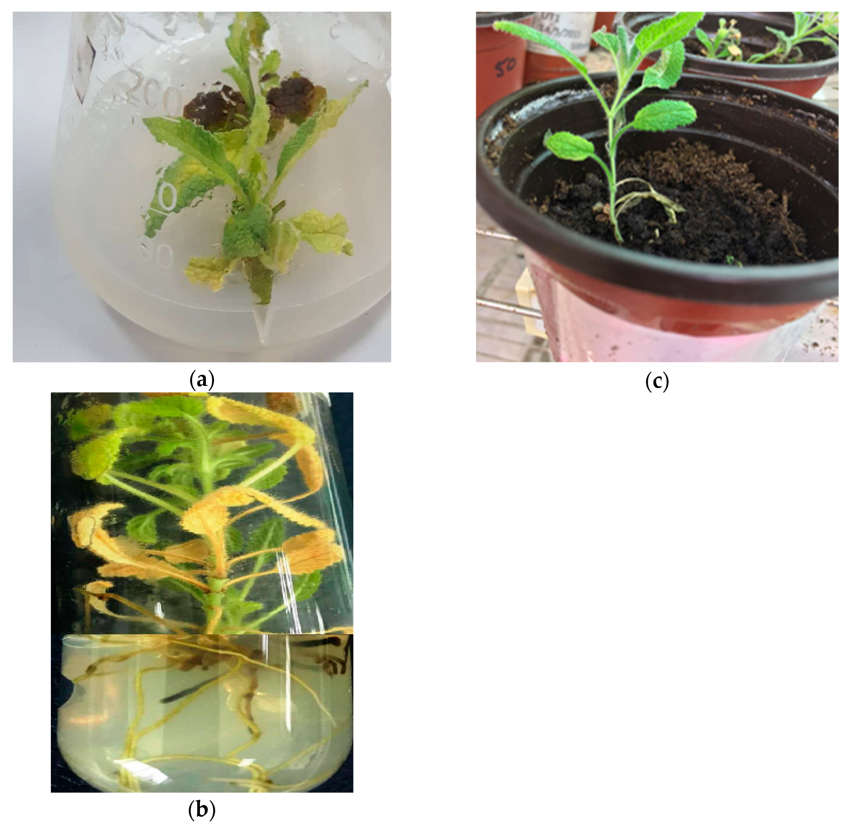
3.2. In Vitro S. dominica Proliferation, Rooting, and Acclimatization

For the in vitro shoot proliferation, the highest shoot numbers of 6.3 shoots/explant were obtained on MS media supplemented with 1.2 mg L−1 of TDZ as shown in Table 1 and Figure 1a. Also, BAP at 1.2 mg L−1 gave 4.8 shoots/explant. The growth regulator kinetin gave only 2.6 shoots at 0.8 mg L−1 (Table 1). Also, the shoot height increased with the cytokinin (CK) level, and the proliferation of shoots increased. Moreover, the callus growth on the shoot base increased when the CK level increased. We can see from Table 1 that TDZ was the best plant hormone in inducing shoot proliferation in the in vitro S. dominica L. Similarly, TDZ maximized the shoot regeneration in Salvia sclarea L. when it was used at 0.5 mg L−1 in combination with IAA [40]. Also, the highest branching of the shoots was achieved in Aeschynanthus pulcher using 3.0 mg L−1 TDZ [41], while the proliferation index in Salvia tomentosa was obtained in hormone-free media or at the low BA level [42]. Like our study, all cytokinin types except kinetin were effective in shoot regeneration in Salvia bulleyana [43]. From the above study, we can notice that plant species, explant type, and plant growth regulators were very important in the determination of the shoot proliferation in S. dominca and other plant species. In our study, a small and neglected callus was observed on the in vitro shoot base, except for when TDZ was used at high concentrations where the callus diameter on the base of the in vitro shoots reached up to 2.18 cm with 2.0 mg L−1 TDZ. Similarly, TDZ at 2.0 mg L−1 produced a higher callus diameter (8.8 mm) on the base of T. polium in vitro shoots [36].
Rooting was achieved at 1.5 mg L−1 of NAA with 6.6 functional roots/explant and 2.76 cm root length, and the shoot height reached up to 4.99 cm as shown in Table 2 and Figure 1b. Other auxins IAA and IBA also gave a high number of roots of 3.5 at 1.5 mg L−1 and 4 at 1.0 mg L−1, respectively, as shown in Table 2. We noticed that higher levels of auxins affected the number of roots adversely. The acclimatization was completely successful for the rooted plants as shown in Figure 1c. This indicates that the rooted in vitro microshoots were hardened in a proper way. In another study, in vitro Teucrium polium shoots were successfully rooted and acclimatized using NAA [36]. Also, the root induction in the woody cuttings of Rosmarinus officinalis L. was achieved using NAA [44]. On the other hand, successful rooting was achieved in Salvia sclarea L. at 0.5 mg L−1 of IAA [40]. In Salvia tomentosa, rooting was recorded at 0.1 mg L−1 IBA, and successful acclimatization was obtained [42]. The auxin type and the used level were the determinants of the root induction in the in vitro cultures. In the present study, NAA gave the most functional roots and can be used successfully for rooting and acclimatization of in vitro S. dominica shoots.

3.3. Induction and Multiplication of S. dominica Callus

Calli of S. dominica were induced successfully using 2,4-D and kinetin at 2.0 and 1.0 mg L−1, respectively. The multiplication of callus was varied according to the hormone used as shown in Table 3, and the 2,4-D at the level of 2.0 mg L−1 was constant in all media used. We can see from Table 3 that BAP at 2.0 mg L−1 gave the best callus fresh weight 5.81 g with friable texture, and the color of callus segments was white to yellow (Figure 2A). Also, kinetin at 2.0 mg L−1 gave 3.61 g of the fresh calli, but the calli were semi friable. In contrast, the high levels of TDZ concentration did not increase the callus fresh weight as kinetin and BAP. Also, the callus segments obtained from TDZ treatment were not friable, and they were in a compact form, then they turned brown in color (Figure 2B). The growth regulator 2,4-D and kinetin were used in the induction of friable calli in Salvia hispanica L [45], Salvia leriifolia [46], and Lantana camara [47]. Furthermore, BA was used in callus induction in Salvia officinalis in combination with NAA [48]. Also, BAP was the best plant growth regulator that increased the friable callus growth in different Salvia species [49,50] and other plant species such as C. gileadensis [30] and Taraxacum officinale [51]. From that, we can conclude that BAP was the best growth regulator to multiply S. dominica calli.

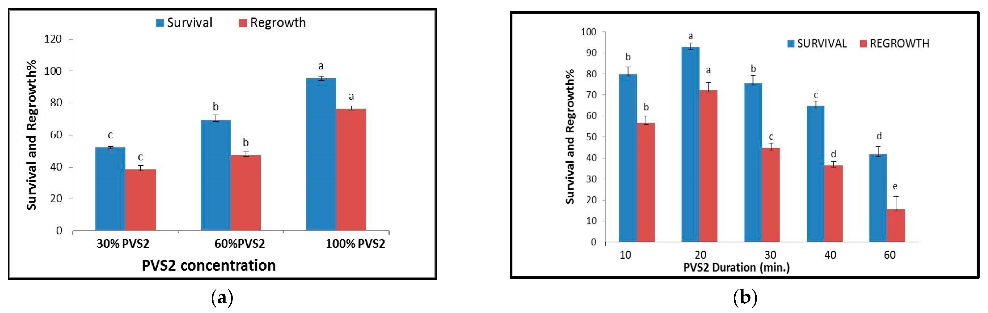
3.4. Cryopreservation of S. dominica Callus

In the cryopreservation experiments, about 95.4% of the survival percentage was recorded for cryopreserved vitrified calli of S. dominica after treatment with pure PVS2 solution for 20 min. Their regrowth percentage was 76.6% as we can see in Figure 3a. Furthermore, about 72.4% of the calli were regrown after treatment in PVS2 for 20 min, as shown in Figure 3b. Meanwhile, a high decrease in the regrowth percentage occurred when the exposure time to PVS2 was extended to 60 min (Figure 3b). The highest recovery percentages (71.8%) were recorded after cryopreservation in the plant vitrification solution PVS2; see Table 4. No recovery rate was obtained in the plant vitrification solution (30% DMSO + 1.0 M sucrose) treatment as shown below in Table 4. Loading solution-type treatments were critical to cryopreserved S. dominica calli, as shown in Table 4. The treatments 1 M sucrose, 10% DMSO + 0.5 M sucrose, and 10% DMSO + 0.75 M sucrose did not exhibit any recovery after being immersed in the liquid nitrogen, while the loading solution treatment (2 M glycerol + 0.4 M sucrose + HF-MS) gave the highest recovery percentage (71.8%) as shown in Table 4. Previous studies also indicated that plant vitrification solutions are used in the cryoprotection process, and most previous studies also indicated that PVS2 was the best vitrification solution [15,51,52]. This is because it has a cryoprotection material that is not lethal to most plant tissues, and the most used and successful PVS2 concentration was 100% in different studies [15,52,53,54]. Furthermore, the duration of incubation in PVS2 was documented in many studies as 30–50 min, and this was critical in many species [55]. The loading solution is also a critical point in the cryoprotection for many plant species and can be used for preparing the plant material for high osmotic stress before immersion in plant vitrification solution [56,57]. In this study, the best exposure time to PVS2 for the vitrified plant material was 20 min, and pure PVS2 was the best concentration used here. Meanwhile, the best vitrification solution used was pure PVS2, and the best loading solution was (0.4 M sucrose + 2 M glycerol + HF media).
In this study, cryopreservation was successful for S. dominica calli. Plant regeneration from the callus was not performed in this study. This may be used in further studies in the future. The callus is considered a valuable genetic plant material of undifferentiated tissue that has all the genetic information [58]. The previous literature documented the use of the callus with cryopreservation methods. Callus cryopreservation was used in some studies without regeneration experiments [59,60]. The ability of the callus regeneration is due to the totipotency potential in the plant cell leading to new plants through direct and indirect embryogenesis [58]. In the plant tissue culture studies, the genetic homogeneity of the in vitro propagated plants in most cases is still constant. The soma-clonal variation in the plant tissue culture is affected by the growth regulators used and the direct and indirect regeneration [61,62]. However, most reports found a low percentage of somaclonal variation occurrence in tissue-cultured plants [63,64]. In this study, no genetic stability was performed. However, some studies have documented the genetic stability after cryopreservation. For example, no genetic variations were detected in the cryopreserved shoots of Thymbra spicata [15], Rubus germplasm [65], or wasabi shoot tips [66] before and after cryopreservation. This study also may open the gate to further research depending on the current data with other details and new approaches.

4. Conclusions

The current study describes a sustainable approach to in vitro propagation and conservation of S. dominica. A successful protocol has been set for the first time for in vitro shoot proliferation, rooting, acclimatization, and conservation via a vitrification procedure in S. dominca. The highest shoot (6.3) and root (6.6) numbers were obtained using 1.2 mg L−1 of TDZ and 1.5 mg L−1 of NAA, respectively. The rooted plants achieved a full acclimatization process. Also, calli were successfully produced with 5.81 g/calli using BAP at 1.5 mg L−1. On the other hand, the highest regrowth 76.6% was achieved after cryopreservation of S. dominica calli using 0.4 M sucrose + 2 M glycerol + HF media as a loading solution for 20 min and 100% PVS2 for 20 min before immersion in the liquid nitrogen. This protocol was easy to handle and produced efficient hardened plants that can be returned to their wild environments as sustainable sources. In addition, a promising conservation method via vitrification was achieved, and the critical points in the S. dominica cryopreservation were determined. So, the used protocol enables us to conserve valuable genetic resources for the long term with high regrowth percentages, which will help in sustainable exploitation of the ex situ conservation of this valuable genetic resource. Furthermore, it will help in conserving plant biodiversity in which the environment serves as a source for human food and shelter. So, the three pillars of sustainability (socioeconomic, environmental, and life) will be achieved.

Author Contributions

Conceptualization and visualization, T.S.A.-Q. and R.A.S.; methodology, T.S.A.-Q. and R.W.T.; software, T.S.A.-Q.; validation, R.A.S., T.S.A.-Q., R.W.T., A.Z. and F.A.-Z.; formal analysis, T.S.A.-Q. and F.A.-Z.; investigation, R.A.S., T.S.A.-Q., A.Z., R.W.T. and F.A.-Z.; resources, T.S.A.-Q., A.Z. and R.A.S.; data curation, R.A.S. and T.S.A.-Q.; writing—original draft preparation T.S.A.-Q. and R.A.S.; writing—review and editing, R.A.S., T.S.A.-Q., A.Z., R.W.T. and F.A.-Z.; supervision, T.S.A.-Q. and R.A.S.; project administration, T.S.A.-Q. and R.A.S. All authors have read and agreed to the published version of the manuscript.

Funding

This research was funded by the Deanship of Scientific Research at the University of Jordan, grant number 2004.

Institutional Review Board Statement

Not applicable.

Informed Consent Statement

Not applicable.

Data Availability Statement

The datasets supporting the results of this article will be freely available upon reasonable request from Rida Shibli and Tamara Al-Qudah.

Acknowledgments

The authors are thankful to the Deanship of Scientific Research at the University of Jordan for funding this project.

Conflicts of Interest

The authors declare no conflict of interest.

References

  1. Niesenbaum, R.A. The integration of conservation, biodiversity, and sustainability. Sustainability 2019, 11, 4676. [Google Scholar] [CrossRef]
  2. Napreenko, M.G.; Antsiferova, O.A.; Aldushin, A.V.; Samerkhanova, A.K.; Aldushina, Y.K.; Baranovskiy, P.N.; Napreenko-Dorokhova, T.V.; Panov, V.V.; Konshu, E.V. New approaches to sustainable management of wetland and forest ecosystems as a response to changing socio-economic development contexts. In Innovations and Traditions for Sustainable Development; Springer: Cham, Switzerland, 2021; pp. 395–416. [Google Scholar]
  3. Borón, V.; Payán, E.; MacMillan, D.; Tzanopoulos, J. Achieving sustainable development in rural areas in Colombia: Future scenarios for biodiversity conservation under land use change. Land Use Policy 2016, 59, 27–37. [Google Scholar] [CrossRef]
  4. Khalil, A.; Hassawi, D.S.; Kharma, A. Genetic relationship among Salvia species and antimicrobial activity of their crude extract against pathogenic bacteria. Asian J. Plant Sci. 2005, 4, 544–549. [Google Scholar] [CrossRef]
  5. Zihlif, M.; Afifi, F.; Abu-Dahab, R.; Abdul Majid, A.M.S.; Somrain, H.; Saleh, M.M.; Naffa, R. The antiangiogenic activities of ethanolic crude extracts of four Salvia species. BMC Complement. Altern. Med. 2013, 13, 358. [Google Scholar] [CrossRef] [PubMed]
  6. Şenkal, B.C.; Uskutoğlu, T.; Cesur, C.; ÖZAVCI, V.; Doğan, H. Determination of essential oil components, mineral matter, and heavy metal content of Salvia virgata Jacq. grown in culture conditions. Turk. J. Agric. For. 2019, 43, 395–404. [Google Scholar] [CrossRef]
  7. Goren, A.C.; Kilic, T.; Dirmenci, T.; Bilsel, G. Chemotaxonomic evaluation of Turkish species of Salvia: Fatty acid compositions of seed oils. Biochem. Syst Ecol. 2006, 34, 160–164. [Google Scholar] [CrossRef]
  8. Taifour, H.; El-Oqlah, A. Jordan Plant Red List; Royal Botanic Garden: Kew, UK, 2014; Volume 1. [Google Scholar]
  9. Taifour, H. Jordan Plant Red List; Royal Botanic Garden: Kew, UK, 2016; Volume 2. [Google Scholar]
  10. Abdallah, M.; Abu-Dahab, R.; Afifi, F. Composition of the essential oils from Salvia dominica L. and Salvia hormium L. grown in Jordan. Jordan J. Pharm. Sci. 2013, 6, 40–46. [Google Scholar] [CrossRef]
  11. Hasan, M.R.; Al-Jaber, H.I.; Al-Qudah, M.A.; Zarga, M.H.A. New sesterterpenoids and other constituents from Salvia dominica growing wild in Jordan. Phytochem. Lett. 2016, 16, 12–17. [Google Scholar] [CrossRef]
  12. Fiore, G.; Nencini, C.; Cavallo, F.; Capasso, A.; Bader, A.; Giorgi, G.; Micheli, L. In vitro antiproliferative effect of six Salvia species on human tumor cell lines. Phytother. Res. Int. J. Devoted Pharmacol. Toxicol. Eval. Nat. Prod. Deriv. 2006, 20, 701–703. [Google Scholar]
  13. Al-Jaber, H.I.; Bdair, O.; Barhoumi, L.M.; Dabaibeh, R.N.; Abu-Zarga, M.H.; Abaza, I.F.; Afifi, F.U. Characterization of chemical components in organs emissions of two Salvia dominica L. populations from Jordan. J. Oleo Sci. 2020, 69, 759–765. [Google Scholar] [CrossRef]
  14. Radomir, A.M.; Stan, R.; Florea, A.; Ciobotea, C.M.; Bănuță, F.M.; Negru, M.; Sumedrea, D.I. Overview of the Success of In Vitro Culture for Ex Situ Conservation and Sustainable Utilization of Endemic and Subendemic Native Plants of Romania. Sustainability 2023, 15, 2581. [Google Scholar] [CrossRef]
  15. Tahtamouni, R.W.; Shibli, R.A.; Al-Abdallat, A.M.; Al-Qudah, T.S.; Younis, L.; Al-Baba, H.; Al-Ruwaiei, H. Cryopreservation of Thymbra spicata L. var. spicata and genetic stability assessment of the cryopreserved shoot tips after conservation. Jordan J. Biol. Sci. 2017, 10, 19–28. [Google Scholar]
  16. Al-Abdallat, A.M.; Shibli, R.A.; Akash, M.W.; Rabbaa, M.; Al-Qudah, T. In vitro preservation of transgenic tomato (Solanum lycopersicum L.) plants overexpressing the stress-related SlAREB1 transcription factor. Int. J. Mol. Sci. 2017, 18, 1477. [Google Scholar] [CrossRef] [PubMed]
  17. Gianguzzi, V.; Barone, G.; Di Gristina, E.; Sottile, F.; Domina, G. Micropropagation of Endemic Endangered Taxa of the Italian Flora: Adenostyles alpina subsp. macrocephala (Asteraceae), as a Case Study. Plants 2023, 12, 1530. [Google Scholar] [CrossRef]
  18. Teixeira, J.C.; Huber, C.D. The inflated significance of neutral genetic diversity in conservation genetics. Proc. Nat. Acad. Sci. USA 2021, 118, e2015096118. [Google Scholar] [CrossRef]
  19. Maynard, D.D.C.; Vidigal, M.D.; Farage, P.; Zandonadi, R.P.; Nakano, E.Y.; Botelho, R.B.A. Environmental, social and economic sustainability indicators applied to food services: A systematic review. Sustainability 2020, 12, 1804. [Google Scholar] [CrossRef]
  20. Salgotra, R.K.; Sharma, M.; Pandotra, P. Biotechnological Interventions for Sustainable Conservation of Plant Genetic Resources in the Scenario of Climate Change. Nat. Resour. Conserv. Res. 2019, 2, 754. [Google Scholar] [CrossRef]
  21. Papafotiou, M.; Vlachou, G.; Martini, A.N. Investigation of the Effects of the Explant Type and Different Plant Growth Regulators on Micropropagation of Five Mediterranean Salvia spp. Native to Greece. Horticulturae 2023, 9, 96. [Google Scholar] [CrossRef]
  22. Cuenca, S.; Amo-Marco, J.B. In vitro propagation of two spanish endemic species of salvia throught bud proliferation. In Vitro Cell. Dev. Biol.-Plant 2000, 36, 225–229. [Google Scholar] [CrossRef]
  23. Skała, E.; Wysokińska, H. In vitro regeneration of Salvia nemorosa L. from shoot tips and leaf explants. In Vitro Cell. Dev. Biol.-Plant 2004, 40, 596–602. [Google Scholar] [CrossRef]
  24. Grigoriadou, K.; Trikka, F.A.; Tsoktouridis, G.; Krigas, N.; Sarropoulou, V.; Papanastasi, K.; Maloupa, E.; Makris, A.M. Μicropropagation and cultivation of Salvia sclarea for essential oil and sclareol production in northern Greece. In Vitro Cell. Dev. Biol.-Plant 2020, 56, 51–59. [Google Scholar] [CrossRef]
  25. Mehalaine, S.; Chenchouni, H. New insights for the production of medicinal plant materials: Ex vitro and in vitro propagation of valuable Lamiaceae species from northern Africa. Curr. Plant Biol. 2021, 27, 100216. [Google Scholar] [CrossRef]
  26. Popova, E.; Kulichenko, I.; Kim, H.H. Critical Role of Regrowth Conditions in Post-Cryopreservation of In Vitro Plant Germplasm. Biology 2023, 12, 542. [Google Scholar] [CrossRef] [PubMed]
  27. Ruta, C.; De Mastro, G.; Tarraf, W.; Ancona, S.; Tagarelli, A.; Ozudogru, A.; Lambardi, M. Long-term preservation of Cicer arietinum L. germplasm by in vitro propagation and cryopreservation. Genet. Resour. Crop Evol. 2020, 67, 263–271. [Google Scholar] [CrossRef]
  28. Murashige, T.; Skoog, F. A revised medium for rapid growth and bioassays with tobacco tissue cultures. Physiol. Plant. 1962, 15, 473–479. [Google Scholar] [CrossRef]
  29. Rabba’a, M.M.; Shibli, R.A.; Shatnawi, M.A. Cryopreservation of Teucrium polium L. shoot-tips by vitrification and encapsulation-dehydration. Plant Cell Tissue Organ Cult. (PCTOC) 2012, 110, 371–382. [Google Scholar] [CrossRef]
  30. Al-Abdallat, A.M.; Adayileh, B.K.; Sawwan, J.S.; Shibli, R.; Al-Qudah, T.S.; Abu-Irmaileh, B.; Albdaiwi, R.N.; Almaliti, J.; Bustanji, Y. Secondary Metabolites Profiling, Antimicrobial and Cytotoxic Properties of Commiphora gileadensis L. Leaves, Seeds, Callus, and Cell Suspension Extracts. Metabolites 2023, 13, 537. [Google Scholar] [CrossRef]
  31. Gu, M.; Li, Y.; Jiang, H.; Zhang, S.; Que, Q.; Chen, X.; Zhou, W. Efficient in vitro sterilization and propagation from stem segment explants of Cnidoscolus aconitifolius (Mill.) IM Johnst, a multipurpose woody plant. Plants 2022, 11, 1937. [Google Scholar] [CrossRef]
  32. Hirakawa, T.; Tanno, S. In vitro propagation of Humulus lupulus through the induction of axillary bud development. Plants 2022, 11, 1066. [Google Scholar] [CrossRef]
  33. Kaviani, B.; Deltalab, B.; Kulus, D.; Tymoszuk, A.; Bagheri, H.; Azarinejad, T. In Vitro Propagation of Pyracantha angustifolia (Franch.) CK Schneid. Horticulturae 2022, 8, 964. [Google Scholar] [CrossRef]
  34. Aghilian, S.; Khajeh-Hosseini, M.; Anvarkhah, S. Evaluation of seed dormancy in forty medicinal plant species. Int. J. Agric. Crop Sci. 2014, 7, 760. [Google Scholar]
  35. Tursun, A.O. The effects of low temperature applications on dormancy of Salvia verticillata L. and Rumex crispus L. seeds. Pak. J. Bot. 2020, 52, 1885–1890. [Google Scholar] [CrossRef] [PubMed]
  36. Al-Qudah, T.S.; Shibli, R.A.; Alali, F.Q. In vitro propagation and secondary metabolites production in wild germander (Teucrium polium L.). In Vitro Cell. Dev. Biol.-Plant 2011, 47, 496–505. [Google Scholar] [CrossRef]
  37. Echeverrigaray, S.; Carrer, R.P.; Andrade, L.B. Micropropagation of Salvia guaranitica Benth. through axillary shoot proliferation. Braz. Arch. Biol. Technol. 2010, 53, 883–888. [Google Scholar] [CrossRef]
  38. Nanos, C.; Tsoulpha, P.; Kostas, S.; Hatzilazarou, S.; Michail, I.; Anastasiadi, V.; Nianiou-Obeidat, I. Asexual Propagation of Greek Salvia officinalis L. Populations Selected for Ornamental Use. Horticulturae 2023, 9, 847. [Google Scholar] [CrossRef]
  39. Mahmoodi, P.; Khajeh-Hosseini, M.; Rashed-Mohassel, M.; Emamipoor, Y. The effect of gibberellic acid and potassium nitrate on seed dormancy of fourteen medicinal plant species (Lamiaceae). Iran. J. Seed Sci. Technol. 2018, 7, 233–242. [Google Scholar]
  40. Ghanbar, T.; Hosseini, B.A.H.M.A.N.; Jabbarzadeh, Z.O.H.R.E.H.; Farokhzad, A.L.I.R.E.Z.A.; Sharafi, A. High-frequency in vitro direct shoots regeneration from axillary nodal and shoot tip explants of clary sage (Salvia sclarea L.). Bulg. J. Agric. Sci. 2016, 22, 73–78. [Google Scholar]
  41. Yang, H.; Yuan, H.; Du, C.; Liang, L.; Chen, M.; Zou, L. Development of a Highly Efficient Shoot Organogenesis System for an Ornamental Aeschynanthus pulcher (Blume) G. Don Using Leaves as Explants. Plants 2022, 11, 2456. [Google Scholar] [CrossRef] [PubMed]
  42. Martini, A.N.; Vlachou, G.; Papafotiou, M. Effect of explant origin and medium plant growth regulators on in vitro shoot proliferation and rooting of Salvia tomentosa, a native sage of the Northeastern Mediterranean Basin. Agronomy 2022, 12, 1889. [Google Scholar] [CrossRef]
  43. Grzegorczyk-Karolak, I.; Hnatuszko-Konka, K.; Krzemińska, M.; Olszewska, M.A.; Owczarek, A. Cytokinin-based tissue cultures for stable medicinal plant production: Regeneration and phytochemical profiling of Salvia bulleyana shoots. Biomolecules 2021, 11, 1513. [Google Scholar] [CrossRef]
  44. Kostas, S.; Kaplani, A.; Koulaouzidou, E.; Kotoula, A.A.; Gklavakis, E.; Tsoulpha, P.; Hatzilazarou, S.; Nianiou-Obeidat, I.; Kanellis, A.K.; Economou, A. Sustainable Exploitation of Greek Rosmarinus officinalis L. Populations for Ornamental Use through Propagation by Shoot Cuttings and In Vitro Cultures. Sustainability 2022, 14, 4059. [Google Scholar] [CrossRef]
  45. Marconi, P.L.; López, M.C.; De Meester, J.; Bovjin, C.; Alvarez, M.A. In vitro establishment of Salvia hispanica L. plants and callus. Biotecnol. Veg. 2013, 13, 203–207. [Google Scholar]
  46. Modarres, M.; Asili, J.; Lahouti, M.; Gangali, A.; Iranshahy, M.; Sahebkar, A. Simultaneous determination of rosmarinic acid, salvianolic acid B and caffeic acid in Salvia leriifolia Benth. root, leaf and callus extracts using a high-performance liquid chromatography with diode-array detection technique. J. Liq. Chromatogr. Relat. Technol. 2014, 37, 1721–1730. [Google Scholar] [CrossRef]
  47. Tahtamouni, R.W.; Shibli, R.A.; Younes, L.S.; Abu-Mallouh, S.; Al-Qudah, T.S. Responses of Lantana Camara Linn. Callus Cultures to Heavy Metals Added to the Culture Media. Jordan J. Biol. Sci. 2020, 13, 551–557. [Google Scholar]
  48. Lemraski, M.G.; Eftekhari, M.; Faraji, M.; Zarrini, S.S. Study of callus induction in common sage (Salvia officinalis L.). Int. J. Agric. Crop Sci. (IJACS) 2014, 7, 386–389. [Google Scholar]
  49. Mederos-Molina, S. In vitro callus induction and plants from stem and petiole explants of Salvia canariensis L. Plant Tissue Cult. 2004, 14, 167–172. [Google Scholar]
  50. Hemmati, N.; Cheniany, M.; Ganjeali, A. Effect of plant growth regulators and explants on callus induction and study of antioxidant potentials and phenolic metabolites in Salvia tebesana Bunge. Bot. Serbica 2020, 44, 163–173. [Google Scholar] [CrossRef]
  51. Martinez, M.E.; Jorquera, L.; Poirrier, P.; Díaz, K.; Chamy, R. Effect of the carbon source and plant growth regulators (PGRs) in the induction and maintenance of an in vitro callus culture of Taraxacum officinale (L) weber Ex FH Wigg. Agronomy 2021, 11, 1181. [Google Scholar] [CrossRef]
  52. Zamecnik, J.; Faltus, M.; Bilavcik, A. Vitrification solutions for plant cryopreservation: Modification and properties. Plants 2021, 10, 2623. [Google Scholar] [CrossRef]
  53. Delgado-Aceves, L.; González-Arnao, M.T.; Santacruz-Ruvalcaba, F.; Folgado, R.; Portillo, L. Indirect somatic embryogenesis and cryopreservation of Agave tequilana Weber cultivar ‘Chato’. Plants 2021, 10, 249. [Google Scholar] [CrossRef]
  54. Sakai, A.; Kobayashi, S.; Oiyama, I. Cryopreservation of nucellar cells of navel orange (Citrus sinensis Osb. var. brasiliensis Tanaka) by vitrification. Plant Cell Rep. 1990, 9, 30–33. [Google Scholar] [CrossRef] [PubMed]
  55. Li, B.-Q.; Feng, C.-H.; Wang, M.-R.; Hu, L.-Y.; Volk, G.; Wang, Q.-C. Recovery patterns, histological observations and genetic integrity in Malus shoot tips cryopreserved using droplet-vitrification and encapsulation-dehydration procedures. J. Biotechnol. 2015, 214, 182–191. [Google Scholar] [CrossRef] [PubMed]
  56. Juan-Vicedo, J.; Ramírez-Luna, J.E.; Piqueras, A.; Casas, J.L. Micropropagation and cryopreservation by vitrification of the Spanish endemic medicinal plant Sideritis leucantha Cav. subsp. leucantha (Lamiaceae). In Vitro Cell. Dev. Biol.-Plant 2021, 57, 1057–1065. [Google Scholar] [CrossRef]
  57. Roque-Borda, C.A.; Kulus, D.; Vacaro de Souza, A.; Kaviani, B.; Vicente, E.F. Cryopreservation of agronomic plant germplasm using vitrification-based methods: An overview of selected case studies. Int. J. Mol. Sci. 2021, 22, 6157. [Google Scholar] [CrossRef]
  58. Efferth, T. Biotechnology applications of plant callus cultures. Engineering 2019, 5, 50–59. [Google Scholar] [CrossRef]
  59. Chenshu, A.; Wang, X.; Yuan, X.; Zhao, B.; Wang, Y. Optimization of cryopreservation of Artemisia annua L. callus. Biotechnol. Lett. 2003, 25, 35–38. [Google Scholar] [CrossRef]
  60. Popova, E.V.; Lee, E.J.; Wu, C.H.; Hahn, E.J.; Paek, K.Y. A simple method for cryopreservation of Ginkgo biloba callus. Plant Cell Tissue Organ Cult. (PCTOC) 2009, 97, 337–343. [Google Scholar] [CrossRef]
  61. Bulbarela-Marini, J.E.; Gómez-Merino, F.C.; Galindo-Tovar, M.E.; Pastelín-Solano, M.C.; Murguía-González, J.; Núñez-Pastrana, R.; Castañeda-Castro, O. Ratio of Somaclonal Variation and the Phytohormonal Content of Citrus× latifolia in Three In Vitro Culture Systems. J. Plant Growth Regul. 2023, 42, 3356–3364. [Google Scholar] [CrossRef]
  62. Biswas, P.; Kumar, N. Application of Molecular Markers for the Assessment of Genetic Fidelity of In Vitro Raised Plants: Current Status and Future Prospects. In Molecular Marker Techniques; Springer: Singapore, 2023; pp. 233–256. [Google Scholar]
  63. Duta-Cornescu, G.; Constantin, N.; Pojoga, D.M.; Nicuta, D.; Simon-Gruita, A. Somaclonal variation—Advantage or disadvantage in micropropagation of the medicinal plants. Int. J. Mol. Sci. 2023, 24, 838. [Google Scholar] [CrossRef]
  64. Taji, A.; Prakash, N.; Lakshmanan, P. In Vitro Plant Breeding; Food Products Press: New York, NY, USA, 2002. [Google Scholar]
  65. Castillo, N.R.F.; Bassil, N.V.; Wada, S.; Reed, B.M. Genetic stability of cryopreserved shoot tips of Rubus germplasm. In Vitro Cell. Dev. Biol.-Plant 2010, 46, 246–256. [Google Scholar] [CrossRef]
  66. Matsumoto, T.; Akihiro, T.; Maki, S.; Mochida, K.; Kitagawa, M.; Tanaka, D.; Niino, T. Genetic stability assessment of Wasabi plants regenerated from long-term cryopreserved shoot tips using morphological, biochemical and molecular analysis. CryoLetters 2013, 34, 128–136. [Google Scholar] [PubMed]
Figure 1. In vitro propagation of in vitro grown S. dominica L.: (a) shoot multiplication; (b) rooting; (c) acclimatization.
Sustainability 15 14218 g001
Figure 2. Callus multiplication of S. dominica. (A) White, yellow, and friable callus grown on MS media supplemented with 2.0 mg L−1 of 2,4-D and BAP. (B) Brown and compact callus grown on MS media supplemented with 1.0 mg L−1 TDZ and 2.0 mg L−1 of 2,4-D.
Sustainability 15 14218 g002
Figure 3. The survival and regrowth as expressed in percentages for the S. dominica calli after cryopreservation by vitrification as influenced by: (a) PVS2 concentrations (30%, 60%, 100%) and (b) different durations of exposure to PVS2 (10, 20, 30, 40, 60 min). The column represents the mean of data entries for each treatment separately ± standard error (the bar). Different letters express that means are significantly different in accordance with Tukey’s HSD test at p ≤ 0.05. The survival and regrowth are shown separately in (a,b).
Sustainability 15 14218 g003
Table 1. In vitro shoot proliferation of S. dominica L. after being treated with different levels of BAP, kinetin, and TDZ after four weeks of incubation in growth room conditions.
Cytokinin TypeConcentration (mg L−1)Number of
Microshoots/Explant
Shoot Height (cm)Callus Diameter
(cm)
BAP y0.0 (control)1.20 ± 0.12 d *1.36 ± 0.11 b0.13 ± 0.02 c
0.42.50 ± 0.15 c2.38 ± 0.090 a0.13 ± 0.01 c
0.82.20 ± 0.12 c2.18 ± 0.080 a0.61 ± 0.07 ab
1.24.80 ± 0.12 a2.46 ± 0.14 a0.84 ± 0.10 a
2.03.0 ± 0.16 b1.44 ± 0.07 b0.52 ± 0.01 b
Kinetin0.0 (control)1.40 ± 0.19 b1.36 ± 0.09 b0.30 ± 0.07 c
0.41.40 ± 0.19 b2.60 ± 0.22 a0.52 ± 0.11 bc
0.82.60 ± 0.18 a1.94 ± 0.35 ab0.72 ± 0.08 b
1.21.30 ± 0.12 b1.98 ± 0.26 ab0.83 ± 0.02 b
2.01.80 ± 0.25 a2.46 ± 0.25 a1.2 ± 0.10 a
TDZ0.0 (control)1.20 ± 0.19 d1.40 ± 0.070 c0.18 ± 0.04 d
0.42.20 ± 0.20 c1.46 ± 0.12 c0.58 ± 0.11 cd
0.82.80 ± 0.12 c2.14 ± 0.20 b0.68 ± 0.10 c
1.26.30 ± 0.30 a2.76 ± 0.19 b1.40 ± 0.05 b
2.04.40 ± 0.18 b3.70 ± 0.11 a2.18 ± 0.18 a
y Each growth regulator for each parameter was analyzed separately in the same column. * Different letters in each column express the data means that are significantly different in accordance with Tukey’s HSD test at p ≤ 0.05. Each data entry is the mean ± standard error.
Table 2. In vitro rooting of S. dominica L. after being treated with different levels of NAA, IAA, and IBA after four weeks of incubation in growth room conditions.
Auxin TypeConcentration (mg L−1)Number of RootsRoot Length
(cm)
Shoot Height
(cm)
NAA y0.0 (control)0.00 ± 0.00 d *0.00 ± 0.00 e1.42 ± 0.04 d
0.51.3 ± 0.21 c0.5 ± 0.08 d1.65 ± 0.06 d
1.02.5 ± 0.17 b0.89 ± 0.09 c2.22 ± 0.85 c
1.56.6 ± 0.34 a2.76 ± 0.08 a4.99 ± 0.15 a
2.02.6 ± 0.16 b1.31 ± 0.16 b2.78 ± 0.19 b
IAA0.0 (control)0.0 ± 0.0 c0.00 ± 0.0 c1.27 ± 0.04 d
0.50.7 ± 0.26 c0.15 ± 0.06 c1.48 ± 0.04 cd
1.01.8 ± 0.32 b0.44 ± 0.05 b1.78 ± 0.09 c
1.53.5 ± 0.22 a1.04 ± 0.06 a2.52 ± 0.11 a
2.00.4 ± 0.16 c0.20 ± 0.09 bc2.19 ± 0.07 b
IBA0.0 (control)0.0 ± 0.00 c0.00 ± 0.0 c1.24 ± 0.03 c
0.52.2 ± 0.20 b0.27 ± 0.05 c1.25 ± 0.02 bc
1.04.0 ± 0.21 a0.42 ± 0.02 b1.44 ± 0.03 b
1.52.8 ± 0.29 b1.07 ± 0.04 a1.87 ± 0.04 a
2.00.5 ± 0.16 c0.24 ± 0.10 b1.22 ± 0.03 a
y Each growth regulator for each parameter was analyzed separately in the same column. * Different letters in each column express the data means that are significantly different in accordance with Tukey’s HSD test at p ≤ 0.05. Each data entry is the mean ± standard error.
Table 3. Callus multiplication of S. dominica on MS media supplemented with different growth regulators TDZ, kinetin, or BAP plus 2.0 mg L−1 of 2,4-D after one month of incubation in the dark.
Cytokinin TypeConcentration (mg L−1)Fresh Weight (g)TextureColor
y Kinetinz C1.11 ± 0.02 d *FriableWhite yellow
0.51.24 ± 0.01 dSemi-friableWhite yellow
1.01.65 ± 0.04 cSemi-friableWhite yellow
1.52.75 ± 0.06 bSemi-friableWhite yellow
2.03.61 ± 0.05 aSemi-friableWhite yellow
BAPz C1.14 ± 0.03 e *FriableWhite yellow
0.51.74 ± 0.08 dFriableWhite yellow
1.02.51 ± 0.05 cfriableWhite yellow
1.53.73 ± 0.07 bFriableWhite yellow
2.05.81 ± 0.10 aFriableWhite yellow
TDZz C1.07 ± 0.01 dFriableWhite yellow
0.51.46 ± 0.08 cCompactBrown
1.02.79 ± 0.03 aCompactBrown
1.51.79 ± 0.04 bCompactBrown
2.01.74 ± 0.09 bCompactBrown
y Each growth regulator for each parameter was analyzed separately in the same column. * Different letters in each column express the data means that are significantly different in accordance with Tukey’s HSD test at p ≤ 0.05. Each data entry is the mean ± standard error. z C: Control is MS media with 2.0 mg L−1 of 2,4-D.
Table 4. The cryopreserved S. dominica callus survival and recovery percentages after treatment with different plant vitrification or loading solution types.
Vitrification Solution Type xSurvival %Recovery %
PVS2 z95.4 ± 4.2 a *71.8 ± 5.5 a
HF-MS + 15%DMSO + 1 M sucrose36.8 ± 5.6 b23.2 ± 3.1 b
HF-MS + 30%DMSO + 1 M sucrose0.0 ± 0.0 d0.0 ± 0.0 d
PVS322.8 ± 2.2 c13.8 ± 2.7 c
Loading Solution Type ySurvival %Recovery %
1.0 M sucrose + HF-MS + 2 M glycerol5.4 ± 5.5 d0 ±0.00 c
0.5 M sucrose + 5% DMSO + HF media33.2 ± 3.9 b8.4 ± 3.2 b
0.75 M sucrose + 5% DMSO + HF media22.2 ± 3.037.4 ± 2.8 b
0.5 M sucrose + 10% DMSO + HF media16.8 ± 7.3 c0 ± 0.00 c
0.75 M sucrose + 10% DMSO + HF media13.4 ± 2.4 cd0 ± 0.00 c
0.4 M sucrose + 2M glycerol + HF media f84.2 ± 2.7 a71.8 ± 5.8 a
x The loading solution used in this experiment was 2 M glycerol + 0.4 M Sucrose + HF-MS. y The vitrification solution used in this experiment was 100% PVS2 for 20 min. z Control in the vitrification solution type experiment. f Control in the loading solution experiment. Each data entry is the mean ± standard error. * Different letters in each column for each solution type express the data means that are significantly different in accordance with Tukey’s HSD test at p ≤ 0.05. Survival and recovery were analyzed separately for the vitrification solution type or loading solution type.
Disclaimer/Publisher’s Note: The statements, opinions and data contained in all publications are solely those of the individual author(s) and contributor(s) and not of MDPI and/or the editor(s). MDPI and/or the editor(s) disclaim responsibility for any injury to people or property resulting from any ideas, methods, instructions or products referred to in the content.

