Article

The Valorization of Agro-Wastes and Stevia Leaves as a Sugar Replacer in Cupcake Formulas: Histological and In Vivo Studies on Diabetic Rats

1
Food Science and Technology Department, Faculty of Agricultural, Al-Azhar University, Cairo 11651, Egypt
2
Department of Food Science, Egyptian Drug Authority, Giza 12511, Egypt
3
Food Technology Department, National Research Center, Cairo 12622, Egypt
4
Department of Biology, College of Science, Princess Nourah bint Abdulrahman University, P.O. Box 84428, Riyadh 11671, Saudi Arabia
5
Food Science and Nutrition Department, College of Food and Agriculture Science, King Saud University, P.O. Box 2460, Riyadh 11451, Saudi Arabia
6
Food Science and Nutrition Department, Faculty of Home Economics, Helwan University, Helwan 11611, Egypt
7
Flavor and Aroma Chemistry Department, National Research Center, Cairo 12622, Egypt
*
Author to whom correspondence should be addressed.
Sustainability 2023, 15(11), 9126; https://doi.org/10.3390/su15119126
Submission received: 20 April 2023 / Revised: 24 May 2023 / Accepted: 2 June 2023 / Published: 5 June 2023
(This article belongs to the Special Issue By-Products of the Agri-Food Industry: Use for Food Fortification)

Abstract

:
One potential solution to enhance the nutritional value of food while addressing environmental concerns is to use bioactive extracts from agro-waste in the food industry. This study aimed to investigate the effects of replacing sucrose with powders made from Stevia leaves (SLP), banana peels (BPP), and carrot leaves (CLP), as well as their mixtures, in cupcakes. Additionally, the study aimed to determine the impact of these substitutes on alloxan-induced diabetic rats fed the cupcakes. Sensory evaluation revealed that up to 60% of sucrose in the cupcake formula could be replaced without significant changes in sensory attributes. Substituting agro-wastes and SLP increased the protein content from 12.86% to 14.26% and the dietary fiber content from 3.65% to 5.60% compared to the control sample. The treated diabetic groups, particularly those fed cupcakes containing SLP-CLP mixture, showed increased body weight gain % and feed intake, reducing serum glucose levels from 427.5 to 180.8 mg/dL after 28 days. The mix of CLP-SLP had the highest additive effect, indicating a significant reduction in various biochemical parameters, including ALT, AST, albumin, urea, uric acid, creatinine, total cholesterol, triglyceride, and LDL, compared to the positive control. No histopathological alterations were detected in the pancreas and liver of diabetic rats fed cupcakes supplemented with SLP-CLP. However, moderate degenerations were observed in the hepatocytes of diabetic rats fed cupcakes fortified with SLP-BPP.

1. Introduction

Consuming functional foods can provide a range of health benefits, particularly for preventing and managing chronic diseases such as type 2 diabetes mellitus (T2DM). These foods are enriched with bioactive ingredients that may improve antioxidant, anti-inflammatory, insulin sensitivity, and anticholesterol activities. Regular consumption of functional foods can therefore be instrumental in managing and preventing T2DM [1].
Fruit and vegetable by-products contain nutraceuticals, such as polyphenols, terpenoids, flavonoids, alkaloids, sterols, pigments, and unsaturated fatty acids, which make them excellent sources of functional foods [2,3]. Studies have shown that agri-food wastes such as peels and leaf extracts have high amounts of flavonoids, which have positive effects on metabolic disorders, such as cardiovascular disease, cancer, obesity, and diabetes [3,4]. These bioactive compounds have been found to reduce fasting blood glucose levels and improve lipid profiles. Furthermore, research by Salwe et al. [5] and Narghare and Dawdle [6] suggests that fruit peels have antibiotic, hypolipidemic, and antioxidant properties. Hence, using postharvest and food processing wastes opens perspectives to use as value-added ingredients and overcome the environmental pollution caused by these wastes.
According to Rebello et al. [7] and Vu et al. [8], banana peels, which are often discarded as agri-food waste, contain antioxidant phenolic compounds that may be beneficial in industrial applications for diabetic food. These compounds have been shown to prevent damage to liver and pancreas tissues associated with hyperglycemia in experiments conducted on Streptozotocin (STZ)-induced diabetic rats [9]. Costa et al. [10] also found that banana peels can impact lipid profile dysfunction in rats by slowing down glucose absorption in the gastrointestinal tract. Additionally, the soluble fiber found in banana peels has been linked to lowered cholesterol levels and a decrease in LDL cholesterol. This effect is mainly due to the ability of banana peels to restore the function of pancreatic tissues by increasing insulin output or inhibiting glucose absorption [11]. Furthermore, cakes enriched with banana fibers have demonstrated a therapeutic protective effect against diabetes [12] and can improve the quality and nutritional properties of butter cake by increasing fiber content [13].
In a study conducted by Goneim et al. [14], it was discovered that carrot leaves’ extract can serve as an excellent natural antioxidant for edible oils. These leaves are a great source of antioxidants that can help combat hyperglycemia and regulate glucose levels and lipids profile. This is because the phenolic compounds present in carrot leaves have a high antioxidant capacity that can prevent damage caused by reactive species [15]. Additionally, carrot leaves contain six times more vitamin C than the root, which can help boost the immune system [16].
Stevia products have a higher sweetening potency and greater customer acceptability than other sugar alternatives due to their antihyperglycemic, antihypertensive, anticaries, anti-inflammatory, and anticancer properties [17]. Stevia is a suitable ingredient for baking as it remains stable even at high temperatures of up to 200 °C [18]. Its versatility is demonstrated in the formulation of low-calorie orange nectar and juice, which was added as a sweetener [19]. Stevia also created mango nectar and passion fruit juice, replacing some sucrose content. These drinks were tested for both sensory and physicochemical properties [20,21].
The current study aims to reveal the additive effect of replacement sugar in cupcake formulas with Stevia leaves, banana peels, and carrot leaves on alloxan-induced diabetic rats. Based on the gradual replacement of sucrose, the highest amounts of agro-wastes and Stevia leaves that could be used with the lowest changes in sensory attributes were submitted for further analysis. Different blood biochemical parameters such as serum blood sugar, liver function parameters, kidney function parameters, and lipid profile parameters, in addition to the histological of diabetic rats’ alterations, occurred in the pancreas and liver of diabetic male albino rats were also studied. Applying food and agro-wastes as a functional additive mixture with Stevia opens perspectives to overcome T2DM consequences and environmental problems due to vast waste masses.

2. Materials and Methods

2.1. Materials

Ripe bananas (Musa Cavendish), a variety of (Williams) and carrot (Daucus carota, Chantene variety), and other ingredients used for preparing cupcake dough are sucrose, butter, fresh eggs, skim milk powder, baking powder, vanilla powder, and cocoa, used in preparing the cupcake dough were purchased from local markets in Cairo, Egypt. Stevia leaves (Stevia rebaudiana) were bought from Sugar Crops Research Institute, Agriculture Research Center, Giza, Egypt. Wheat flour (72% extraction) was obtained from the South Cairo Mills Company, Cairo, Egypt. All chemicals used in this study were purchased from El-Gamhouria Trading Chemicals and Drugs Company, Cairo, Egypt.

2.2. Preparation of Peels and Leaves Powder

Ripe bananas and carrots (20 kg each) were washed to remove dirt and left to drainage at room temperature for 30 min. The banana peels and carrot leaves were separated from the pulp and roots and weighed to determine their weight. Banana peels account for about 37% of fresh bananas, while carrot leaves represent about 45% of the total weight of the whole fresh plant. Both the peels and leaves were cut into small pieces and then dried at 50 °C ± 5 °C for 24 h using a hot air dryer. The dried banana peels and carrot leaves were ground to produce powders in a grinder (Braun JB3060 Tribute Collection Blender, Neu-Isenburg, Germany) and then packed in plastic bags and kept at −18 °C in a deep freezer until further utilization. The Stevia green fresh leaves were cleaned and dried at 50 °C ± 5 °C for 24 h using a hot air dryer. Once dried, it was milled, then sieved (60 meshes), and kept in closed plastic bags in the laboratory until analyzed.

2.3. Preparation of Cupcake Samples

The cupcake was prepared according to the method described by Bedoya-Perales and Steel [22] with slight modifications using the formulas presented in Table 1. The control cupcake sample was prepared; the sugar was mixed with homogenate eggs and butter containing vanilla for 20 min using a standard hand mixer (MK-H4-W, Panasonic Co, Petaling Jaya, Malaysia). Wheat flour and baking powder were added in small portions and mixed for 5 min, then baked at 180 °C ± 5 °C in an electrical oven (Vipinho 0448, Perfecta, Curitiba, Brazil) for 30–35 min. The produced cupcake samples were left at room temperature for 2 h. To cool, store in a refrigerator at 4 °C until further analysis. To prepare the supplemented cupcake samples, sugar was replaced by BPP, CLP, SLP, and mixed SLP-BPP and SLP-CLP (1:1 w/w) at different levels (20, 40, 60, and 80%), which represented 4, 8, 12, and 16% from the total formula of the cupcake. Table 1 presents the formulations of the cupcake samples supplemented with 12% agro-wastes and Stevia powder, representing the highest amount that could be added with the lowest changes in sensory attributes.

2.4. Sensory Properties of Cupcake Samples

The sensory properties of cupcakes were evaluated after cooling by well-trained 26 panelists from the Food Science and Technology Department, Faculty of Agriculture, Al-Azhar University, and the Flavor and Aroma Chemistry Department, Food Industries and Nutrition Division, National Research Centre, Cairo, Egypt, using a 9-point hedonic scale (9 = like immensely, 5 = neither like nor a dislike, and 1 = dislike extremely). Six sensory attributes were evaluated (appearance, crust color, crumb color, taste, aroma, and overall acceptability) according to the method described by Lee [23]. Randomly coded samples were served to panelists individually. Between samples, participants were instructed to rinse their palates. There was enough room to handle the samples and questionnaire, and the evaluation time was not limited.

2.5. Chemical Analysis of the Cupcake Samples

The moisture, protein, fat, ash, and fiber of the best samples evaluated by the sensory analysis were determined [24]. The total carbohydrates were calculated by difference. Reducing sugars were estimated by 3, 5-dinitrosalicylic acid (DNS) method using D (-) fructose as standard. To determine the total sugars, the phenolsulphoric acid method was used. The nonreducing sugars were calculated by subtracting the reducing sugars from the total soluble sugars [25].

2.6. Animals

Forty-eight male albino rats averaging 120–140 g and aged 1–2 months were sourced from the Animal House Colony of the Egyptian Organization for Biological and Vaccines, Giza, Egypt. Before the experiment, the animals were given a standard laboratory diet of food and water for a week to help them to acclimate and confirm appropriate development and behavior. Solid-bottomed cages were used to distribute and house the animals in a chemical-free, temperature-controlled at 21 ± 2 °C, 60 ± 5 °C relative humidity, and artificially lit (12 h dark/light cycle) room. All animals were handled humanely and used following the national law, the UK’s Animals (Scientific Procedures) Act, 1986 and its supporting guidelines, and EU Directive 2010/63/EU for animal research (Publication No. 85-23, revised 1985).

2.7. Diabetes Induction

Diabetic rats were induced by intraperitoneal injection after fasting for 12 h with alloxan solution at a rate of 0.12 mL/100 g body weight according to the method described by Desia and Bhide [26]. Alloxan buffer was prepared by adding 7.5 mL of 5.7% glacial acetic acid to 92.5 mL of 8.2% sodium acetate solution, according to Malaisse [27] and Mostafavinia et al. [28]. After 2 h of injection, all rats were fed a normal basal diet for 1 week before starting the experimental feed on cupcake diet samples (to induce hyperglycemia).

2.8. Diet Composition

The synthetic base diet was formulated as casein (150 g/kg diet), unsaturated fat (100 g/kg diet), sucrose (220 g/kg diet), maize starch (440 g/kg diet), cellulose (40 g/kg diet), salt mixture (40 g/kg diet), and vitamin mixture (10 g/kg diet) as stated by AOAC [24] and Mohammed et al. [29]. The AIN-93M diet was a reference for formulating salt and vitamin mixtures [30].

2.9. Experimental Design

During 28 days of the experimental period, 48 rats were organized into eight groups of six, each of which was characterized as follows based on the best sensory formula:
Group 1: Negative control (−) fed on basal diet.
Group 2: Positive diabetic rats’ control (+) fed on basal diet.
Group 3: Diabetic rats fed on a cupcake control diet (100% sugar).
Group 4: Diabetic rats fed on a cupcake diet containing 12% BPP.
Group 5: Diabetic rats fed on a cupcake diet containing 12% CLP.
Group 6: Diabetic rats fed on a cupcake diet containing 12% SLP.
Group 7: Diabetic rats fed on a cupcake diet containing 12% mixed SLP and BPP (1:1 w/w).
Group 8: Diabetic rats fed on a cupcake diet containing 12% mixed SLP and CLP (1:1 w/w).

2.10. Blood Samples Collection

At the end of the experimental period (28 days) and under ether anesthesia, blood samples were collected from the retro-orbital sinus plexus from all rats after being fasted for 12 h. The samples were placed in dry and clean centrifuge tubes and allowed to clot for 1–2 h at 37 °C. The samples were separated by centrifugation at 5000 rpm for 10 min to separate the serum and determine blood parameters.

2.11. Determination of Biochemical Parameters

The enzymatic colorimetric technique determined glucose [31], total cholesterol, HDL, LDL, and triglycerides [32,33,34,35]. Colorimetric methods were used to assess aspartate aminotransferase (AST), alanine aminotransferase (ALT) activities [36,37], and plasma albumin A [38] as indices of liver function. As indications of renal function, colorimetric methods measured creatinine, urea, and uric acid [39,40,41].

2.12. Histological Examination

Autopsy samples were taken from the liver and pancreas of rats in different groups and fixed in 10% formal saline for 24 h. Washing was carried out in tap water, and then serial dilutions of alcohol (methyl, ethyl, and absolute ethyl) were used for dehydration. Specimens were cleared in xylene and embedded in paraffin at 56 °C in a hot air oven for 24 h. Paraffin beeswax tissue blocks were prepared for sectioning at 4 microns thickness by a sledge microtome. The obtained tissue sections were collected on glass slides, deparaffinized, and stained by hematoxylin and eosin stain for examination through the electric light microscope [42].

2.13. Statistical Analysis

The data were statistically analyzed using SPSS (version 16.0 software Inc., Chicago, IL, USA). Treatment means were compared using the least significant differences (LSD) at 0.05 probability and standard error levels.

3. Results and Discussion

3.1. Chemical Analysis of Cupcake Samples

Table 2 displays the chemical composition results of various samples, including BPP, CLP, SLP, cupcake control, and cupcake-supplemented samples. These samples contained 12% of wastes and Stevia powders, with the highest added amount, resulting in minimal changes in sensory properties. The crude protein content in SLP was significantly the highest (16.25%), while BPP was the lowest (13.19%). Meanwhile, CLP showed increased dietary fiber (16.42%) and ash content (15.97%). It is noteworthy that BPP had the lowest dietary fiber (14.29%) and highest fat contents (19.11%), as given in Table 2. By using agro-wastes and SLP as substitutes for sucrose in cupcake recipes, there was a noticeable decrease in the overall amount of sugars, including reducing and nonreducing sugars (as given in Table 2). The previous results agreed with the available literature [25,43,44].
The addition of agro-wastes (BPP and CLP) and SLP to the cupcakes resulted in significant increases in crude protein, ash, and crude fiber contents, compared to the control. However, the supplemented cupcakes had lower levels of total carbohydrates, which improved their caloric value, especially when sucrose was partially replaced with a mixture of BPP, CLP, and SLP (60%). These findings suggest that the use of these alternative ingredients can enhance the nutritional quality of baked goods. To our knowledge, the aim of using BPP and CLP in food products in the literature was to enhance fiber by replacing flour in bakeries. Therefore, replacing sugar in the current study is a potential goal for T2DM patients in addition to increasing fiber content. In their study, Erukainure et al. [12], Ahmed et al. [45], and dos Santos et al. [46] incorporated BPP and CLP in cakes as a replacement for wheat flour. This was performed to enhance the fiber content, while still maintaining sucrose as the primary sweetener, which accounted for 20–30% of the recipe. The effect of agro-wastes and Stevia powders on the chemical composition of the final product agrees with Bin Mohd Zaini et al. [47], Leite et al. [48], and Marcinek and Krejpcio [49], who reported the proximate compositions of the above-dried agro-wastes and leaves. Consequently, cupcakes supplemented with 12% of CLP showed a higher ash content (3.31%) and dietary fiber (5.60%), followed by CLP-SLP (1:1 w/w) mixture 2.79 and 5.52%, respectively. On the other hand, cupcake samples containing 12% SLP had the highest crude protein and fat content (14.26 and 14.31%), followed by the CLP-SLP mixture (13.90 and 14.91%).

3.2. Sensory Analysis of Cupcake Samples

Samples of cupcakes were tested with different combinations of SLP, BPP, and CLP as a substitute for the main sweetener sucrose. These substitutes were added in varying percentages of 4–16% of the basic formula. The samples were then evaluated for their sensory attributes. It was found that the best activity against T2DM with the least changes in sensory attributes was achieved with a 60% replacement of sucrose, representing 12% of the total formula (Table 3). The fortified samples showed significant changes compared to the control. However, the samples fortified with 12% BPP followed by a 12% CLP-SLP mixture were the most preferred after the control. In the literature, the acceptability of sponge cake fortified with carrot leaves decreased to 79.2% compared to the control [46]. Along the same line, supplementation cake with BPP reduced its acceptability from excellent to very good [45]. Elsebaie and Mostafa [50] also revealed a significant decrease in cake sensory properties when adding Stevia from 10 to 30%. The aforementioned published studies agreed with the results of the present study, which aimed to use the highest amounts of agro-wastes and Stevia with the minimum changes in sensory attributes to exploit their bioactivity against T2DM, add fibers, and successfully replace sucrose.

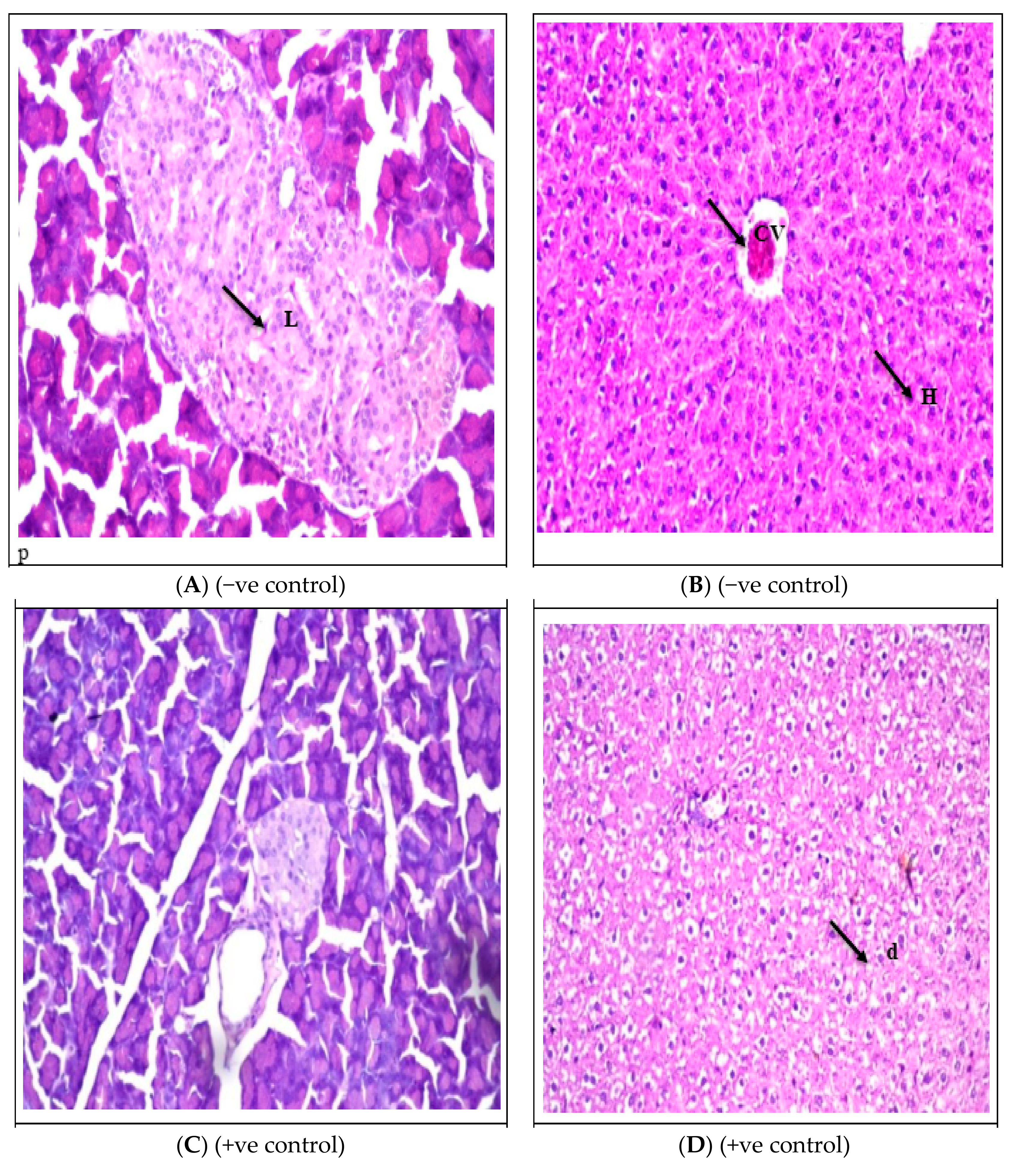
3.3. Effect of Supplemented Cupcakes on the Body Weight Gain and Feed Intake Parameters

Compared to the positive control group fed on a standard basal diet or fed on control cupcake, a significant increase (p < 0.05) in body weight gain (BGW%) of all the rat groups fed on cupcake samples containing BPP, CLP, SLP, and their mixtures, while a significant decrease (p < 0.05) as compared to the negative group (Table 4). The positive control group fed on a standard basal diet or a control cupcake showed a lower BGW% (−7.56 and −8.77%, respectively) than the negative group (+27.29%). Despite the increase in BGW% in groups supplemented with BPP (+7.25%), CLP (+8.18%), and SLP (+11.67%), as compared to the positive control group fed on a standard basal diet or fed on control cupcake, but still lower than the negative control (Table 4). An interesting additive effect could be noted for BWG% in the groups treated with BPP-SLP and CLP-SLP mixtures (1:1 w/w). However, the diabetic groups fed with CLP-SLP (+16.13%) and BPP-SLP (+14.36%) had a more significant increase in BGW% than the positive diabetic control group, which agreed with the differences in the proximate analysis of agro-wastes, where CLP has the highest fiber content as provided in Table 1.
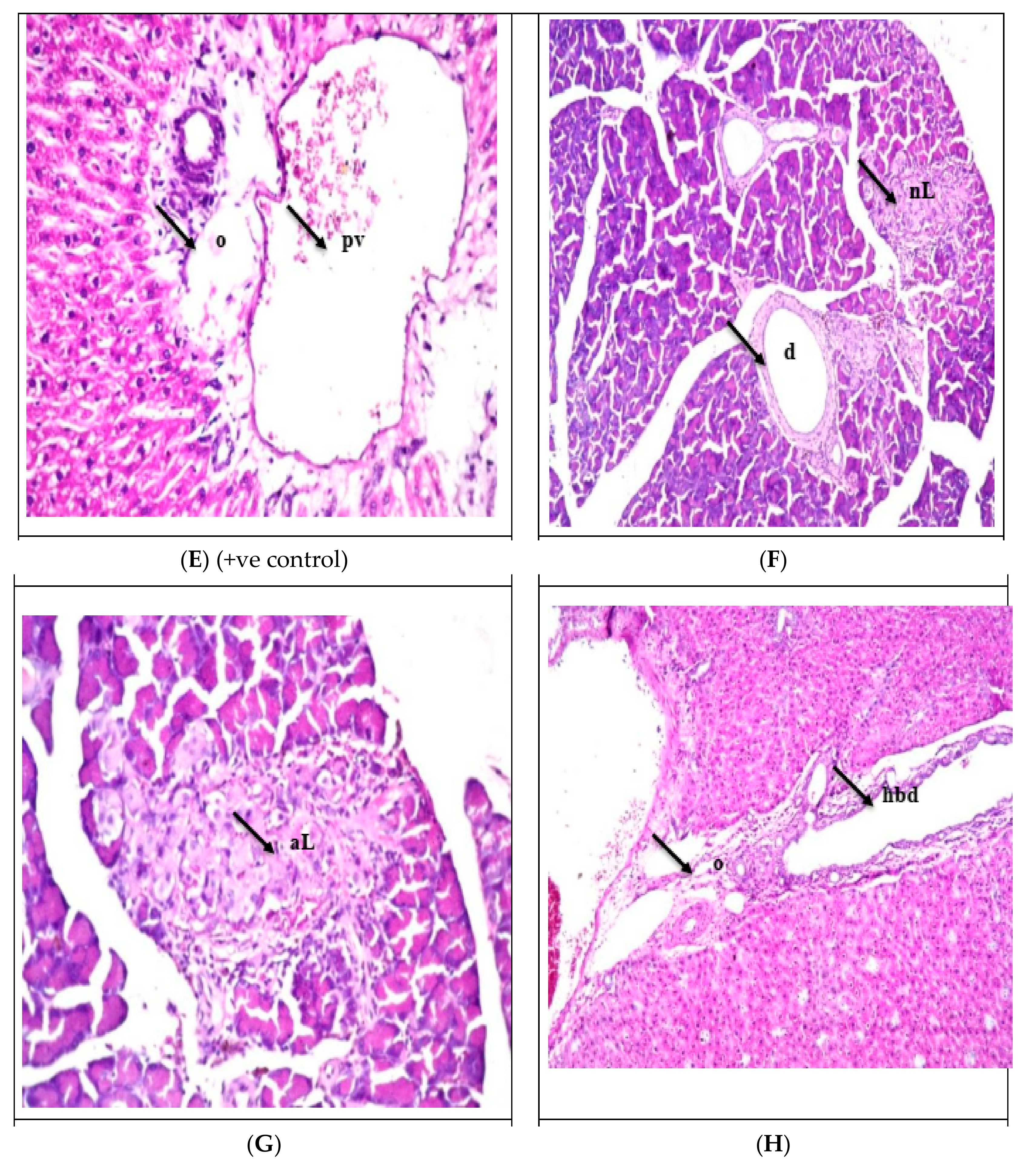
The effect of feeding on cupcake samples containing agro-wastes and Stevia on feed intake was recorded and listed in Table 5. The obtained results cleared a significant decrease (p < 0.05) in feed intake of the diabetic control group (+) fed on a standard basal diet (74.03 g/rat/28 days) and the diabetic rats’ group fed on cupcake control (75.35 g/rat/28 days) as compared with the negative group control (91.56 g/rat/28 days). The feed intake of treated groups with supplemented agro-wastes and Stevia powders (79.9, 80.61, and 79.70 g/rat/28 days) was significantly lower than the negative group but higher than the positive diabetic control and the diabetic rats fed on cupcake control (100% sugar) groups (Table 5). The results above agreed with the BWG% discussed above, with the lowest feed intake amount for the CLP-SLP mixture (78.23 g/rat/28 days), followed by the BPP-SLP mixture (78.90 g/rat/28 days).
According to Meliala et al. [51], the results align with our findings, showing an increase in weight gain for the normal control group and a decrease for the diabetic group after 3 weeks of treatment. Interestingly, the diabetic groups that were fed with banana peel flakes gained more weight significantly than the diabetic control group (p < 0.05). Moreover, there was no significant difference (p > 0.05) in food intake between the normal and diabetic groups [51]. The same pattern was observed in STZ-induced diabetic rats treated with total and soluble banana peel fibers, where the diabetic control group had the lowest body weight, while the normal group had the highest. Additionally, the diabetic groups that consumed different types of fibers had less food intake daily than the diabetic control group, indicating that dietary fiber intake may help reduce the symptoms of polyphagia in T2DM mice [52]. Along the same line, carrot supplementation induced a time-dependent body weight gain-fed high-fat diet in different groups [53]. To our knowledge, nothing was reported in the literature concerning the effect of dried carrot leaves on nutritional or biochemical parameters. Consistent with the results of the present study, Ahmad and Ahmad [54] indicated that aqueous extract from leaves of Stevia rebaudiana produced a significant (p  <  0.05) reduction in body weight and body weight gain of the rats compared to the negative control. Additionally, Stevia sweetener delivery decreased the amount of feed and water consumed by diabetic rats compared to control groups.
Rats’ bodies may weigh less due to less glucose in the diet being metabolized or consuming less food overall [55]. This decrease in weight in the rats receiving Stevia extract may be attributable to the high concentration of stevioside, which decreased the rats’ food consumption [56]. This result concurs with earlier studies that demonstrated a beneficial relationship between the drop in feed intake and the dose of stevioside administered to the rats and the body weight growth percentage [57].
The effect of feeding cupcake samples containing Stevia and agro-wastes on the fasting glucose level (mg/dL) of diabetic rats was determined weekly through the experimental period (28 days), and the results are listed in Table 6. Compared to the positive diabetic control group (427.5 mg/dL) and diabetic control cupcake (374.7 mg/dL), a significant decrease (p < 0.05) in glucose levels of all diabetic groups fed on the supplemented cupcake with Stevia and agro-wastes, ranging from 195.1 to 180.9 mg/dL at the end of the experimental period. However, as expected, the negative control group had the least glucose level (91.8 mg/dL) at the end of the testing period, as given in Table 6. After 4 weeks of the experiment, the lowest glucose level was recorded for the CL-SLP mixture due to the additive effect compared to using SLP or CLP separately in the cupcakes formula (Table 6).
In line with Meliala et al. [51], standard diets containing banana peel flakes have been found to significantly reduce blood glucose levels in comparison to a diabetic control group. Additionally, Louis et al. [4] observed a similar trend in diabetic rats fed with a diet supplemented with carrot powder, which resulted in lower blood glucose levels in comparison to the diabetic control group. In a study involving diabetic rats, Stevia aqueous extract significantly restored fasting blood glucose levels towards normal from the 1st week to the 8th-week experiment [54]. Previous research has shown that stevioside is effective in controlling blood sugar levels by improving insulin secretion, sensitivity, and utilization in insulin-deficient rats [58]. Moreover, another study suggests that Stevia extract contains certain biomolecules that could potentially make the insulin receptor more sensitive to the hormone or stimulate the islets of Langerhans beta cells to release insulin. Both of these actions could help improve the body’s enzymes that break down carbohydrates and restore normal blood glucose levels [59]. Stevia rebaudiana leaves’ extract has been found to reduce random and fasting blood glucose levels in rats by reviving the β-cells of the pancreas, reactivating the glycogen synthase system, enhancing insulin secretion, and increasing liver glycogen level [60].
Table 7 presents the results of a study on the effects of cupcake samples containing Stevia and agro-wastes on serum liver functions, ALT, AST, and albumin of alloxan-induced diabetic rats. The study found that the positive control group, which consisted of diabetic rats fed on a standard diet or cupcake control, had significantly higher levels (p < 0.05) of ALT and AST (64.1 and 65.5 mg/dL, respectively, for the standard diet group and 61.9 and 62.7 mg/dL, respectively, for the cupcake control group) compared to the negative control group (34.7 and 32.9 mg/dL, respectively). However, supplementing the cupcake with Stevia and agro-wastes and Stevia led to reduced AST and ALT concentrations compared to the positive control group, but they were still higher than the negative control group (Table 7). Among the supplemented groups examined, SLP alone or in an additive mixture with CLP showed the lowest ALT and AST concentrations. The study also found a significant decrease (p < 0.05) in the albumin level of the positive control group (4.5 mg/dL) and cupcake control group (4.7 mg/dL) compared to the negative control group (6.9 mg/dL). The supplemented cupcake samples had a lower albumin content than the negative control group (5.0, 5.4, 5.6, 5.3, and 5.8 mg/dL) but higher than the positive control group. The highest albumin level was observed in the additive effect of the SLP-CLP mixture, which was higher than the cupcake control, the positive control, or the samples supplemented with Stevia or other separate agro-wastes.
In line with the above results, Assi et al. [61] showed that the administration of Stevia extract for STZ-induced diabetic rats significantly reduced hepatic parameters (ALT and AST). Increased levels of these biomarkers point to increased tissue permeability, injury, or necrosis brought on by the production of free radicals in all tissues due to protein glycosylation and glucose autooxidation [62]. The unaltered food intake and plasma ALT and AST levels during the fortification of a high-fructose diet with carrot juice imply that the oral gavage administration did not cause stress to experimental animals [63]. The impact of banana fruit and peel on liver function enzymes of diabetic rats with induced hepato-toxicity was discovered by Zakaria et al. [64]. Comparing the diabetic hepatotoxic group’s serum ALT, AST and ALP activities to the corresponding value in the normal control group revealed a substantial increase (p < 0.05) in each enzyme activity. The fast release of these enzymes from the cytoplasm into the blood circulation following plasma membrane rupture and cellular injury may cause increased serum AST and ALT levels. Elevated ALT is seen as a more sensitive signal and is typically accompanied by increased AST. High serum transaminases are thought to be a sign of hepatic damage [65]. The mean value of ALT, AST, and ALP levels significantly decreased (p < 0.05) when diabetic hepatotoxic rats were given dried banana fruit and its dried peel compared to the positive control group. According to the research by Mosa and Kkalil [66], eating dried or fresh banana peels may change a patient’s risk of developing acute liver failure.
The significant reduction in albumin in the diabetic control (positive) compared to the negative control or treated groups with supplemented cupcakes is due to the glycation of renal matrix proteins causing alterations in kidney architecture and increased basement membrane permeability, resulting in nephropathy [67]. These gradual alterations build up over time, eventually leading to renal failure. Preventing protein glycation can be very helpful in preventing the formation of such alterations. Microproteinuria and albuminuria, vital clinical indicators of diabetic nephropathy, and/or enhanced protein catabolism could explain the decrease in total protein and albumin percentage [68].
Based on the findings given in Table 8, the diabetic groups that consumed cupcake samples containing agro-wastes and Stevia powders demonstrated a significant reduction (p < 0.05) in urea, uric acid, and creatinine compared to the positive control group and the diabetic rats that consumed regular cupcakes. The SLP-CLP mixture added to the cupcakes showed the best results, with urea, uric acid, and creatinine levels of 47.65, 4.94, and 1.16 mg/dL, respectively, due to the additive effect of the ingredients. The negative group had the lowest kidney function parameters among all the tested groups (as shown in Table 8).
In agreement with the above results, treatment of diabetic rats with Stevia extract ex-pressed a significant decrease in renal indicators (urea and creatinine), as Assi et al. [61] reported. When diabetic rats were fed a diet supplemented with banana fruit and its peel, the mean values of uric acid, creatinine, and urea decreased significantly (p < 0.05) compared to the positive control group. Furthermore, neither the banana fruit nor its peel caused any discernible alterations in renal functioning among the treatment groups [64]. Rats exposed to gentamicin consistently had higher serum urea, uric acid, and creatinine levels. It was discovered that these levels (p ≤ 0.01) considerably decreased in a dose-dependent way in the groups receiving carrot extract, which was also supported by histological observations [69]. It was revealed that circulating lipids bind to and become trapped by the extracellular matrix, where they are oxidized, increasing the reactive oxygen species generation, which could damage the structure and function of diabetic kidneys [70]. Therefore, treatment with flavorings in nano-encapsulated and nonencapsulated forms inhibited lipid alterations, which could be one explanation for its possible renoprotective activity.
The results presented in Table 9 show that diabetic groups who consumed cupcakes fortified with agro-wastes and Stevia powders experienced a significant decrease (p < 0.05) in total cholesterol, triglyceride, and LDL levels. This decrease was particularly noticeable in cupcakes that were supplemented with the CLP-SLP additive mixture. In comparison to the positive diabetic control and cupcake control groups, the total cholesterol, triglyceride, and LDL levels of the positive control group decreased from 197.5, 115.1, and 70.8 mg/dL to 130.5, 60.9, and 47.6 mg/dL, respectively, for the diabetic group fed on cupcakes supplemented with CLP-SLP mixture. In contrast, there was a significant increase (p < 0.05) in HDL levels for diabetic groups treated with BPP, CLP, SLP, and their mixtures (27.6, 31.2, 32.9, 30.2, and 36.7 mg/dL, respectively) compared to the positive diabetic control and cupcake control groups (17.9 and 19.7 mg/dL, respectively). This lipid-lowering effect could potentially prevent cardiovascular disease in diabetes, which is also supported by the decreased glucose levels in the treated groups, as discussed in Table 6.
The previous findings agreed with Soleti et al. [53], where a high-fat diet with carrot supplement showed antihyperglycemic and lipid-lowering activities due to its effect on improving pancreatic β-cells function. It has been established that various metabolic and regulatory abnormalities that occur during diabetes lead to hyperlipidemic conditions in people with diabetes, while increased TC, LDL-C, and TG levels in the blood cause diabetic dyslipidemia [71]. Moreover, Wang et al. [49] revealed that the serum TC, TG, LDL-C, and HDL-C levels of the diabetic mice were significantly increased compared with the negative control group. After supplementing banana fibers, the lipid profile parameters decreased significantly. Treatment of diabetic rats with Stevia extract caused a significant decrease in serum cholesterol and TG compared to untreated rats. In contrast, diabetic rats treated with Stevia extract showed a significant elevation in the HDL level compared to control diabetic rats [61].

3.4. Effect of Cupcakes Supplementation on the Liver and Pancreas Histopathology

The negative control group displayed normal pancreas and liver structures, with no histological alterations, as indicated by Figure 1A,B and Table 10. On the other hand, severe alterations were observed in the positive control group, with degenerations in both pancreas and liver structures, as shown in Figure 1C–E and Table 10. When diabetic rats were fed on the cupcake control sample, moderate alterations were found in pancreas structures due to degenerations. However, normal hepatic structures were observed, as displayed in Figure 1F–J and Table 10. In the case of cupcakes supplemented with BPP, moderate changes were detected in the structure of the pancreas islet of Langerhans, but no degenerations were noted in hepatocytes (Figure 1K,L and Table 10). Likewise, feeding diabetic rats with cupcakes containing CLP revealed mild to moderate changes in the liver, but no effects were observed in the pancreas islet of Langerhans, as detected in Figure 1M–P and Table 10. Additionally, no significant histopathological changes were detected in the pancreas, and only moderate changes were observed in the liver in diabetic rats fed on cupcakes containing SLP (Figure 1Q–S and Table 10). Interestingly, a mixture of SLP and BPP in cupcakes caused no histopathological alteration in the pancreas, while moderate degenerations were detected in the hepatocytes (Figure 1T,U and Table 10). The most exciting finding was that diabetic rats fed on cupcakes supplemented with a mixture of SLP and CLP showed no histopathological alterations in the pancreas and liver, as shown in Figure 1V–X and Table 10. These results suggest that the additive effect of the supplements reduces T2DM complications.
The above findings agreed with Wang et al. [52], where feeding diabetic rats with banana fibers noticeably reduced hepatocellular steatosis, fewer lipid droplets, relatively less inflammatory cell infiltration, and ameliorative effects on liver histopathology in diabetic mice. On the other hand, the number and volume of pancreatic islets in groups fed on banana fibers increased, and the pancreatic tissue lesions were improved. Along the same line, the histologic evaluation of the hepatic tissues performed by Assi et al. [61] showed a significant decrease (p < 0.05) in liver injuries in the diabetic group treated with Stevia compared to the diabetic control rats.

4. Conclusions

Using agro-wastes (CLP and BPP) and SLP as the main sweetener in cupcakes instead of sucrose, can solve an environmental issue and improve various nutritional and biochemical parameters in diabetic rats. When diabetic rats consumed cupcakes with these supplements, they experienced a significant increase in body weight gain and feed intake. Additionally, the CLP-SLP mixture resulted in a significant reduction in all biochemical parameters studied. No histopathological alterations were detected in the pancreas when consuming SLP-BPP and CLP-SLP. However, the hepatocytes of diabetic rats that consumed cupcakes with a BPP-SLP mixture showed moderate degeneration. This approach of replacing sugar with agro-wastes and Stevia can enhance the nutritional values of food products, prevent complications for T2DM patients, and solve potential environmental problems.

Author Contributions

Conceptualization, project administration, investigation, and writing—original draft, B.S. and M.E.-W.; extraction, methodology, formal analysis, statistical analysis, and data curation, M.E.-W., B.S., S.E.-B. and H.A.; histology, S.E.-B.; writing—review and editing, A.F.; and funding acquisition, H.Y. and M.E. All authors have read and agreed to the published version of the manuscript.

Funding

This work was funded by Princess Nourah bint Abdulrahman University, Researchers Supporting Project number (PNURSP2023R23).

Institutional Review Board Statement

The Ethical Committee of Medical Research approved the animal study protocol at the Egyptian National Research Centre (Approval no. 1451022023).

Informed Consent Statement

Not applicable.

Data Availability Statement

The data presented in this study are available in this article.

Acknowledgments

The authors appreciate the Researcher Support Program (PNURSP2023R23) at Princess Nourah bint Abdulrahman University, Riyadh, Saudi Arabia, for the provided facilities.

Conflicts of Interest

The authors declare no conflict of interest.

References

  1. Alkhatib, A.; Tsang, C.; Tiss, A.; Bahorun, T.; Arefanian, H.; Barake, R.; Khadir, A.; Tuomilehto, J. Functional Foods and Lifestyle Approaches for Diabetes Prevention and Management. Nutrients 2017, 9, 1310. [Google Scholar] [CrossRef] [PubMed] [Green Version]
  2. Ayala-Zavala, F.; Vega, V.; Rosasdomingues, C.; Palafox, H.; Villarodriguez, A.; Wasim, D.; Dàvila, E.; Gonzàlez, A. Agro-industrial potential of exotic fruit by products as a source of food additives. Food Res. Int. 2011, 44, 1866–1874. [Google Scholar] [CrossRef]
  3. O’Shea, N.; Arendty, K.; Gallagher, E. Dietary fiber and phytochemical characteristics of fruit and vegetable by products and their recent applications as novel ingredients in food products. Innov. Food Sci. Emerg. Technol. 2012, 16, 1–10. [Google Scholar] [CrossRef]
  4. Louis, X.L.; Raj, P.; McClinton, K.J.; Yu, L.; Suh, M.; Netticadan, T. Supplementation of Type 1 Diabetic Rats with Carrot Powder Lowers Blood Glucose without Improving Cardiac Structure and Function. Prev. Nutr. Food Sci. 2018, 23, 115–121. [Google Scholar] [CrossRef]
  5. Salwe, K.J.; Sachdev, D.O.; Bahurupi, Y.; Kumarappan, M. Evaluation of antidiabetic, hypolipedimic and antioxidant activity of hydro alcoholic extract and leaves and fruit peels of Punica granatum in Onale Wister albino rats. J. Nat. Sci. Bid Med. 2015, 6, 56–62. [Google Scholar] [CrossRef] [Green Version]
  6. Narghare, V.V.; Dawdle, S.O.C. In vitro antioxidant, hypoglycemic and oral glucose tolerance test of banana peels. Alexandria J. Med. 2017, 53, 237–243. [Google Scholar] [CrossRef] [Green Version]
  7. Rebello, G.; Ramos, M.; Pertuzatti, B.; Barcia, M.T.; Castillo, N.; Hermosfn, I. Flour of banana peel as a source of antioxidant phenolic compounds. Food Res. Int. 2014, 55, 397–403. [Google Scholar] [CrossRef]
  8. Vu, H.T.; Scarlett, C.J.; Young, Q.V. Changes of phytochemicals and antioxidants capacity of banana peels during the ripening process with and without ethylene treatment. Sci. Hortic. 2019, 253, 255–262. [Google Scholar] [CrossRef]
  9. Putta, S.; Silakabattini, K.; Kumar, J. Protective Effect of Tylophora India against Streptozotocin Induced Pancreatic and Liver dysfunction in Westar Rats. Biomed. Pharmacol. J. 2020, 13, 1755–1763. [Google Scholar] [CrossRef]
  10. Costa, T.O.B.; Silveira, C.O.S.; Pereira, M.C.; Helbia, E. Lipid and glycemic profile of rats fed on diets with banana peel and banana stalk. Demetra Food Nutr. Health 2018, 13, 263–273. [Google Scholar]
  11. Malviya, N.; Jains, S.; Malviya, S. Antidiabetic potential of medicinal plants. Acta Pol. Pharm. 2010, 67, 113–118. [Google Scholar]
  12. Erukainure, O.L.; Ebuehi, O.A.T.; Adeboyejo, F.O.; Okafor, E.N.; Hafizur, R.M.O.; Aliyu, M.O.; Elemo, G.N. Anti-diabetic and hypoglycemic properties and fiber enriched cake in Alloxan–induced diabetic rats. Mediterr. J. Nutr. Metab. 2013, 6, 135–141. [Google Scholar] [CrossRef]
  13. Sodchit, C.; Tochampa, W.; Kongbangkerd, T.; Singanusong, R. Effect of banana peel cellulose as a dietary fiber supplement on baking and sensory qualities of butter cake. Songklanakarin J. Sci. Technol. 2013, 35, 641–646. [Google Scholar]
  14. Goneim, G.; Ibrahim, F.; El-Shehawy, S. Carrot leaves: Antioxidative and nutritive values. J. Food Dairy Sci. 2011, 2, 201–211. [Google Scholar] [CrossRef]
  15. Eugenio, M.H.A.; Pereira, R.G.F.; Ao de Abreu, W.C.; Pereira, M.C.A. Phenolic compounds and antioxidant activity of tuberous root leaves. Int. J. Food Prop. 2017, 20, 2966–2973. [Google Scholar] [CrossRef]
  16. AICR: American Institute for Cancer Research. Available online: https://www.aicr.org/ (accessed on 18 December 2022).
  17. Gandhi, S.; Gat, Y.; Arya, S. Natural sweeteners: Health benefits of Stevia. Foods Raw Mater. 2018, 6, 392–402. [Google Scholar] [CrossRef]
  18. Panpatil, V.V.; Polasa, K. Assessment of Stevia (Stevia rebaudiana)–natural sweetener: A review. J. Food Sci. Technol. 2008, 45, 467–473. [Google Scholar]
  19. Maya, D.; Ritu, P. Formulation of fruit (orange juice) and whey based beverages flavoured with different herbs using natural sweetener as Stevia. Int. J. Sci. Educ. 2016, 4, 5975–5979. [Google Scholar]
  20. Cadena, R.S.; Cruz, A.G.; Netto, R.R. Sensory profile and physicochemical characteristics of mango nectar sweetened with high intensity sweeteners throughout storage time. Food Res. Int. 2013, 54, 1670–1679. [Google Scholar] [CrossRef] [Green Version]
  21. Rocha, I.F.; Bolini, H.M. Passion fruit juice with different sweeteners: Sensory profile by descriptive analysis and acceptance. Food Sci. Nutr. 2015, 3, 129–139. [Google Scholar] [CrossRef]
  22. Bedoya-Perales, N.S.; Steel, C.J. Effect of the concentrations of maltogenic α-amylase and fat on the technological and sensory quality of cakes. Food Sci. Technol. 2014, 34, 760–766. [Google Scholar] [CrossRef] [Green Version]
  23. Lee, J.H. Physicochemical and sensory characteristics of sponge cakes with Rubus coreanus powder. Prev. Nutr. Food Sci. 2015, 20, 204–209. [Google Scholar] [CrossRef] [PubMed] [Green Version]
  24. AOAC. Official Methods of Analysis of AOAC International, 18th ed.; AOAC International: Gaithersburg, MD, USA, 2011. [Google Scholar]
  25. Abou-Arab, E.; Abou-Arab, A.; Abu-Salem, F. Physico-chemical assessment of natural sweeteners steviosides produced from stevia rebudiana bertoni plant. J. Food Dairy Sci. 2009, 34, 11037–11057. [Google Scholar] [CrossRef]
  26. Desia, N.S.; Bhide, S.A. Hypoglycemic effect of Hamiltonia sauveolens. Indian. J. Med. 1985, 81, 86–91. [Google Scholar]
  27. Malaisse, W.J. Alloxan toxicity to the Pancreatic B-cell: A new hypothesis. Biochem. Pharmacol. 1982, 31, 3527. [Google Scholar] [CrossRef]
  28. Mostafavinia, M.; Amini, A.; Ghorishi, S.K.; Pouriran, R.; Bayat, M.O. The effects of dosage and the routes of administrations of Streptozotocin and alloxan on induction rate of type1 diabetes mellitus and mortality rate in rats. Lab. Anim. Res. 2016, 32, 160–165. [Google Scholar] [CrossRef] [Green Version]
  29. Mohammed, D.M.; Ahmed, K.A.; Desoukey, M.A.; Sabry, B.A. Assessment of the antiulcer properties of Lawsonia inermis L. leaves and its nano-formulation against prolonged effect of acute ulcer in rats. Toxicol. Rep. 2022, 9, 337–345. [Google Scholar] [CrossRef]
  30. Reeves, P.G.; Nielsen, F.H.; Fahey, G.C., Jr. AIN-93 purified diets for laboratory rodents: Final report of the American Institute of Nutrition ad hoc writing committee on the reformulation of the AIN-76A rodent diet. J. Nutr. 1993, 123, 1939–1951. [Google Scholar] [CrossRef] [Green Version]
  31. Trinder, P. Determination of glucose in blood using glucose oxidase with an alternative oxygen acceptor. Ann. Clin. Biochem. 1969, 6, 24–27. [Google Scholar] [CrossRef]
  32. Richmond, W. Preparation and properties of a cholesterol oxidase from Nocardia sp. and its application to the enzymatic assay of total cholesterol in serum. Clin. Chem. 1973, 19, 1350–1356. [Google Scholar] [CrossRef]
  33. Allain, C.C.; Poon, L.S.; Chan, C.S.; Richmond, W.F.P.C.; Fu, P.C. Enzymatic determination of total serum cholesterol. Clin. Chem. 1974, 20, 470–475. [Google Scholar] [CrossRef]
  34. Lopes-Virella, M.F.; Stone, P.; Ellis, S.; Colwell, J.A. Cholesterol determination in high-density lipoproteins separated by three different methods. Clin. Chem. 1977, 23, 882–884. [Google Scholar] [CrossRef]
  35. Wieland, H.; Seidel, D. A simple specific method for precipitation of low density lipoproteins. J. Lipid Res. 1983, 24, 904–909. [Google Scholar] [CrossRef]
  36. Reitman, S.; Frankel, S. A colorimetric method for the determination of serum glutamic oxalacetic and glutamic pyruvic transaminases. Am. J. Clin. Pathol. 1957, 28, 56–63. [Google Scholar] [CrossRef]
  37. Babson, A.L.; Greeley, S.J.; Coleman, C.M.; Phillips, G.E. Phenolphthalein monophosphate as a substrate for serum alkaline phosphatase. Clin. Chem. 1966, 12, 482–490. [Google Scholar] [CrossRef]
  38. Doumas, B.T.; Watson, W.A.; Biggs, H.G. Albumin standards and the measurement of serum albumin with bromcresol green. Clin. Chim. Acta 1971, 31, 87–96. [Google Scholar] [CrossRef]
  39. Fawcett, J.; Scott, J. A rapid and precise method for the determination of urea. J. Clin. Pathol. 1960, 13, 156–159. [Google Scholar] [CrossRef] [Green Version]
  40. Schirmeister, J. Determination of creatinine level. Dtsch. Med. Wschr. 1964, 89, 1940–1947. [Google Scholar]
  41. Barham, D.; Trinder, P. An improved color reagent for the determination of blood glucose by the oxidase system. Analyst 1972, 97, 142–145. [Google Scholar] [CrossRef]
  42. Banchroft, J.D.; Stevens, A.; Turner, D.H. Theory and Practice of Histological Techniques, 4th ed.; Churchill Livingstone: London, UK, 1996. [Google Scholar]
  43. Ragab, M.; Osman, M.F.; Khalil, M.E.; Gouda, M.S. Banana (Musa sp.) peels as a source of pectin and some food nutrients. J. Sustain. Agric. Sci. 2016, 42, 88–102. [Google Scholar] [CrossRef]
  44. El-Sayed, S.S.F. Impact of some agronomical practices on carrot productivity and nutritional value. J. Plant Prod. 2021, 12, 365–376. [Google Scholar] [CrossRef]
  45. Ahmed, Z.; El-Sharnouby, G.; EL-Waseif, M. Use of Banana Peel as a By-Product to Increase the Nutritive Value of the Cake. J. Food Dairy Sci. 2021, 12, 87–97. [Google Scholar] [CrossRef]
  46. Dos Santos, A.C.; Yassunaka, N.N.; Ruiz, S.P.; Schneider, V.V.A.; Visentainer, J.V.; Madrona, G.S. Sensory and physicochemical study of carrot leaf sponge cake. Braz. J. Food Res. 2013, 4, 41–46. [Google Scholar] [CrossRef] [Green Version]
  47. Bin Mohd Zaini, H.; Bin Sintang, M.D.; Dan, Y.N.; Ab Wahab, N.; Bin Abdul Hamid, M.; Pindi, W. Effect of addition of banana peel powder (Musa balbisiana) on physicochemical and sensory properties of fish patty. Br. Food J. 2019, 121, 2179–2189. [Google Scholar] [CrossRef]
  48. Leite, C.W.; Boroski, M.; Boeing, J.S.; Aguiar, A.C.; França, P.B.; de Souza, N.E.; Visentaine, J.V. Chemical characterization of leaves of organically grown carrot (Dacus carota L.) in various stages of development for use as food. Ciênc. Tecnol. Aliment. Campinas 2011, 31, 735–738. [Google Scholar] [CrossRef] [Green Version]
  49. Marcinek, K.; Krejpcio, Z. Stevia rebaudiana Bertoni–chemical composition and functional properties. Acta Sci. Pol. Technol. Aliment. 2015, 14, 145–152. [Google Scholar] [CrossRef]
  50. Elsebaie, E.M.; Mostafa, S.M.I. Utilization of Stevia Leaves Powder in Reduced Calorie Cake. J. Food Dairy Sci. Mansoura Univ. 2018, 9, 83–86. [Google Scholar] [CrossRef]
  51. Meliala, A.; Sumiwi, Y.A.A.; Narwidina, P.; Rini, S.L.S.; Setyaningsih, W. Banana peel flakes alleviate blood glucose and stress in a dose-dependent manner. Int. J. Pharm. Pharm. Sci. 2020, 12, 75–81. [Google Scholar] [CrossRef]
  52. Wang, M.; Yang, F.; Yan, X.; Chao, X.; Zhang, W.; Yuan, C.; Zeng, Q. Anti-diabetic effect of banana peel dietary fibers on type 2 diabetic mellitus mice induced by Streptozotocin and high-sugar and high-fat diet. J. Food Biochem. 2022, 46, 14275. [Google Scholar] [CrossRef]
  53. Soleti, R.; Coué, M.; Trenteseaux, C.; Hilairet, G.; Fizanne, L.; Kasbi-Chadli, F.; Mallegol, P.; Chaigneau, J.; Boursier, J.; Krempf, M.; et al. Carrot Supplementation Improves Blood Pressure and Reduces Aortic Root Lesions in an Atherosclerosis-Prone Genetic Mouse Model. Nutrients 2021, 13, 1181. [Google Scholar] [CrossRef]
  54. Ahmad, U.; Ahmad, R.S. Anti-diabetic property of aqueous extract of Stevia rebaudiana Bertoni leaves in Streptozotocin-induced diabetes in albino rats. BMC Complement. Altern. Med. 2018, 18, 179. [Google Scholar] [CrossRef] [Green Version]
  55. Chang, J.C.; Wu, M.C.; Liu, I.M.; Cheng, J.T. Increase of insulin sensitivity by stevioside in fructose-rich chow-fed rats. Horm. Metab. Res. 2005, 37, 610–616. [Google Scholar] [CrossRef]
  56. Awney, H.; Massoud, M.; El-Maghrabi, S. Long-term feeding effects of stevioside sweetener on some toxicological parameters of growing male rats. J. Appl. Toxicol. 2011, 31, 431–438. [Google Scholar] [CrossRef]
  57. Assaei, R.; Mokarram, P.; Dastghaib, S.; Darbandi, S.; Darbandi, M.; Zal, F.; Akmali, M.; Ranjbar Omrani, G.H. Hypoglycemic effect of aquatic extract of Stevia in pancreas of diabetic rats: PPARγ-dependent regulation or antioxidant potential. Avicenna J. Med. Biotech. 2016, 8, 65–74. [Google Scholar]
  58. Geuns, J.M.C.; Buyse, J.; Vankeirsbilck, A.; Temme, E.H.M. Metabolism of stevioside by healthy subjects. Exp. Biol. Med. 2007, 232, 164–173. [Google Scholar] [CrossRef]
  59. Hossain, M.S.; Alam, M.B.; Asadujjaman, M.; Islam, M.M.; Rahman, M.A.; Islam, M.A.; Islam, A. Anti-hyperglycemic and anti hyperlipidemic effects of different fractions of stevia rebaudiana leaves in alloxan-induced diabetic rats. Int. J. Pharm. Sci Res. 2011, 2, 1722–1729. [Google Scholar]
  60. Akbarzadeh, S.; Eskandari, F.; Tangestani, H.; Bagherinejad, S.T.; Bargahi, A.; Bazzi, P.; Daneshi, A.; Sahrapoor, A.; O’Connor, W.J.; Rahbar, A.R. The effect of Stevia rebaudiana on serum omentin and visfatin level in stz-induced diabetic rats. J. Diet Suppl. 2015, 12, 1–12. [Google Scholar] [CrossRef] [Green Version]
  61. Assi, A.A.; Abd El-Hamid, D.H.; Abdel-Rahman, M.S.; Ashry, E.E.; Bayoumi, S.A.; Asmaa, M.; Ahmed, A.M. The Potential Efficacy of Stevia Extract, Glimepiride and Their Combination in Treating Diabetic Rats: A Novel Strategy in Therapy of Type 2 Diabetes Mellitus. Egypt. J. Basic Clin. Pharmacol. 2020, 10, 101455. [Google Scholar] [CrossRef]
  62. Chaitanya, K.V.; Pathan, A.A.K.; Mazumdar, S.S.; Chakravarthi, G.P.; Parine, N.; Bobbarala, V. Role of oxidative stress in human health. An overview. J. Pharm Res. 2010, 3, 330–1333. [Google Scholar]
  63. Mahesh, M.; Pandey, H.; Raja Gopal Reddy, M. Carrot Juice Consumption Reduces High Fructose-Induced Adiposity in Rats and Body Weight and BMI in Type 2 Diabetic Subjects. Nutr. Metab. Insights 2021, 14. [Google Scholar] [CrossRef]
  64. Zakaria, R.; Abelbaky, M.S.; Abo-Raya, A.O. Effect of Banana (Musa balbisiana) Fruits and Its Peels on Acute Hepatotoxicity with Diabetic Rats. Egypt. J. Appl. Sci. 2020, 35, 143–157. [Google Scholar]
  65. Ha, W.S.; Kim, C.K.; Song, S.H.; Kang, C.B. Study on mechanism of multistep hepatotumorigenesis in rat: Development of hepatotumorigenesis. J. Vet. Sci. 2001, 2, 53–58. [Google Scholar] [CrossRef] [PubMed]
  66. Mosa, Z.; Kkalil, A. The effect of banana peels supplemented diet on acute liver failure rats. Ann. Agric. Sci. 2015, 60, 373–379. [Google Scholar] [CrossRef] [Green Version]
  67. Forbes, J.M.; Cooper, M.E.; Oldfield, M.D.; Thomas, M. C Role of advanced glycation end products in diabetic nephropathy. J. Am. Soc. Nephrol. 2003, 14, 254–258. [Google Scholar] [CrossRef] [Green Version]
  68. Bakris, G.L.; Mangrum, A.; Copley, J.B.; Vicknair, N.; Sadler, R. Effect of calcium channel or β-blockade on the progression of diabetic nephropathy in African Americans. Hypertension 1997, 29, 744–750. [Google Scholar] [CrossRef]
  69. Sodimbaku, V.; Pujari, L.; Mullangi, R.; Marri, S. Carrot (Daucus carota L.): Nephroprotective against gentamicin-induced nephrotoxicity in rats. Indian J. Pharmacol. 2016, 48, 122–127. [Google Scholar] [CrossRef] [Green Version]
  70. Trevisan, R.; Dodesini, A.R.; Lepore, G. Lipids and renal disease. J. Am. Soc. Nephrol. 2006, 17, 145–147. [Google Scholar] [CrossRef] [Green Version]
  71. Abou Baker, D.H.; Mohammed, D.M. Polyphenolic rich fraction of Physalis peruviana calyces and its nano emulsion induce apoptosis by caspase 3 up-regulation and G2/M arrest in hepatocellular carcinoma. Food Biosci. 2022, 50, 102007. [Google Scholar] [CrossRef]
Figure 1. Effect of replacement sugar in cupcake formula with stevia leaves and agro-wastes (banana peels and carrot leaves) on the liver and pancreas histopathology in T2DM mice. (A) The pancreas of the rats in group 1 displayed a typical histological structure, with the islet of Langerhans cells (L) functioning as the endocrine portion and the surrounding akin (a) serving as the exocrine. (B) In group 1, the rat liver exhibits a normal histological structure with the central vein (cv) and surrounding hepatocytes (H) in the parenchyma. (C) In group 2, there were areas of rat tissue that displayed atrophy and a decrease in the size of Langerhans cells in the islets. (D) In group 2, the rat’s liver displays hepatocyte degeneration (d) throughout the parenchyma. (E) In group 2, the rat liver displays severe dilatation of the portal vein (pv) accompanied by edema (o) in the portal area. (F) In group 3, the rat’s pancreas exhibits atrophy and necrobiotic changes in the islet of Langerhans cells (nL), along with the dilation of the pancreatic duct (d). (G) In group 3, the rat’s pancreas exhibited atrophy of the islet of Langerhans (aL), which can be identified through magnification. (H) In group 3, the rat’s liver displays portal vein dilatation alongside bile duct dilatation and hyperplasia (hbd), edema (o), and a small number of inflammatory cells (arrow) infiltrating the portal area. (I) In group 3, the rat liver exhibits a concentration of inflammatory cells in the parenchyma, as indicated by an arrow. (J) In group 3, there are some individual hepatocytes showing fatty change, as indicated by an arrow. (K) In group 4, the rat’s pancreas indicates necrobiotic changes (pointed by an arrow) in a few islets of Langerhans cells, along with the expansion of pancreatic (d) ducts. (L) In group 4, there is evidence of liver damage with a dilated portal vein (PV) and edema (O) in the portal area. (M) In group 5, the rat’s pancreas displays duct dilation (d). (N) In group 5, the rat’s pancreas is magnified to identify the fatty change in the acinar lining epithelium, as indicated by the arrow. (O) The rat’s pancreas from group 5 exhibits congestion in its stromal blood vessels. (P) In group 5, the rat’s liver exhibits dilatation of both the central vein (cv) and portal vein (pv). (Q) In group 6, the rat’s pancreas displays intact islets of Langerhans cells, as indicated by the arrow. (R) In group 6, the rat’s liver exhibits hepatocyte degeneration, as the arrow indicates. (S) In the sixth group, the rat’s liver displays portal vein (pv) congestion and edema, along with a small number of inflammatory cells in the portal area. (T) In group 7, the rat’s pancreas exhibits a histological structure that appears normal and intact, as indicated by the arrow. (U) In group 7, the rat’s liver displays hepatocyte degeneration around the central vein, as indicated by the arrow. (V) The pancreas from group 8 rats displays a typical histological structure of Langerhans cells (L) in the islets. (W) The liver sample from group 8 exhibits a normal histological structure. (X) In group 8, the rat’s liver exhibits dilatation and congestion in both the central vein (cv) and portal vein (pv).
Sustainability 15 09126 g001aSustainability 15 09126 g001bSustainability 15 09126 g001cSustainability 15 09126 g001dSustainability 15 09126 g001e
Table 1. The formulations of cupcake samples.
Ingredients * (g)Cupcake Treatments **
Cupcake (Control)12% BPP12% CLP12% SLP12% BPP and SLP12% CLP and SLP
WF 72%353535353535
BPP0120060
CLP0012006
SLP0001266
Sugar (sucrose)2088888
Butter191919191919
Fresh eggs101010101010
Skim milk powder444444
Crude cocoa1.31.31.31.31.31.3
Baking powder0.50.50.50.50.50.5
Vanilla0.20.20.20.20.20.2
Water (mL)101010101010
Total ingredients100100100100100100
* WF, wheat flour; BPP, banana peels powder; CLP, carrot leaves powder; SLP, Stevia leaves powder. ** Mixed SLP, BPP, or CLP in equal proportions (1:1 w/w).
Table 2. Proximate chemical composition of raw materials and cupcake samples on a dry weight basis.
Chemical Composition (%)Raw Materials **Proximate Chemical Composition % of Cupcake Samples (M ± SE) *
BPPCLPSLPCupcake ControlCupcake with 12% BPPCupcake with 12% CLPCupcake with 12% SLPCupcake with 12% BPP and SLP ***Cupcake with 12% CLP and SLP
Moisture9.13 ± 0.19 A8.87 ± 0.23 A8.95 ± 0.20 A22.61 ± 0.33 a22.59 ± 0.37 a22.51 ± 0.29 a22.55 ± 0.27 a22.63 ± 0.37 a22.67 ± 0.33 a
Crude protein13.19 ± 0.23 A15.34 ± 0.22 B16.25 ± 0.20 C12.86 ± 0.17 a13.45 ± 0.23 ab13.52 ± 0.22 b14.26 ± 0.23 c13.85 ± 0.19 bc13.90 ± 0.20 bc
Ether extract19.11 ± 0.18 C15.26 ± 0.13 B4.59 ± 0.15 A13.89 ± 0.11 a16.19 ± 0.13 d15.69 ± 0.10 c14.31 ± 0.12 a15.35 ± 0.11 bc14.91 ± 0.10 b
Ash13.40 ± 0.13 B15.97 ± 0.14 C8.28 ± 0.11 A1.39 ± 0.03 a2.99 ± 0.05 e3.31 ± 0.06 f2.33 ± 0.05 b2.64 ± 0.04 c2.79 ± 0.03 d
Dietary fiber14.29 ± 0.17 A16.42 ± 0.19 C15.22 ± 0.18 B3.65 ± 0.08 a5.35 ± 0.09 b5.60 ± 0.10 b5.46 ± 0.09 b5.39 ± 0.08 b5.52 ± 0.10 b
Total sugars15.71 ± 0.19 A3.67 ± 0.11 C14.78 ± 0.16 B20.19 ± 0.33 a9.82 ± 0.09 b8.45 ± 0.13 d9.75 ± 0.09 b9.79 ± 0.09 b9.08 ± 0.23 c
Reducing5.46 ± 0.14 A1.19 ± 0.07 C4.65 ± 0.10 B0.09 ± 0.03 f0.67 ± 0.13 a0.14 ± 0.04 e0.51 ± 0.04 c0.60 ± 0.12 b0.34 ± 0.09 d
Nonreducing10.25 ± 0.15 A2.48 ± 0.09 C10.13 ± 0.12 B20.10 ± 0.33 a9.15 ± 0.09 bc8.31 ± 0.19 d9.24 ± 0.10 b9.19 ± 0.09 bc8.74 ± 0.14 cd
Other total carbohydrates24.30 ± 0.13 C33.34 ± 0.17 B40.88 ± 0.20 A48.02 ± 0.25 b52.20 ± 0.23 a53.43 ± 0.22 a53.89 ± 0.23 a52.98 ± 0.21 a53.80 ± 0.23 a
M ± SE *, means ± standard error for chemical composition; the means within the same row having different superscripts are significantly varied (p < 0.05). ** BPP, banana peels powder; CLP, carrot leaves powder; SLP, Stevia leaves powder. *** Mixed SLP, BPP, or CLP in equal proportions (1:1 w/w).
Table 3. Sensory evaluation of cupcake samples supplemented with agro-wastes and Stevia.
Sensory PropertiesSensory Properties of Cupcake Samples (M ± SE) *
Cupcake ControlCupcake with 12% BPPCupcake with 12% CLPCupcake with 12% SLPCupcake with 12% BPP and SLPCupcake with 12% CLP and SLP
Appearance9.06 ± 0.33 c8.24 ± 0.37 b7.86 ± 0.29 a7.71 ± 0.27 a8.04 ± 0.37 ab7.97 ± 0.33 ab
Crust color8.79 ± 0.17 c8.11 ± 0.23 b7.80 ± 0.22 ab7.60 ± 0.23 a7.75 ± 0.19 ab7.90 ± 0.20 ab
Crumb color8.55 ± 0.11 c8. 17 ± 0.13 c7.35 ± 0.10 ab7.25 ± 0.12 a7.15 ± 0.11 a7.61 ± 0.10 b
Taste8.86 ± 0.13 c8.18 ± 0.15 b7.15 ± 0.16 a7.10 ± 0.15 a7.81 ± 0.14 b7.89 ± 0.13 b
Aroma9.11 ± 0.08 d8.10 ± 0.09 c7.75 ± 0.10 ab7.46 ± 0.09 a7.89 ± 0.08 bc7.95 ± 0.10 bc
Overall acceptability8.84 ± 0.25 d8.18 ± 0.23 c7.60 ± 0.22 ab7.39 ± 0.23 a7.81 ± 0.21 bc7.89 ± 0.23 bc
M ± SE *, means ± standard error for sensory properties; the means within the same row having different superscripts are significantly varied (p < 0.05).
Table 4. The impact of cupcake samples containing agro-wastes and Stevia on the body gain (g).
GroupsBody Weight (g) through 4 Weeks *BWG ** (g/Rat) through 4 WeeksBWG (%)
Initial Body Weight (g)After 1 weekAfter 2 weeksAfter 3 weeksFinal Body Weight
Control (−)138.10 ± 1.50 a149.90 ± 2.10 c158.40 ± 2.40 e167.35 ± 2.40 e175.80 ± 2.40 e37.70 ± 1.12 h+27.29
Control (+)136.10 ± 1.50 a134.66 ± 2.60 a131.20 ± 2.40 a127.20 ± 2.40 a125.80 ± 2.60 a−10.30 ± 1.11 b−7.56
Cupcake (Control)
100% sugar
139.10 ± 1.80 a136.00 ± 2.40 a132.50 ± 2.30 a130.20 ± 2.30 a126.90 ± 2.20 a−12.20 ± 1.09 a−8.77
Cupcake with 12% BPP138.95 ± 1.51 a140.70 ± 2.30 b142.60 ± 2.20 b146.75 ± 2.20 b149.10 ± 2.40 b+10.15 ± 1.13 c+7.25
Cupcake with 12% CLP139.20 ± 1.20 a141.60 ± 2.40 b143.50 ± 2.30 b147.70 ± 2.30 b150.60 ± 2.40 b+11.40 ± 1.10 d+8.18
Cupcake with 12% SLP138.80 ± 1.10 a142.15 ± 2.50 b147.10 ± 2.30 bc151.55 ± 2.30 bc155.00 ± 2.30 bc+16.20 ± 1.12 e+11.67
Cupcake with 12% BPP and SLP139.90 ± 1.20 a144.00 ± 2.40 b150.50 ± 2.30 c156.50± 2.30c160.00 ± 2.40 c+20.10 ± 1.10 f+14.36
Cupcake with 12% CLP and SLP138.80 ± 1.51 a143.00 ± 2.30 b150.80 ± 2.20 c156.10 ± 2.20 c161.20 ± 2.40 c+22.40 ± 1.13 g+16.13
M ± SE *, means ± standard error for the value of body weight gain (g/rat); the means within the same column having different superscripts are significantly varied (p < 0.05). ** BWG, body weight gain.
Table 5. The impact of cupcake treatments containing agro-wastes and Stevia on the feed intake (g).
GroupsFeed Intake (g/6 rats)
After 1 WeekAfter 2 WeeksAfter 3 WeeksAfter 4 WeeksTotal Feed Intake (g/6 Rats/28 Days)Total Feed Intake (g/Rat/28 Days)
Control (−)123.7 * ± 0.58 b134.1 ± 0.58 c141.4 ± 0.58 c150.2 ± 0.58 c549.4 ± 0.58 c91.56 ± 0.58 c
Control (+)114.1 ± 0.48 a112.6 ± 0.58 a110.7 ± 0.58 a106.8 ± 0.58 a444.2 ± 0.58 a74.03 ± 0.58 a
Cupcake Control (100% sugar)116.9 ± 0.50 a114.2 ± 0.58 ab111.6 ± 0.58 a109.4 ± 0.58 a452.1 ± 0.58 a75.35 ± 0.58 a
Cupcake with 12% BPP116.7 ± 0.58 a118.8 ± 0.58 b120.4 ± 0.58 b123.5 ± 0.79 b479.4 ± 0.58 b79.90 ± 0.58 b
Cupcake with 12% CLP117.8 ± 0.47 a119.6 ± 0.58 b121.9 ± 0.58 b124.4 ± 0.58 b483.7 ± 0.58 b80.61 ± 0.58 b
Cupcake with 12% SLP116.1 ± 0.79 a119.2 ± 0.58 ab120.3 ± 0.58 b122.6 ± 0.58 b478.2 ± 0.58 b79.70 ± 0.58 b
Cupcake with 12% BPP and SLP116.1 ± 0.58 a117.2 ± 0.58 ab119.0 ± 0.58 b121.1 ± 0.51 b473.4 ± 0.58 b78.90 ± 0.58 b
Cupcake with 12% CLP and SLP114.1 ± 0.85 a116.0 ± 0.58 ab118.2 ± 0.58 b121.1 ± 0.58 b469.4 ± 0.58 b78.23 ± 0.58 b
M ± SE *, means ± standard error for the value of body weight gain (g/rat); the means within the same column having different superscripts are significantly varied (p < 0.05).
Table 6. The effect of supplemented cupcake on serum blood glucose level (mg/dL).
GroupsGlucose Level (mg/dL)
InitialAfter 1 WeekAfter 2 WeeksAfter 3 WeeksFinal After 4 WeeksA Rate Change of Glucose (mg/dL)Percentage Change of Glucose (%)
Control (−)80.7 ± 2.32 a85.6 ± 1.12 a88.8 ± 1.41 a90.6 ± 1.52 a91.8 ± 1.52 a+11.10+13.75
Control (+)310.6 ± 3.43 bc353.3 ± 2.31 g398.6 ± 2.37 e409.5 ± 2.44 e427.5 ± 2.44 e+116.9+37.63
Cupcake Control (100% sugar)318.7 ± 3.11 cd336.4 ± 2.44 f354.9 ± 2.45 d366.7 ± 2.30 d374.7 ± 2.30 d+56+17.57
Cupcake with 12% BPP302.9 ± 2.51 b229.6 ± 2.03 bc208.5 ± 2.12 b199.7 ± 1.99 bc195.1 ± 1.99 c−107.8−35.58
Cupcake with 12% CLP326.1 ± 3.08 d293.6 ± 1.99 e212.3 ± 1.90 b200.1 ± 1.87 c189.9 ± 1.87 bc−136.20−41.76
Cupcake with 12% SLP325.8 ± 2.89 d265.7 ± 1.81 d224.5 ± 1.79 c201.2 ± 1.97 c186.9 ± 1.97 bc−138.9−42.63
Cupcake with 12% BPP and SLP312.2 ± 2.37 bc230.5 ± 1.67 bc207.7 ± 1.84 b197.5 ± 1.92 bc190.8 ± 1.92 c−121.4−38.88
Cupcake with 12% CLP and SLP325.7 ± 3.24 d240.5 ± 2.05 cd208.6 ± 2.39 b190.9 ± 2.09 b180.8 ± 2.09 b−144.9−44.48
M ± SE, means ± standard error for serum glucose level; the means within the same column having different superscripts are significantly varied (p < 0.05).
Table 7. The effect of supplemented cupcake on liver function parameters.
GroupLiver Function Parameters
ALT (mg/dL)AST (mg/dL)Albumin (g/dL)
Control (−)34.7 ± 2.10 a32.9 ± 2.30 a6.9 ± 0.40 e
Control (+)64.1 ± 2.20 e65.5 ± 2.32 e4.5 ± 0.30 a
Cupcake Control (100% sugar)61.9 ± 2.35 e62.7 ± 2.30 e4.7 ± 0.50 a
Cupcake with 12%BPP46.1 ± 2.20 d48.5 ± 2.25 d5.0 ± 0.30 b
Cupcake with 12%CLP43.7 ± 2.30 bc42.4 ± 2.35 bc5.4 ± 0.20 c
Cupcake with 12%SLP42.3 ± 2.10 bc41.6 ± 2.05 bc5.6 ± 0.25 cd
Cupcake with 12% BPP and SLP44.8 ± 2.15 cd43.2 ± 2.15 cd5.3 ± 0.30 c
Cupcake with 12% CLP and SLP41.9 ± 2.40 b39.9 ± 2.35 b5.8 ± 0.25 d
Normal levelUp to 41Up to 376–8
M ± SE, means ± standard error for serum ALT, AST, and albumin; the means within the same column having different superscripts are significantly varied (p < 0.05).
Table 8. The effect of supplemented cupcakes on kidney function parameters.
GroupsKidney Function Parameters (mg/dL)
UreaUric AcidCreatinine
Control (−)29.51 ± 1.44 a3.11 ± 0.14 a0.50 ± 0.02 a
Control (+)67.98 ± 2.33 e7.92 ± 0.12 f1.97 ± 0.08 g
Cupcake Control (100% sugar)65.66 ± 2.65 e7.51 ± 0.17 e1.89 ± 0.07 f
Cupcake with 12%BPP53.72 ± 1.20 d5.81 ± 0.18 d1.39 ± 0.09 e
Cupcake with 12%CLP50.23 ± 1.31 c5.32 ± 0.12 c1.31 ± 0.08 d
Cupcake with 12%SLP49.81 ± 1.11 bc5.09 ± 0.13 bc1.25 ± 0.07 c
Cupcake with 12%BPP and SLP51.84 ± 1.11 c5.54 ± 0.17 cd1.27 ± 0.09 cd
Cupcake with 12% CLP and SLP47.65 ± 1.12 b4.94 ± 0.19 b1.16 ± 0.06 b
Normal value15–452.5–4.50.3–1.2
M ± SE, means ± standard error for serum urea, uric acid, and creatinine; the means within the same column with different superscripts significantly varied (p < 0.05).
Table 9. The effect of supplemented cupcakes on lipid profile parameters.
GroupLipid Profile Parameters (mg/dL)
Total CholesterolTriglycerideHDLLDL
Control (−)95.7 ± 3.12 a55.8 ± 1.32 a29.6 ± 1.11 d47.3 ± 1.10 a
Control (+)197.5 ± 3.11 f115.1 ± 3.13 f17.9 ± 2.15 a70.8 ± 2.13 d
Cupcake Control (100% sugar)185.1 ± 3.15 e110.8 ± 2.76 f19.7 ± 2.13 b68.4 ± 2.90 d
Cupcake with 12% BPP148.2 ± 2.52 d80.4 ± 3.12 e27.6 ± 1.45 c54.7 ± 1.06 c
Cupcake with 12% CLP139.7 ± 2.44 c63.7 ± 2.23 bc31.2 ± 1.71 de50.8 ± 1.47 b
Cupcake with 12% SLP135.9 ± 2.33 b65.4 ± 2.11 c32.9 ± 1.44 e49.5 ± 1.65 b
Cupcake with 12% BPP and SLP141.1 ± 2.31 c70.4 ± 2.12 d30.2 ± 0.33 d52.6 ± 1.62 bc
Cupcake with 12% CLP and SLP130.5 ± 1.11 b60.9 ± 2.23 b36.7 ± 0.90 f47.6 ± 1.48 ab
Normal valueUp to 140Up to 100Up to 40Up to 60
M ± SE, means ± standard error for serum total cholesterol, triglyceride, HDL, and LDL; the means within the same column with different superscripts significantly varied (p < 0.05).
Table 10. The severity of histopathological alterations in the liver and pancreas of different experimental groups *.
OrganHistopathological Alteration Group
12345678
PancreasAtrophy in islet of Langerhans-+++++++----
Ductal cystic alteration--++++---
Acinar degeneration ----++---
Congestion in blood vessels----++---
LiverDegeneration in hepatocytes-+++---++++-
Portal reaction-+++++++++--
Congestion in veins-++++++-++-+
Focal inflammatory cells aggregation in the parenchyma--------
Hyperplasia--+-----
* +++ severe, 75 → 100%; ++ moderate, 50 → 75%; + mild, 25 → 50%; —nil, 0 → 25%.
Disclaimer/Publisher’s Note: The statements, opinions and data contained in all publications are solely those of the individual author(s) and contributor(s) and not of MDPI and/or the editor(s). MDPI and/or the editor(s) disclaim responsibility for any injury to people or property resulting from any ideas, methods, instructions or products referred to in the content.

