Article

Skin Brightening Efficacy of Grammatophyllum speciosum: A Prospective, Split-Face, Randomized Placebo-Controlled Study

by
Verisa Chowjarean
1,2,
Preeyaporn Plaimee Phiboonchaiyanan
1,3 and
Saraporn Harikarnpakdee
1,4,*
1
Cosmeceutical Research, Development and Testing Center, College of Pharmacy, Rangsit University, Pathum Thani 12000, Thailand
2
Department of Pharmaceutical Technology, College of Pharmacy, Rangsit University, Pathum Thani 12000, Thailand
3
Department of Pharmacology, College of Pharmacy, Rangsit University, Pathum Thani 12000, Thailand
4
Department of Industrial Pharmacy, College of Pharmacy, Rangsit University, Pathum Thani 12000, Thailand
*
Author to whom correspondence should be addressed.
Sustainability 2022, 14(24), 16829; https://doi.org/10.3390/su142416829
Submission received: 9 November 2022 / Revised: 2 December 2022 / Accepted: 12 December 2022 / Published: 15 December 2022
(This article belongs to the Special Issue Green Cosmetics: Syncing Nature and Hygiene)

Abstract

:
Grammatophyllum speciosum Blume is widely used as a traditional herbal medicine throughout Asia. The purpose of the study was to investigate the clinical skin-whitening effect and in vitro evaluation of the underlying mechanism of G. speciosum pseudobulb ethanolic extract (GSE). The study examined the inhibitory effects of GSE on B16F10 tyrosinase activity, melanin content, and mushroom tyrosinase. The GSE was developed into a hydrogel formulation and evaluated for its stability. A randomized, double-blind, placebo-controlled study of hydrogel containing GSE was conducted on healthy volunteers to examine the skin irritation and skin-whitening effect using Maxameter® MX 18 and Visioface® RD. GSE significantly inhibited the formation of melanin in B16F10 cells without affecting the tyrosinase enzyme and mushroom tyrosinase. After 6 months, the hydrogel containing a 0.5% (w/w) GSE formulation showed good physicochemical stability. There was no skin irritation caused by GSE hydrogel in participants. GSE hydrogel significantly increased melanin reduction activity by 8.285% after 56 days of treatment, whereas the hydrogel base was −0.949%. The results revealed that G. speciosum decreased melanogenesis in B16F10 cells and increased melanin-reduction activity in our clinical study. Hence, G. speciosum could be used in skincare products as a form of dermatological-whitening agent.

1. Introduction

Melanin is essential for the formation of skin pigment, and it shields the skin from the harmful effects of ultraviolet light [1,2]. The excessive generation of melanin and its subsequent accumulation are the causes of a wide variety of skin conditions, including freckles, melasma, age spots, and hyperpigmentation syndrome [1]. In the first two stages of melanin synthesis, tyrosinase is an enzyme that plays an essential role [2,3]. It metabolizes L-tyrosine into L-DOPA (a product of o-diphenol) and L-DOPA into the equivalent o-quinone [3]. In the market for skin-lightening agent additives, kojic acid, hydroquinone, arbutin, and ascorbic acid are all currently used because these substances inhibit the production of melanin [1]. However, these substances exhibit poor penetration into the skin and have negative side effects such as skin cancer and irritation. As a result, there has been an explosion of research into natural plant extracts that are suitable for topical use [3,4].
The giant-sized orchid in the family Orchidaceae is known as Grammatophyllum speciosum Blume, or Waan-Phet-Cha-Hueng, in Thailand. It is indigenous to the tropical rainforests of Southeast Asia, and can be found in Burma, Indonesia, Laos, Malaysia, and the Philippines, in addition to Thailand [5,6]. In traditional Thai medicine, a decoction made from G. speciosum is used to cure conditions such as bronchitis and sore throat. Traditional Thai medicine lists its roots as being used as a bug bite treatment. In addition, the stem of the G. speciosum plant can be used to treat skin rashes and abscesses, fever, as well as anemia. When used topically, the extract of pseudobulb can help relieve the pain caused by scorpion poison (Heterometrus laoticus) [6]. A number of biologically active compounds have been identified in the pseudobulb of G. speciosum, such as glucosyloxybenzyl derivatives of (R)-2-benzyl- malic acid, and of (R)-eucomic acid, grammatophyllosides A-D, cronupapine, vandateroside II, gastrodin, vanilloloside, orcinol glucoside, and isovitexin [7]. Since gastrodin was found in a high concentration, it was used as an analytical marker in our study [8]. In previous research, G. speciosum pseudobulb ethanolic extract (GSE) was analyzed to determine its total phenolic content [9], as well as its biological activities for cosmetic properties, such as the inhibition of elastase and collagenase [10]; its antioxidant properties were also determined using an assay on 2, 2′-azino-bis (3-ethylbenzthiazoline-6-sulfonic acid) (ABTS), 2,2-Diphenyl-1-picrylhydrazyl (DPPH), and superoxide radical scavenging activities [9]. The total phenolic content of GSE was 48.2 ± 0.4 mg EGCG equivalent/g. The extract was able to scavenge DPPH, ABTS, and superoxide anion radicals, indicating the antioxidative property of GSE [9]. GSE showed greater potential to inhibit elastase activity than EGCG, while also showing mild anti-collagenase properties [10]. In in vitro cell culture studies, GSE was subjected to studies on the stimulation of wound healing of human skin fibroblast cells and the stemness of human keratinocytes [9,11]. The results indicated that GSE was able to increase cell migration to close wounds [9]. GSE boosted the stem cell phenotypes of human keratinocyte cells by upregulated stem cell molecular markers CD133, ALDH1A1, and stemness regulatory protein β-catenin [11], which are crucial elements in the regeneration process.
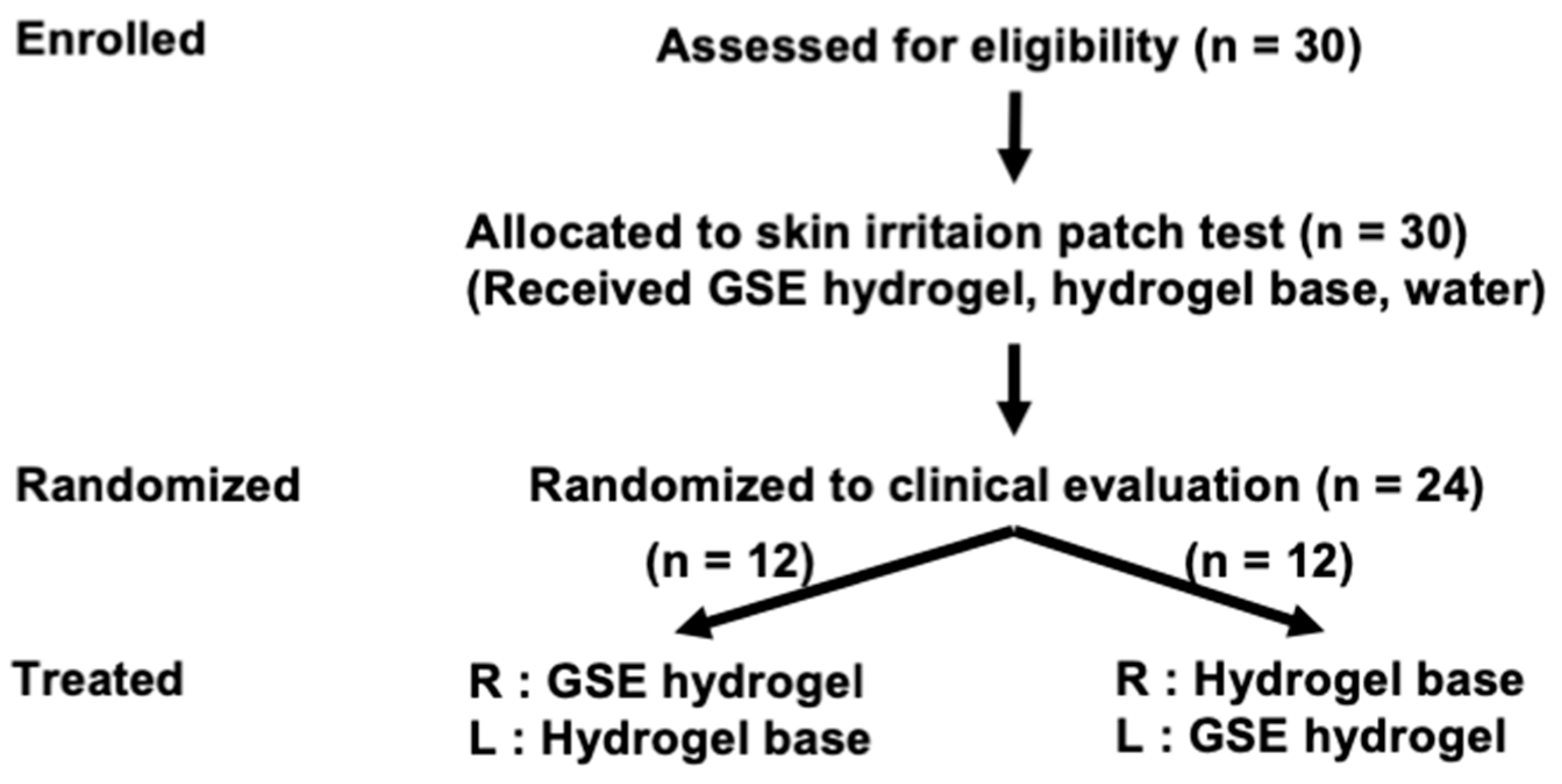
There has not been sufficient research into the application of G. speciosum in cosmeceutical products. Therefore, we performed a randomized, double-blind, placebo-controlled trial of an anti-aging serum for skin containing GSE [10]. The facial application of GSE serum was proven to safely promote skin distensibility by reducing skin viscoelasticity and wrinkle volume in healthy participants [10]. Nonetheless, there is a dearth of research concerning the antimelanogenic and therapeutic applications of this medicinal plant topically. Based on the previous investigation, the research team proposed the production of a cosmetic hydrogel with skin whitening and other advantageous features, as shown in the study flow (Figure 1). The purpose of this research was to examine anti-tyrosinase enzyme activity and melanin content in an in vitro non-cell-based assay, as well as in B16F10 melanoma cells of GSE. The GSE was subsequently developed into a hydrogel formulation for topical use. Clinical research on G. speciosum’s capability to reduce or decelerate dark spots, freckles, and melasma was studied. A randomized, double-blind, placebo-controlled trial of a hydrogel containing GSE was performed in healthy volunteers to investigate its skin-irritation and skin-whitening effects (Figure 1). Knowledge acquired from this study could help implement the extracts of G. speciosum for inclusion as an anti-melanogenesis agent in skin-whitening products. In support of previous research, the dual effects of G. speciosum’s whitening and anti-aging properties [10] pose challenges to the use of natural products as green cosmetics in sustainable cosmeceuticals.

2. Materials and Methods

2.1. Plant Sample Preparation and Extraction

G. speciosum (at least 3-year-old plants) was cultivated in the Phanom Sarakham District of Chachoengsao Province, Thailand. After harvest, G. speciosum pseudobulbs were rinsed with distilled water. Then, they were chopped into small pieces and dried in the shade at 45 °C. Using a high-speed disintegrator, dried materials were reduced to a fine powder. The dried samples were finely crushed and steeped for three days at room temperature in 95% (w/w) ethyl alcohol at the ratio of 1:9 w/v (dry extract:ethanol). The procedure was then carried out three times in this manner. The ethanolic solution of the extract was evaporated using a rotary evaporator [11]. The following equation can be used to determine the yield percentage:
%   y i e l d = m a s s   o f   v a c u u m d r i e d   G .   s p e c i o s u m   e x t r a c t m a s s   o f   d r i e d   G .   s p e c i o s u m × 100

2.2. Quality Control of GSE

High-performance liquid chromatography (HPLC) was used to analyze GSE (Agilent Technologies, Santa Clara, CA, USA) according to a method developed by Chowjarean et al., 2018 [8]. As an analytical marker, gastrodin, a key component of the extract, was employed. The validation of botanical identification procedures was carried out following AOAC international guidelines [12]. Prior to injection, a sample stock solution (1 mg/mL) was prepared using ethanol as a solvent. The stock solution was then filtered using a Whatman nylon syringe filter (0.22 μm). The presence of gastrodin was determined by contrasting the retention time and spectrum of GSE with a reference for gastrodin (% purity > 98%) (Chengdu Biopurify Phytochemicals, Chengdu, China). The amount of gastrodin in the GSE was determined by comparing the peak area of gastrodin in the GSE to a standard of gastrodin.
The mobile phase consisted of two different solvents: solvent A (water) and solvent B (acetonitrile). There was a gradual shift from 100% solvent A to 99% solvent A for 24 min, followed by a shift to 80% solvent A (6 min), to 20% solvent A (5 min), and ultimately back to 100% solvent A (15 min). An Inertsil ODS-3 C18 (4.6 × 150 mm, 5 μm) HPLC column was employed. The temperature of the column was 25 °C, with a 1.0 mL/min flow rate. The injection volume was 20 μL. DAD was set to a wavelength range of 200–380 nm, with a monitoring wavelength of 220 nm. The Agilent Chem station software was used to compute the results.

2.3. Mushroom Tyrosinase Enzyme Assay

The substrate utilized in this process was L-DOPA, and the kojic acid standard served as the positive control. Three groups of samples were studied: (1) GSE solutions in methanol:water (80:20 v/v), (2) kojic acid standard solutions in methanol:water (80:20 v/v), and (3) control (solvent methanol:water 80:20 v/v). Then, 80 μL of GSE solutions, kojic acid, or the control were added into 40 μL of pH 6.8 phosphate buffer. This was then followed by the addition of 40 μL of mushroom tyrosinase (480 units/mL) in phosphate buffer (pH 6.8). After that, we added 40 μL of a 0.85 mM L-DOPA substrate in a phosphate buffer to each well before placing it in an incubator at 25 °C for 10 min. An analytical wavelength of 492 nm was used in a microplate reader (Bio-Rad, Model 680, Tokyo, Japan) to observe the formation of DOPAchrome in the wells. The test was carried out three times for accuracy. The following equation can be used to determine the percent inhibition of tyrosinase activity:
%   I n h i b i t i o n = A B C D A B × 100
  • A: absorbance of blank solution;
  • B: absorbance of blank solution, but without enzyme;
  • C: absorbance of sample solution;
  • D: absorbance of sample solution, but without enzyme.

2.4. Cell Culture

B16F10 melanoma cells (ATCC, Manassas, VA, USA) were grown in Dulbecco’s modified Eagle medium (DMEM) containing 10% fetal bovine serum (FBS), 100 U/mL penicillin, and 100 μg/mL streptomycin, and maintained in an incubator with 5% CO2 (Thermo, Model 3111, Waltham, MA, USA) and 37 °C humidity. To harvest the cells, 0.25% (w/v) trypsin and 0.06 mM EDTA in phosphate-buffered saline were added. The cells (passage 10–20) were grown to a semiconfluent state prior to harvesting. After being resuspended in complete DMEM, the cells were then counted using a hemacytometer to determine their total number.

2.4.1. Cell Viability Assay

The cytotoxicity of GSE and kojic acid was determined in the B16F10 cell using sulforhodamine B (SRB) assay [13]. Cells (1 × 10⁴ cells) were cultured in a 96-well plate and incubated overnight. The cells were incubated with GSE (0.0001–1 mg/mL) or kojic acid for 72 h. Cells were then fixed, rinsed, and colored. The absorbance was detected using the microplate reader (Bio-Rad, Model 680, Japan) at 540 nm. The vitality of the cells was compared to those of a control group that had been treated with 0.1% absolute ethanol in a medium. The following equation was used to determine the percentages of viable cells in each sample:
%   C e l l   v i a b i l i t y = A B × 100
  • A: absorbance of the sample;
  • B: absorbance of the control.

2.4.2. Measurement of Intracellular Melanin Content

The procedure used to measure the melanin content was described in the previous publication, with a few small modifications [14]. Cells (1 × 10⁵ cells) were cultured onto 6-well plates, and the plates were left in an incubator at 37 °C overnight. After that, the samples were added, and the mixture was left to incubate for 24 h. The old medium was replaced with a new one for another 48 h. Intracellular melanin was determined in the cells. The cells were incubated for 1 h in 500 μL of 2 N NaOH at 60 °C. The solutions were collected, and a microplate reader (Bio-Rad, Model 680, Japan) was used to measure the absorbance of the solution at 450 nm. The melanin level was calculated and compared to the melanin standard. To determine the actual quantity of melanin produced from the same number of cells, the melanin content of the treatment was calculated by dividing the total amount of protein. The total protein was measured by the Bradford dye-binding assay [14]. The following is a calculation that was used to determine the percentages of the relative ratio of the melanin content:
%   R e l a t i v e   r a t i o   o f   m e l a n i n   c o n t e n t = M t M c × 100
  • Mt: ratio of melanin and the total amount of protein in the sample;
  • Mc: ratio of melanin and the total amount of protein in the control.

2.4.3. Measurement of Cellular Tyrosinase Activity

We performed an analysis of tyrosinase activity using a slightly modified version of the method that was published by Manosroi et al. [14]. In brief, treated cells were lysed and centrifuged for 10 min at 15,000 rpm. The supernatants were then collected and incubated with 0.05% L-DOPA in 50 mM sodium phosphate buffer (pH 6.8) at 37 °C for 2 h. The absorbance of dopachrome was measured using a microplate reader (Bio-Rad, Model 680, Japan) at 490 nm. A comparison was made to the level of enzyme activity with the mushroom tyrosinase standard. The total protein was also measured. The enzyme activity of the sample was analyzed and compared with the control. The following is a calculation that was used to determine the percentages of the relative ratio of the tyrosinase activity:
%   R e l a t i v e   r a t i o   o f   t h e   t y r o s i n a s e   a c t i v i t y = T t T c × 100
  • Tt: ratio of tyrosinase activity and the total amount of protein in the sample;
  • Tc: ratio of tyrosinase activity and the total amount of protein in the control.

2.5. Stability and Physicochemical Properties of the GSE

GSE’s physicochemical qualities, such as color, solubility, and pH, were assessed. The heating–cooling cycle test was used to study the chemical and physical stability of GSE. These thermal stress tests can provide helpful information regarding the stability prognosis in the early phases of the crude extract development process, such as when they are used for screening purposes [15]. During this test, the GSE was reserved at 4 ± 2 °C for 48 h and then switched to 45 ± 2 °C for 48 h. Following the completion of 6 cycles, the physical and chemical characteristics, as well as the total amount of gastrodin, were re-analyzed.

2.6. Topical GSE Hydrogel Stability

The hydrogel base and GSE hydrogels were formulated with cosmetic-grade ingredients. The physicochemical parameters of GSE hydrogel, including its appearance, color, odor, pH, viscosity, and gastrodin content, were investigated. According to ASEAN guidelines, the accelerated stability of the GSE hydrogel was investigated at 40 ± 2 °C and 75 ± 5% relative humidity (RH) for 6 months. Evaluations of the stability were carried out at 0, 3, and 6 months.

2.7. Approval of the Clinical Trial Protocol

This study was a double-blind, randomized, placebo-controlled trial comparing GSE hydrogel to water-based hydrogel as the control. The clinical trial was conducted following the declarations of Helsinki and Tokyo on human subjects and medical research. Before the recruitment of the participants, the study protocol was submitted to and approved by Western University’s ethical committee, Thailand (Project identification code: HE-WTU542787).

2.8. Recruitment of Healthy Volunteers

Thirty Thai volunteers aged 25–55 years (two males and twenty-eight females) were recruited (Figure 2). This study excluded volunteers with skin disorders or hypersensitive skin. There were no pregnant or breastfeeding women among the recruited volunteers. Before completing a consent form, all of the selected volunteers were fully briefed about the study’s procedures.

2.9. Skin Irritation Evaluation

A closed-chamber patch test was conducted by professional investigators and healthcare assistants in order to clinically examine the consequences of skin irritation. A certified pharmacist provided the randomization code of each patch test, which was blinded to the physician and assistants. Each participant received an adhesive, non-woven fabric from 3M (Thailand), a gauze pad (1 × 1 inch), placed on their backs for 24 h. The participant received 3 patches containing 0.5% (w/w) GSE hydrogel, hydrogel base, and water. A certified specialist and physicians assessed irritation 1 and 3 days after the removal of the patches. The assessment score by International Contact Dermatitis Research Group (ICDRG) was used to evaluate irritation level [16]. After a 10-day washout interval, as previously conducted by Chowjarean et al., 2018. [10], to eliminate the effects of previous treatment, the operation was repeated for confirmation.

2.10. Clinical Efficacy Evaluation

Randomized double-blind, placebo-controlled trials were carried out to establish the clinical effects of GSE hydrogel on human skin. Twenty-four participants who had no allergic symptoms in the previous week were chosen from the patch test. According to the interview data, all participants had not been exposed to steroids for 4 weeks. Twelve subjects were randomly assigned to each of the two groups. Trial participants, investigators, care providers, outcome assessors, and data analysts were all blinded. The first group applied GSE hydrogel on the right side of their faces and base hydrogel (control hydrogel) to the left. The second group applied GSE hydrogel on the left side of their faces and base hydrogel (control hydrogel) to the right. The participants were instructed to use the sample two times a day (in the morning and before bed). During the treatment time, we suggested that the participants shield the area being evaluated topically from sunlight, UV rays, and any other skin irritants. Moreover, the individuals were prohibited from smoking and alcohol consumption.
Maxameter® MX 18 RD (Courage and Khazaka, Cologne, Germany) and Visioface® RD (Courage and Khazaka, Cologne, Germany) were used to evaluate skin whitening on treated skin at baseline (Day 0) and 14, 28, 42, and 56 days following application. Volunteers intended to wear the mask to identify the measuring points (the mask has two space circles on the left and right faces). Then, 10 randomization points were recorded for analysis. The experimental space was maintained at 25 °C and 40–60% RH. The ability of the GSE hydrogel to reduce melanin content was calculated using the following equation [17], and the calculation formula below was used to determine the percentage of a sample to reduce melanin content or efficacy:
%   R e d u c t i o n   a c t i v i t y   o r   e f f i c a c y = M d 0 M d m M d 0 × 100
  • Md0: the melanin content on the baseline (D0);
  • Mdm: the melanin content on D14, D28, D42, and D56 (measuring days).

2.11. Statistical Analysis

The data from in vitro experiments are shown as the means ± standard deviations (SDs) (n = 3). The human study results are presented as the means ± standard errors of measurement (SEMs). A one-way ANOVA test and post-hoc analysis were used to analyze the in vitro parameters. For the purpose of the clinical investigation, a repeated measure ANOVA and a paired t-test were utilized. Statistically significant values were considered at p < 0.05 values, which were calculated using the SPSS program, version 21 by IBM Corp., Armonk, NY, USA.

3. Results

3.1. Plant Sample Preparation, Extraction, and Quality Control of G. speciosum

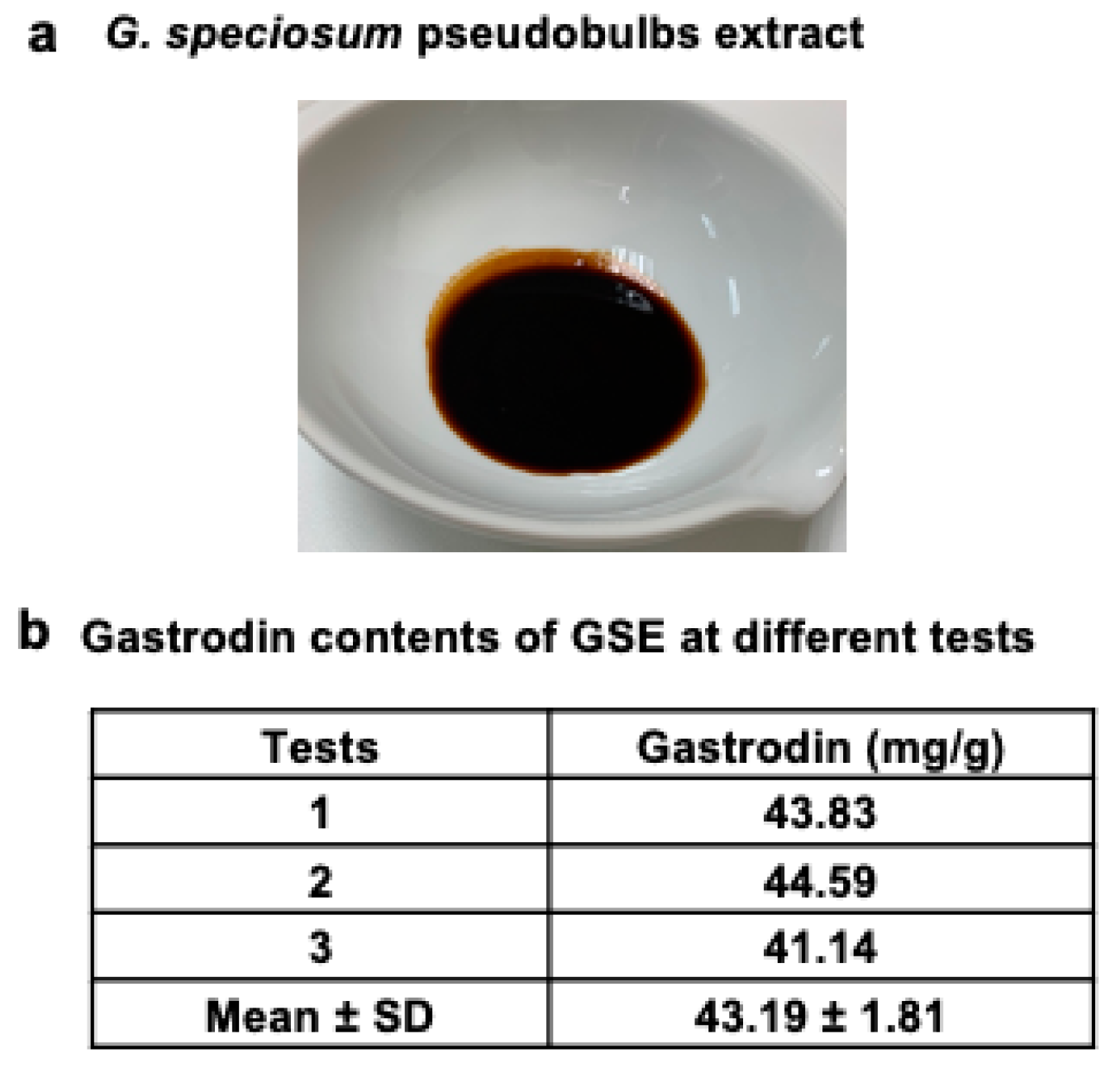
G. speciosum Blume pseudobulbs yielded 6.49% w/w of the initial dry weight. G. speciosum was subjected to milling and extraction, which resulted in an herb-to-extract ratio (HER) of 1:9. The extract of G. speciosum was thick and showed a dark brown color (Figure 3a). A validated approach [8] was used to analyze the quantity of gastrodin in three separate GSE tests. Tests 1, 2, and 3 yielded gastrodin concentrations of 43.83, 44.59, and 41.14 mg/g, respectively (Figure 3b), which were not statistically different (p > 0.05). The results suggested that there were no significant differences in gastrodin levels across batches.

3.2. Effect of GSE on In Vitro Mushroom Tyrosinase Activity

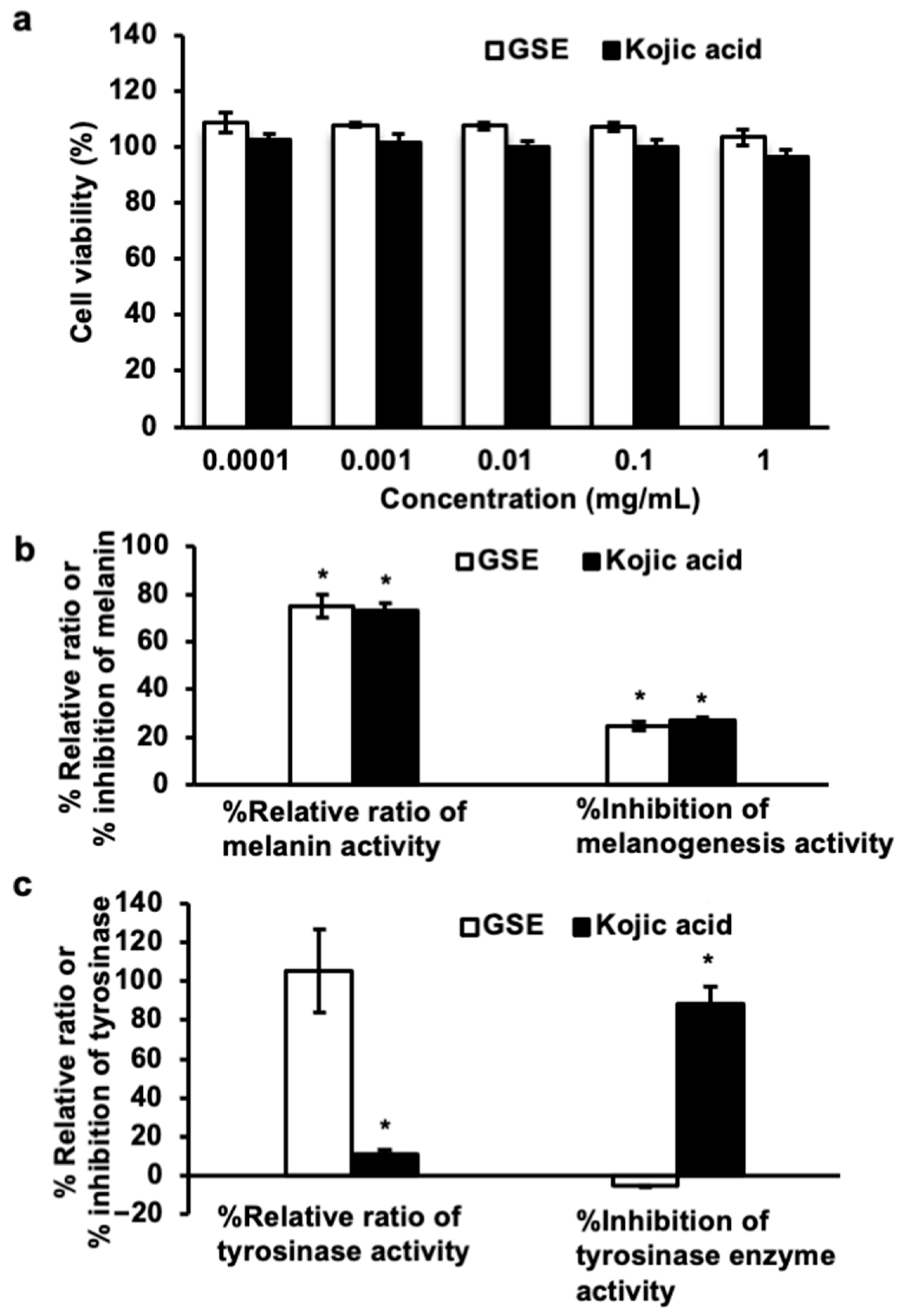
Anti-tyrosinase activity was evaluated in vitro in a non-cell-based manner. The activity of the tyrosinase enzyme was not inhibited by GSE at a concentration of 10 mg/mL, whereas kojic acid (68 μg/mL) inhibited tyrosinase enzyme activity by 73.91 ± 1.48%.

3.3. Effect of GSE on the Viability of B16F10 Cells

To establish an appropriate concentration for the melanogenesis experiment, the GSE and kojic acid (positive control) cytotoxicity assays were performed on B16F10 melanoma cells. Figure 4a shows the cytotoxic effects of GSE and kojic acid at 0.0001 to 1 mg/mL concentrations. The cell viability of GSE and kojic acid at 1 mg/mL (high concentration) was 103.36 ± 2.89 and 96.46 ± 2.77%, respectively. The results demonstrated that GSE and kojic acid at all dosages (0.0001–1 mg/mL) were non-toxic to B16F10 cells. Consequently, 1 mg/mL was chosen as the optimal GSE concentration for further evaluation in the melanin-content experiment and for tyrosinase enzyme activity.

3.4. Effect of GSE on Melanin Content in B16F10 Cells

A melanogenesis assay of GSE was studied in B16F10 melanoma cells. Because B16F10 melanoma cells are simple to cultivate and have the ability to generate melanin like normal human melanocytes, they are commonly used as a model for evaluating the prevention of melanogenesis by drugs [18]. Kojic acid is a melanogenesis inhibitor that prevents tautomerization of the melanin synthesis pathway by chelating with Cu2+ in tyrosinase [19]. Therefore, kojic acid was employed as a positive control. It was determined that 1 mg/mL of GSE suppressed the melanin production to 24.69 ± 1.82%, while kojic acid, at the same concentration, showed a melanin reduction of 26.81 ± 1.79% (Figure 4b). The relative ratios of melanin content were calculated and presented as a percentage. The percentages of relative ratios for melanin content of GSE and kojic acid were 75.31 ± 4.73 and 73.19 ± 1.28%, respectively. The GSE inhibited melanin by 0.92 times more than the control. As such, GSE appears to effectively inhibit the production of melanin in the B16F10 melanoma cell line.

3.5. Effect of GSE on Tyrosinase Enzyme Activity in B16F10 Cells

Tyrosinase is important in the production of melanin, the pigment that produces skin color. Tyrosinase is involved in the initial two stages of melanogenesis. Tyrosinase activity can be increased to increase melanin production [2,3]. Cellular tyrosinase activity was assessed by seeding B16F10 cells and treating them with GSE and kojic acid (positive control) at concentrations of 1 mg/mL. GSE or kojic-acid-treated proteins extracted from these cells were then combined with L-DOPA. Relative tyrosinase inhibitory effects are presented in Figure 4c. The percentages of GSE and kojic acid that suppress tyrosinase enzyme activity were −5.36 ± 0.43 and 88.96 ± 8.69%, respectively. Therefore, it is evident that GSE did not exhibit a potent ability to decrease tyrosinase activity. Based on these results, GSE was shown to be efficient at inhibiting the formation of melanin without affecting tyrosinase; the precise mechanism of this should be investigated further.

3.6. Stability and Physicochemical Characteristics of the GSE

Analyses of the physicochemical properties of the GSE extract solution were conducted, and results of accelerated stability tests showed that GSE extract solutions in propylene glycol 400 were stable (Table 1). The 0.5% (w/w) GSE was dissolved in propylene glycol 400, producing a clear solution without any precipitates. Consequently, propylene glycol 400 was selected as the co-solvent for the ensuing hydrogel.

3.7. The Formulation Development and Physical and Chemical Stability of GSE-Containing Hydrogels

After an in vitro evaluation of the GSE’s potential skin-whitening activity was conducted, topical hydrogel was developed for a preliminary clinical study. The GSE was dissolved completely in 0.5% (w/w) propylene glycol 400 to produce a transparent solution. Thus, the GSE hydrogel formulation included propylene glycol 400 as a co-solvent. Table 2 lists additional ingredients used in the formulation of the topical hydrogel. The physicochemical properties of GSE hydrogel, including its appearance, pH, and viscosity, are listed in Table 3. According to ASEAN guidelines, all formulations were stable at 40 °C and 75% RH for 6 months. In addition to its good physicochemical stability, a GSE hydrogel formulation at 0.5% (w/w) extract was chosen for further skin-irritation testing in human volunteers.

3.8. Skin Irritation Testing

Thirty volunteers were evaluated for skin irritation using the closed-patch skin irritation test. The hydrogel, either with or without GSE, was patched evenly across the right side of the back of each participant for a period of 24 h. The level of irritancy was observed using the evaluated scale from the ICDRG. After 24 and 72 h, both the water-based control and the GSE hydrogel showed no skin irritation compared to the hydrogel base (Table 4). The whitening properties of this hydrogel were then tested on human volunteers.

3.9. Skin Whitening Testing

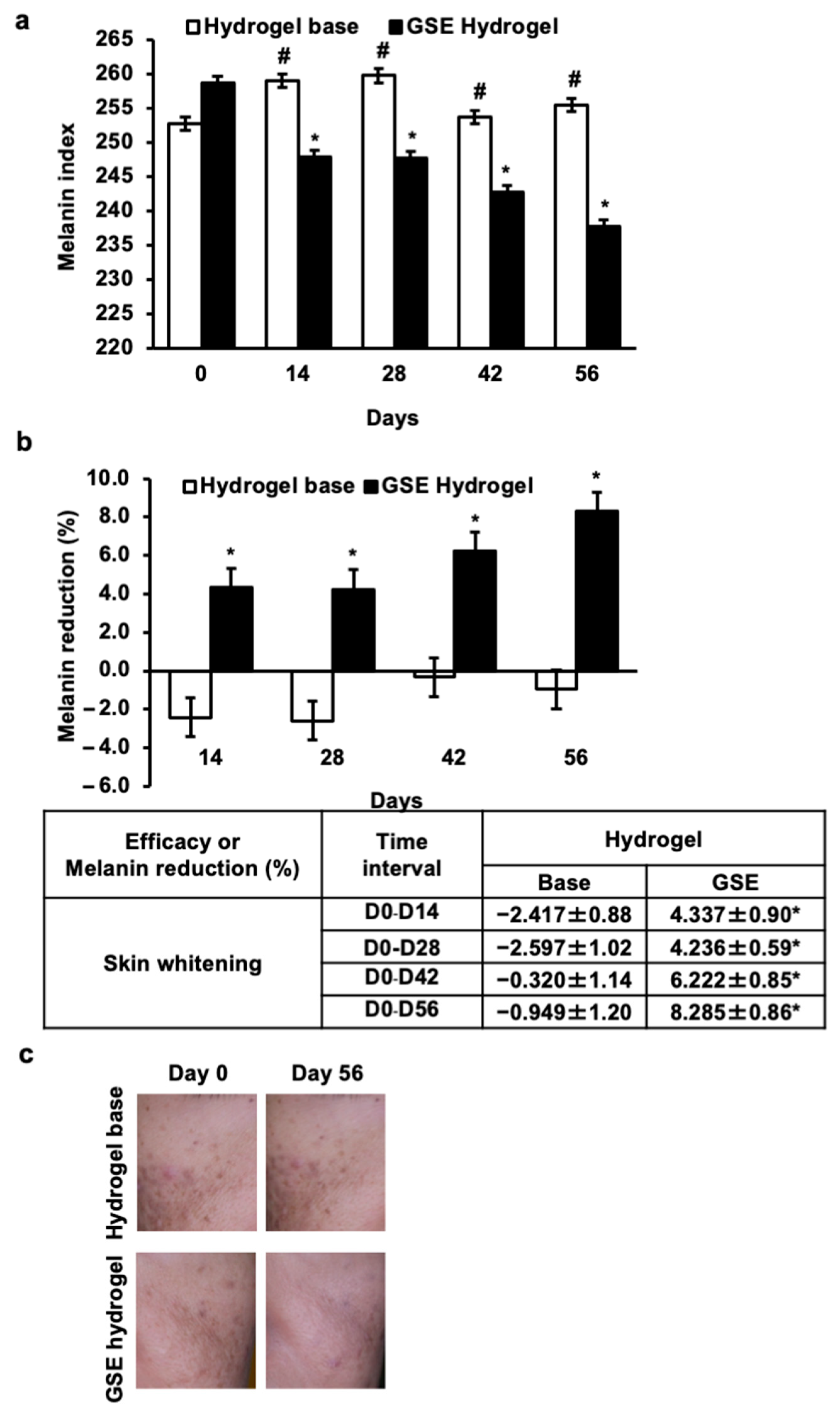
The results demonstrated that the average percent melanin index after the application of GSE hydrogel was 258.60, 247.82, 247.66, 242.71, and 237.70, while that of the hydrogel base was 252.67, 258.96, 259.71, 253.94, and 255.42 for 0, 14, 28, 42, and 56 days, respectively (Figure 5a). The results showed that the average percentage of efficacy or melanin reduction after treatment with the GSE hydrogel was 4.337, 4.239, 6.222, and 8.285%, whereas the hydrogel base was −2.147, −2.597, −0.320, and −0.949% for 0, 14, 28, 42, and 56 days, respectively (Figure 5b). GSE hydrogel also achieved its greatest degree of melanin reduction activity of 8.285% after 56 days of testing. Treatment with the GSE hydrogel resulted in a considerable reduction in melanin levels within two weeks (14 days) of treatment (Figure 5b). Figure 5c demonstrates that, after 56 days of using GSE hydrogel, the skin was visibly brighter and smoother than it had been at the beginning of the study. During and after the study, no volunteers were found to exhibit any adverse effects. These results demonstrate that GSE effectively lightened the skin of human participants.

4. Discussion

In order to evaluate the possible skin-whitening characteristics of G. speciosum pseudobulb extract, the goal of this work was to examine the extract’s ability to prevent melanogenesis in B16F10 melanoma cells. In addition, this study developed a stable topical formulation for assessing the efficacy of the whitening effect of G. speciosum pseudobulb in human subjects.
In this study, GSE acted as a whitening agent due to its ability to decrease melanogenesis activity in B16F10 melanoma cells by reducing melanin synthesis (Figure 4b). Kojic acid was chosen to serve as the positive control in this study because it is currently used in the cosmetics industry as a skin-lightening agent [19]. However, it penetrates the skin poorly and can cause major adverse effects, such as skin inflammation [20]. The discovery of safety compounds from natural sources such as GSE is required. As a result of GSE’s ability to prevent melanogenesis in B16F10 melanoma cells (Figure 4b), it was chosen as a candidate for use in skin-lightening formulations and then tested on humans.
GSE was developed into a cosmetic termed “GSE hydrogel”, which underwent an evaluation of its physicochemical properties and a stability test. Additionally, 0.5% GSE hydrogel was formulated in 12% PEG 400, 2.5% HPMC, and 1% Microcare PHC® (Table 2). For 6 months, the GSE hydrogel displayed good physical and chemical stability, indicating that this GSE hydrogel formulation was appropriate for 56-day testing in skin irritation and skin whitening. Our GSE hydrogel showed no skin irritation compared to the hydrogel base, which is concordant with our previous report on GSE serum [10]. However, this GSE hydrogel is preferable to the previous formulation (GSE serum) since its viscosity remains unchanged after 6 months, indicating a longer shelf life. After 56 days of application, the melanin index value reduced repeatedly and dramatically following the application of the GSE hydrogel, with the greatest reduction being 8.285% (Figure 5b). Previous research has shown that GSE has a high potential for anti-elastase activity, antioxidant activity, increasing human skin fibroblast migration, and enhancing stemness in human keratinocytes [11]. It has been demonstrated that the clinical application of GSE serum promotes skin distensibility by decreasing skin viscoelasticity and wrinkle volume in healthy subjects [10]. According to these findings, this study is the first to report that the clinical use of GSE effectively lightened the skin of human volunteers. The fact that G. speciosum has both skin-lightening and anti-aging properties supports the notion that it would be an excellent candidate for use in cosmetics as a skin-whitening and anti-aging agent.
In the screening of putative melanogenesis inhibitors, mushroom tyrosinase is typically the enzyme that is used as the target [21]. According to the results, GSE did not inhibit mushroom tyrosinase enzyme activity and was unable to inhibit tyrosinase activity in B16F10 melanoma cells (Figure 4c). However, the GSE was found to be effective at inhibiting the synthesis of melanin. These results are consistent with clinical results showing that GSE hydrogel decreased epidermal melanin content. It has been postulated that the oxidizing effect of UV irradiation plays a role in the process of melanogenesis [1]. Redox agents can also have an influence on skin pigmentation by interacting with copper on tyrosinase or o-quinones to suppress the oxidative polymerization of melanin precursors. These interactions can take place either inside the tyrosinase enzyme or within the skin [22]. Antioxidants can also prevent pre-existing melanin from being directly photooxidized [23]. Vitamins E, C, and niacinamide are common antioxidants found in skin-lightening formulas [23,24]. Gastrodin, a phenolic glycoside [p-(hydroxymethyl) phenyl-b-D-glucopyranoside] discovered in GSE, may play an important function as a skin-lightening agent. According to research, gastrodin is an effective antioxidant thanks to its ability to increase SOD levels [8,9,25,26]. It is possible to inhibit melanogenesis through its antioxidant activity. However, the precise mechanism requires additional investigation.
We intend to use a non-hazardous solvent extraction technique that is safe for humans and the environment and simple to implement in large-scale manufacturing. Ethanol was selected, and the yield of the 95% pseudobulbs G. speciosum ethanolic extract was high (6.49% w/w of the initial dry weight) compared to an earlier report using methanolic extract (1.86% w/w of the initial dry weight) [5]. GSE demonstrated a high level of antioxidant activity in preliminary antioxidant tests [9]. As a result, we intend to investigate GSE further regarding its biological activities in order to support its use in cosmeceutical products. For the purpose of extract quality control, we attempted to identify the GSE analytical marker. The analytical marker should have a high concentration in GSE in order to compare its concentration across batches. Since gastrodin was found in a high concentration, it was used as an analytical marker in our study [8].

5. Conclusions

The study proved systematically that GSE has the capacity to reduce epidermal melanin content both in vitro and in clinical study. Hence, G. speciosum has the potential to be utilized in the dermatological industry as a lightening agent in skincare products. A stable and safe GSE hydrogel with a long shelf life for industrial applications was also obtained. This study focuses on the combined effects of all compounds in GSE; therefore, exploring the related bioactive mechanism behind the synergism is recommended for further study.

Author Contributions

Conceptualization, V.C. and S.H.; methodology, V.C., P.P.P. and S.H.; validation, V.C., P.P.P. and S.H.; formal analysis, V.C., P.P.P. and S.H.; investigation, V.C., P.P.P. and S.H.; resources, V.C. and S.H.; data curation, V.C. and S.H.; writing—original draft preparation, V.C., P.P.P. and S.H.; writing—review and editing, V.C., P.P.P. and S.H.; visualization, V.C., P.P.P. and S.H.; supervision, V.C. and S.H.; project administration, P.P.P.; funding acquisition, V.C. and S.H. All authors have read and agreed to the published version of the manuscript.

Funding

This research was funded by the Research Institute of Rangsit University (Grant no. 80/2561), the Thailand Research Fund (Grant no. MRG6280011), and the Cosmeceutical Research, Development and Testing Center, College of Pharmacy, Rangsit University (Grant no. CDRT-1600-01-2021).

Institutional Review Board Statement

The study was conducted in accordance with the Declaration of Helsinki, and the protocol was approved by the Ethics Committee of Western University’s ethical committee, Thailand (Project identification code: HE-WTU542787).

Informed Consent Statement

All the subjects gave their informed consent for inclusion before they participated in the study.

Data Availability Statement

Not applicable.

Conflicts of Interest

The authors declare no conflict of interest.

References

  1. Kumari, S.; Tien Guan Thng, S.; Kumar Verma, N.; Gautam, H.K. Melanogenesis Inhibitors. Acta Derm. Venereol. 2018, 98, 924–931. [Google Scholar] [CrossRef] [Green Version]
  2. D’Mello, S.A.N.; Finlay, G.J.; Baguley, B.C.; Askarian-Amiri, M.E. Signaling Pathways in Melanogenesis. Int. J. Mol. Sci. 2016, 17, 1144. Available online: https://www.mdpi.com/1422-0067/17/7/1144 (accessed on 1 November 2022). [CrossRef] [Green Version]
  3. Solano, F. Photoprotection and Skin Pigmentation: Melanin-Related Molecules and Some Other New Agents Obtained from Natural Sources. Molecules 2020, 25, 1537. [Google Scholar] [CrossRef] [Green Version]
  4. Hu, Y.; Zeng, H.; Huang, J.; Jiang, L.; Chen, J.; Zeng, Q. Traditional Asian Herbs in Skin Whitening: The Current Development and Limitations. Front. Pharmacol. 2020, 11, 982. Available online: https://www.frontiersin.org/articles/10.3389/fphar.2020.00982 (accessed on 1 November 2022). [CrossRef]
  5. Nontachaiyapoom, S.; Sasirat, S.; Manoch, L. Symbiotic seed germination of Grammatophyllum speciosum Blume and Dendrobium draconis Rchb. f., native orchids of Thailand. Sci. Hortic. 2011, 130, 303–308. Available online: http://www.sciencedirect.com/science/article/pii/S030442381100330X (accessed on 1 November 2022). [CrossRef]
  6. Uawonggul, N.; Chaveerach, A.; Thammasirirak, S.; Arkaravichien, T.; Chuachan, C.; Daduang, S. Screening of plants acting against Heterometrus laoticus scorpion venom activity on fibroblast cell lysis. J. Ethnopharmacol. 2006, 103, 201–207. [Google Scholar] [CrossRef]
  7. Sahakitpichan, P.; Mahidol, C.; Disadee, W.; Chimnoi, N.; Ruchirawat, S.; Kanchanapoom, T. Glucopyranosyloxybenzyl derivatives of (R)-2-benzylmalic acid and (R)-eucomic acid, and an aromatic glucoside from the pseudobulbs of Grammatophyllum speciosum. Tetrahedron 2013, 69, 1031–1037. Available online: http://www.sciencedirect.com/science/article/pii/S0040402012018005 (accessed on 1 November 2022). [CrossRef]
  8. Chowjarean, V.; Sucontphunt, A.; Vchirawongkwin, S.; Charoonratana, T.; Songsak, T.; Harikarnpakdee, S.; Tengamnuay, P. Validated RP-HPLC method for quantification of gastrodin in ethanolic extract from the pseudobulbs of Grammatophyllum speciosum blume. Malays. J. Anal. Sci. 2018, 22, 219–226. [Google Scholar]
  9. Harikarnpakdee, S.; Chowjarean, V. Grammatophyllum speciosum Ethanolic Extract Promotes Wound Healing in Human Primary Fibroblast Cells. Int. J. Cell Biol. 2018, 2018, 7836869. [Google Scholar] [CrossRef] [Green Version]
  10. Chowjarean, V.; Phiboonchaiyanan, P.P.; Harikarnpakdee, S.; Tengamnuay, P. A natural skin anti-ageing serum containing pseudobulb ethanolic extract of Grammatophyllum speciosum: A randomized double-blind, placebo-controlled trial. Int. J. Cosmet. Sci. 2019, 41, 548–557. [Google Scholar] [CrossRef]
  11. Chowjarean, V.; Nimmannit, U.; Chaotham, C.; Eaknai, W.; Sucontphunt, A.; Plaimee, P.P.; Tengamnuay, P.; Chanvorachote, P. Grammatophyllum speciosum Extract Potentiates Stemness in Keratinocyte Cells. Chiang Mai J. Sci. 2018, 45, 237–248. [Google Scholar]
  12. AOAC. Guideline for Dietary Supplements and Botanicals. 2013. Available online: http://www.eoma.aoac.org/app_k.pdf (accessed on 1 November 2022).
  13. Kanlayavattanakul, M.; Lourith, N.; Chaikul, P. Jasmine rice panicle: A safe and efficient natural ingredient for skin aging treatments. J. Ethnopharmacol. 2016, 193, 607–616. [Google Scholar] [CrossRef] [PubMed]
  14. Manosroi, A.; Chaikul, P.; Abe, M.; Manosroi, W.; Manosroi, J. Melanogenesis of methyl myristate loaded niosomes in B16F10 melanoma cells. J. Biomed. Nanotechnol. 2013, 9, 626–638. [Google Scholar] [CrossRef]
  15. Estanqueiro, M.; Conceição, J.; Amaral, M.H.; Santos, D.; Silva, J.B.; Lobo, J.M.S. Characterization and stability studies of emulsion systems containing pumice. Braz. J. Pharm. Sci. 2014, 50, 361–369. [Google Scholar] [CrossRef] [Green Version]
  16. Ivens, U.; Serup, J.; O’goshi, K. Allergy patch test reading from photographic images: Disagreement on ICDRG grading but agreement on simplified tripartite reading. Skin Res. Technol. 2007, 13, 110–113. [Google Scholar] [CrossRef]
  17. Whangsomnuek, N.; Mungmai, L.; Mengamphan, K.; Amornlerdpison, D. Efficiency of Skin Whitening Cream Containing Etlingera elatior Flower and Leaf Extracts in Volunteers. Cosmetics 2019, 6, 39. Available online: https://www.mdpi.com/2079-9284/6/3/39 (accessed on 1 November 2022). [CrossRef] [Green Version]
  18. Zheng, Y.; Lee, E.H.; Lee, S.Y.; Lee, Y.; Shin, K.O.; Park, K.; Kang, I.J. Morus alba L. root decreases melanin synthesis via sphingosine-1-phosphate signaling in B16F10 cells. J. Ethnopharmacol. 2023, 301, 115848. [Google Scholar] [CrossRef]
  19. Saeedi, M.; Eslamifar, M.; Khezri, K. Kojic acid applications in cosmetic and pharmaceutical preparations. Biomed. Pharmacother. 2019, 110, 582–593. Available online: https://www.sciencedirect.com/science/article/pii/S0753332218367477 (accessed on 1 November 2022). [CrossRef]
  20. García-Gavín, J.; González-Vilas, D.; Fernández-Redondo, V.; Toribio, J. Pigmented contact dermatitis due to kojic acid. A paradoxical side effect of a skin lightener. Contact Dermat. 2010, 62, 63–64. [Google Scholar] [CrossRef]
  21. Matos, P.; Paranhos, A.; Batista, M.T.; Figueirinha, A. Synergistic Effect of DIBOA and Verbascoside from Acanthus mollis Leaf on Tyrosinase Inhibition. Int. J. Mol. Sci. 2022, 23, 13536. [Google Scholar] [CrossRef]
  22. Briganti, S.; Camera, E.; Picardo, M. Chemical and instrumental approaches to treat hyperpigmentation. Pigment. Cell Res. 2003, 16, 101–110. [Google Scholar] [CrossRef] [PubMed]
  23. Ebanks, J.P.; Wickett, R.R.; Boissy, R.E. Mechanisms regulating skin pigmentation: The rise and fall of complexion coloration. Int. J. Mol. Sci. 2009, 10, 4066–4087. [Google Scholar] [CrossRef] [PubMed]
  24. Sarkar, R.; Arora, P.; Garg, K.V. Cosmeceuticals for Hyperpigmentation: What is Available? J. Cutan. Aesthetic Surg. 2013, 6, 4–11. [Google Scholar] [CrossRef] [PubMed]
  25. Zhao, X.; Zou, Y.; Xu, H.; Fan, L.; Guo, H.; Li, X.; Li, G.; Zhang, X.; Dong, M. Gastrodin protect primary cultured rat hippocampal neurons against amyloid-beta peptide-induced neurotoxicity via ERK1/2-Nrf2 pathway. Brain Res. 2012, 1482, 13–21. [Google Scholar] [CrossRef]
  26. Huang, Q.; Shi, J.; Gao, B.; Zhang, H.Y.; Fan, J.; Li, X.J.; Fan, J.Z.; Han, Y.H.; Zhang, J.K.; Yang, L.; et al. Gastrodin: An ancient Chinese herbal medicine as a source for anti-osteoporosis agents via reducing reactive oxygen species. Bone 2015, 73, 132–144. [Google Scholar] [CrossRef] [PubMed]
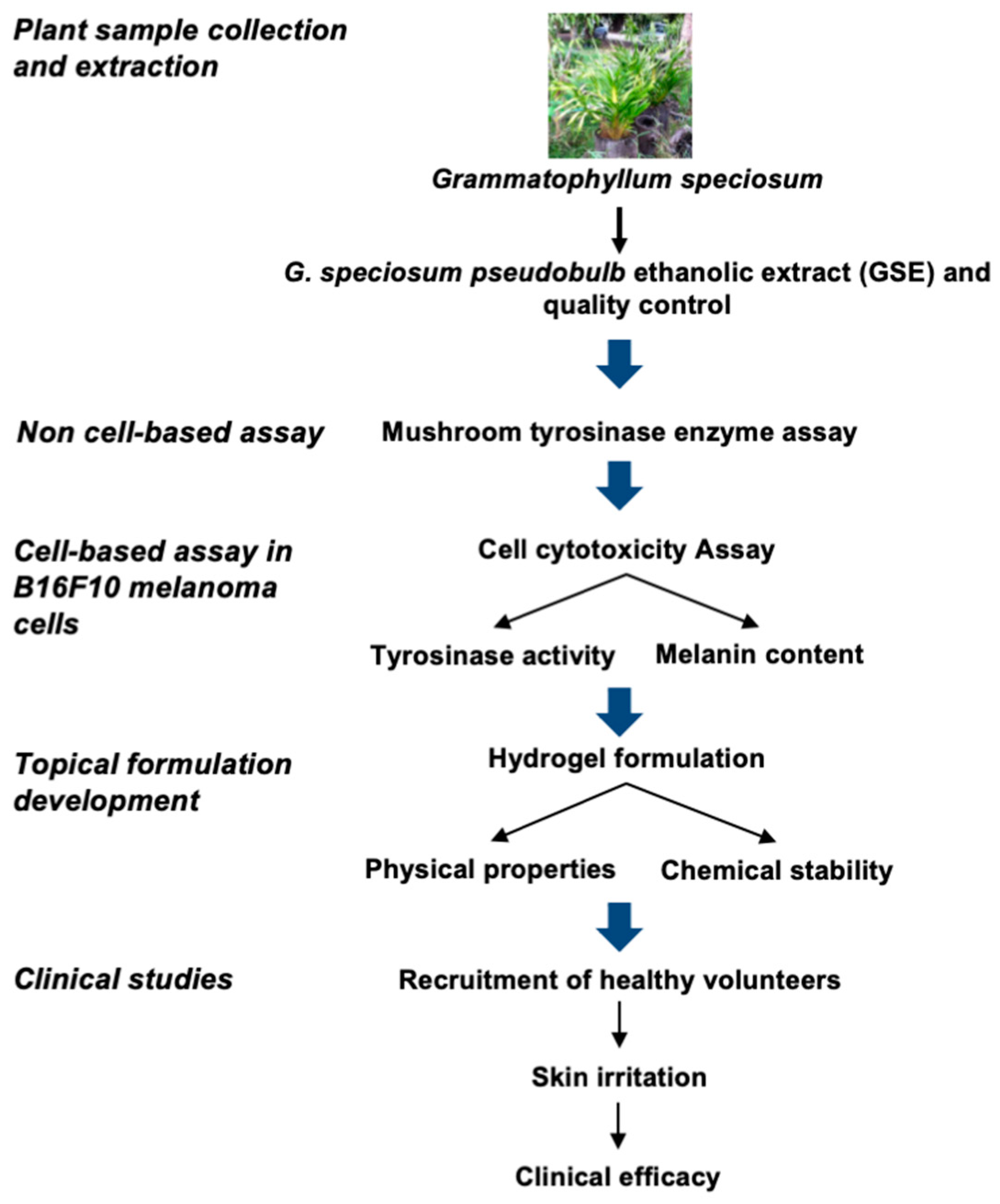
Figure 1. Study flow chart.
Sustainability 14 16829 g001
Figure 2. Disposition of the subject. R, right treatment area; L, left treatment area.
Sustainability 14 16829 g002
Figure 3. (a) G. speciosum pseudobulb ethanolic extract (GSE). (b) Gastrodin contents of GSE in three different samples. Data are presented as mean ± SD for triplicate samples.
Sustainability 14 16829 g003
Figure 4. (a) Cytotoxicity assay performed on B16F10 melanoma cells after treatment with GSE and kojic acid at concentrations ranging from 0.0001–1 mg/mL. The sulforhodamine B (SRB) assay was used to evaluate the viability of the cells. The percentage values in the treated cells were compared to those in the control cells. (b) The graph depicts the percentage of the relative ratio of melanin concentration and suppression of melanogenesis activity in B16F10 melanoma cells treated with GSE and kojic acid. (c) The graph shows the percentage of relative ratio of tyrosinase activity and the suppression of tyrosinase enzyme activity in B16F10 melanoma cells treated with GSE and kojic acid. Data are presented as mean ± SD for triplicate samples. * p < 0.05 shows a significant difference from the control.
Sustainability 14 16829 g004
Figure 5. Clinical evaluation of the skin whitening effect of GSE hydrogel. Melanin levels were measured using a Mexameter on the faces of 24 volunteers at Day 0 (before treated with hydrogel) and Day 56 (after being treated with a hydrogel base or GSE hydrogel formulation twice daily for 56 days). (a) The average melanin index value of GSE hydrogel and hydrogel base at the baseline (Day 0) and following the treatment for 14, 28, 42, and 56 days are presented. * p < 0.05 shows a significant difference from the GSE hydrogel versus Day 0 (before treatment). # p < 0.05 shows a significant difference from the hydrogel base versus Day 0 (before treatment). (b) Percentage of melanin reduction (%) of GSE hydrogel. Values presented as means ± SEMs were obtained from the volunteers on the same day. * p < 0.05 shows a significant difference from the hydrogel base. (c) Photos of a volunteer.
Sustainability 14 16829 g005
Table 1. Stability of the GSE before and after the accelerated tests.
ParametersGSE
Before HC 1After HC 1
Gastrodin content (μg/g)21.53 ± 0.5522.06 ± 0.80
ColorBrownish yellowBrownish yellow
pH5.35.3
1 HC; heating–cooling cycle (n = 3, value = mean + S.D.).
Table 2. Ingredients of base and GSE hydrogel.
% (w/w)
GSEPEG 400HPMCMicrocare PHC® 1DI Water
Base-122.5184.5
GSE hydrogel0.5122.5184.0
1 Microcare PHC; Phenoxyethanol (and) Chlorphenesin (and) Glycerin.
Table 3. Stability of the 0.5% (w/w) GSE hydrogel formulation.
0.5% (w/w)
GSE Hydrogel
Condition 40 °C 75% RH
Gastrodin Content (μg/g)pHViscosity 1 (cps)
0 months (D0)22.86 ± 0.985.315537.53 ± 20.49
3 months (D90)22.93 ± 0.535.355585.58 ± 30.35
6 months (D180)22.57 ± 0.725.285507.18 ± 80.73
1 Spindle no. 94, 30 rpm and % torque > 50 (n = 3, value = mean ± S.D.).
Table 4. International Contact Dermatitis Research Group (ICDRG) value and skin irritation reaction observed for GSE hydrogel.
Test SubstancesICDRG Value
First TimeSecond Time
24 h72 h24 h72 h
Hydrogel base0000
GSE hydrogel0000
DI water0000
Publisher’s Note: MDPI stays neutral with regard to jurisdictional claims in published maps and institutional affiliations.