Article

Airborne Pollen, Allergens, and Proteins: A Comparative Study of Three Sampling Methods

by
Chiara Suanno
1,†,
Silvia Sandrini
2,†,
Iris Aloisi
1,
Paola De Nuntiis
2,
Maria Cristina Facchini
2,
Stefano Del Duca
1,3,* and
Delia Fernández-González
2,4
1
Department of Biological, Geological and Environmental Sciences, University of Bologna, Via Irnerio 42, 40126 Bologna, Italy
2
National Research Council of Italy-Institute of Atmospheric Sciences and Climate (CNR-ISAC), Via Gobetti 101, 40129 Bologna, Italy
3
Interdepartmental Centre for Agri-Food Industrial Research, University of Bologna, Via Quinto Bucci 336, 47521 Cesena, Italy
4
Department Biodiversity and Environmental Management, University of Léon, Campus Vegazana, S/n, 24007 Léon, Spain
*
Author to whom correspondence should be addressed.
These authors contributed equally to this work.
Sustainability 2022, 14(19), 11825; https://doi.org/10.3390/su141911825
Submission received: 29 July 2022 / Revised: 31 August 2022 / Accepted: 15 September 2022 / Published: 20 September 2022

Abstract

:
Nowadays, there is a wide range of different methods available for the monitoring of pollen and allergens, but their relative efficiency is sometimes unclear, as conventional pollen monitoring does not thoroughly describe pollen allergenicity. This study aims to evaluate airborne pollen, allergen, and protein levels, associating them with meteorological and chemical parameters. The sampling was performed in Bologna (Italy) during the grass flowering period, with three different devices: a Cyclone sampler (CS), a Dicothomous sampler (DS), and a Berner impactor (BI). Total proteins were extracted from aerosol samples, and grass allergens Phl p 1 and Phl p 5 were quantified by ELISA. Airborne Poaceae pollen concentrations were also evaluated, using a Hirst-type trap. Proteins and allergens collected by CS resulted about ten times higher than those collected by the other two instruments, possibly due to their different cut-offs, while DS and BI results appeared consistent only for the total proteins collected in the fine fraction (1.3 vs. 1.6 μg/m3). Airborne proteins correlated neither with Poaceae pollen nor with its aeroallergens, while aeroallergens correlated with pollen only in the coarse particulate, indicating the presence of pollen-independent aeroallergens in the fine particulate, promoted by high wind speed.

1. Introduction

It is widely known that certain airborne pollen types contain small molecules called allergens [1,2,3]. Most allergens are proteins or glycoproteins with a molecular weight between 5 and 100 kDa that are not associated with pathogens or parasites but can nonetheless activate the immune system and promote the production of antibodies against them in sensitised subjects [4]. When pollen allergens contact the respiratory mucosa of allergic patients, they activate an IgE-mediated immune response, causing the symptomatology of rhinoconjunctivitis or allergic asthma, by type I hypersensitivity mechanisms [3]. The impossibility to cure pollen allergy implies that allergic patients need to avoid the exposition to their relevant allergens to safeguard their quality of life [5]. Hence, it is essential for them to be informed on the daily atmospheric concentrations of allergenic pollen, and for this reason a continuous airborne pollen monitoring network has been put in place in many countries in the last century [6,7,8].
However, the correlation between airborne pollen levels and allergic symptoms is not always clear. A possible explanation of this inconsistency is that the exposure to one pollen type does not linearly correlate with the exposure to its allergens, for three main reasons: (I) the same pollen type can have variable allergen concentrations (i.e., pollen potency); (II) airborne debris of the source plant could carry the same allergens of its pollen; (III) pollen allergens can be released in the atmosphere and disperse independently from pollen [8,9,10,11]. The latter phenomenon is still largely unknown, but there is proof that allergens can be transferred from pollen grains to smaller airborne particles such as air pollutants by leaching or contact [12,13,14,15]. In peculiar atmospheric conditions, e.g., during thunderstorms, allergens could also be released by osmotic rupture of the cell wall or by elution, and thus can also be carried by starch granules, vesicles, Ubish bodies, or other organelles [8,16,17,18]. Contrarily to the intact pollen grains, the diameter of which ranges between 10 and 100 μm, these smaller allergen carriers can go undetected by traditional pollen monitoring, they can remain in the air for longer periods, and they have the potential to reach the lower human airways [10,13,19,20]. The inhalation of released pollen allergens could also help to explain the aetiology of pollen-induced allergic asthma, since inhaled pollen grains are too broad to enter the lower airways, and thus cannot directly cause the inflammation of their mucosa that leads to asthma [21]. This inflammation can be more problematic if the allergen carrier to the lower airways is an air pollutant [12,22,23].
Regardless of their carrier and biological nature, airborne allergens are a relevant component of the bioaerosol, making up to ~5% of urban air particulate matter [24,25], such that they are also included in air quality indices [26,27]. In the last decade, there has been a growing interest in the measurement of airborne pollen allergen concentrations, to compare it to the atmospheric levels of their respective pollen grains. These studies found out that the concentration of pollen allergens transported by smaller carriers is independent from the airborne pollen concentration, and that there is often a mismatch between a pollen type’s peak and its allergens’ peaks [9,28,29,30,31,32,33,34].
To accurately sample and analyse airborne allergens, it is necessary to employ volumetric air samplers with a lower diameter cut-off than those employed for pollen sampling, using different sampling substrates and with higher air volume intakes [8,32]. In fact, while pollen grains belong to the aerosol fraction with a diameter greater than 10 μm (PM10), the other pollen allergens carriers have smaller, more variable sizes, and they can even be found in the finest particles fraction (PM2.5 and PM1). So far, several studies on airborne allergens have been carried out using air samplers originally designed to collect small airborne particles such as fungal spores or air pollutants [8]. The most common instruments employed for these studies are the Cyclone samplers [35,36,37,38,39,40,41] and sieve (or cascade) impactors, in particular the Andersen sampler [42] and the high-volume ChemVol impactor [20,43,44,45]. While in almost all these studies the aeroallergen sampler is operated in parallel and compared with a Hirst-type pollen trap, to date only one work compared the efficiency of two different instruments, the Cyclone sampler and the ChemVol impactor, to collect airborne allergens [32], revealing that the first is more sensitive to lower concentrations of airborne allergens and records higher particle concentrations than the latter. Assessing the capture efficiency of the existing samplers is pivotal to compare the results of studies employing different devices and to explore other possible capture techniques.
Once the aeroallergens are captured by the sampler, they can be identified and quantified by ELISA. However, this technique allows for the analysis of one allergen at time, and the processed sample cannot be employed for further analysis. This implies that only one or two allergens can be investigated per sampling campaign because of the small size of daily aerosol samples; hence, it is necessary to select the allergens of interest for the monitoring [8].
While many different allergenic species are clinically relevant in Italy, Poaceae pollen represents the largest allergen source all over Europe, and it is responsible for pollen allergies in more individuals than any other flowering plant family, with the most allergenic pollen belonging to the genera Phleum, Dactylis, and Lolium [46,47]. Group I and group V grass pollen allergens are particularly problematic, with Phl p 1 (group I) and Phl p 5 (group V) from Phleum pratense L. having the highest sensitisation rates in Poaceae allergic subjects [45], so that anti-Phl p 1 and anti-Phl p 5 antibodies are considered good markers for grass allergy in adults [48]. Because of their relevance, and owing to the possibility for grass allergens to be released in the air during mowing, there is growing interest in monitoring the atmospheric presence of Phl p 1 and Phl p 5 allergens [45,49,50], but the literature is still scarce, and no data are available for their airborne dynamics in Italy; hence, they are good candidates for aeroallergen monitoring.
The general aim of this study was to assess which factors influence the quantification of airborne allergens. This was achieved by setting three specific goals: (I) comparing the total protein concentrations recorded by three different aerosol samplers (Cyclone, Dichotomous, and Berner) and the size distribution of their carriers, and evaluating which technical features of the samplers might have influenced the results; (II) comparing the airborne concentrations of Poaceae pollen and airborne proteins with the concentrations of selected grass aeroallergens (Phl p 1 and Phl p 5) to enhance possible correlations and evaluate the possible sources of the allergens; (III) correlating the chemical composition of fine and coarse particulate matter collected by the samplers, the protein concentrations, and the concurrent meteorological data to assess whether aeroallergen concentrations were influenced by abiotic factors.

2. Materials and Methods

2.1. Study Area

Bologna (54 m asl) is located in the Po Plain at the base of the Apennine Ridge, a vast flat area in the northern part of Italy, which biogeographically belongs to the Continental Region. The area is characterised by a sub-continental climate (Köppen-Geiger Cfa) with cold winters and hot summers (typical monthly mean temperatures ranging from 1 to 26 °C), high humidity levels (typical monthly mean relative humidity ranging from 60% to 84%), and low wind intensities (typical annual mean wind intensities of about 2 m/s). The annual mean precipitation is between 760–800 mm with a xerothermic period in July and August.

2.2. Pollen Sampling

Pollen was sampled with the Hirst-type sampler Lanzoni VPPS2000 (Lanzoni S.r.l., Bologna, Italy), in compliance with the European standard [51]. The pollen sampler was placed close to the other air samplers employed in this study and operated with an air throughput of 10 L/min. The sampling tape was collected each week and cut in 7 daily samples, that were then stained with basic fuchsin and mounted on glass slides. Poaceae pollen was then identified and counted in light microscopy using an objective with 40× magnification lens. Pollen counts were converted into daily atmospheric concentrations (grains/m3).

2.3. Particulate Matter Sampling

Sampling was performed from March to June 2015. Three different samplers were employed for the collection of airborne particles:
  • One low-volume Cyclone Burkard sampler (CS) (Burkard Manufacturing Co, Rickmansworth, UK), which is a continuous, wind-oriented volumetric sampler using single reverse-flow miniature Cyclone technology to collect airborne particles. Air was aspired with a speed of 16.5 L/min, and particles were collected directly into a 1.5 mL vial, which was substituted every 24 h of sampling.
  • Two five-stage Berner impactors (BIs) (Hauke GmbH, Wien, Austria), which are volumetric cascade samplers that allow the separation of the particles collected according to their aerodynamic diameter [52,53]. Both BIs had cut-off sizes (d50, the square root value of Stokes number) [54] at 0.14, 0.42, 1.2, 3.5, and 10 μm in aerodynamic diameter and were operated with a constant air flow rate of 80 L/min. One BI (BI1) was employed to collect samples for protein analysis, using Tedlar foils as sampling surfaces on each stage. The other BI (BI2) was loaded with aluminium foils for carbonaceous aerosol analysis, and with Tedlar foils cut in half and placed on top of the aluminium foils, covering 50% of their surface, for ion-chromatographic analysis [55]. Samples were collected every 24 h.
  • One high-volume Dichotomous air sampler (DS) (Model 310 Universal Air Samples (UAS), MSP Corporation, Shoreview, MN, USA), that is a volumetric impactor able to separate the collected particles into two groups, “coarse” and “fine”, based on an aerodynamic diameter threshold [56]. In this study, the aerodynamic diameter ranged from 1 to 10 μm for the coarse fraction and from 0.1 to 1 μm for the fine fraction. The sampler was operated with a constant air flow of 300 L/min, and particles were collected on quartz fibre filters. Samples were collected every 24 h.
All these samplers were located close to each other on a terrace at 25 m above ground level on top of ISAC-CNR building in the northern part of the urban area of Bologna. Sampling days were selected as the days with the highest airborne pollen concentrations in Bologna in the past 10 years, based on data provided by ARPAE (www.arpae.it, accessed on 16 February 2015).

2.4. Extraction and Quantification of Airborne Proteins

The presence of different sampling substrates required the employment of specific protocols for each sampler. Airborne proteins present in the particulate samples were extracted as follows:
  • Samples collected by the CS in 1.5 mL vials were centrifuged for 1 min at 18,000× g. Total proteins were then extracted at room temperature for 2 h with 120 µL of phosphate buffer (50 mM pH 7.0) supplemented with 150 mM NaCl, 3 mM EDTA, 0.005% Tween 20, and 125 mM ammonium bicarbonate [28,40,57,58]. The extract was separated by centrifugation at 2000× g for 10 min, and then the supernatant was lyophilised and resuspended in 100 μL of 50 mM Tris-HCl, pH 7.5.
  • Samples collected on Tedlar supports by BI1 were resuspended in sterile bi-distilled water supplemented with a protease inhibitor cocktail (Merck KGaA, Darmstadt, Germania). Tedlar samples were sonicated twice on ice and stirred overnight at 4 °C. Tedlar foils were removed and the extract was lyophilised and re-suspended in 100 μL of 50 mM Tris-HCl, pH 7.5.
  • Quartz fibre filters used in the DS were submerged in a solution that allowed the maximum recovery of proteins (2 mM Tris-HCl pH 7.8 + 0.0001% Tween + protease inhibitor cocktail). Samples were sonicated twice on ice and stirred overnight at 4 °C. Afterwards, filter material and debris were removed from the solution by centrifugation for 10 min, 5000× g, at 4 °C. Samples were lyophilised and resuspended in 100 μL of 50 mM Tris-HCl, pH 7.5.
Since to the authors’ knowledge there are no published protocols for protein extraction from the BI and the DS sampling surfaces, a preparatory study was carried out to define the extraction protocols that allowed the maximum recovery of proteins from Tedlar foils and quartz fibres, so that the comparison of sampling efficiencies among different instruments would not be biased by different extraction efficiencies.
All extracts were analysed for total protein concentration by BCA assay (Merck KGaA) performed in 96-well microplates and calibrated with solutions of BSA dissolved in the corresponding extraction solution. The absorbance was measured at 560 nm on a microplate photometer (VICTOR™ X3 Multilabel Plate Reader, PerkinElmer, Milan, Italy). Blanks were assayed with the same procedure of the samples. It is known that some substances including most reducing agents interfere at even low concentrations in the BCA assay. The list of interfering compounds includes ammonium sulphate, which is a major component of atmospheric particulate matter, hence present in the filter extracts in concentrations above the 1.5 mM threshold of interference. For this reason, the interference of the ammonium sulphate was tested by preparing a series of ammonium sulphate solutions at increasing concentrations from 0 to 50 mM in TrisHCl. This allowed to quantify the amount of interference due to ammonium sulphate alone as a function of its concentration in the extracts, as detailed in the Supplementary Materials (Figure S1). Of course, this correction cannot take into account the existence of synergic effects between chemical interferents, but it is helpful to keep into account the effect of the most abundant among the potential interfering substances.
The ELISA Double Sandwich method, previously described by Takahashi et al. [58], was used as modified by Moreno-Grau et al. [40] and Fernández et al. [37,38]. Briefly, Phl p 1 and Phl p 5 content in extracted samples was quantified by double antibody sandwich ELISA, using as standard allergens purified from timothy-grass (Phleum pratense L.) pollen extract by a combination of ion-exchange chromatography and gel filtration, as previously described [59]. ELISA plates (Greiner, Frickenhausen, Germany) were coated overnight with 0.25 µg/well of monoclonal antibodies anti-Phl p 1 (7A8 and 9F6) or anti-Phl p 5 (10B1 and 10C12) (Bial Industrial Farmacéutica, Porto, Portugal) at room temperature in a moist chamber, and then blocked with 200 µL/well of PBS-BSA-T (1% bovine serum albumin and 0.05% Tween 20 in PBS). Afterwards, plates were incubated with 100 µL per well of total proteins extracted from the airborne samples, or purified pollen allergens at growing concentrations starting from a stock Phl p 1 of 1000 ng/mL and Phl p 5 of 2.5 ng/mL, followed by serial dilutions in PBS-BSA-T.
Plates were then incubated with 100 µL/well of biotinylated mouse anti-Phl p 1 monoclonal antibody 9A4 at 0.4 µg/mL concentration, or biotinylated rabbit anti-Phl p 5 polyclonal antibody at 0.125 µg/mL concentration (Bial Industrial Farmaceutica), and then with 100 µL/well of streptavidin-conjugated peroxidase at 0.25 µg/mL concentration. All the above-mentioned incubations were performed at 37 °C for 1 h, with 3 washes of 200 µL per well of PBS-BSA-T between successive steps. Finally, the assay was developed by adding 200 µL/well of o-phenylenediamine (Sigma-FastTM tablet sets, Merck KGaA) and incubating the plate for 30 min at room temperature in the dark. This reaction was stopped by adding 50 μL 3M H2SO4 and the absorbance was then measured at 492 nm.
The standard curve was constructed from nine data points using a four-parameter logistic curve fit. Phl p 1 and Phl p 5 concentrations in the samples were then interpolated from the linear portion of the standard curve and expressed in ng/mL.
In each case, the final curve was obtained as an average of the two absorbance values, in order to minimise potential error. Samples were analysed in triplicate and daily concentrations were interpolated from the standard curve and expressed in picograms per cubic metre of air.

2.5. Meteorological Data

Hourly data for atmospheric temperature (T), maximum (T max) and minimum (T min) temperatures (°C), rainfall (R; mm), relative humidity (RH; %), and wind speed (WS; m/s) were supplied by the Regional Agency for Prevention, Environment and Energy of Emilia-Romagna (ARPAE).

2.6. Chemical analysis of PM

The chemical composition of particulate matter collected by the BI2 has been determined as described in [60]. Briefly, Tedlar substrates were extracted in 10 mL of MilliQ water for 30 min in an ultrasonic bath and analysed by ion chromatography for the quantification of water-soluble inorganic species and organic acids (acetate, formate, methanesulfonate (MSA), oxalate). A total organic carbon (TOC) analyser (Shimadzu Italia S.r.l., Milano, Italy) was used for the determination of water-soluble organic carbon (WSOC). Total carbon (TC) was quantified on fractions of the aluminium foils by evolved gas analysis with a Multi N/C2100 analyser (Analytik, Jena, Germany). In this method, all carbonaceous matter (organic, carbonate and elemental carbon, EC) is converted to CO2 [61] and TC is measured as total evolved CO2 by a Non-Dispersive Infrared (NDIR) analyser.
Carbon content (WSOC and TC) and concentration of water-soluble inorganic and organic ions were analysed also on quartz fibre filters collected by the DC, with the same analytical techniques used for BI2, following the procedure described in detail by Decesari et al. [62]. Briefly, fractions of PM1 and PM1–10 were water-extracted from the quartz fibre filters, filtered, and analysed by ionic chromatography and the TOC analyser, while fractions of the same filters were directly analysed for their content in TC by evolved gas analysis.

2.7. Statistical Analysis

Sampling days when the studied parameter was impossible to quantify (i.e., under the detection limit (DL) of the employed instrument/technique) were excluded from comparative analyses.
The Shapiro–Wilk normality test was applied to the collected data to assess their normal distribution (p-value < 0.05). Hence, the relationship between the concentration of total proteins, total pollen, and Phl p 5 with the aerosol chemical composition and the main meteorological variables was investigated through a linear regression analysis, which provided the Pearson correlation coefficients (R) for each couple of parameters, separately for fine fraction, coarse fraction, and PM10. A multivariate analysis of variance (MANOVA) was carried out in RStudio [63] to compare the protein concentrations recorded by the different samplers, excluding days with <DL values, using the manova () function with default settings from the dplyr library. To assess the significance (p-value < 0.05) of differences between BI and DS samplers at the beginning and at the end of the sampling campaign, a two-way ANOVA was performed in RStudio using anova () function with default settings, followed by a pairwise t-test using the function pairwise.t.test () with Bonferroni correction. PCA was performed in RStudio using the function dudi.pca ().

3. Results

3.1. Airborne Proteins and Allergens Concentrations

Average protein content of samples collected by the DS over the entire sampling period was 1.21 µg/m3 for the fine particles fraction and 0.35 µg/m3 for the coarse particle fraction. Both stages recorded relative minimums of protein concentrations towards the end of March, April, and June, with the absolute minimum on 1 April for the coarse fraction (0.03 µg/m3) and on 26 June for the fine fraction (0.03 µg/m3). The maximum protein concentration instead was recorded on 19 March (6.72 µg/m3) and 10 June (0.72 µg/m3) for the fine and the coarse samples, respectively (Table 1, Figure 1A).
The five stages of the BI1 were characterised by strong differences in airborne protein concentrations, and for several days the protein content in all five stages was under the detection limits. In detail, the mean protein concentration was 0.31, 0.68, 0.74, 0.16, and 0.11 µg/m3 for stages 0.14, 0.42, 1.2, 3.5, and 10 µm aerodynamic diameter, respectively. All stages were characterised by minimum values (<0.05 µg/m3) recorded at the end of the sampling season (Table 1, Figure 1B), but stages 3.5 and 10 tended to collect less proteins than the others, while the highest protein values were recorded by the 0.42 stage on 15 March (1.878 µg/m3).
The highest protein concentration per sample was observed in the CS samples, characterised by a mean protein content of 61.97 µg/m3. Minimum and maximum protein concentrations were 10.89 and 232.95 µg/m3 on 29 April and 27 June, respectively (Table 1, Figure 1C). Average concentrations obtained for total airborne proteins are summarised in Table 1 for the three samplers, together with their concentration range.
According to the Shapiro–Wilk test, protein concentration data are normally distributed, with a good concordance between mean and median values (Table 1). No significant difference was detected by ANOVA between the total protein concentrations recorded by the three samplers. The majority of protein concentrations recorded by the DS and the BI1 ranged between 0 and 2 µg/m3, with higher values and variability recorded by the DS fine fraction and the BI1 0.42 and 1.2 µm stages compared to the other fractions.
In order to separately compare what was observed in the fine and coarse fractions of aerosol, the first three stages of the Berner impactor (from 0.05 up to 1.2 μm of aerodynamic diameter) were summed to get the protein concentration in the PM1.2 fraction, while the sum of the fourth and fifth stages corresponded to the coarse sample of the DS (1 < d < 10 μm). Taking into account the slight difference in the cut-off diameter (1 μm vs. 1.2 μm), total protein concentration for the fine fraction appears to be consistent between the two samplers (1.3 vs. 1.6 μg/m3) (Table 2). On the other hand, the average protein mass concentration in the coarse fraction was 0.22 μg/m3 for the DS and 0.38 μg/m3 for the BI1.
Finally, protein concentrations from all the different stages were summed up for both the BI1 and the DS to evaluate the protein content of the PM10 fraction, which resulted slightly higher in the particulate collected by the BI1 compared to the DS (Table 2).
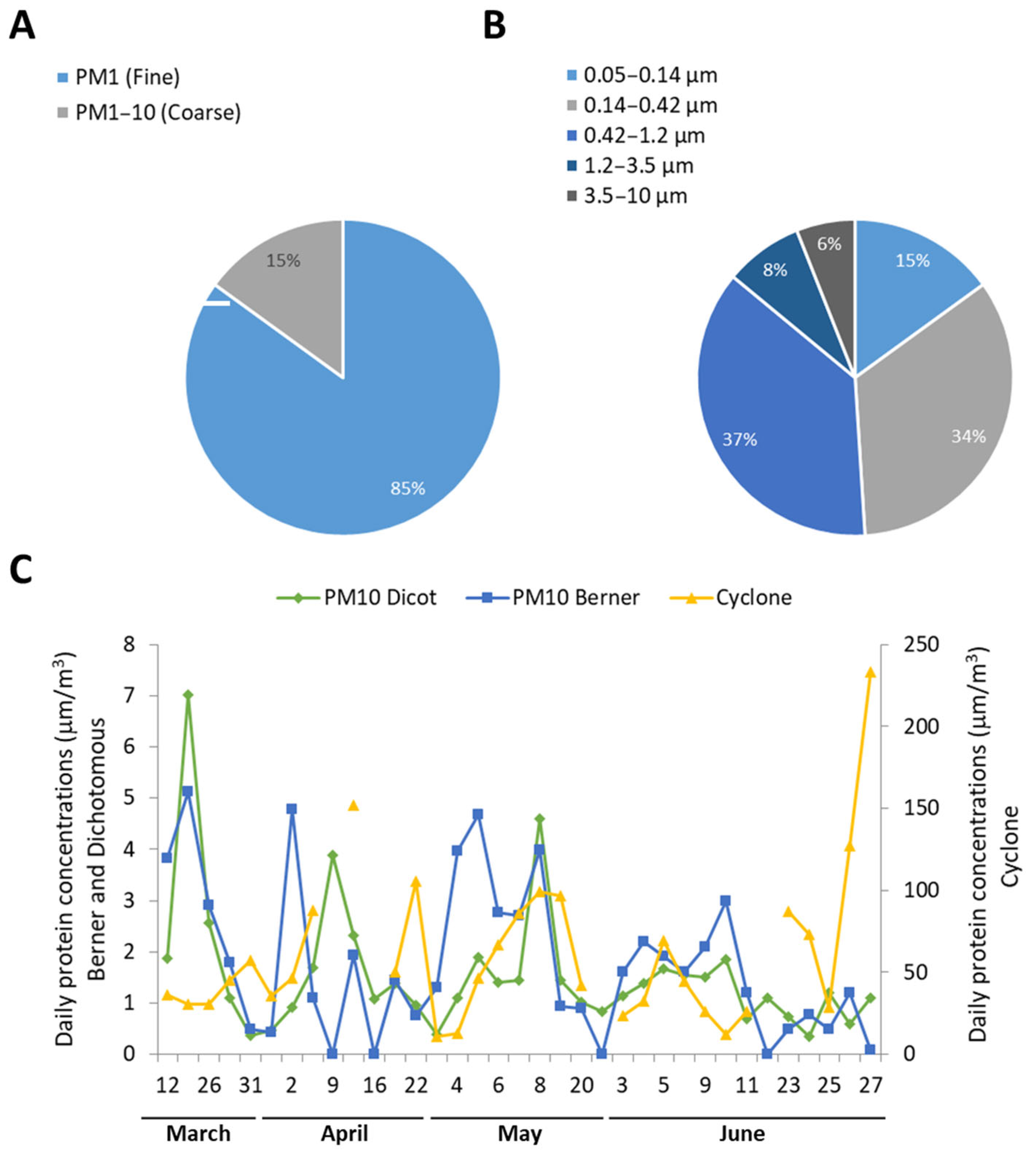
The analysis of protein concentrations in the stages of the DS and the BI1s showed a particle-size-dependent distribution (Figure 2A). In the fine fraction of aerosol, 85% of the total PM10 proteins collected by the DS sampler were detected, with the remaining 15% found in the coarse fraction. The same trend was observed for the BI1, where the protein concentration in the PM1.2 fraction accounted for 86% of total proteins, with the second (0.14–0.42 μm) and third stages (0.42–1.2 μm) containing the highest amount of proteins, 34 and 37%, respectively (Figure 2A).
According to BI1 and DS, there is a turning point in the sampling campaign, with a significant (p-value < 0.01) difference in protein content between the initial period (12 March–12 May), characterised by slightly higher airborne protein concentrations and a certain inter- and intra-sampler variability, and a second period (13 May–27 June) characterised by lower and more constant protein levels. This was in relation to more stable meteorological conditions in Bologna during that period, in particular for what concerns wind speed and relative humidity, with respect to the first half of the campaign.
The time trend of daily protein concentrations obtained by the CS (Figure 1C) did not match with those of the other two samplers, which on the contrary were in better agreement not only for general trend over the months (Figure 1A,B), but also for the daily protein concentrations, although with a low correlation (R = 0.4 for the PM10 fraction). Differences in daily protein concentrations between BI1 and DS were more evident when considering fine and coarse fractions separately: the correlation coefficient was still 0.4 for PM1, while no correlation existed for PM1–10 (Figure 2C).
No allergen among the investigated was detected in any of the stages of the BI1. Of the two allergens considered, Phl p 1 was detected only in proteins collected by the CS, while it was below the detection limit in the other samplers. Phl p 5, on the contrary, was found in CS samples and in both fine and coarse fractions of the DS (Table 2), but less than 1% of total Phl p 5 sampled was in the PM10 fraction of the DS, and only 0.1% was in the PM1 of the DS, while the vast majority of the allergen was collected by the CS.
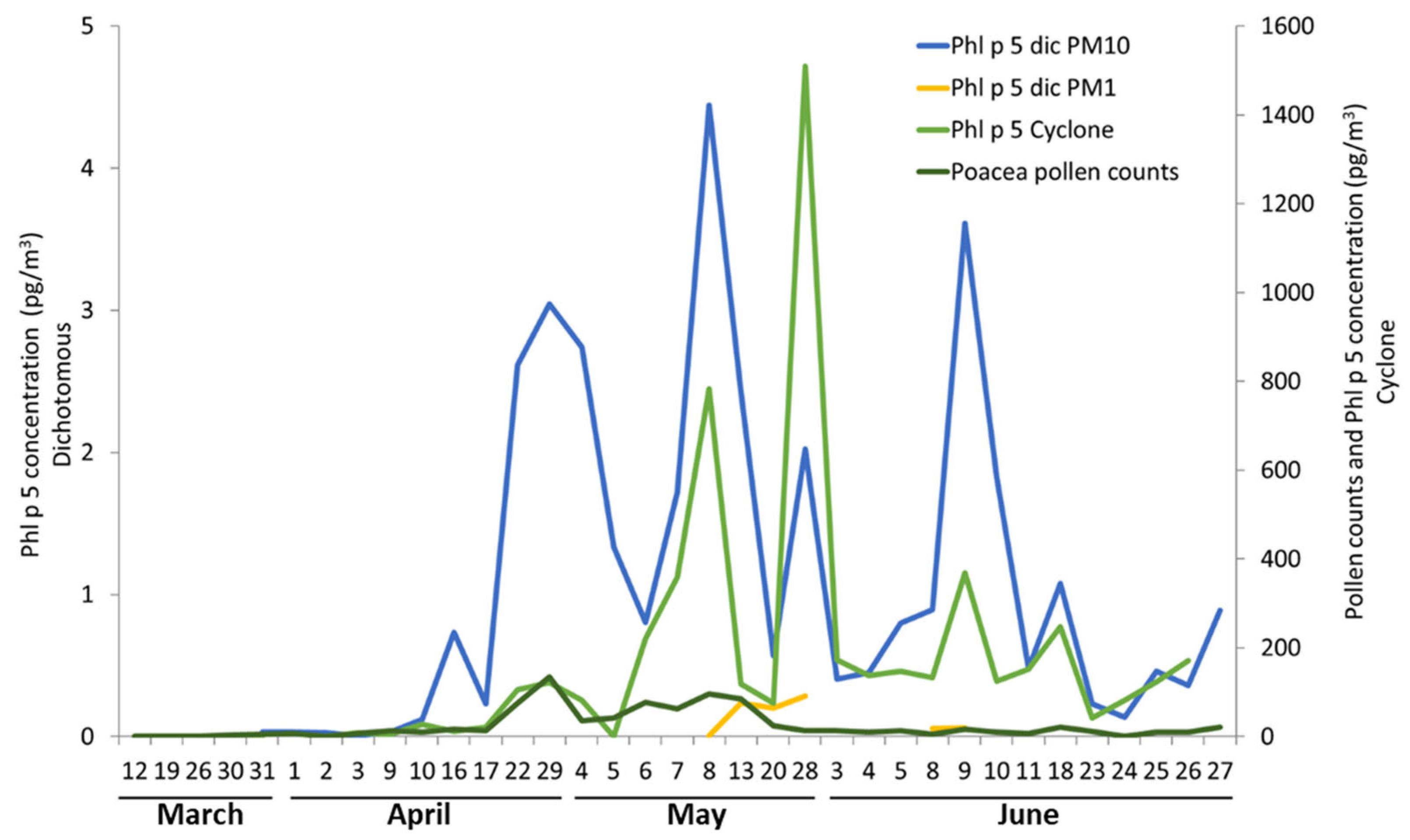
The temporal trend of Phl p 5 in the PM1 and PM10 fractions (Figure 3) highlighted the first part of the campaign during which Phl p 5 was not detected, followed by an increase in its concentrations from mid-April. Maximum concentrations corresponded to the main pollination period, from the middle of April to the middle of May, with a peak on 29 April. After this period, allergens continued to show relatively high concentrations, while total pollen counts remained low.
As for total proteins, the Phl p 5 allergen concentrations were also the highest in the CS samples (179 pg/m3), ascribable to the collection of entire pollen grains [8]. Interestingly, a small fraction of the total Phl p 5 allergens was detectable in the atmospheric fine PM (<1 μm) as well, presumably in the form of free allergens.

3.2. Relationships between Biological, Chemical, and Meteorological Parameters

Airborne protein concentrations were compared with meteorological parameters and with the results of chemical analysis for the DS and the BIs samples. Among the aerosol chemical components analysed for the DS samples, both in the fine and in the PM10 fraction, total proteins displayed the highest correlation (R = 0.85 and 0.82, respectively) with the water-soluble organic fraction of aerosol and with some of its components, mainly formate and oxalate (only in the fine fraction, since these species characterise the PM1 fraction). This correlation is present but weaker for BI fine and PM10 fractions (R = 0.58 and 0.65), but for this sampler there is also a positive correlation between proteins and WSOC in the coarse fraction (R = 0.57). A lower correlation (R = 0.42 in the fine fraction and R = 0.59 in PM10) was observed for proteins collected by the DS with the total fraction of organic carbon (OC), which comprises both the water-soluble and water-insoluble fractions, suggesting a higher correlation with the secondary fraction of organic carbon. For the BI samples, total protein concentrations display the highest correlation with nitrate and sulphate in the coarse fraction (R = 0.87 and 0.81, respectively) and with ammonium in all the fractions (R = 0.65 in the fine PM, R = 0.77 in the coarse PM, and R = 0.76 in the PM10). A high degree of correlation was also observed between total proteins and the secondary inorganic components of aerosol collected by the DS, mainly nitrate, sulphate, and ammonium, both in the fine (R = 0.78, 0.64, and 0.80, respectively) and in the PM10 fractions (R = 0.69, 0.46, and 0.40, respectively). On the contrary, airborne proteins correlated with Poaceae pollen grains neither in the coarse nor in the fine fraction of both samplers.
Atmospheric levels of total proteins in the PM10 fraction correlated neither with Poaceae pollen concentrations nor with the allergen Phl p 5. A significant negative correlation was also observed between total proteins and Phl p 5 in the fine fraction (R = −0.62) of the DS, while a positive correlation was observed between total Poaceae pollen airborne concentrations and Phl p 5 both in the coarse (PM1–10, R = 0.70) and in the PM10 fraction (R = 0.72) of the DS, but not in its fine fractions.
The allergen Phl p 5 was negatively correlated with all the aerosol components considered in the fine fraction of the DS, while it showed a positive correlation with TC, OC, and EC (R = 0.63, 0.63, and 0.44, respectively). The only positive correlation for Phl p 5 in the fine fraction, although slight, was observed with wind speed (R = 0.46), which on the contrary was negatively correlated with all the other aerosol components for its overall dispersing effect. Temperature did not correlate with allergen concentration, differently from what reported in literature [20]. Moreover, a negative correlation was observed between Phl p 5 concentrations in the fine PM and relative humidity (R = −0.53). No significant correlations (both positive and negative) were observed for Phl p 5 and the other chemical and meteorological parameters considered in the PM10 fraction of DS, where Phl p 5 positively correlated only with total Poaceae pollen.
Figure 4 shows that the trend of protein concentrations in the fine particulate follows the dynamics of carbon-based and nitrogen-based compounds, with a slight delay, for both BIs and DS. However, the presence of Phl p 5 does not coincide with peaks in the protein content of the fine particulate (Figure 4B).
A PCA was performed to analyse the composition of the particulate matter collected by the different stages of the DS and the BI (Figure 5 and Figure 6). In both cases, the diversification between the coarse particulate samples is driven by Cl, Mg, and Na levels, that are usually sea or road salts [64], and Ca, that usually comes from crustal/mineral dust or human activities [64,65]; the fine particulate appears to be more homogeneous according to all the evaluated parameters, and especially to Cl, Mg, Ca, and Na, except for some outliers (e.g., 19 March and 30 March). Variations in the protein content characterise the intermediate (stage 3 (0.42–1.2 μm) of the BI) and fine (stage 2 (0.14–0.42 μm) of the BI, fine fraction of the DS) particulate. Moreover, total proteins and Phl p 5 have negative and positive loading scores on the PC1 (Figure 6), respectively, suggesting again a poor correlation between the two variables, with Phl p 5 being more variable among coarse samples.

4. Discussion

4.1. Different Samplers Have Different Protein Collection Efficiencies

This study compared three different types of aerosol samplers that use the two most common aeroallergen sampling methods [8]: a cyclonic sampler (CS) and two high-volume cascade impactors (BIs and DS). The most relevant difference between the two groups is that the single-vial CS did not allow to distinguish between different particle sizes, having a high collection efficiency for particles down to 1 µm and an upper limit mainly defined by its inlet and its sampling velocity, which allows to collect cell debris or even whole microorganisms such as Poaceae pollen grains (diameter over 30 µm) [66]. The larger maximum dimensions of the particles sampled by the CS could be the explanation for the significantly higher protein concentrations recorded by this sampler compared to the total proteins collected by BI1 and DS.
Moreover, among the CS, the DS, and the BIs, there are significant differences in air intake velocity. In fact, CS is a low-volume sampler with an air throughput of 16.5 L/min, that is 1/5 of the BIs one and 1/18 of the DS one. The present results suggest that lower aspiration volumes allow a higher efficiency in airborne proteins collection, with the lower average protein concentration recorded by the DS and the highest by the CS (Table 2). This efficiency was, however, variable on a daily basis, with CS constantly recording higher protein concentrations than the other two samplers, while the DS appeared more efficient in collecting proteins in the fine particulate than the BS (Figure 2). Although coarse and fine PM samples were influenced by the same variables for both DS and BI (Figure 5 and Figure 6), the results they provided on protein concentrations and aerosol chemical composition were not in agreement even though they rely on the same sampling strategy. However, both the samplers indicated a strong positive correlation of airborne proteins levels with WSOC, especially in the fine and PM10 fractions, and with the secondary inorganic components of aerosol (nitrate, sulphate, and ammonium) in all fractions, which is expected because of their biological nature. Moreover, the DS and the BI were in better agreement on the protein concentrations found in the fine fraction, which is the vast majority of the proteins collected during the campaign, a result in line with previous studies demonstrating that fine PM accounts for the majority of both free and bound airborne amino acids [67].
When evaluating the aeroallergen concentrations, the CS again proved to be the most efficient, collecting both the Poaceae allergens investigated, while the DS only sampled the most abundant, Phl p 5, and the BI was not able to capture any of the examined aeroallergens. A possible explanation to the low efficiency in aeroallergens collection of both the aerosol samplers with the lowest particle diameter cut-off is that such allergens might have been associated to particles larger than 10 μm, e.g., Poaceae pollen grains, that are instead more easily captured by the CS. The absence of all the aeroallergens in the BI1 samples instead is probably due to the combination of the lower aspiration velocity of the BI1 compared to the DS, and to the fractioning of the sampled particulate, which could have lowered the allergen quantity per stage under the detection limit of the ELISA test. In fact, BI1 sampled a daily air volume of about 115 m3, which is roughly 1/4 of the air volume collected daily by the DS (430 m3), and the particulate in this air volume was subdivided into five fractions, contrarily to the DS that only had two fractions. To verify this hypothesis, the BI should be operated for longer sampling intervals, so that it can process an air volume comparable to the DS, and the samples from the different stages should be analysed both separated and pooled.
In the DS samples, Phl p 5 resulted as more concentrated in the coarse fraction, possibly associated with plant or pollen fragments and other coarse airborne particles. While the timing of the allergen peaks was mainly in concordance between the CS and the DS, the peaks’ intensity and the time trend largely variated between the two samplers (Table 2, Figure 3). This again can be explained by the possibility for the CS to sample whole pollen grains, while the DS can only collect smaller particles, with lower allergen content.
The discrepancies among the results obtained by the different samplers operated in parallel highlight the impossibility to directly compare the outcomes of studies carried out with different instruments, air intake velocity, and number of sampling stages.

4.2. Airborne Pollen and Airborne Protein Levels Are not Representative of Aeroallergen Concentrations

Atmospheric levels of total proteins in the PM10 fraction of the DS correlated neither with Poaceae pollen concentrations nor with the allergen Phl p 5. The fact that Poaceae pollen was not a relevant source of airborne proteins during the studied period was expected since total airborne proteins derive from many sources, e.g., other pollen types, plant debris, insects, spores, and anthropogenic sources such as biomass combustion residues [67,68]. Furthermore, Phl p 5 and total proteins in the fine fraction of the DS showed a negative correlation (R = −0.62), indicating that the aeroallergen was not a common component of the airborne proteins, and leading to the consideration that its atmospheric increment might be favoured by factors that reduce the emissions of other proteins in the fine aerosol. PCA (Figure 6) confirms poor correlation between the two variables, and reveals a higher variability of Phl p 5 among coarse samples. This could be explained assuming that coarse samples can also contain collapsed pollen grains or their fragments [69], which could cause a swift increase in their allergen content. While according to this study total airborne proteins do not seem to describe the atmospheric dynamics of specific airborne allergens of the season, and cannot be assumed as a proxy for Phl p 1 and Phl p 5, it is not clear whether they could mirror the total aeroallergen concentrations. It is apparent that proteins present in the aerosol need a thorough characterisation, also aimed to explain the slight delay between total carbon and the nitrogen compounds (NO3, proteins) in the atmosphere [67,68].
Poaceae pollen and Phl p 5 airborne concentration trends were not consistent, with high Phl p 5 peaks corresponding to low Poaceae pollen loads towards the end of the sampling campaign for both the DS and the CS (Figure 3). The increase of pollen allergen levels at the end of the pollen season is a phenomenon already described in literature [37,61], but in this case the hypothesis of a release of allergenic proteins outside the pollen grain favoured by high atmospheric humidity [17] seems not to be corroborated, since there was a negative correlation between Phl p 5 and atmospheric humidity. This is in contrast with what hypothesised by several studies that instead highlighted a massive protein and allergen release from pollen grains under humid conditions [16,45,70], but it is in line with other case studies on airborne pollen allergen levels [69]. It is important to notice that studies positively correlating atmospheric humidity and pollen allergen concentrations mainly investigate thunderstorm events, where there are many other concurrent factors contributing to allergen release [21,71]. Moreover, high relative humidity is a constant feature of the Po valley climate and was not influenced by storms or precipitations during the sampling period; hence, it is possible that in this case low humidity favoured aeroallergen dispersal avoiding its wet deposition.
On the other hand, the positive correlation between Phl p 5 and wind velocity suggests that the latter could have played a role in the mechanical rupture of pollen grains [69], and it might have contributed to keeping the free aeroallergens suspended in the air in the fine aerosol fraction, and to the long-range transport of such allergens.
Since Phl p 5 showed a relatively high correlation with Poaceae pollen in the coarse fraction of the DS, while no significant correlation was found between Phl p 5 concentrations in the fine aerosol fractions and total Poaceae pollen grains, it is possible that aeroallergens in the coarse aerosol are associated with pollen debris and orbicules [72] or dehydrated, shrunk pollen [69]; hence, they correlate with the airborne pollen concentrations, while aeroallergens in the fine aerosol are likely free allergens or allergens associated with smaller particulate that may originate from both pollen or plant debris such as mowed grass and have a longer atmospheric lifetime and different dynamics than pollen grains.
Additionally, group V allergens such as Phl p 5 are common within the Poaceae family; hence, it is also plausible that, in time, the variation in species composition of flowering Poaceae led to the dispersal of pollen grains from species with a higher content of Phl p 5 or its homologs [73]. Since Phl p 5 had a positive correlation with wind speed, it could be also speculated that the wind could have favoured long-distance dispersal events of Poaceae pollen with higher pollen potency [45].
Furthermore, significant and positive correlations of Phl p 5 with some of the aerosol chemical components (OC, EC, TC) observed in the coarse fraction suggest a preferential association of aeroallergens with combustion particles as also found by Namork et al. [74] through Scanning Electronic Microscopy observations of ambient air filters, or more in general to organic particulate pollutants such as soot, black/elemental carbon (BC/EC), and diesel exhaust particles [75,76].
The variety of possible explanations for the mismatch between pollen concentrations and pollen allergens underlines the importance of the implementation of molecular approaches to airborne pollen monitoring in order to identify pollen grains to the species level and evaluate their pollen potency. Moreover, this study corroborates the idea that an aeroallergen monitoring of major pollen allergens should be put in place to support the traditional aerobiological monitoring, and that pollen allergy diagnosis based on IgE levels against a specific allergen should be implemented to allow pollen allergic subjects a better management of their symptoms.

5. Conclusions

In conclusion, airborne pollen allergen quantification is influenced by factors intervening in different times, from the allergen production, to its release in the atmosphere, to its transport, and eventually to its collection by an air sampler.
Earlier events, such as the quantity of allergen biosynthesised by the pollen grains, are more difficult to investigate because they are influenced by several concurrent factors occurring during the plant’s life. The causes of the allergens’ release by pollen grains in the atmosphere, instead, are starting to be comprehended, and appear to be biological and mechanical processes mainly caused by high environmental humidity and wind speed [16,45,69,70]. In this study, in particular, the airborne concentrations of a major grass allergen, Phl p 5, showed a positive correlation with wind velocity in the fine fraction, while this correlation was not found for the coarse particulate. This might imply that the wind is involved in either the release or the transport of airborne allergens, as free molecules or associated to nanometric carriers. On the other hand, Phl p 5 levels did not significantly correlate with humidity, temperature, nor with the inorganic components of the aerosol in either fraction of the PM.
Contrarily, in both fine and coarse particulate, daily concentrations of Phl p 5 had a strong positive correlation with those of airborne Poaceae pollen, which is likely their primary source. However, pollen grains cannot be assumed as indicators of the airborne presence of the allergen, since the time trends of their concentrations in the atmosphere were visibly different. In the coarse fraction, the allergen levels also correlated with those of airborne OC and TC, indicating an association with other organic compounds, while this was not true for the allergens in the fine fraction. On the other hand, in neither fraction was Phl p 5 correlated with airborne protein concentrations, showing even a strong negative correlation with them in the coarse fraction, and indicating that total airborne proteins cannot be assumed as proxy for monitoring a specific allergen. Due to the difficulties in characterising the organic components of the aerosol, identifying a precise source of aeroallergens is still unfeasible, and more basic research on pollen grains’ rupture and allergen release is strongly needed.
Additionally, an aspect more controllable and heavily influencing the quantification of airborne allergens is the capture efficiency of the air samplers. In fact, in this study it is apparent that instruments collecting larger particles such as the CS record on average higher aeroallergen levels than those with a lower particle cut-off, possibly due to the capture of whole pollen grains. This allowed the CS to record allergens that went undetected by the DS and the BI (Phl p 1). On the other hand, instruments with many sampling stages might fraction the collected aeroallergens in small quantities that are below the ELISA detection limit, up to the point that the presence of the aeroallergen is missed entirely, as in the case of the BI. It is possible to conclude that the selection of the air sampler must be adapted to the scope of the study: if the intention is to quantify all the possible aeroallergens present in the atmosphere, even when carried by large pollen debris or entire pollen grains, then the CS represents the best choice. However, when the focus of the study is to evaluate the presence of aeroallergens in the particulate matter with a diameter under 10 µm, high-volume cascade impactors with only two stages such as the Dichotomous can provide more accurate results.

Supplementary Materials

The following supporting information can be downloaded at: www.mdpi.com/article/10.3390/su141911825/s1, Figure S1: Evaluation of the interference by ammonium sulphate in the BCA protein quantification.

Author Contributions

Conceptualisation, M.C.F., S.D.D., D.F.-G.; methodology, C.S., S.S., I.A., P.D.N.; software, C.S.; validation, S.S.; formal analysis, C.S., S.S., I.A., P.D.N.; investigation, C.S., S.S.; resources, M.C.F., S.D.D., D.F.-G.; data curation, C.S., S.S.; writing—original draft preparation, C.S., S.S.; writing—review and editing, I.A., S.D.D., D.F.-G.; visualisation, C.S., S.S., I.A., P.D.N., M.C.F., S.D.D., D.F.-G.; supervision, M.C.F., S.D.D., D.F.-G.; project administration, M.C.F., S.D.D., D.F.-G.; funding acquisition, M.C.F., S.D.D., D.F.-G. All authors have read and agreed to the published version of the manuscript.

Funding

APC was funded by the project PRIN 2015 ISIDE to S.D.D.

Institutional Review Board Statement

Not applicable.

Informed Consent Statement

Not applicable.

Data Availability Statement

Not applicable.

Conflicts of Interest

The authors declare no conflict of interest. The funders had no role in the design of the study; in the collection, analyses, or interpretation of data; in the writing of the manuscript; or in the decision to publish the results.

References

  1. D’Amato, G.; Vitale, C.; Sanduzzi, A.; Molino, A.; Vatrella, A.; D’Amato, M. Allergenic Pollen and Pollen Allergy in Europe. Allergy 2017, 62, 976–990. [Google Scholar] [CrossRef] [PubMed]
  2. Migueres, M.; Dávila, I.; Frati, F.; Azpeitia, A.; Jeanpetit, Y.; Lhéritier-Barrand, M.; Incorvaia, C.; Ciprandi, G. Types of sensitization to aeroallergens: Definitions, prevalences and impact on the diagnosis and treatment of allergic respiratory disease. Clin. Transl. Allergy 2014, 4, 16. [Google Scholar] [CrossRef] [PubMed]
  3. Ozdoganoglu, T.; Songu, M. The burden of allergic rhinitis and asthma. Ther. Adv. Respir. Dis. 2012, 6, 11–23. [Google Scholar] [CrossRef] [PubMed]
  4. Shah, R.; Grammer, L.C. Chapter 1: An overview of allergens. Allergy Asthma Proc. 2012, 33 (Suppl. S1), 2–5. [Google Scholar] [CrossRef]
  5. Zuberbier, T.; Lötvall, J.; Simoens, S.; Subramanian, S.V.; Church, M.K. Economic burden of inadequate management of allergic diseases in the European Union: A GA2LEN review. Allergy 2014, 69, 1275–1279. [Google Scholar] [CrossRef]
  6. Bousquet, J.; Khaltaev, N.; Cruz, A.A.; Denburg, J.; Fokkens, W.J.; Togias, A.; Zuberbier, T.; Baena-Cagnani, C.E.; Canonica, G.W.; Van Weel, C.; et al. Allergic Rhinitis and its Impact on Asthma (ARIA) 2008. Allergy 2008, 63 (Suppl. S86), 8–160. [Google Scholar] [CrossRef]
  7. Levetin, E. Methods for aeroallergen sampling. Curr. Allergy Asthma Rep. 2004, 4, 376–383. [Google Scholar] [CrossRef]
  8. Suanno, C.; Aloisi, I.; Fernández-González, D.; Del Duca, S. Monitoring techniques for pollen allergy risk assessment. Environ. Res. 2021, 197, 111109. [Google Scholar] [CrossRef] [PubMed]
  9. Agarwal, M.K.; Swanson, M.C.; Reed, C.E.; Yunginger, J.W. Airborne ragweed allergens: Association with various particle sizes and short ragweed plant parts. J. Allergy Clin. Immunol. 1984, 74, 687–693. [Google Scholar] [CrossRef]
  10. Cecchi, L. From pollen count to pollen potency: The molecular era of aerobiology. Eur. Respir. J. 2013, 42, 898–900. [Google Scholar] [CrossRef]
  11. Mothes, N.; Horak, F.; Valenta, R. Transition from a Botanical to a Molecular Classification in Tree Pollen Allergy: Implications for Diagnosis and Therapy. Int. Arch. Allergy Immunol. 2004, 135, 357–373. [Google Scholar] [CrossRef]
  12. Behrendt, H.; Becker, W.M.; Fritzsche, C.; Sliwa-Tomczok, W.; Tomczok, J.; Friedrichs, K.H.; Ring, J. Air Pollution and Allergy: Experimental Studies on Modulation of Allergen Release from Pollen by Air Pollutants. Int. Arch. Allergy Immunol. 1997, 113, 69–74. [Google Scholar] [CrossRef]
  13. D’Amato, G. Airborne paucimicronic allergen-carrying particles and seasonal respiratory allergy. Allergy 2001, 56, 1109–1111. [Google Scholar] [CrossRef] [PubMed]
  14. D’Amato, G.; Cecchi, L.; Bonini, S.; Nunes, C.; Annesi-Maesano, I.; Behrendt, H.; Liccardi, G.; Popov, T.; Van Cauwenberge, P. Allergenic pollen and pollen allergy in Europe. Allergy 2007, 62, 976–990. [Google Scholar] [CrossRef]
  15. De Linares, C.; de la Guardia, C.D.; Lugilde, D.N.; Alba, F. Airborne Study of Grass Allergen (Lol p 1) in Different-Sized Particles. Int. Arch. Allergy Immunol. 2010, 152, 49–57. [Google Scholar] [CrossRef] [PubMed]
  16. Grote, M.; Vrtala, S.; Niederberger, V.; Valenta, R.; Reichelt, R. Expulsion of allergen-containing materials from hydrated rye grass (Lolium perenne) pollen revealed by using immunogold field emission scanning and transmission electron microscopy. J. Allergy Clin. Immunol. 2000, 105, 1140–1145. [Google Scholar] [CrossRef] [PubMed]
  17. Grote, M.; Valenta, R.; Reichelt, R. Abortive pollen germination: A mechanism of allergen release in birch, alder, and hazel revealed by immunogold electron microscopy. J. Allergy Clin. Immunol. 2003, 111, 1017–1023. [Google Scholar] [CrossRef] [PubMed]
  18. Prado, N.; De Linares, C.; Sanz, M.L.; Gamboa, P.; Villalba, M.; Rodríguez, R.; Batanero, E. Pollensomes as Natural Vehicles for Pollen Allergens. J. Immunol. 2015, 195, 445–449. [Google Scholar] [CrossRef] [PubMed]
  19. Després, V.R.; Huffman, J.A.; Burrows, S.M.; Hoose, C.; Safatov, A.S.; Buryak, G.; Fröhlich-Nowoisky, J.; Elbert, W.; Andreae, M.O.; Pöschl, U.; et al. Primary biological aerosol particles in the atmosphere: A review. Tellus B Chem. Phys. Meteorol. 2012, 64, 15598. [Google Scholar] [CrossRef]
  20. Galan, C.; Antunes, C.; Brandao, R.; Torres, C.; Garcia-Mozo, H.; Caeiro, E.; Ferro, R.; Prank, M.; Sofiev, M.; Albertini, R.; et al. Airborne olive pollen counts are not representative of exposure to the major olive allergen Ole e 1. Allergy 2013, 68, 809–812. [Google Scholar] [CrossRef] [Green Version]
  21. Harun, N.-S.; Lachapelle, P.; Douglass, J. Thunderstorm-triggered asthma: What we know so far. J. Asthma Allergy 2019, 12, 101–108. [Google Scholar] [CrossRef]
  22. Kang, I.G.; Ju, Y.H.; Jung, J.H.; Ko, K.P.; Oh, D.K.; Kim, J.H.; Lim, D.H.; Kim, Y.H.; Jang, T.Y.; Kim, S.T. The Effect of PM10 on Allergy Symptoms in Allergic Rhinitis Patients during Spring Season. Int. J. Environ. Res. Public Health 2015, 12, 735–745. [Google Scholar] [CrossRef]
  23. Taylor, P.E.; Flagan, R.C.; Valenta, R.; Glovsky, M.M. Release of allergens as respirable aerosols: A link between grass pollen and asthma. J. Allergy Clin. Immunol. 2002, 109, 51–56. [Google Scholar] [CrossRef]
  24. Pöschl, U. Atmospheric Aerosols: Composition, Transformation, Climate and Health Effects. Angew. Chem. Int. Ed. 2005, 44, 7520–7540. [Google Scholar] [CrossRef]
  25. Shiraiwa, M.; Selzle, K.; Pöschl, U. Hazardous components and health effects of atmospheric aerosol particles: Reactive oxygen species, soot, polycyclic aromatic compounds and allergenic proteins. Free Radic. Res. 2012, 46, 927–939. [Google Scholar] [CrossRef]
  26. Di Filippo, P.; Pomata, D.; Riccardi, C.; Buiarelli, F.; Gallo, V.; Quaranta, A. Free and combined amino acids in size-segregated atmospheric aerosol samples. Atmos. Environ. 2014, 98, 179–189. [Google Scholar] [CrossRef]
  27. Poruthoor, S.K.; Dasgupta, A.P.K.; Genfa, Z. Indoor Air Pollution and Sick Building Syndrome. Monitoring Aerosol Protein as a Measure of Bioaerosols. Environ. Sci. Technol. 1998, 32, 1147–1152. [Google Scholar] [CrossRef]
  28. Aloisi, I.; Del Duca, S.; De Nuntiis, P.; Maray, A.M.V.; Mandrioli, P.; Gutiérrez, P.; Fernández-González, D. Behavior of profilins in the atmosphere and in vitro, and their relationship with the performance of airborne pollen. Atmos. Environ. 2018, 178, 231–241. [Google Scholar] [CrossRef]
  29. Fernández-González, D.; Maray, A.M.V.; Parrado, Z.G.; Barrera, R.M.V.; Gutiérrez, P.; De Nuntiis, P.; Mandrioli, P. Are the profilins an important component in the atmosphere? Ole e 2-like panallergen. Aerobiologia 2019, 35, 165–175. [Google Scholar] [CrossRef]
  30. García-Sánchez, J.; Trigo, M.D.M.; Recio, M. Extraction and quantification of Ole e 1 from atmospheric air samples: An optimized protocol. Chemosphere 2019, 225, 490–496. [Google Scholar] [CrossRef]
  31. Plaza, M.P.; Alcázar, P.; Hernández-Ceballos, M.A.; Galán, C. Mismatch in aeroallergens and airborne grass pollen concentrations. Atmos. Environ. 2016, 144, 361–369. [Google Scholar] [CrossRef]
  32. Plaza, M.P.; Alcázar, P.; Velasco-Jiménez, M.J.; Galán, C. Aeroallergens: A comparative study of two monitoring methods. Aerobiologia 2017, 33, 363–373. [Google Scholar] [CrossRef]
  33. Schäppi, G.F.; Suphioglu, C.; Taylor, P.E.; Knox, R.B. Concentrations of the major birch tree allergen Bet v 1 in pollen and respirable fine particles in the atmosphere. J. Allergy Clin. Immunol. 1997, 100, 656–661. [Google Scholar] [CrossRef]
  34. Spieksma, F.T.M.; Kramps, J.A.; Plomp, A.; Koerten, H.K. Grass-pollen allergen carried by the smaller micronic aerosol fraction. Grana 1991, 30, 98–101. [Google Scholar] [CrossRef]
  35. Brennan, G.L.; Potter, C.; de Vere, N.; Griffith, G.W.; Skjøth, C.A.; Osborne, N.J.; Wheeler, B.; McInnes, R.N.; Clewlow, Y.; Barber, A.; et al. Temperate airborne grass pollen defined by spatio-temporal shifts in community composition. Nat. Ecol. Evol. 2019, 3, 750–754. [Google Scholar] [CrossRef]
  36. Cariñanos, P.; Casares-Porcel, M.; de la Guardia, C.D.; Aira, M.J.; Belmonte, J.; Boi, M.; Elvira-Rendueles, B.; De Linares, C.; Fernández-Rodriguez, S.; Maya-Manzano, J.M.; et al. Assessing allergenicity in urban parks: A nature-based solution to reduce the impact on public health. Environ. Res. 2017, 155, 219–227. [Google Scholar] [CrossRef]
  37. Fernández-González, D.; González-Parrado, Z.; Maray, A.M.V.; Valencia-Barrera, R.M.; Camazón-Izquierdo, B.; De Nuntiis, P.; Mandrioli, P. Platanus pollen allergen, Pla a 1: Quantification in the atmosphere and influence on a sensitizing population. Clin. Exp. Allergy 2010, 40, 1701–1708. [Google Scholar] [CrossRef]
  38. Fernández-González, D.; Rajo, F.J.R.; Parrado, Z.G.; Barrera, R.M.V.; Jato, V.; Grau, S.M. Differences in atmospheric emissions of Poaceae pollen and Lol p 1 allergen. Aerobiologia 2011, 27, 301–309. [Google Scholar] [CrossRef]
  39. Fernández-González, M.; Álvarez-López, S.; González-Fernández, E.; Aira, M.J.; Rodríguez-Rajo, F.J. Cross-reactivity between the Betulaceae family and fallout in the real atmospheric aeroallergen load. Sci. Total Environ. 2020, 715, 136861. [Google Scholar] [CrossRef]
  40. Moreno-Grau, S.; Elvira-Rendueles, B.; Moreno, J.; García-Sánchez, A.; Vergara, N.; Asturias, J.A.; Arilla, M.; Ibarrola, I.; Seoane-Camba, J.A.; Suárez-Cervera, M. Correlation between Olea europaea and Parietaria judaica pollen counts and quantification of their major allergens Ole e 1 and Par j 1-Par j 2. Ann. Allergy Asthma Immunol. 2006, 96, 858–864. [Google Scholar] [CrossRef]
  41. Rodríguez-Rajo, F.J.; Jato, V.; González-Parrado, Z.; Elvira-Rendueles, B.; Moreno-Grau, S.; Vega-Maray, A.; Fernández-González, D.; Asturias, J.; Suárez-Cervera, M. The combination of airborne pollen and allergen quantification to reliably assess the real pollinosis risk in different bioclimatic areas. Aerobiologia 2011, 27, 1–12. [Google Scholar] [CrossRef]
  42. De Linares, C.; Plaza, M.P.; Valle, A.M.; Alcázar, P.; de la Guardia, C.D.; Galán, C. Airborne Cupressaceae Pollen and Its Major Allergen, Cup a 1, in Urban Green Areas of Southern Iberian Peninsula. Forests 2021, 12, 254. [Google Scholar] [CrossRef]
  43. Buters, J.T.M.; Weichenmeier, I.; Ochs, S.; Pusch, G.; Kreyling, W.; Boere, A.J.F.; Schober, W.; Behrendt, H. The allergen Bet v 1 in fractions of ambient air deviates from birch pollen counts. Allergy 2010, 65, 850–858. [Google Scholar] [CrossRef] [PubMed]
  44. Buters, J.T.M.; Thibaudon, M.; Smith, M.; Kennedy, R.; Rantio-Lehtimäki, A.; Albertini, R.; Reese, G.; Weber, B.; Galan, C.; Brandao, R.; et al. Release of Bet v 1 from birch pollen from 5 European countries. Results from the HIALINE study. Atmos. Environ. 2012, 55, 496–505. [Google Scholar] [CrossRef]
  45. Buters, J.; Prank, M.; Sofiev, M.; Pusch, G.; Albertini, R.; Annesi-Maesano, I.; Antunes, C.; Behrendt, H.; Berger, U.; Brandao, R.; et al. Variation of the group 5 grass pollen allergen content of airborne pollen in relation to geographic location and time in season. J. Allergy Clin. Immunol. 2015, 136, 87–95.e6. [Google Scholar] [CrossRef]
  46. Burbach, G.J.; Heinzerling, L.M.; Edenharter, G.; Bachert, C.; Bindslev-Jensen, C.; Bonini, S.; Bousquet, J.; Bousquet-Rouanet, L.; Bousquet, P.J.; Bresciani, M.; et al. GA 2 LEN skin test study II: Clinical relevance of inhalant allergen sensitizations in Europe. Allergy 2009, 64, 1507–1515. [Google Scholar] [CrossRef]
  47. Singh, M.B.; Bhalla, P.L. Hypoallergenic derivatives of major grass pollen allergens for allergy vaccination. Immunol. Cell Biol. 2003, 81, 86–91. [Google Scholar] [CrossRef]
  48. Sekerkova, A.; Polackova, M.; Striz, I. Detection of Phl p 1, Phl p 5, Phl p 7 and Phl p 12 Specific IgE Antibodies in the Sera of Children and Adult Patients Allergic to Phleum Pollen. Allergol. Int. 2012, 61, 339–346. [Google Scholar] [CrossRef]
  49. Schäppi, G.F.; Monn, C.; Wüthrich, B.; Wanner, H.-U. Direct Determination of Allergens in Ambient Aerosols: Methodological Aspects. Int. Arch. Allergy Immunol. 1996, 110, 364–370. [Google Scholar] [CrossRef]
  50. Plaza, M.P.; Alcázar, P.; Oteros, J.; Galán, C. Atmospheric pollutants and their association with olive and grass aeroallergen concentrations in Córdoba (Spain). Environ. Sci. Pollut. Res. 2020, 27, 45447–45459. [Google Scholar] [CrossRef]
  51. EN16868 Ambient Air—Sampling and Analysis of Airbone Pollen Grains and Fungal Spores for Networks Related to Allergy—Volumetric Hirst Method. 2019. Vol. CEN/TS 168. Available online: https://standards.iteh.ai/catalog/standards/cen/5f1349aa-f4cc-430a-978e-3044737e3f28/en-16868-2019 (accessed on 7 October 2022).
  52. Štefancová, L.; Schwarz, J.; Mäkelä, T.; Hillamo, R.; Smolík, J. Comprehensive Characterization of Original 10-Stage and 7-Stage Modified Berner Type Impactors. Aerosol Sci. Technol. 2011, 45, 88–100. [Google Scholar] [CrossRef]
  53. Berner, A.; Lürzer, C.; Pohl, F.; Preining, O.; Wagner, P. The size distribution of the urban aerosol in Vienna. Sci. Total Environ. 1979, 13, 245–261. [Google Scholar] [CrossRef]
  54. Hinds, W.C. Aerosol Technology: Properties, Behavior, and Measurement of Airborne Particles, 2nd ed.; John Wiley & Sons, Ltd.: Hoboken, NJ, USA, 1999. [Google Scholar]
  55. Matta, E.; Facchini, M.C.; Decesari, S.; Mircea, M.; Cavalli, F.; Fuzzi, S.; Putaud, J.-P.; Dell’Acqua, A. Mass closure on the chemical species in size-segregated atmospheric aerosol collected in an urban area of the Po Valley, Italy. Atmos. Chem. Phys. 2003, 3, 623–637. [Google Scholar] [CrossRef]
  56. Poor, N.; Clark, T.; Nye, L.; Tamanini, T.; Tate, K.; Stevens, R.; Atkeson, T. Field performance of dichotomous sequential PM air samplers. Atmos. Environ. 2002, 36, 3289–3298. [Google Scholar] [CrossRef]
  57. Jato, V.; Rodríguez-Rajo, F.J.; González-Parrado, Z.; Rendueles, B.E.; Moreno-Grau, S.; Maray, A.M.V.; González, D.F.; Asturias, J.A.; Suárez-Cervera, M. Detection of airborne Par j 1 and Par j 2 allergens in relation to Urticaceae pollen counts in different bioclimatic areas. Ann. Allergy Asthma Immunol. 2010, 105, 50–56. [Google Scholar] [CrossRef]
  58. Takahashi, Y.; Ohashi, T.; Nagoya, T.; Sakaguchi, M.; Yasueda, H.; Nitta, H. Possibility of real-time measurement of an airborne Cryptomeria japonica pollen allergen based on the principle of surface plasmon resonance. Aerobiologia 2001, 17, 313–318. [Google Scholar] [CrossRef]
  59. Arilla, M.C.; Ibarrola, I.; Eraso, E.; Aguirre, M.; Martínez, A.; Asturias, J.A. Quantification in mass units of group 1 grass allergens by a monoclonal antibody-based sandwich ELISA. Clin. Exp. Allergy 2001, 31, 1271–1278. [Google Scholar] [CrossRef]
  60. Sandrini, S.; van Pinxteren, D.; Giulianelli, L.; Herrmann, H.; Poulain, L.; Facchini, M.C.; Gilardoni, S.; Rinaldi, M.; Paglione, M.; Turpin, B.J.; et al. Size-resolved aerosol composition at an urban and a rural site in the Po Valley in summertime: Implications for secondary aerosol formation. Atmos. Chem. Phys. 2016, 16, 10879–10897. [Google Scholar] [CrossRef]
  61. Gelencsér, A.; Mészáros, T.; Blazsó, M.; Kiss, G.Y.; Krivácsy, Z.; Molnár, A.; Mészáros, E. Structural Characterisation of Organic Matter in Fine Tropospheric Aerosol by Pyrolysis-Gas Chromatography-Mass Spectrometry. J. Atmos. Chem. 2000, 37, 173–183. [Google Scholar] [CrossRef]
  62. Decesari, S.; Facchini, M.C.; Carbone, C.; Giulianelli, L.; Rinaldi, M.; Finessi, E.; Fuzzi, S.; Marinoni, A.; Cristofanelli, P.; Duchi, R.; et al. Chemical composition of PM10 and PM1 at the high-altitude Himalayan station Nepal Climate Observatory-Pyramid (NCO-P) (5079 m a.s.l.). Atmos. Chem. Phys. 2010, 10, 4583–4596. [Google Scholar] [CrossRef] [Green Version]
  63. RStudio Team. RStudio: Integrated Development for R. Available online: http://www.rstudio.com/ (accessed on 16 June 2022).
  64. Belis, C.A.; Karagulian, F.; Larsen, B.R.; Hopke, P.K. Critical review and meta-analysis of ambient particulate matter source apportionment using receptor models in Europe. Atmos. Environ. 2013, 69, 94–108. [Google Scholar] [CrossRef]
  65. Tørseth, K.; Hanssen, J.E.; Semb, A. Temporal and spatial variations of airborne Mg, Cl, Na, Ca and K in rural areas of Norway. Sci. Total Environ. 1999, 234, 75–85. [Google Scholar] [CrossRef]
  66. Calvo, A.I.; Baumgardner, D.; Castro, A.; Fernández-González, D.; Vega-Maray, A.M.; Valencia-Barrera, R.M.; Oduber, F.; Blanco-Alegre, C.; Fraile, R. Daily behavior of urban Fluorescing Aerosol Particles in northwest Spain. Atmos. Environ. 2018, 184, 262–277. [Google Scholar] [CrossRef]
  67. Matos, J.T.V.; Duarte, R.M.B.O.; Duarte, A.C. Challenges in the identification and characterization of free amino acids and proteinaceous compounds in atmospheric aerosols: A critical review. TrAC Trends Anal. Chem. 2016, 75, 97–107. [Google Scholar] [CrossRef]
  68. Kang, H.; Xie, Z.; Hu, Q. Ambient protein concentration in PM10 in Hefei, central China. Atmos. Environ. 2012, 54, 73–79. [Google Scholar] [CrossRef]
  69. Grewling, Ł.; Bogawski, P.; Kostecki, Ł.; Nowak, M.; Szymańska, A.; Frątczak, A. Atmospheric exposure to the major Artemisia pollen allergen (Art v 1): Seasonality, impact of weather, and clinical implications. Sci. Total Environ. 2020, 713, 136611. [Google Scholar] [CrossRef]
  70. Taylor, P.E.; Jonsson, H. Thunderstorm asthma. Curr. Allergy Asthma Rep. 2004, 4, 409–413. [Google Scholar] [CrossRef]
  71. Hughes, D.D.; Mampage, C.B.A.; Jones, L.M.; Liu, Z.; Stone, E.A. Characterization of Atmospheric Pollen Fragments during Springtime Thunderstorms. Environ. Sci. Technol. Lett. 2020, 7, 409–414. [Google Scholar] [CrossRef]
  72. Ruggiero, F.; Bedini, G. Systematic and morphologic survey of orbicules in allergenic angiosperms. Aerobiologia 2018, 34, 405–422. [Google Scholar] [CrossRef]
  73. Matthiesen, F.; Løwenstein, H. Group V allergens in grass pollens. II. Investigation of group V allergens in pollens from 10 grasses. Clin. Exp. Allergy 1991, 21, 309–320. [Google Scholar] [CrossRef]
  74. Namork, E.; Johansen, B.V.; Løvik, M. Detection of allergens adsorbed to ambient air particles collected in four European cities. Toxicol. Lett. 2006, 165, 71–78. [Google Scholar] [CrossRef]
  75. Knox, R.B.; Suphioglu, C.; Taylor, P.; Desai, R.; Watson, H.C.; Peng, J.L.; Bursill, L.A. Major grass pollen allergen Lol p 1 binds to diesel exhaust particles: Implications for asthma and air pollution. Clin. Exp. Allergy 1997, 27, 246–251. [Google Scholar] [CrossRef]
  76. Ormstad, H. Suspended particulate matter in indoor air: Adjuvants and allergen carriers. Toxicology 2000, 152, 53–68. [Google Scholar] [CrossRef]
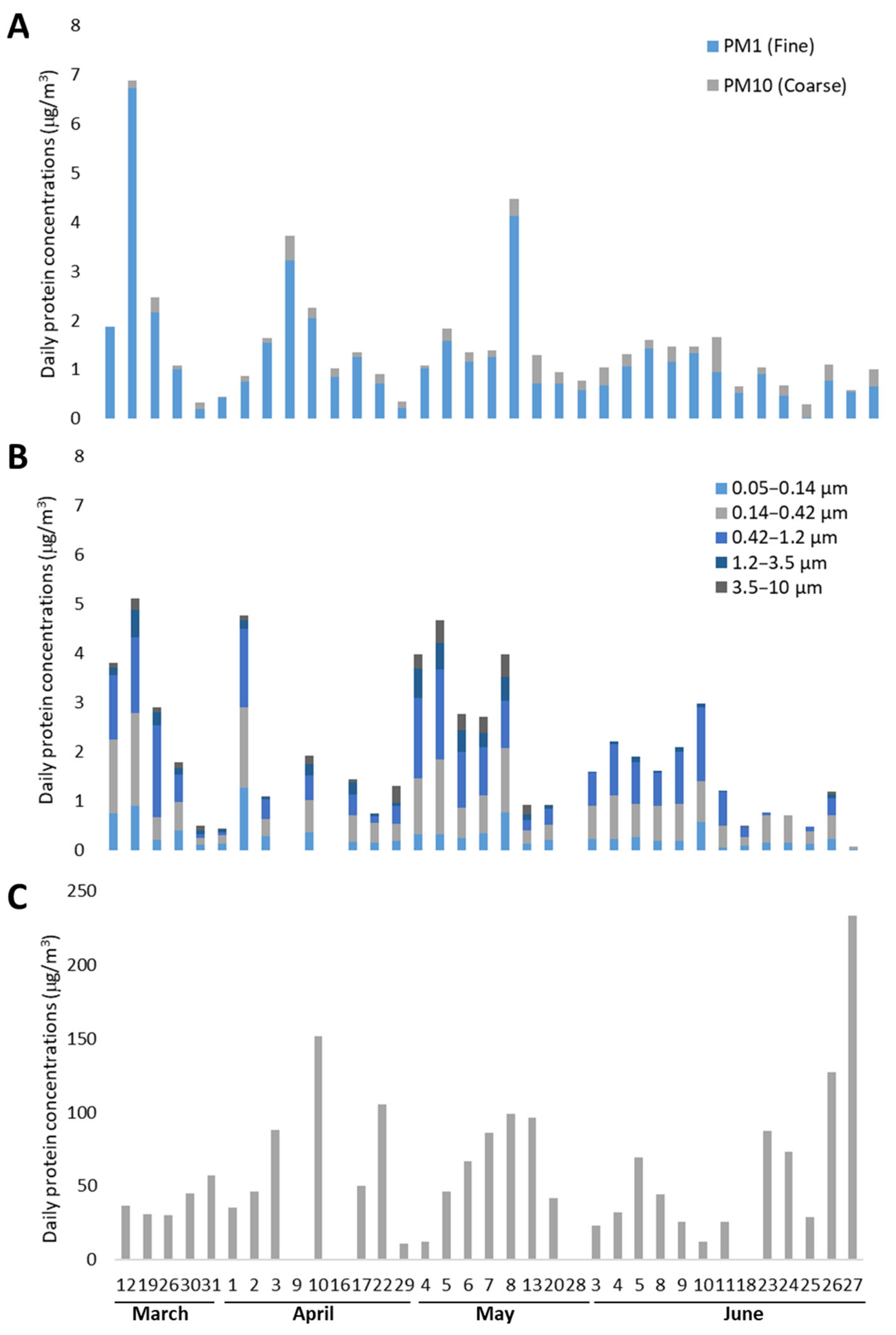
Figure 1. Daily concentrations of total airborne proteins, expressed as μg/m3, collected by the Dichotomous sampler (A), the Berner impactor (B), and Cyclone sampler (C) during the sampling campaign in Bologna in 2015. Missing values in (B,C) are days with a protein concentration under the ELISA detection limit.
Sustainability 14 11825 g001
Figure 2. Comparison of the protein concentrations in the different aerosol fractions sampled by the Dichotomous, Berner, and Cyclone samplers in Bologna in 2015. Relative concentrations of airborne proteins in different particle sizes are represented for the Dichotomous sampler (A) and for the Berner Impactor (B). The time trend of daily protein concentrations (μg/m3) recorded by the three samplers during the campaign is reported in (C), with the scale for the Dichotomous and Berner samples on the left y-axis and the scale for the Cyclone samples on the right y-axis.
Sustainability 14 11825 g002
Figure 3. Comparison between daily fluctuations in airborne Poaceae pollen concentration (pollen grains/m3) recorded by the Hirst-type pollen trap and airborne Phl p 5 concentrations (pg/m3) recorded by the Dichotomous and the Cyclone samplers during the sampling campaign in Bologna in 2015. Phl p 5 concentrations are plotted separately for the fine (PM1) and the coarse (PM1–10) particulate collected by the Dichotomous sampler, and their scale is reported on the left y-axis, while the scale for Phl 5 concentrations recorded by the Cyclone and for the pollen grains concentrations is reported on the right y-axis.
Sustainability 14 11825 g003
Figure 4. Temporal dynamics of the carbon-based and nitrogen-based components of the fine (diameter ≤ 1.2 μm) aerosol sampled by the Berner impactor (A) and the fine (diameter ≤ 1 μm) aerosol sampled by the Dichotomous sampler (B) in Bologna in 2015. The scale for the airborne daily concentration of water-soluble organic carbon (WSOC), total carbon (TC), and nitrates (NO3) is reported on the left y-axes in both graphs, while the scale for the daily airborne protein concentration is reported on the right y-axis.
Sustainability 14 11825 g004
Figure 5. Principal Component Analysis (PCA) biplot of the concentrations of the aerosol components (inorganic and organic components, total proteins) recorded by the Berner impactor during the sampling campaign in Bologna in 2015, assuming the sampling days as samples. Axes represent the principal components Dim1, explaining 48.3% of the variance, and Dim2, explaining 15.7% of the variance. Colours indicate the different stages of the impactor, collecting airborne particles in the following range of diameters: 1 = 0.05–0.14 μm; 2= 0.14–0.42 μm; 3 = 0.42–1.2 μm; 4 = 1.2–3.5 μm; 5 = 3.5–10 μm.
Sustainability 14 11825 g005
Figure 6. Principal Component Analysis (PCA) biplot of the concentrations of the aerosol components (inorganic and organic components, total proteins, and Phl p 5) recorded by the Dichotomous sampler during the sampling campaign in Bologna in 2015, assuming the sampling days as samples. Axes represent the principal components Dim1, explaining 39.1% of the variance, and Dim2, explaining 17.6% of the variance. Colours indicate the particulate diameter, with C = Coarse (1–10 μm) and F = Fine (≤1 μm).
Sustainability 14 11825 g006
Table 1. Average, minimum, and maximum values (μg/m3) for the proteinscollected by Dichotomous, Berner, and Cyclone samplers.
Dichotomous SamplerBerner ImpactorCyclone Sampler
Particle cut-off, μm (Ø)0.1–1
(Fine)
1–10
(Coarse)
0.140.421.23.510
Average protein concentration (μg/m3) 1.280.220.310.680.740.160.1161.97
Minimum protein concentration (μg/m3)0.030.030.020.060.050.020.0110.89
Maximum protein concentration (μg/m3)6.720.721.271.881.880.600.47232.95
Median protein concentration (μg/m3)0.950.190.230.590.670.080.0646.41
Table 2. Average concentrations of total proteins (μg/m3) and Phl p 5 allergen (pg/m3), measured in samples collected by Dichotomous, Berner, and Cyclone samplers, respectively. <DL = under the instrument detection limit.
Dichotomous SamplerBerner ImpactorCyclone Sampler
FractionPM1PM1–10PM10PM1.2PM1.2–10PM10
Average Phl p 5 concentrations (pg/m3)0.131.051.08<DL<DL<DL179
Average total protein concentrations (μg/m3)1.280.221.501.640.382.0261.97
Publisher’s Note: MDPI stays neutral with regard to jurisdictional claims in published maps and institutional affiliations.