Article

Long-Term COVID: Case Report and Methodological Proposals for Return to Work

by
Ginevra Malta
1,*,†,
Luigi Cirrincione
1,*,†,
Fulvio Plescia
1,
Marcello Campagna
2,
Claudia Montagnini
3 and
Emanuele Cannizzaro
1
1
Department of Health Promotion, Mother and Child Care, Internal Medicine and Medical Specialties, “G. D’Alessandro”, Università degli Studi di Palermo, 90127 Palermo, Italy
2
Dipartimento di Scienze Mediche e Sanità Pubblica, Università degli Studi di Cagliari, 09123 Cagliari, Italy
3
Ministero della Difesa, Via Cortonese n° 5, 06124 Perugia, Italy
*
Authors to whom correspondence should be addressed.
These authors contributed equally to this work.
Sustainability 2022, 14(15), 9332; https://doi.org/10.3390/su14159332
Submission received: 24 June 2022 / Revised: 25 July 2022 / Accepted: 26 July 2022 / Published: 29 July 2022

Abstract

:
Almost two years after the beginning of the SARS-CoV-2 pandemic, the knowledge of which in the infectious and therapeutic spheres is constantly evolving, attention paid to the medicolegal aspects linked to this emergency phenomenon has mainly focused on the liability implications falling on healthcare personnel. With regard to the medicolegal assessment of the outcomes of COVID-19 illness, although it is a procedure that is commonly used, and although references in the assessment tables in force have been adhered to, a specific assessment protocol has not been standardized that takes into account, from an objective point of view, the degree of severity of the long-term residual outcomes and their impact on the social and working lives of subjects. This shortcoming appears to be attributable to the immediate need to categorize the results of COVID-19, but, in our opinion, it deserves an in-depth study and protocols to enable evaluation committees to draw up an assessment as precisely as possible and that is free of gaps, which could be the subject of legal disputes. The aim of the present work, in light of a worldwide problem, is to arrive at specific and univocal evaluation criteria for COVID-19 disease outcomes, applicable in different operational contexts of reemployment.

1. Introduction

The outcomes of COVID-19 infection/disease have proved to be heterogeneous over time, and their evaluation has been fraught with multiple aspects linked to continually emerging scientific evidence [1,2]. This is related both to the severity of the clinical picture suffered by the patients, such as with regard to their ages, and the existence of risk factors both intrinsic (such as genetic factors [3] and comorbidities [4] linked to the work performed [5]) and extrinsic (climatic, environmental, and organizational factors [6]).
It is clear that these results are worthy of evaluation in strict relation to the specific situation of each subject, and, therefore, it is also worthwhile to consider the actual activities carried out and the time commitment of the same. This aspect, as the pandemic continues, has become even more important if we consider fragile individuals, i.e., those with comorbidities that also require greater protection within the workplace [7].
A variety of measures have been taken in this regard, ranging from the provision of social distancing and protective barriers to the application of smart working, and this is because, for the same injury event, fragile individuals have more pronounced outcomes [8,9].
The various social security systems operating in the Italian state refer to different regulations that sometimes converge in order to fill any gaps dictated by their excessive age. With regard to ordinary evaluations, if this creates confusion and divergences that lead to legal disputes—even more so with regard to the evaluation of post-COVID outcomes—such divergences will become evident, especially when taking into account the serious social impact of the emergency phenomenon [10].
From a scientific point of view, knowledge about the long-term outcomes of SARS-CoV-2 infection is constantly evolving and is being studied daily, especially in terms of permanence and, therefore, chronicity [11].
The increasingly widespread practice of reasoning by virtue of methodological principles, and not on the basis of cases, fully expresses the systematic frieze in which to set, even today, more than two years after the beginning of the emergency condition [12], the polarized debate on the procedural gears introduced into the regulatory framework by the twenty-year-long table system used in the various social security fields that, today, in the opinion of the writers, needs a specific addendum for the outcomes of COVID-19, providing for targeted evaluations not only with regard to the type of outcomes but also to the occupational status of the subjects. Recent studies have focused on the long-term sequelae of COVID-19, especially on pulmonary and neurological complications [13,14] but also on other items, for example, mental health after lockdown periods [15].

2. Clinical Case

2.1. Work Context

The subject, aged 33 years at the time, in the period of 10 March 2020–21 April 2020, worked in the morning hours at the medical clinics of Codogno; he visited patients with symptomatic COVID-19 (suspected and confirmed), and in the afternoon, hours he took care of patients at the RSA, where there were people suffering from COVID-19 beyond the capacity of the system, given the difficulties in finding suitable beds in local hospitals. He also worked night shifts and was on-call for any medical emergencies. The type of work carried out can be classified as prompt assistance services, as well as also shiftwork with related stress complications [16]; it consists of the examination of outpatients and inpatients, with the management of the therapies administered, the placement of therapeutic aids where required (oxygen masks), peripheral venous access, bladder catheters, etc. As already mentioned, at the time, although Ministry of Defense doctors and healthcare workers were provided with personal protective equipment (PPE); prevention and containment measures; and other protocols adopted to prevent the spread of SARS-CoV-2 infection [17,18], the facilities for managing patients with possible respiratory failure were not optimal, certainly not because of a lack of facilities, but because of the unpredictability of the emergency pandemic event, the limited knowledge of the ways in which SARS-CoV-2 is transmitted [19], and the relative efficacy of anti-infection protocols to be adopted [20], so much so that, in view of the large number of general practitioners who fell ill or died, the Mayor set up the General Medicine Outpatient Clinic where doctors would visit the sick in the morning to replace colleagues who had fallen ill, died, or were unable to perform their duties due to a lack of PPE.

2.2. Instrumental Assessments

Chest X-ray report (at 1 month): “…The radiological examination of the chest, performed in double projection, shows some thin parenchymal striae projectively in the likely location of the anterior basal segment of the left lower lobe, compatible with inflammatory outcomes. The cardiomediastinal profile is within normal limits…”;
HR chest CT scan report (at 3 months): “…Reason for examination: post-COVID-19 check; Report: Chest study performed without mdc, completed by 2D MPR reconstructions in the sagittal and coronal planes. Residual ground-glass areola of about 7 mm in the posterior segment of the left lower lobe and minimal attenuation, tenuous, ground-glass in the antero-medial segment of the right lower lobe, in parascissural. Subtle bi-apical fibrotic findings. No pleuro-pericardial effusions. Pervious trachea and bronchi. No ilo-mediastinal or axillary lymphadenomegalies. Thymic residue. Conclusions: residual ground glass in outcomes of COVID-19 relata pneumonia…”;
Spirometry report (at 3 months) (Table 1): “…History: see previous—previous COVID infection (…omissis…) EO: ET symmetrical hypo-expandable; MV slightly reduced in the C parenchymal (…omissis…) FR: restrictive ventilatory defect of marked degree for the age. Br vilatation test not significant. Current diagnosis: outcomes of COVID infectious process; fibrotic pulmonary outcomes/functional restrictive S./residual asthenia (…omissis…)”.
Olfactometric examination report (at 4 months): “…History: reported hyposmia and ageusia post COVID-19. Objective examination: clear nasal passages. Olfactometry by sniff-test. In order to examine the pure olfactory modality, the following substances are sprayed into the nasal passages under positive pressure: coffee, nitrobenzene, eucalyptol. No response sensation is evoked. In order to examine the gustatory olfactory modality, the following substances are delivered by positive pressure spray: vanillin, ammonia, acetic acid. No response sensation is evoked. To examine the olfactory-trigeminal modality, the following substances are sprayed: camphor, menthol, thymol. No response sensation is evoked. Conclusions: anosmia and hypogeusia…”;
Chest CT scan report (at 1 year): “…pleuro-parenchymal fibrotic findings at the upper lung apices. In the posterior basal segment of the LIS, presence of a millimetric areola of the type “pGGO” of about 6 mm, currently undetermined due to its small size and as such to be re-evaluated in 12 months. Small lymph node in minor scissure. Specific anterior mantle micronodulation in the upper lingula. no interstitial disease or COPD. Pervious large bronchi. No gross mediastinal lymph nodes visible without mdc. No pleural or pericardial effusion. Normal heart. Slight dorsal somatic deformities in the mid-section…”;
Spirometry report (at 1 year) (Table 2): “…CO diffusion reduced (…omissis…) discrete restrictive defect…”;
Report ENT examination and OST test (at 1 year and 1 month): “…The objective examination does not show expansive lesions or deviations of the internal structures of the nose that could impede the correct nasal airflow. The functional evaluation was carried out in an olfactometry laboratory using both subjective and objective methods. The subjective olfactometric examination, carried out using the Olfactory Smart Threshold test (OST) was positive for anosmia. The objective olfactometric examination, obtained by means of olfactory evoked potentials, at the time of the examination showed anosmia both at the functional evaluation of the olfactory threshold and at the functional evaluation of uni- and cross-modal olfactory perception. At high stimulatory concentrations, some peripheral electrophysiological signals are also detected, which however are abnormal in form, amplitude and duration without appreciable bulbar and central correlates. In the absence of stimulation there are isolated, exclusively peripheral signals. I recommend re-evaluation in 6 months and olfactory rehabilitation…”;
Current state: Since 3 June up to the present (17 August 2021), the patient reports anosmia and ageusia; respiratory difficulties with prolonged exercise (required by his profession); difficulty in carrying out sports activities previously practiced (water polo, swimming, and gym) due to easy fatigability; dyspnea upon greater exertion; a tendency to apathy along with difficulty in staying in closed places or with more people; and frequent nocturnal awakenings.
Clinical examination: Subject is lucid, mnestic, oriented in time and space, fluent in speech, and cognizant of their mental context; the re-evocation of their work experience at the RSA evokes an alteration of their mood, with a tendency toward deflection and a conscious alteration of the tone of their voice into a flat, monotone timbre.
Thorax: Hypoexpansibility of lung bases, MV reduced basally and apically bilaterally, FVT weakened basally and apically bilaterally.
Sense organs: For the assessment of taste and smell, please refer to the specialist examinations and instrumental tests.
First of all, it should be noted that, in accordance with the guidelines in force at the time, the diagnosis [21] and the monitoring of the disease were carried out as the patient said, the former by means of a molecular swab [22,23] at the immediate presentation of symptoms [24] (despite the precautions taken [25]), and the latter by means of serious radiographic examinations [26].

2.3. Assessment of Causation

The current assessment of outcomes resulting from COVID-19 infection is a highly debatable subject. However—apart from the flood of regulatory acts issued by multiple bodies—Article 42, paragraph 2 of Decree Law 18/2020 (the so-called “Cura Italia” decree) established that COVID-19 infection contracted “during work” constitutes an accident at work, laying the foundations for the continuation of the assessment procedures due to victims, equating a violent cause to the virulent cause.
Transferring the civilian issue to the military, it should be remembered that the height of the pandemic was a very serious situation where the military, from all branches, were deployed in national and international territories to protect citizens with consequent exposure to the risk of contagion.
Moreover, it must be acknowledged in a clearer manner that the rules of military interest, which correlate an injury or death with service-related events related to the activity performed, express the legal concepts of preponderant cause/concause, which allow for an even more linear assessment of the facts in question. In fact, established case law equates the cause of service with the preponderant and necessary contributing cause, identified in those service factors that—in a prevalent manner with respect to factors extraneous to the service—have contributed (albeit by influencing the further and worsening nosological course) to necessarily determining the harmful effect; necessarily because, had such factors been absent, the effect would have been different or would not have occurred.
In this regard—as already mentioned—Article 64 of Presidential Decree No. 1092/1973 establishes that “there is a right to preferential pension treatment for civil servants suffering from infirmity or injuries leading to impairment of personal integrity, ascribable to one of the categories of Table A annexed to Law no. 313/1968, provided that they are the cause of inability to work and are dependent on facts of service according to a causal or concausal, efficient and decisive relationship”. The second paragraph of the same article specifies that service-related events are those arising from the fulfillment of service obligations or those resulting from orders received or carried out spontaneously in connection with employment. Naturally, as dictated by medicolegal methodology, the classic, canonical criteria for ascertaining the causal link must be satisfied.
In the case of COVID-19 disease, given the real danger of contagion, the General Inspectorate of Military Health also applied and included within the case provided for by Article 1880 of Legislative Decree 66/2010 (Code of Military Regulations) traumatic injuries produced by infectious causes (in short, the so-called “new” Model C, Ex Law 157/52, which also provides for the rapid assessment of causal links in cases of infectious origin at the initiative of the Office, i.e., by the administration to which the person belongs). Therefore, it is a procedure that is facilitated, in comparison to the ordinary procedure (at the request of the person concerned), for the purposes of recognizing the causal link between the condition related to the service obligation and the resulting damage.
A number of circulars have been issued on this subject, i.e., on the classification of the causal relationship between the fact of service and infirmity due to a violent cause (no longer only traumatic in nature), even in cases where—in the absence of the hospitalization of the soldier—it is possible to recognize a relationship of responsibility between the service and the morbidity attributable to an infectious cause (see IGSM/UPSG/2020-169).
In this respect, the following concepts are defined:
  • The term “occasion of work” means any exposure to risk attributable to the performance of service activities, directly or indirectly, irrespective of the employee’s voluntary conduct: this circumstance (i.e., the occasion of work) is relevant for accidents protected by INAIL.
  • With regard to “occupational risk”, it is necessary that the service activity carried out resulted in a “specific risk” for the employee arising from the particular conditions of the service activity carried out;
  • An accident can also occur if there is an “aggravated generic occupational risk”, i.e., a risk which, although common to the general population, places a greater burden on the worker.
Points 2 and 3 include, according to the circulars, rescue operations, hospital activities, public order services, protection of public safety, or any other service carried out in a particular environmental situation.
It follows that the virulent cause (given the current scientific and factual evidence) is recognized—for the Armed Forces—as a cause of service in two respects:
  • Direct causation: Coronavirus injury is directly service-related in light of explicit service instructions/orders issued by the military authority;
  • Preponderant causation: The specific activity contributed efficiently and decisively to the onset of the disease.
In these cases, we repeat, the injury to the apparatuses of the greatest tropism caused by SARS-CoV-2 (nervous [27] and respiratory [28]) is the cause of impairment to psychophysical integrity (or death) contracted following exposure due to service obligations, since the job involves exposure to a specific risk and must, therefore, be assessed as a responsible cause of the disease [29]. This hypothesis of a causal link is reinforced by the high statistical risk of contracting the infection (and the disease) during prolonged periods of exposure, even if the viral load is low. In this regard, a formula has also been proposed by the scientific community for assessing the risk of infection, which is a function of exposure time and the magnitude of the viral load [30].
On the basis of a logical, scientific judgment correlated to the high risk of exposure to the virus—as well as to the examination of classic, consolidated medical–legal criteria on the relationship between causes—as applied to the case in question (the chronological and pathogenetic criteria [31] of damage suitability, the qualitative/quantitative adequacy of damage continuity, the exclusion of other causes, etc.), the following can be stated: Between the causal exposure being held responsible and the disease form under examination, and with regard to the scientific literature data, which, among other things, mark out the average time duration of the infection/malignant disease [32], it is reasonable to conclude that the person concerned contracted COVID-19 disease due to (or, at least, due to the concomitant cause of) the service they performed and that they were, moreover, burdened by the specific risk intrinsic to medical activity [33].
From the ISTAT report of 2 March 2022 [34], it can be seen that the first Italian case of COVID-19 was reported in Lombardy on 20 February 2020. The geographical spread of the pandemic was limited in the southern and island regions, on average higher in the central regions than in the south, and very high in the northern regions. In total, 91% of excess mortality found on a national average in March 2020 was concentrated in areas with a high pandemic spread: 3271 municipalities and 37 northern provinces. (Figure 1).
In these provinces, deaths from all causes more than doubled compared to the 2015–2019 average in March. If we compare the period from 20 February to 31 March of the indicated time bands, deaths rose from 26,218 to 49,351 (+23,133); just over half of this increase (52%) was made up of deaths reported to the COVID-19 Integrated Surveillance System (12,156).
Within this grouping, the provinces most affected by the epidemic have paid a very high price in terms of human lives, with triple-digit percentage increases in deaths in March 2020 compared to March 2015–2019: Bergamo (568%), Cremona (391%), Lodi (371%), Brescia (291%), Piacenza (264%), Parma (208%), Lecco (174%), Pavia (133%), Mantua (122%), Pesaro, and Urbino (120%).
These data confirm the very high risk of contagion to which medical staff (military and civilian [35]) working in the same areas were subjected during the period of service under review, which almost coincided with the beginning of the state of emergency.
In the case in question, with regard to the aforementioned medicolegal method of assessing the causal link, it should be noted that, with regard to the chronological criterion, the subject manifested the first symptoms of the disease at a time congruent with the current scientific literature on the incubation period of the virus with respect to contact, which is perfectly consistent with the period of service performed. With regard to the qualitative and quantitative “adequacy” criteria, it is clear that the morphological characteristics and tropism of COVID-19 appear to satisfy those criteria, having acted on apparatuses that, today, are the site of congruous outcomes in light of the statistical data available on the effects and outcomes present in a population of subjects of the same age and anthropological and physical characteristics. Similarly, as described above, the modal and phenomenological continuity criteria can be considered to be satisfied. In terms of the criteria of scientific, epidemiological–statistical, and circumstantial admissibility, it seems superfluous to expand on them given the continuation of the current pandemic emergency (and, in any case, see the data previously reported regarding the period of service). The clinical, anamnestic, and exclusion criteria were also met given the results of the diagnostic, monitoring, and therapeutic tests carried out: the illnesses found were attributable exclusively to the COVID-19 virus in the absence of further ascertainable causes/concauses of infection that could be linked to circumstances other than those of service.

2.4. The Evaluation of Outcomes

In the present case, the systems affected by the results of the virulent cause appear to be the respiratory and nervous systems, and they appear to be chronic [36]. The instrumental investigations carried out show that there are residual symptoms:
  • Severe respiratory failure;
  • Anosmia with ageusia.
These findings are not surprising given the available scientific literature, and they appear to be worthy of further study prior to their evaluation.
The main route of transmission of SARS-CoV-2 is aerogenous [37]. It has been shown that transmission can also occur via aerosols (particles <5 μ in diameter) in enclosed, poorly ventilated rooms, possibly through air conditioning circuits. Another possible mode of transmission is through contact of the hands with contaminated surfaces or objects and then inadvertently bringing the hands to the eyes, nose, or mouth [38].
SARS-CoV-2 (like SARS-CoV-1) has a tropism for the cells of many organs and systems, in particular, those of the lung, which express the ACE2 receptor. As the lung is the main target organ of SARS-CoV-2, the presence of viral particles replicating in the cells of this organ causes widespread alveolar damage, which, in severe forms, leads to severe respiratory failure (ARDS).
The most common outcomes of COVID-19 lung disease are fibrotic and, thus, pathophysiologically irreversible outcomes resulting in chronic impairment of respiratory function as in other occupational pneumopathies, such as those from pesticide exposure, resulting in fibrotic phenomena [39]. According to various studies, these outcomes have also been observed in young patients with a variability between 30 and 75% and with residual restrictive respiratory syndrome even after rehabilitation therapy.
The existence of pneumonia is suggested by the initial appearance of fever associated with cough and dyspnea and can be confirmed by chest imaging. These respiratory symptoms may be associated with myalgia, diarrhea, ageusia, and anosmia. In particular, these last two symptoms, given the substantial nonspecificity of the additional symptoms mentioned above, are useful in the differential diagnosis with other respiratory disorders that present similar manifestations.
As for neurological and systemic outcomes, apart from the direct invasion of the virus into neuronal tissue, these may be related to the systemic inflammatory response due to the activation of innate immunity, leading in severe cases to a “cytokine storm”, which is responsible for acute thromboembolic complications (more related to vaccine somministration [40]) and post-infectious inflammatory neurological diseases that probably involve both the cell-mediated immune reaction and the humoral immune system. The direct invasion of SARS-CoV-2 into the nervous system is probably due to two independent mechanisms: one by a retrograde trans-neuronal pathway and the other by hematogenous invasion, with transcytosis or a direct infection of blood–brain barrier endothelial–epithelial cells (BBB) facilitated by their expression of angiotensin-converting enzyme (ACE) receptor 2 [41].
The anosmia is partially explained by the mechanism of trans-neuronal retrograde transport; this is probably due to both the impaired nerve transmission and the inflammation of olfactory fissures with consequent obstructive outcomes, which prevent odor molecules from reaching the olfactory epithelium [42], as well as altered perception due to the direct invasion of olfactory sensory neurons. From the primary olfactory sensory neurons, SARS-CoV-2 can use the axonal mechanism of active retrograde transport to reach the anterior cranial fossa through the lamina cribrosa of the ethmoidal bone as a gateway for CNS invasion.
Anosmia is also included in post-infectious neurological syndromes (PINS).
To date, peripheral PINS cases associated with SARS-CoV-2 infection consist of reports of Guillain-Barré polyneuritic syndrome, the Miller Fisher ophthalmoparesis triad, ataxia and areflexia, and anosmia and ageusia. Interestingly, in these cases, pulmonary involvement was relatively mild, and RT-PCR for SARS-CoV-2 in cerebrospinal fluid was negative, providing support to the hypothesis of an aberrant immune response rather than direct viral invasion as the underlying mechanism [43]. However, in other cases, such symptoms were attributed to direct virus action.
The persistence of anosmia–ageusia varies from four months post-infection to (probably) chronicity (given, of course, the short period of case studies).
Instrumentally, anosmia is assessed using the olfactory smart threshold test (OST), which is an objective test for assessing olfactory capacity and, more precisely, the olfactory threshold, which is also cross-modal [44].

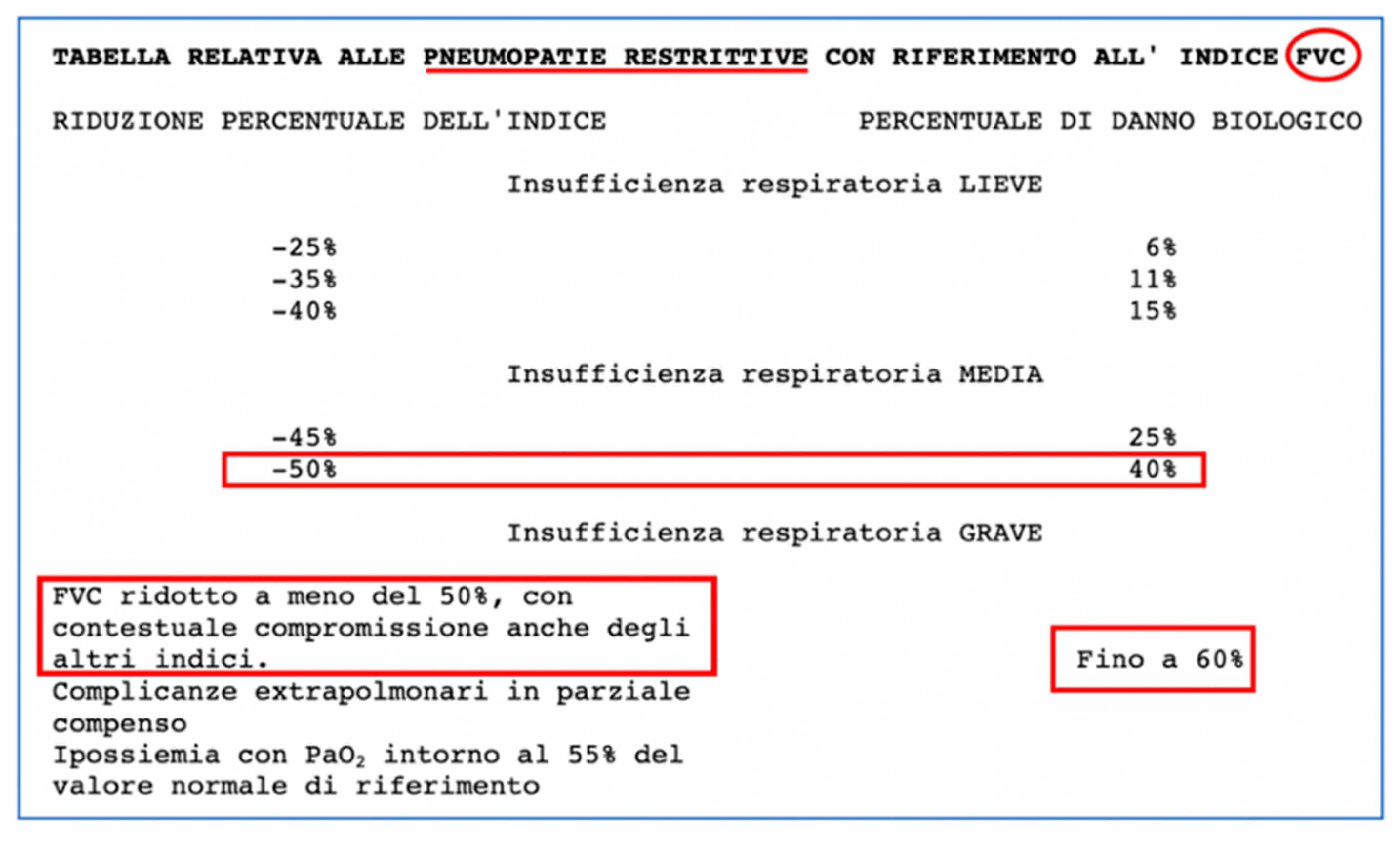
2.5. Assessment of Respiratory Function

The result of the last spirometry check carried out is reported below as an instrumental assessment relevant to the medicolegal classification of the injury/residual damage to the respiratory function:

2.6. Spirometry (1 Year Later)

The examination shows a restrictive dysventilatory syndrome clinically classified as “moderate”. This is mainly due to a reduction in the FVC and FEV1 indices, with a consequent percentage increase in the Tiffeneau Index (EV1/FVC). The other indices are also compromised at baseline and, after Salbutamol tests, show a slight improvement; in any case, they are significantly reduced compared to what can be seen in subjects of the same age. This is also reflected in the reduction of DLCO values in relation to interstitial disease. This picture was already appreciable at the spirometric examination carried out 4 months after recovery, showing “restrictive ventilatory defect of a marked degree for the age”.
With regard to the medicolegal evaluation of the respiratory deficit, proceeding with an analogical criterion for the site and the results (with respect to the military tables to be applied, which are very synthetic for many apparatuses and organs) and for a precise quantification of the degree of respiratory insufficiency, this allows us to make a brief reference to the procedural indications set out in Annex No. 2—Part A of the Ministerial Decree of 12 July 2000, which, although applicable to different cases, provides us with some useful evaluation details for the purposes of the functional evaluation of the consequences of the case in question—see Figure 2.
This allows us to define, in the context of the medical–legal field, that we are dealing with a medium degree of respiratory insufficiency, taking into account the FVC values reported in spirometry, i.e., an FVC equal to 3.36 against normal values between 4.14 and 6.15. In fact, considering an average normal value equal to 5.145, the result is equivalent to a reduction in the index of about 50% compared to an average/normal value. An average reference value is a value that renders the centripetal tendency of the reference range of values considered to be a normal range. This range must be considered in relation to variable factors that, in the clinical field, are mainly represented by age, sex, and race. Case in point, the central value of the range was considered, taking into account that the subject is 35 years old, an age at which, in healthy subjects, the optimal test result would be the selected value [45,46].
This can be seen even more clearly in the pictograms showing the “z-scores” (observed value–predicted value/standard deviation) relative to the intervals of normal values in the global spirometry shown above, from which it is possible to appreciate the reduction in DLCO (the alveolar–capillary diffusion of carbon monoxide, which is the index used in the evaluation of restrictive dysventalitic syndromes) (Figure 3). The z-score is a mathematical combination of the difference between the observed and theoretical value and the interindividual variability that provides a single number that takes into account the expected variability of lung function (in relation to age and height) compared to a comparable group of healthy subjects.
The above, together with the alteration of other indices in relation to the normal values provided (taking into account the young age of the subject and, therefore, to a slight extent, the low susceptibility to improvement given the time that has already elapsed) it appears reasonable to assess the permanent invalidity of the stabilized sequelae of the respiratory illness at 43%. This assessment is consistent with the table in force for civil invalidity pursuant to the Ministerial Decree of 5 February 1992, which (Table 3), as is well known, estimates the damage in terms of reduction of the G.L.C., as is the case with service causality; the said reference table makes it possible to estimate the permanent invalidity of clinical–functional conditions, similar to those under assessment in Table Voice No. 6014—Pulmonary Tuberculosis—Parenchymal or Pleural Fibrosis Outcomes with Moderate Respiratory Failure. The damage range is 41–50%.
This quantification also finds its scientific/documental rationale in the report from the last CT scan performed on 26 May 2021, where pleuro-parenchymal fibrotic findings in the pulmonary apices, as well as further alterations underway in the follow-up for complete definition, were found and, as already explained, in the alteration of multiple spirometric indices, which, as in many post-COVID patients, testify to the concrete value of the so-called clinical–radiological dissociation.
In the military tables (Presidential Decree No. 834 of 30 December 1981, “Definitivo riordinamento delle pensioni di guerra, in attuazione della delegazione prevista dall’art. 1 della legge 23 settembre 1981, n. 533”, these injuries would appear to be classifiable in Table A—Category VII, Entry No 18: “Bilateral basal pleurisy, or extensive results of unilateral pleurisy of suspected TB nature, given the organ, the results and the bilateral nature of the injuries (documented CT)”.
Presidential Decree No. 834 of 30 December 1981 further specifies that “…For chronic bronchopathies, assignment to a category higher than the 7th category provided for in table A must be made on the basis of the extent of emphysema and the reduction in respiratory capacity (medium-marked-serious), determined by spirometric examination or gas analysis…”.
Furthermore, in point 10 of the fifth category of the table, we find:
“pulmonary tuberculosis in the state of extensive, but clinically stabilised outcomes, again after stratigraphic assessment, when these do not by their extent lead to serious disruption of respiratory function”.
For this reason, in the light of the results of the overall instrumental examinations, and since there are no coinciding or comparable items, for obvious reasons, given the time when the tables were drawn up, it seems more appropriate to fix the permanent invalidity attributed to the respiratory disease and its results/posthumous effects at Table A—Category VI.
On the other hand, it should also be borne in mind that—based on the constant jurisprudence of the Court of Auditors, according to whom the extent of the impairment of the general working capacity of each of the disabilities referred to in Tables A and B must be included in fairly precise percentage ranges of damage (Table B, 11–20%; VIII ctg Table A, 21–30%; I ctg Table A, 91–100%)—the VI ctg of Table A effectively corresponds to a reduction in G.L.C. equal to 41–50%, which is the percentage band that all assessments cited herein seem to confirm as permanent invalidity, which correlates to the sequelae of the respiratory illness under examination.

2.7. Assessment of Anosmia/Ageusia

For the evaluation of the anosmia/ageusia, we relied on the results of the last instrumental examinations performed and on the olfactory smart threshold test and the evoked potentials test (an objective measure of the sensory deficit) as instrumental investigations relevant to the medicolegal classification of the residual damage/injury to olfactory–geusical functions.

2.8. Olfactory Smart Threshold Test (OST) and Evoked Potentials (1 Year and 1 Month Later)

Bearing in mind that, in this case, cross-modal perception means the inseparable interaction between the olfactory and taste senses, the examination conducted (OST) and—as per the report of the same day—the objective olfactometric examination obtained by means of evoked potentials, concluded with a picture of anosmia, both in the function of the olfactory threshold and in the function of uni- and cross-modal olfactory perception, with, therefore, a complete impairment of the cross-modal interaction inherent to the geusia, thus motivating the current ageusia (Figure 4).
The aforementioned deficit was also assessed with visual–mnestic tests, i.e., with the help of a cotton swab with an aqueous solution in different compositions (sugars, sodium chloride, acetic acid) and, placed on a quadrant of the protruded tongue, the subject was asked to identify the type of taste sensation perceived, describing it as sweet, salty, sour, or bitter. The subject, in the absence of visual stimuli of the substance, did not correctly identify the type of taste.
From a physiopathological point of view, this outcome appears to be due to a neurotropism of COVID-19, which biochemically simulates neuritis [47,48] or, as explained above, causes damage related to inflammatory outcomes [49].
In terms of assessment, in accordance with the Ministerial Decree of 5 February 1992, anosmia corresponds to Table Voice No. 6801, with a damage percentage of 20.
That is, with a unique value given the predictable cross-modal interaction sequelae noted above and already known in the literature.
Although this value is not present, configured as a “one-off” (percentages between 11 and 20%) in the military tables (Presidential Decree No. 834 of 30 December 1981, “Definitivo riordinamento delle pensioni di guerra, in attuazione della delegega prevista dall’art. 1 della legge 23 settembre 1981, no. 533), this injury can be classified by analogy in terms of the physiopathological criteria in Table A—Category VI, Entry No. 18 with respect to neuritis and its permanent outcomes. In any case, these criteria can be combined, in accordance with Table F, with disability from respiratory pathology as above and, therefore, be assimilated into the same table and category identified.

3. Discussion

The case of our patient, aged 33, a military doctor who contracted COVID at work, was relevant in two respects:
  • The outcomes observed, i.e., complete, permanent anosmia and respiratory difficulties more than a year after recovery from the disease, showed a medium-to-severe functional incidence in relation to the subject’s age, as well as anticipating what is now the much-discussed picture of so-called long-term COVID.
  • The assessment of the aforementioned outcomes required the use of multiple assessment tables that illustrate the current difficulties in assessing outcomes resulting from SARS-CoV-2 infection, in this case, in light of the aforementioned codicil and regulatory elements.
  • In Italy, this assessment requires the use of the current reference standards listed below:
    -
    Inps: Ministerial Decree 05/02/1992;
    -
    Inail: D.M. 12/07/2000;
    -
    Ministry of Defense: Tables annexed to Presidential Decree No. 915 of 23 December 1978 “Consolidated text of the regulations on war pensions”, tables annexed to Presidential Decree No. 834 of 30 December 1981 “Definitivo riordinamento delle pensioni di guerra, in attuazione della delegazione prevista dall’art. 1 della legge 23 settembre 1981, n. 533”.
With regard to the outcomes taken into consideration for the case examined, the tables show significant differences in evaluation, especially with regard to anosmic outcomes (Table 3).
As an addendum to the above, see the INAIL document “The fact sheets of the health superintendency, COVID-19 outcomes: evaluation indications. by Central Health Superintendence”. In this regard, although it is possible to agree on the application of a principle of proportionality between the sum obtained from the tabular value of the individual injuries and the indemnifiable permanent biological damage, the current classification of COVID-19 outcomes appears reductive given the occupational differences between the subjects with injuries that inflict a lesser degree of impairment, which can be assessed in a range of tabular values: between 1 and 5% in injuries with a greater degree of impairment, which can be placed in the wide assessment range equal to or greater than 8% of the table and, finally, “intermediate injuries”, assessable in the measure of 6–7% of biological damage.
For example, in the case of workers in the construction industry or, as in the present case, in the health sector, the deficits resulting from complete anosmia include an inability to detect the possible presence of gas; therefore, a dangerous condition may arise.
Similarly, in the face of other factors affecting the same apparatus, respiratory failure may impair the ability to work of COVID-19-recovered individuals in a way that is different from the normal outcomes of an occupational pneumopathy.
In view of the above, despite the fact that the table system is based on the fact that there is no different indemnity for damage depending on the work carried out, from an occupational point of view and in accordance with the principle of fairness for citizens, it is impossible not to take into account the different impact of the results depending on the task carried out, thus enhancing the concept of “specific risk” to which certain categories of workers are exposed.
In our opinion, a basic evaluation system of outcomes should be structured at least on an experimental basis, which, on the basis of employment indicators (and, therefore, multiplication factors), could quantify the final damage by taking into account the personalization it deserves.
In order to do this, we propose to draw up a list of workers most at risk from COVID-19 and the relative impacts that the outcomes may have on their specific jobs, i.e., to outline risk classes (Table 4).
This methodological proposal appears to merit a multidisciplinary approach by specialists in forensic medicine and occupational medicine, generating an evaluation commission possibly supplemented by specialists from other clinical branches.
This committee will not draw up an ordinary report with table entries and a quantification of the damage, but rather, a reasoned technical opinion explaining the type of work carried out by the person, the damage suffered, and the specific functional incidence of the latter in the performance of the work activity.

Author Contributions

Data curation, G.M. and C.M.; Methodology, G.M. and E.C.; Supervision, M.C. and E.C.; Writing—original draft, G.M. and L.C.; Writing—review & editing, L.C., F.P. and E.C. All authors have read and agreed to the published version of the manuscript.

Funding

The authors did not receive funding for this publication.

Institutional Review Board Statement

Not applicable.

Informed Consent Statement

No informed consent was required.

Data Availability Statement

Not applicable.

Acknowledgments

The authors gratefully acknowledge the support of all authors.

Conflicts of Interest

The authors declare no conflict of interest.

References

  1. Arimany-Manso, J.; Martin-Fumadó, C. Medico-legal issues regarding from the COVID-19 pandemic. Med. Clin. (Engl. Ed.). 2020, 155, 344–346. [Google Scholar] [CrossRef] [PubMed]
  2. da Silveira Gorman, R.; Syed, I.U. Connecting the Dots in Emerging Mast Cell Research: Do Factors Affecting Mast Cell Activation Provide a Missing Link between Adverse COVID-19 Outcomes and the Social Determinants of Health? Med. Sci. 2022, 10, 29. [Google Scholar] [CrossRef]
  3. Anastassopoulou, C.; Gkizarioti, Z.; Patrinos, G.P.; Tsakris, A. Human genetic factors associated with susceptibility to SARS-CoV-2 infection and COVID-19 disease severity. Hum. Genom. 2020, 14, 40. [Google Scholar] [CrossRef] [PubMed]
  4. Washburn, T.; Diener, M.L.; Curtis, D.S.; Wright, C.A. Modern slavery and labor exploitation during the COVID-19 pandemic: A conceptual model. Glob. Health Action 2022, 15, 2074784. [Google Scholar] [CrossRef] [PubMed]
  5. Costantino, C.; Cannizzaro, E.; Alba, D.; Conforto, A.; Cimino, L.; Mazzucco, W. SARS-CoV-2 pandemic in the Mediterranean area: Epidemiology and perspectives. EuroMediterr. Biomed. J. 2020, 15, 102–106. [Google Scholar]
  6. Gambaro, E.; Gramaglia, C.; Marangon, D.; Azzolina, D.; Probo, M.; Rudoni, M.; Zeppegno, P. The Mediating Role of Gender, Age, COVID-19 Symptoms and Changing of Mansion on the Mental Health of Healthcare Workers Operating in Italy during the First Wave of the COVID-19 Pandemic. Int. J. Environ. Res. Public Health 2021, 18, 13083. [Google Scholar] [CrossRef]
  7. Müller, I.; Mancinetti, M.; Renner, A.; Bridevaux, P.-O.; Brutsche, M.H.; Clarenbach, C.; Garzoni, C.; Lenoir, A.; Naccini, B.; Ott, S.; et al. Frailty assessment for COVID-19 follow-up: A prospective cohort study. BMJ Open Respir. Res. 2022, 9, e001227. [Google Scholar] [CrossRef] [PubMed]
  8. Moretti, A.; Menna, F.; Aulicino, M.; Paoletta, M.; Liguori, S.; Iolascon, G. Characterization of Home Working Population during COVID-19 Emergency: A Cross-Sectional Analysis. Int. J. Environ. Res. Public Health 2020, 17, 6284. [Google Scholar] [CrossRef]
  9. Langmaid, L.; Ratner, L.; Huysman, C.; Curran, S.; Uluer, A. Supporting the Medically Fragile: Individualized Approach to Empowering Young Adults with Chronic Disease during the COVID-19 Pandemic. J. Adolesc. Health 2020, 67, 453–455. [Google Scholar] [CrossRef] [PubMed]
  10. Bellotti, L.; Zaniboni, S.; Balducci, C.; Grote, G. Rapid Review on COVID-19, Work-Related Aspects, and Age Differences. Int. J. Environ. Res. Public Health 2021, 18, 5166. [Google Scholar] [CrossRef]
  11. Martimbianco, A.L.C.; Pacheco, R.L.; Bagattini, M.; Riera, R. Frequency, signs and symptoms, and criteria adopted for long COVID-19: A systematic review. Int. J. Clin. Pract. 2021, 75, e14357. [Google Scholar] [CrossRef]
  12. Carenzo, L.; Costantini, E.; Greco, M.; Barra, F.L.; Rendiniello, V.; Mainetti, M.; Bui, R.; Zanella, A.; Grasselli, G.; Lagioia, M.; et al. Hospital surge capacity in a tertiary emergency referral centre during the COVID-19 outbreak in Italy. Anaesthesia 2020, 75, 928–934. [Google Scholar] [CrossRef] [PubMed]
  13. Desai, A.D.; Lavelle, M.; Boursiquot, B.C.; Wan, E.Y. Long-term complications of COVID-19. Am. J. Physiol. Cell Physiol. 2022, 322, C1–C11. [Google Scholar] [CrossRef] [PubMed]
  14. Antoniou, K.M.; Vasarmidi, E.; Russell, A.-M.; Andrejak, C.; Crestani, B.; Delcroix, M.; Dinh-Xuan, A.T.; Poletti, V.; Sverzellati, N.; Vitacca, M.; et al. European Respiratory Society Statement on Long COVID-19 Follow-Up. Eur. Respir. J. 2022, 2102174, Epub ahead of print. [Google Scholar] [CrossRef]
  15. Bourmistrova, N.W.; Solomon, T.; Braude, P.; Strawbridge, R.; Carter, B. Long-term effects of COVID-19 on mental health: A systematic review. J. Affect. Disord. 2022, 299, 118–125. [Google Scholar] [CrossRef]
  16. Cannizzaro, E.; Cirrincione, L.; Mazzucco, W.; Scorciapino, A.; Catalano, C.; Ramaci, T.; Ledda, C.; Plescia, F. Night-Time Shift Work and Related Stress Responses: A Study on Security Guards. Int. J. Environ. Res. Public Health 2020, 17, 562. [Google Scholar] [CrossRef] [PubMed] [Green Version]
  17. Tang, S.; Mao, Y.; Jones, R.M.; Tan, Q.; Ji, J.S.; Li, N.; Shen, J.; Lv, Y.; Pan, L.; Ding, P.; et al. Aerosol transmission of SARS-CoV-2? Evidence, prevention and control. Environ. Int. 2020, 144, 106039. [Google Scholar] [CrossRef]
  18. Cirrincione, L.; Plescia, F.; Ledda, C.; Rapisarda, V.; Martorana, D.; Lacca, G.; Argo, A.; Zerbo, S.; Vitale, E.; Vinnikov, D.; et al. Technical note 1 COVID-19 Pandemic: New Prevention and Protection Measures. Sustainibility 2022, 14, 4766. [Google Scholar] [CrossRef]
  19. Tabah, A.; Ramanan, M.; Laupland, K.B.; Buetti, N.; Cortegiani, A.; Mellinghoff, J.; Conway Morris, A.; Camporota, L.; Zappella, N.; Elhadi, M.; et al. Personal protective equipment and intensive care unit healthcare worker safety in the COVID-19 era (PPE-SAFE): An international survey. J. Crit. Care 2020, 59, 70–75. [Google Scholar] [CrossRef]
  20. Vitale, E.; Vella, F.; Filetti, V.; Cirrincione, L.; Indelicato, G.; Cannizzaro, E.; Rapisarda, V. How to prevent SARS-CoV-2 transmission in the agri-food industry during the first pandemic wave: Effects on seroprevalence. Appl. Sci. 2021, 11, 10051. [Google Scholar] [CrossRef]
  21. Pontone, G.; Scafuri, S.; Mancini, M.E.; Agalbato, C.; Guglielmo, M.; Baggiano, A.; Muscogiuri, G.; Fusini, L.; Andreini, D.; Mushtaq, S.; et al. Role of computed tomography in COVID-19. J. Cardiovasc. Comput. Tomogr. 2021, 15, 27–36. [Google Scholar] [CrossRef] [PubMed]
  22. Cheng, M.P.; Papenburg, J.; Desjardins, M.; Kanjilal, S.; Quach, C.; Libman, M.; Dittrich, S.; Yansouni, C.P. Diagnostic testing for severe acute respiratory syndrome-related Coronavirus-2: A narrative review. Ann. Intern. Med. 2020; published online ahead of print. [Google Scholar] [CrossRef] [Green Version]
  23. World Health Organization. Antigen-Detection in the Diagnosis of SARS-CoV-2 Infection Using Rapid Immunoassays; Interim Guidance; World Health Organization: Geneva, Switzerland, 2020; Available online: https://www.who.int/publications/i/item/antigen-detection-in-the-diagnosis-of-sars-cov-2infection-using-rapid-immunoassays (accessed on 24 June 2022).
  24. Desai, A.N.; Patel, P. Stopping the Spread of COVID-19. J. Am. Med. Assoc. (JAMA) 2020, 323, 1516. [Google Scholar] [CrossRef] [Green Version]
  25. Adams, J.G.; Walls, R.M. Supporting the Health Care Workforce during the COVID-19 Global Epidemic. J. Am. Med. Assoc. (JAMA) 2020, 323, 1439–1440. [Google Scholar] [CrossRef] [Green Version]
  26. World Health Organization. Clinical Management of Severe Acute Respiratory Infection When Coronavirus (2019-nCoV) Infection Is Suspected; Interim Guidance; World Health Organization: Geneva, Switzerland, 2019. [Google Scholar]
  27. Carod-Artal, F.J. Neurological complications of coronavirus and COVID-19. Rev. Neurol. 2020, 70, 311–322. (In Spanish) [Google Scholar] [CrossRef]
  28. Puelles, V.G.; Lütgehetmann, M.; Lindenmeyer, M.T.; Sperhake, J.P.; Wong, M.N.; Allweiss, L.; Chilla, S.; Heinemann, A.; Wanner, N.; Liu, S.; et al. Multiorgan and Renal Tropism of SARS-CoV-2. N. Engl. J. Med. 2020, 383, 590–592. [Google Scholar] [CrossRef]
  29. Di Nunno, N.; Esposito, M.; Argo, A.; Salerno, M.; Sessa, F. Pharmacogenetics and Forensic Toxicology: A New Step towards a Multidisciplinary Approach. Toxics 2021, 9, 292. [Google Scholar] [CrossRef] [PubMed]
  30. Cirrincione, L.; Rapisarda, V.; Mazzucco, W.; Provenzano, R.; Cannizzaro, E. SARS-CoV-2 and the Risk Assessment Document in Italian Work; Specific or Generic Risk Even If Aggravated? Int. J. Environ. Res. Public Health 2021, 18, 3729. [Google Scholar] [CrossRef] [PubMed]
  31. Sangiorgio, V.; Parisi, F. A multicriteria approach for risk assessment of COVID-19 in urban district lockdown. Saf. Sci. 2020, 130, 104862. [Google Scholar] [CrossRef]
  32. Carpenter, C.R.; Mudd, P.A.; West, C.P.; Wilber, E.; Wilber, S.T. Diagnosing COVID-19 in the Emergency Department: A Scoping Review of Clinical Examinations, Laboratory Tests, Imaging Accuracy, and Biases. Acad. Emerg. Med. 2020, 27, 653–670. [Google Scholar] [CrossRef]
  33. Cirrincione, L.; Rapisarda, V.; Ledda, C.; Vitale, E.; Provenzano, R.; Cannizzaro, E. Considerations on the Update of the Risk Assessment Document during the Pandemic State by COVID-19 in Italy. Front. Public Health 2021, 9, 655927. [Google Scholar] [CrossRef]
  34. Available online: https://www.istat.it/it/files//2022/03/Report_ISS_ISTAT_2022_tab3.pdf (accessed on 24 June 2022).
  35. Gad, M.; Kazibwe, J.; Quirk, E.; Gheorghe, A.; Homan, Z.; Bricknell, M. Civil-military cooperation in the early response to the COVID-19 pandemic in six European countries. BMJ Mil. Health 2021, 167, 234–243. [Google Scholar] [CrossRef] [PubMed]
  36. Baig, A.M. Chronic COVID syndrome: Need for an appropriate medical terminology for long-COVID and COVID long-haulers. J. Med. Virol. 2021, 93, 2555–2556. [Google Scholar] [CrossRef] [PubMed]
  37. Salian, V.S.; Wright, J.A.; Vedell, P.T.; Nair, S.; Li, C.; Kandimalla, M.; Tang, X.; Porquera, E.M.C.; Kalari, K.R.; Kandimalla, K.K. COVID-19 Transmission, Current Treatment, and Future Therapeutic Strategies. Mol. Pharm. 2021, 18, 754–771. [Google Scholar] [CrossRef] [PubMed]
  38. Carraturo, F.; del Giudice, C.; Morelli, M.; Cerullo, V.; Libralato, G.; Galdiero, E.; Guida, M. Persistence of SARS-CoV-2 in the environment and COVID-19 transmission risk from environmental matrices and surfaces. Environ. Pollut. 2020, 265 Pt B, 115010. [Google Scholar] [CrossRef]
  39. Ledda, C.; Cannizzaro, E.; Cinà, D.; Filetti, V.; Vitale, E.; Paravizzini, G.; Di Naso, C.; Iavicoli, I.; Rapisarda, V. Oxidative stress and DNA damage in agricultural workers after exposure to pesticides. J. Occup. Med. Toxicol. 2021, 16, 1. [Google Scholar] [CrossRef] [PubMed]
  40. Bilotta, C.; Perrone, G.; Adelfio, V.; Spatola, G.F.; Uzzo, M.L.; Argo, A.; Zerbo, S. COVID-19 Vaccine-Related Thrombosis: A Systematic Review and Exploratory Analysis. Front. Immunol. 2021, 12, 729251. [Google Scholar] [CrossRef] [PubMed]
  41. Beyerstedt, S.; Casaro, E.B.; Rangel, É.B. COVID-19: Angiotensin-converting enzyme 2 (ACE2) expression and tissue susceptibility to SARS-CoV-2 infection. Eur. J. Clin. Microbiol. Infect. Dis. 2021, 40, 905–919. [Google Scholar] [CrossRef] [PubMed]
  42. Eliezer, M.; Hautefort, C.; Hamel, A.-L.; Verillaud, B.; Herman, P.; Houdart, E.; Eloit, C. Sudden and Complete Olfactory Loss of Function as a Possible Symptom of COVID-19. JAMA Otolaryngol. Head Neck Surg. 2020, 146, 674–675. [Google Scholar] [CrossRef] [Green Version]
  43. Avenali, M.; Martinelli, D.; Todisco, M.; Canavero, I.; Valentino, F.; Micieli, G.; Alfonsi, E.; Tassorelli, C.; Cosentino, G. Clinical and Electrophysiological Outcome Measures of Patients with Post-Infectious Neurological Syndromes Related to COVID-19 Treated with Intensive Neurorehabilitation. Front. Neurol. 2021, 12, 643713. [Google Scholar] [CrossRef]
  44. Mazzatenta, A.; Neri, G.; D’Ardes, D.; De Luca, C.; Marinari, S.; Porreca, E.; Cipollone, F.; Vecchiet, J.; Falcicchia, C.; Panichi, V.; et al. Smell and Taste in Severe COVID-19: Self-Reported vs. Testing. Front. Med. 2020, 7, 589409. [Google Scholar] [CrossRef]
  45. Miller, M.R.; Quanjer, P.H.; Swanney, M.P.; Ruppel, G.; Enright, P.L. Interpreting lung function data using 80% predicted and fixed thresholds misclassifies more than 20% of patients. Chest 2011, 139, 52–59. [Google Scholar] [CrossRef] [PubMed]
  46. Quanjer, P.H.; Stanojevic, S.; Cole, T.J.; Baur, X.; Hall, G.L.; Culver, B.H.; Enright, P.L.; Hankinson, J.L.; Ip, M.S.M.; Zheng, J.; et al. Multi-ethhnic reference values for spirometry for the 3–95 years age range: The Global Lung Function 2012 equations. Eur. Respir. J. 2012, 40, 1324–1343. [Google Scholar] [CrossRef]
  47. Scoppettuolo, P.; Borrelli, S.; Naeije, G. Neurological involvement in SARS-CoV-2 infection: A clinical systematic review. Brain Behav. Immun. Health 2020, 5, 100094. [Google Scholar] [CrossRef] [PubMed]
  48. Mao, L.; Jin, H.; Wang, M.; Hu, Y.; Chen, S.; He, Q.; Chang, J.; Hong, C.; Zhou, Y.; Wang, D.; et al. Neurologic manifestations of hospitalized patients with coronavirus disease 2019 in Wuhan, China. JAMA Neurol. 2020, 77, 6683–6690. [Google Scholar] [CrossRef] [Green Version]
  49. Hu, B.; Huang, S.; Yin, L. The cytokine storm and COVID-19. J. Med. Virol. 2021, 93, 250–256. [Google Scholar] [CrossRef] [PubMed]
Figure 1. ISTAT report on COVID-19 spread in Italy, regionally and geographically.
Sustainability 14 09332 g001
Figure 2. Reference to respiratory functional evaluation from Annex No. 2—Part A of the Ministerial Decree of 12/07/2000.
Sustainability 14 09332 g002
Figure 3. DLCO values in spirometry 1 year after COVID-19 infection.
Sustainability 14 09332 g003
Figure 4. Anosmia/ageusia evaluation in OST test VOCs for the functional assessment of the stimulation response: olfactory/olfactory - olfactory/trigeminal and olfactory/gustatory 1 year after COVID-19 infection.
Sustainability 14 09332 g004
Table 1. Spirometry 3 months after COVID-19 infection.
Forced Vital Capacity
ParameterUMDescriptionTeorTest#2%TeorPost#3%Test#2
Best VCFLBest CVT5.143.62703.66+1.2
FVCLForced vital capacity5.143.62703.66+1.2
FEV1LVolume exhaled after 1 s4.263.30773.29−0.3
PEFL/sPeak expiratory flow9.748.869110.59+19.5
PIFL/sPeak inspiratory flow 5.34 6.20+16.1
FEV1/FVC%%FEV1 as a percentage of FVC81.191.111289.7
FEV1/VC%%FEV1 as a percentage of VC81.1107.7133107.3
FEF25–75%L/sAverage exp. flow 25–75% FVC4.734.22894.53+7.5
MEF 75%L/sExposed flow at 25% of FVC8.378.751049.43+7.8
MEF 50%L/sExposed flow at 50% of FVC5.424.80895.17+7.9
MEF 25%L/sExposed flow at 75% of FVC2.472.11852.11+0.3
FET 100%sForced breathing time 2.1 2.6+21.7
PEFrL/minPeak expiratory flow (L/min)584.4531.691635.2+19.5
Lung Volumes and Ventilatory Profile
ParameterUMDescriptionTeorTest#1%Teor
IVCLInspiratory vital capacity5.393.0657
ERVLExpiratory reserve volume1.550.9662
IRVLInspiratory reserve volume 1.43
VEL/minVentilation espiratory minute 20.00
RfL/minRespiratory frequency 29.50
VtLCurrent volume 0.68
VT/Ti-Mean inspiratory flow 0.85
Ti/Ttot-Ti/Ttot ratio 0.39
Table 2. Spirometry 1 year after COVID-19 infection.
SVC PRE
MisNormalPred% PredZ Score
ERVL0.88-1.5556-
ICL1.21-3.8731-
VCL2.084.46–6.305.3839−5.89
IRVL0.81----
VTL (btps)0.397----
PREPOST DB (Salbutamol 400 mcg)
MisNormalPred% PredZ ScoreMisVar%Variation% PredZ Score
FVCL3.364.14–6.155.1465−2.923.360.00065−2.92
FEV1L3.233.43–5.104.2676−2.033.240.01076−2.00
FEV1/FVC%%96.169.3–92.981.11192.1096.50.301192.14
PEFL/S8.797.75–11.739.7490−0.798.810.02090−0.77
FEF 25–75%L/S4.343.02–6.444.7392−0.376.001.66381271.22
MEF 25%L/S2.401.19–3.762.4797−0.103.200.80331290.93
MEF 50%L/S4.683.25–7.595.4286−0.566.752.07441251.01
MEF 75%L/S8.015.56–11.188.3796−0.218.530.5371020.09
FEV6L0.00 ---0.000.00---
FEV1/FEV6%%0.0 ---0.00.0---
FEV1/VCmax%%96.169.3–92.981.11192.1096.50.301192.14
DLCO Test Results
MisNormalPred% PredZ score
DLCOmL/min/mmHg24.1028.07–41.9535.0169−2.58
DLCO corrmL/min/mmHg24.1028.07–41.9535.0169−2.58
DLCO/VAmL/min/mmHg/L5.593.51–6.284.891140.83
VAL4.316.00–8.307.1560−4.06
TLC (DLCO)L4.486.15–8.457.3061−4.03
DLCO 3eqmL/min/mmHg21.0128.07–41.9535.0160−3.32
Test Results N2 Multiple Breath Washout
MisNormalPred% PredZ score
FRCL4.192.44–4.423.431221.27
TLC (N2 WO)L6.686.15–8.457.3091−0.89
RV (N2 WO)L3.321.20–2.551.881773.51
RV/TLV (N2 WO)%49.718.2–36.227.21824.11
FRC/TLC (N2 WO)%62.839.9–62.050.91231.76
LCI-11.395.49–6.586.0318916.25
VCL3.364.46–6.305.3862−3.60
Table 3. Comparison of different reference evaluations for most long COVID outcomes.
OutcomeAssessments
INPSINAILMdD
Anosmia20%850–40%
(Item neurosis/neuritis—VI cat)
Moderate Respiratory Distress41–50%Up to 40%40–30%
(Item neurosis/neuritis—VII cat)
Table 4. Example of risk stratification according to worker category.
Type of WorkerRisk Classes—Impact of Long-Term COVID Damage on Job Activities (Considering Pulmonary and Neurological Damage)
FarmersHigh
Health workersHigh, especially for shift work and subcategories such as anesthetists working with gases
Administrative staffLow
Publisher’s Note: MDPI stays neutral with regard to jurisdictional claims in published maps and institutional affiliations.

