Article

Nano-Iron and Nano-Zinc Induced Growth and Metabolic Changes in Vigna radiata

Department of Botany, Faculty of Life Sciences, Maharshi Dayanand University, Rohtak 124001, India
*
Author to whom correspondence should be addressed.
Sustainability 2022, 14(14), 8251; https://doi.org/10.3390/su14148251
Submission received: 24 April 2022 / Revised: 20 June 2022 / Accepted: 29 June 2022 / Published: 6 July 2022

Abstract

:
The widespread industrial use and consequent release of nanosized iron (nFe3O4) and zinc oxide (nZnO) particles into the environment have raised concerns over their effects on living organisms, including plants. These nanoparticles are the source of their respective metal ions and although plants require both Fe and Zn ions for proper growth, excessive levels of these metals are toxic to them. A better understanding of the effects of these nanoparticles on plants also offers an opportunity for their useful applications in agriculture. The present work evaluates the changes in seed germination, plant growth, photosynthetic capacity, levels of biomolecules and antioxidant enzymes in Vigna radiata (L.) Wilczek when grown in the presence of nFe3O4 (size 1–4 nm) and nZnO (size 10–20 nm) and compared to the control plants. The plants were raised hydroponically for up to 14 days at two different concentrations of nanoparticles, viz. 10 and 100 mg/L. Inductively coupled plasma mass spectrometry (ICP-MS) results established that V. radiata can accumulate Fe and Zn in shoots with high efficiency. The results indicated that nFe3O4 had a favourable effect on V. radiata, whereas no apparent benefit or toxicity of nZnO was observed at the tested concentrations.

1. Introduction

Since the last decade, nanotechnology has found applications in almost every industry including the pharmaceutical, food and packaging, agriculture, ceramics, cosmetics, water and air purification industries. Increased application of nanotechnology is coupled with a simultaneous release of nanoparticles into the environment, provoking the scientists to assess the risks associated with their exposure [1]. In this regard, various studies carried out on plants, animals and microorganisms have indicated the presence of both negative as well as positive effects from nanoparticles. In plants, nanoparticles have been shown to improve seedling germination percentages and rates, as well as root and shoot length and vegetative biomass. On the other hand, reduction in plant growth, chlorophyll content and induction of oxidative stress has also been observed. The effects are largely dependent on nanoparticle type, size, concentration, exposure method, duration of exposure and even on the species type exposed to the nanoparticles [2]. Since plants are an important link in the food chain, the effect of various nanoparticles on their growth and development has been an active area of research that is fast catching the attention of plant and environmental scientists alike.
In this context, studying the effects of nanoparticles on Vigna radiata (L.) Wilczek (mung bean) deserves special attention since it is an agriculturally, nutritionally and economically important pulse crop from the legume family. Moreover, the plant is recommended to be used for phytotoxicity testing by the Organisation for Economic Co-operation and Development (OECD). Earlier, both stimulatory as well as inhibitory effects of metal and metal oxide nanoparticles on this plant have been reported. Foliar spray of titanium dioxide nanoparticles (nTiO2) at a 10 mg/L concentration for 14-day-old mung bean plants significantly improved the shoot and root length, root area, root nodule, chlorophyll content and total soluble leaf protein of the plants [3]. Priming of V. radiata seeds with magnesium oxide nanoparticles (nMgO) at 100 mg/L enhanced seedling germination and seedling vigour [4]. Manganese oxide nanoparticles (nMn3O4, 133 mg/dL) increased the root length and chlorophyll contents of the V. radiata plants grown in sandy loam soil but decreased the shoot length and protein content [5]. In a pot study conducted over 10 days, silver nanoparticles (nAg; 50 mg/L) decreased the chlorophyll, carotenoids, phenol and flavonoid contents but improved necrosis in V. radiata leaves [6]. Toxicity of copper oxide nanoparticles (nCuO) to V. radiata grown in artificial medium supplemented with nCuO for 21 days was investigated by Prakash et al., (2014) [7]. The authors observed significant reduction in root length, shoot length, biomass and chlorophyll contents with concomitant increase in lipid peroxidation, ROS generation and proline and hydrogen peroxide contents of the plants. Lignification of root cells and modulations in the expression of superoxide dismutase, catalase and ascorbate peroxidase genes in roots of the treated plants were also reported. These effects of nCuO were more pronounced at higher concentrations, specifically at 100, 200 and 500 mg/L. In a separate study, Jahagirdar et al., (2020) [8] reported that the biogenically synthesised Cu nanoparticles promoted germination of V. radiata seeds but had an adverse effect on root growth at 100 mg/L.
The impact of iron and zinc oxide nanoparticles (nFe3O4 and nZnO) on V. radiata has also attracted increasing interest, as both are among the most widely used metal oxide nanoparticles (around 100 tons/year globally) with widespread applications in catalysis, biomedicine, sensing, water treatment, etc. [9]. Furthermore, both are essential nutrient elements of the plants and could be a potential source of their respective ionic forms, needed for normal growth of plants, but at the same time may exert toxic effects at higher doses. A stimulatory effect of nZnO at 20 mg/L but growth retardation beyond this concentration on V. radiata grown in nanoparticle-supplemented agar media was reported by Pramod et al., (2011) [10]. In a separate study, Rani et al., (2020) [11] also confirmed the stimulatory effect of nZnO and nMgO on V. radiata seed germination and chlorophyll contents at a 5 mg/kg concentration of nanoparticles. Amooaghaie et al., (2016) [12] reported that nZnO at a 50 mg/L concentration increased fresh weight and dry weight of shoot and chlorophyll content in other plants such as tomato and wheat, but not any significant effect was observed at a 100 mg/L concentration of nZnO. Zero-valent nFe also had a stimulatory effect on shoot and root elongation of hydroponically cultured V. radiata at 600 mg/L and displayed no inhibition on seed germination at doses of 0–1000 mg/L [13]. Similar results were reported by Sun et al., (2020) [14], where nano-iron oxide did not present any significant phytotoxicity to mung bean even at 1000 mg/L. Mahavar et al., (2018) [15] analysed the effect of ZnO and Fe2O3 nanoparticles on the growth of mung bean plants using an agar overlay method at two different concentrations, i.e., 50 and 100 mg/L. After a 28-day study, the authors observed the inhibitory effects of nZnO on the fresh weight and of nFe2O3 on the Chl a and carotenoid contents of the plant. Overall, the interactions of nFe3O4 and nZnO with the mung bean have been studied only superficially, with most of the studies being centred around the morphological responses and no attempts have thus far been made for studying these interactions at physiological and biochemical levels specifically covering the markers of the oxidative stress. Since nanoparticles are well known to generate oxidative stress in plants [16,17], further studies on this aspect are needed to fill the existing knowledge gaps. Simultaneously, better understanding of the effects of these nanoparticles on various metabolic processes and the stress physiology of V. radiata may also offer an opportunity for their useful applications in the field of agriculture.
Hence, considering the increased release of nFe3O4 and nZnO in the natural environment and their inevitable interaction with plants, the present work reports the effects of these nanomaterials on the morphology, physiology and metabolism of V. radiata. The specific parameters taken into account for this purpose are germination, growth, photosynthetic capacity, metabolite contents, lipid peroxidation and antioxidant status of the plants.

2. Materials and Methods

2.1. Chemicals

All the chemicals used were of analytical grade and purchased from HiMedia Laboratories Pvt. Ltd., Mumbai; Merck Life Science Pvt. Ltd., Mumbai; Rankem Laboratory Reagent, New Delhi; and Ranbaxy Fine Chemicals Limited, New Delhi. Certified seeds of V. radiata were procured from Chaudhary Charan Singh Haryana Agricultural University, Hisar. The study protocol complied with the relevant institutional, national and international guidelines and legislation.

2.2. Synthesis and Characterisation of Nanoparticles

To prepare nFe3O4, the procedure given by Maity and Aggarwal (2007) [18] was followed. The two iron salts, 0.32 g of FeCl3.6H2O and 0.16 g FeCl2.4H2O, were dissolved in 40 mL of deionised water and the solution was heated at 80 °C for 1 h with continuous stirring. Thereafter, 5.0 mL of NH4OH (30% w/v) was quickly added to it. The resulting suspension was stirred vigorously for another 1 h and then the solution was allowed to cool at room temperature. Magnetic decantation was used to separate the nFe3O4 precipitate formed, which was then washed five times with distilled water.
The nZnO was prepared as per the procedure of Moghaddam et al., (2009) [19]. An aqueous solution of Zn(NO3)2.6H2O (0.045 M) was prepared at room temperature. In a separate beaker, 0.09 M aqueous solution of NaOH was heated to 55 °C under continuous magnetic stirring. Thereafter, the Zn(NO3)2.6H2O solution was added to it dropwise under vigorous stirring for 2 h. The precipitated nZnO was washed thrice with distilled water and ethanol and dried at 60 °C.
The size of the synthesised nanoparticles was analysed with a transmission electron microscope (TEM) and the crystal structure was confirmed by X-ray diffraction (XRD) analysis. TEM (Hitachi: H-7500, 120 kV) and XRD (Panalytical X’Pert Pro, Malvern, UK ) analyses were carried out at the Sophisticated Analytical Instrumentation Facility (SAIF) at Punjab University, Chandigarh. Functional groups of synthesised nFe3O4 and nZnO were analysed by fourier transform infrared spectroscopy (FTIR, Bruker AlphaII) at the Department of Zoology, Maharshi Dayanand University, Rohtak.

2.3. Exposure of V. radiata to Nanoparticles and Treatment Design

Effect of nFe3O4 and nZnO on V. radiata was studied at two different concentrations, i.e., 10 and 100 mg/L, making four treatment groups in total. The concentrations of both the nanoparticles were finalised after a careful survey of the literature, which revealed that a good number of studies on ZnO and Fe3O4 nanoparticles’ interaction with plants were carried out in the range from 2–100 mg/L. Seeds of V. radiata were surface sterilised with 0.1% mercuric chloride, washed 3–4 times with distilled water and twenty seeds for each treatment group were soaked separately in aqueous suspensions of nFe3O4 and nZnO for 12 h. Twenty seeds corresponding to the control group were soaked in plain distilled water without the addition of any nanoparticle. Thereafter, the seeds were transferred to the Petri plates lined with a double layer of germination paper, irrigated with 20.0 mL of the respective soaking solution and placed in an incubator at 25 ± 2 °C in the dark for 24 h. Seed germination as observed by the emergence of the radicle out of the seed coat was recorded for all groups. The % of seed germination was calculated as the number of seeds germinated/total number of seeds × 100. Five replicates were set for control for all the treatment groups, making a total of 100 seeds/group.
The germinated seeds (20 seeds per beaker) were further placed on a wire mesh kept above the beakers filled with nanoparticle-spiked Hoagland solution in such a way that their roots were in contact with the nutrient solution. Control plants were grown in plain Hoagland solution without the nanoparticles. Beakers were placed in the laboratory next to the full glass window and continuously aerated with a pump to maintain the oxygen level. To maintain the volume of the nutrient solution at a constant, the water loss due to evaporation was replenished every day. After 14 days of growth, root length, shoot length and plant biomass of all the treated and untreated plants were recorded. Other parameters such as chlorophyll, carbohydrate, protein contents and activities of antioxidant enzymes in the aboveground parts were also recorded in all the treated plants and compared with the control. Dry weight was determined after keeping the plants at 70 °C for 24 h. Each beaker with 20 plants was considered as one sampling unit or replicate. Experimental data from five replicates for each condition, i.e., for control as well as for each of the treatment groups, were collected separately, making the total number of samples n = 100/group. Finally, the average data from each replicate was used for performing statistical analysis.

2.4. Chlorophyll Content

Each 14-day-old V. radiata plant had a single pair of leaves in which chlorophyll content was estimated by the procedure followed by Siva and Benita (2016) [20]. Fresh leaf material (500 mg) was ground with 80% acetone (20 mL) for 5 min in a prechilled mortar and pestle. The extract thus formed was centrifuged at 4 °C for 25 min at 8000 rpm. The supernatant was made up to 50 mL with 80% acetone and used for chlorophyll measurement.

2.5. Gas Exchange and Chlorophyll Fluorescence

Gas exchange parameters including net photosynthesis, stomatal conductance and transpiration rate were measured in 14-days-old nanoparticle-treated and untreated V. radiata plants using a portable gas exchange system (LCi-SD, ADC BioScientific Ltd., Hoddesdon, UK). Measurements were made on fully expanded leaves between 10:30 and 12:30 A.M. at 22 ± 0.8 °C. The photosynthetic photon flux density and CO2 concentration at the time of measurements were 200 ± 10 µmol m−2 s−1 and 300 ppm, respectively.
A chlorophyll fluorometer (OS30p, Opti-Sciences, Hudson, USA) was used to measure chlorophyll fluorescence in the same leaves which were used to measure the gaseous exchange parameters. The leaves were placed in the dark for 30 min to calm down the reaction centres and the minimum (Fo) and maximum (Fm) chlorophyll fluorescence were measured. Maximum quantum efficiency of PSII was calculated from Fv/Fm = ((Fm – Fo)/Fm.

2.6. Carbohydrate and Protein Content

Carbohydrate estimation was performed as per the protocol stated by Dubois et al., (1956) [21]. Plant material (1.0 g) from each treatment was homogenised with 3.0 mL of sodium phosphate buffer (50 mM, pH 7.0) and centrifuged at 10,000 rpm for 10 min. The supernatant (100 µL) was mixed thoroughly with 1.0 mL of extraction buffer, 1.0 mL of 5% phenol and 5.0 mL of concentrated sulfuric acid. All tubes were placed at 37 °C for 10 min. The intensity of the colour developed was noted at 490 nm and the total carbohydrates were interpolated from the standard curve of D-glucose in the range of 10–100 µg/mL.
Protein content of the shoots was measured by the method of Lowry et al., (1951) [22] using the standard curve of BSA (20–100 µg/mL) and the results were expressed as µg protein/g of plant material.

2.7. Lipid Peroxidation

Lipid peroxidation releases malondialdehyde (MDA) as one of the final products in amounts proportional to the level of stress present. The amount of MDA produced was determined by the method of Heath and Packer (1968) [23] and expressed as µM MDA/g fresh weight.

2.8. Antioxidant Status

Metallic nanoparticles are known to generate oxidative stress in plants by generating various types of damaging reactive oxygen species (ROS) such as O2˙, OH˙ and H2O2. Plants can scavenge these ROS primarily by activating their antioxidant enzyme machinery. Hence, in the present work, activities of superoxide dismutase (SOD), catalase (CAT), glutathione peroxidase (GPx), glutathione reductase (GR) and glutathione-S-transferase (GST) were measured to assess the level of oxidative stress faced by V. radiata plants upon exposure to nFe3O4 and nZnO.
Plant extracts were prepared by grinding 0.5 g of plant tissue in 5 mL of potassium phosphate buffer (0.1 M, pH 7.0) in a prechilled mortar and pestle. The homogenate was then centrifuged at 15,000 rpm at 4 °C for 10 min. The supernatant obtained was used as a source of enzyme for further experimentation. For each assay, potassium phosphate buffer was set as blank and the control reaction mixture had all constituents except plant extract.
The SOD activity was determined by measuring its capacity to inhibit the photoreduction of NBT according to the method of Beauchamp and Fridovich (1971) [24]. The reaction mixture containing the supernatant (1.0 mL), 0.1 mM EDTA (0.5 mL), 13 mM methionine (0.5 mL), 75 μM NBT (0.5 mL) and 2 μM riboflavin (0.5 mL) was incubated at room temperature for 10 min. Thereafter, the absorbance of the reaction mixture was measured at 560 nm. The 50% inhibition of the reaction between riboflavin and NBT in the presence of methionine was taken as one unit of SOD activity.
The modified method of Aebi (1983) [25] was used for the CAT assay. The assay mixture included 1.5 mL of 0.01 M potassium phosphate buffer (pH 7.0), 1.2 mL of 150 mM H2O2 and 0.5 mL of plant extract. A decrease in absorbance at 240 nm was recorded up to 2.5 min at 30 s intervals. A unit of catalase activity was defined as the amount of enzyme that degraded 1 μmole of H2O2 per min under the standard assay conditions.
The GR assay was performed as per the procedure of David and Richard (1983) [26]. Plant extract (0.1 mL), 2.0 mL potassium phosphate buffer (0.12 M, pH 7.2), EDTA (0.1 mL), sodium azide (0.1 mL) and oxidised glutathione (0.1 mL) were mixed with distilled water (0.6 mL) and kept at room temperature for 3 min. Thereafter, 0.1 mL NADPH was added and absorbance was read at 340 nm at intervals of 15 sec up to 3 min. One GR unit was defined as the amount of enzyme that oxidises 1.0 µM of β-NADPH per min under the standard assay conditions.
The GPx assay was conducted according to the procedure stated by Rani et al., (2004) [27]. The reaction mixture containing 0.4 mL potassium phosphate buffer (0.4 M, pH 7.0), 0.1 mL sodium azide (10 mM), 0.2 mL reduced glutathione (4 mM), 0.1 mL H2O2 (2.5 mM), 0.2 mL of water and 0.5 mL of plant extract was incubated at 37 °C for 0, 30, 60 and 90 s. Thereafter, the reaction was terminated with the addition of 0.5 mL of 10% TCA and the reaction mixture was centrifuged at 8000 rpm. After centrifugation, 3.0 mL buffer and 1.0 mL of DTNB (0.04% DTNB in 1% sodium citrate) reagent were added to 2.0 mL of supernatant. The absorbance of the coloured solution was measured at 412 nm. One GPx unit was defined as the amount of enzyme that produced 1.0 µM of oxidised glutathione per min under the standard assay conditions.
The GST activity was determined by the procedure proposed by Habig et al., (1974) [28]. The enzyme assay was based on the capacity of the enzyme to form the complex of GSH and CDNB. The reaction mixture contained GSH (0.1 mL), CDNB (0.1 mL) and 2.7 mL potassium phosphate buffer. The reaction started after the addition of enzyme extract (0.1 mL) and change in the absorbance at 340 nm was recorded spectrophotometrically for 3 min. One unit of GST was considered to conjugate one mol CDNB with reduced glutathione per min at 25 °C.

2.9. Inductively Coupled Plasma Mass Spectroscopy (ICP-MS) Analysis

The uptake of nanoparticles by the plants was confirmed by ICP-MS. Plants were removed from the hydroponics culture on the 14th day of treatment. They were washed thoroughly three times with distilled water and placed in an oven preheated to 80 °C to dry the plant material. To 0.5 g of dried sample, 10 mL HNO3 and 3 mL H2O2 were added in the digestion vessel. The temperature of the vessel was slowly raised to 100 °C over 40 min and then allowed to stand at 100 °C for another 15 min. The vessel was allowed to cool off to room temperature and subjected to ICP-MS analysis at Sigma Test and Research Centre, Delhi.

2.10. Statistical Analysis

A completely randomised design was used. The data were analysed by one-way analysis of variance (ANOVA) using IBM SPSS Statistics 26 software with post hoc analysis using Tukey’s test at the p < 0.05 confidence level. For each treatment, the results were expressed as the mean of five different replicates and the standard error (S.E.) values were used to represent the variability of data in graphs.

3. Results and Discussion

3.1. Characterisation of Nanoparticles

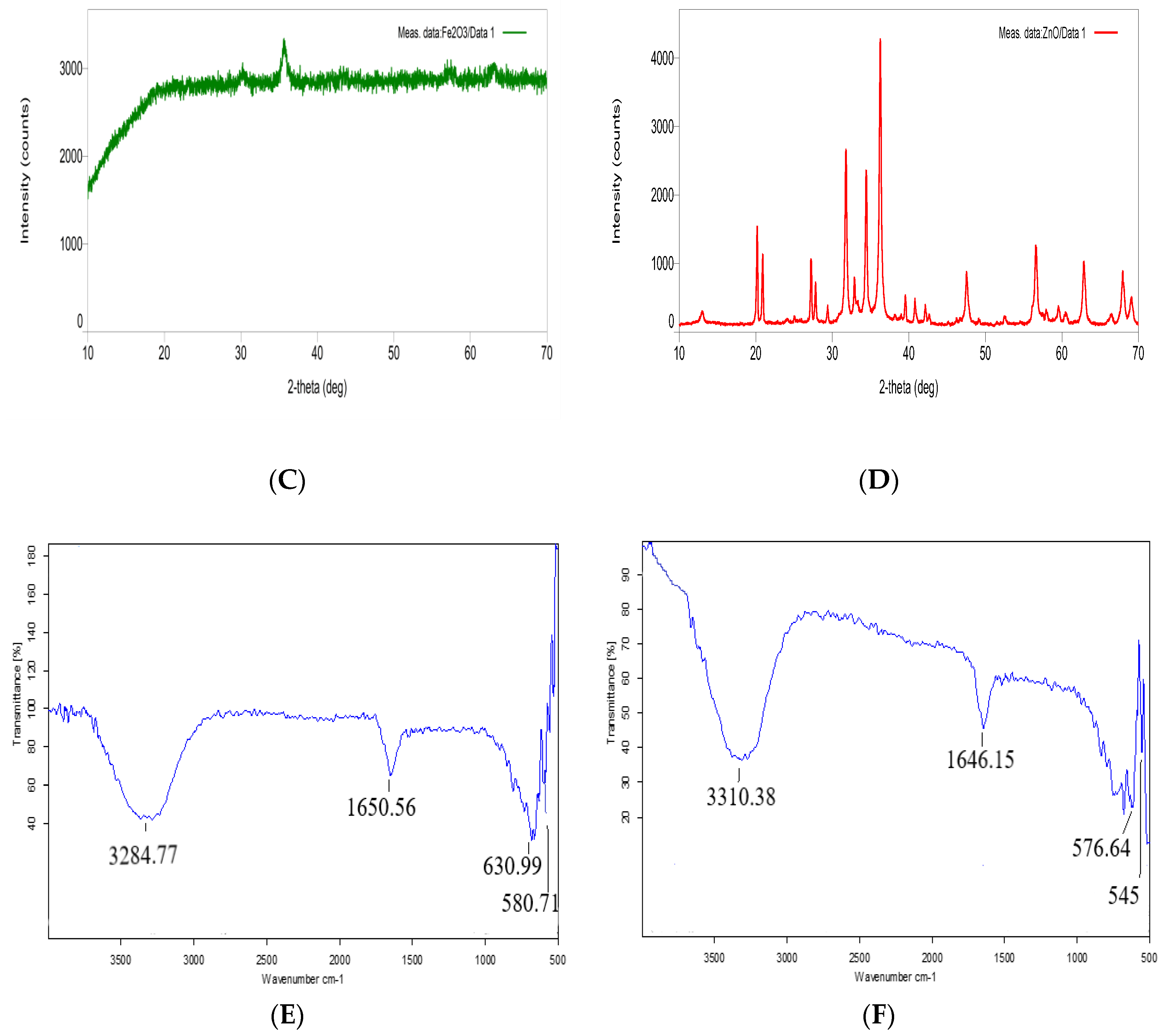
TEM analysis (Figure 1A,B) showed that the synthesised nFe3O4 and nZnO were polydispersed, but were within a very narrow size range of 1–4 and 10–20 nm, respectively. XRD analysis revealed the information about the crystal structure. The peaks obtained in the XRD graph for nFe3O4 were in accordance with the reference XRD (JCPDS card No. 89-0691). For nFe3O4 (Figure 1C), the peaks were at 30°, 35.2°, 43.1°, 53.4°, 57.1° and 62.8° at the 2θ plane with hkl values of 220, 311, 400, 422, 511 and 440, respectively. Distinct peaks for the produced nanoparticles were revealed by FTIR spectra of nFe3O4 and nZnO in Figure 1E,F, respectively. Peaks at 630.99 and 580.71 cm−1 in Figure 1E were due to the stretching vibrations of Fe–O bonds in the crystalline lattice of nFe3O4 [29], whereas peaks at 545 and 576 cm−1 in Figure 1F showed stretching of the Zn–O group [30]. The bending and stretching of hydroxyl groups (-OH) were attributed to the peaks near 1650, 3280 and 3300 cm−1.
Peaks for nZnO (Figure 1D) were observed at 31.67°, 34.31°, 36.14°, 47.40°, 56.52°, 62.73°, 66.28°, 67.91° and 69.03° at the 2θ plane with hkl values of 100, 002, 101, 102, 110, 103, 200, 112 and 201, respectively, and agreed with the reference XRD (JCPDS No: 36-1451).

3.2. Bioaccumulation of nFe3O4 and nZnO

The presence of metallic Fe and Zn in the V. radiata shoots were analysed by ICP-MS. As shown in Table 1, V. radiata shoots accumulated a significantly higher amount of metals at 100 mg/L compared to control plants as well as the plants treated with a 10 mg/L concentration of nanoparticles. About 8 and 83% of the given nFe3O4 and about 27 and 90% of the given nZnO accumulated in the aboveground parts of 14-day-old V. radiata plants at 10 and 100 mg/L concentrations, respectively, confirming that both the nanoparticles were able to enter the plants. Comparatively higher % accumulation at 100 mg/L showed that most of the nanoparticles absorbed by roots were translocated to the shoots. A concentration-dependent increase in the accumulation of Fe and Zn was observed in several other plant species. Bandyopadhyay et al., (2015) [31] reported that when alfalfa plants were grown in nZnO-spiked soil, the accumulation of nZnO in the roots, shoots and leaves increased as the concentration of nZnO was increased from 250 to 500 to 750 mg/kg.
Zinc accumulation in pea plants also increased proportionally to the concentration of nZnO treatment [32]. The highest iron content was accumulated by shoots of peanut plants at 10, 250 and 1000 mg/kg treatments of nFe2O3 [33]. Iron accumulation also increased in 1, 2 and 3 mg/L nFe3O4-treated Ocimum basilicum plants as compared to the control plants [34].
Apart from the concentration, another factor which greatly influences the accumulation of nanoparticles by plants is the size of the nanoparticles. Generally, nanoparticles of 40–50 nm dimensions are able to penetrate into the plant tissues [35]. Higher % accumulation of nFe3O4 and nZnO in V. radiata might be attributed to the small size and narrow size range of the synthesised nanoparticles.

3.3. Seed Germination and Plant Growth

The ability of nanoparticles to affect seed germination was tested independently for both nFe3O4 and nZnO at two different concentrations (10 and 100 mg/L). The nFe3O4 as well as nZnO did not affect seed germination in V. radiata as 100% germination was recorded in all the treated seeds, which was similar to the control seeds. Corresponding results were reported by Lin and Xing (2007) [36] in radish, oilseed rape, lettuce and cucumber at 20, 200 and 2000 mg/L of nZnO and by Wang et al., (2011) [37] in rye grass and pumpkin at 30 and 100 mg/L concentrations of nFe3O4.
In the present study, nFe3O4 showed a concentration-dependent increase in the growth of V. radiata plants on the 14th day of treatment. Shoot length of nFe3O4-treated plants increased by 13% and 29% whereas root length increased by 10% and 27% at 10 and 100 mg/L nFe3O4 concentrations, respectively as compared to the control plants (Table 2). Similarly, plant fresh weight increased by 36% at 10 mg/L and 58% at 100 mg/L of nFe3O4 and dry weight increased by 61% at 10 mg/L and 78% at 100 mg/L of nFe3O4. nFe3O4 at both concentrations was conducive to the growth of plants, but significant stimulation of shoot length at 100 mg/L and of fresh weight at both 10 and 100 mg/L was achieved. Similar results were reported by Elfeky et al., (2013) [34], as they showed that sweet basil plant height and plant fresh and dry weight increased via nFe3O4 at the concentrations of 1, 2 and 3 mg/L. Plaksenkova et al., (2019) [38] reported that nFe3O4 increased the shoot length, root length and plant biomass in Eruca sativa at 1, 2 and 4 mg/L concentrations of nanoparticles. An increase in fresh weight and dry weight of chickpea by nFe3O4 was reported by Pawar et al., (2019) [39] at 4, 8 and 12 mg/L concentrations of nanoparticles. Compared to the control plants, the nZnO slightly increased the root length, shoot length, fresh weight and dry weight of V. radiata plants at a 10 mg/L concentration, though the % stimulation was not significant with the values being 8, 3, 15 and 18, respectively. However, at a concentration of 100 mg/L, nZnO growth parameters were slightly inhibited (Table 2), with root length and fresh and dry weight of V. radiata plants decreasing by 4, 7 and 9%, respectively, with no effect on shoot length. Clearly, nZnO produced both desirable and undesirable effects on V. radiata, but the changes were not significant and may be considered as small and inappreciable. The obtained results are in agreement with other studies reported in the literature. Boonyanitipong et al., (2011) [40] reported that root length of rice seedlings decreased at 500 and 1000 mg/L concentrations of nZnO. Lee et al., (2013) [41] also reported that nZnO at 2000 mg/L decreased both the root length and root biomass of Fagopyrum esculentum.
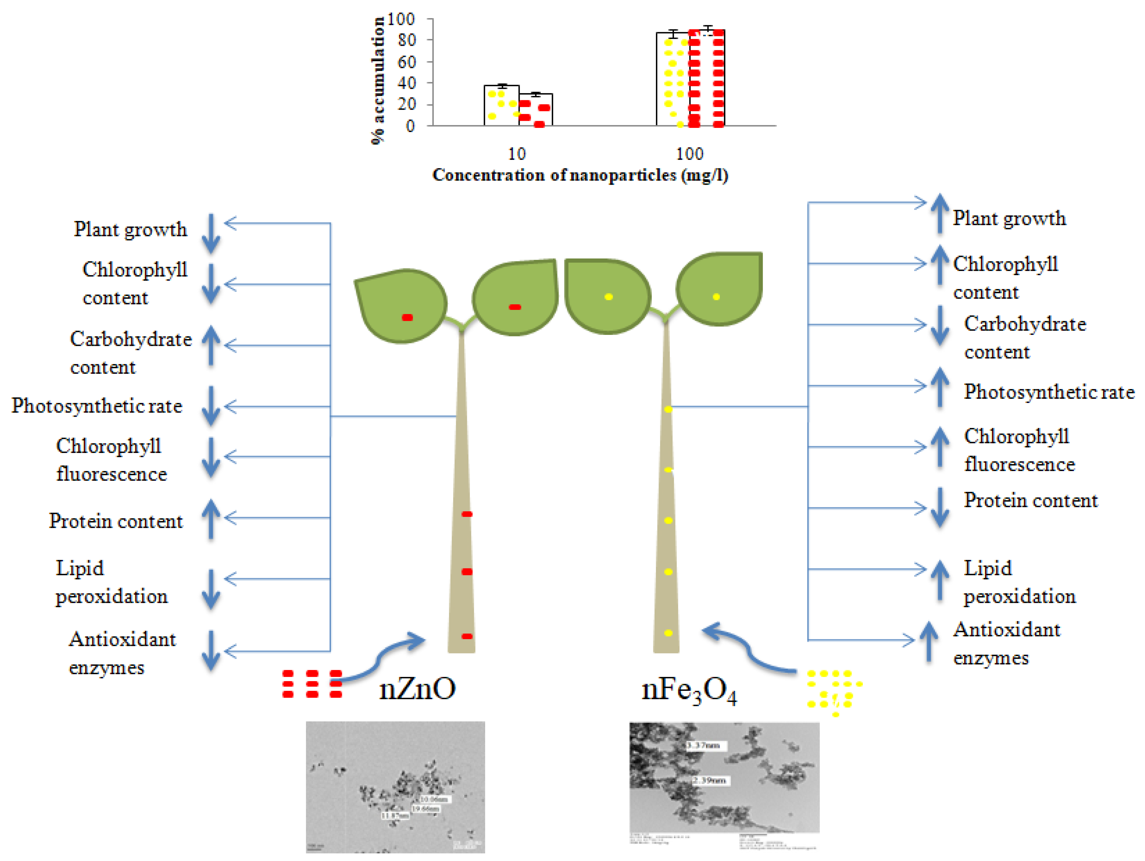
The stimulatory effect of nFe3O4 on plant growth may be due to various reasons, such as the nanoparticles leading to the production of OH radicals which trigger the cell wall loosening, release the tensional stress in the cell and increase the elongation of the cell [42]. Changes in growth may also be linked to the changes in the chlorophyll content of the plant, which is explained next in Section 3.4. Structural distortions and inhibition of root growth in maize upon exposure to nZnO (1.0 mg/mL) were attributed to nZnO-induced deep invaginations resembling a “tunnel” in the primary root tip [43]. Previous studies reported that the toxicity of nZnO is not due to their nanosize but due to the production of zinc ions [44]. Wang et al., (2016) [45] linked the reduced biomass of nZnO (300 mg/L)-treated Arabidopsis plants to decreased chlorophyll contents, and hence, photosynthetic efficiency of the treated plants. A summarised view of the changes in growth, levels of biomolecules and antioxidant enzymes in the nFe3O4- and nZnO-treated V. radiata plants is presented in Scheme 1.

3.4. Chlorophyll Content

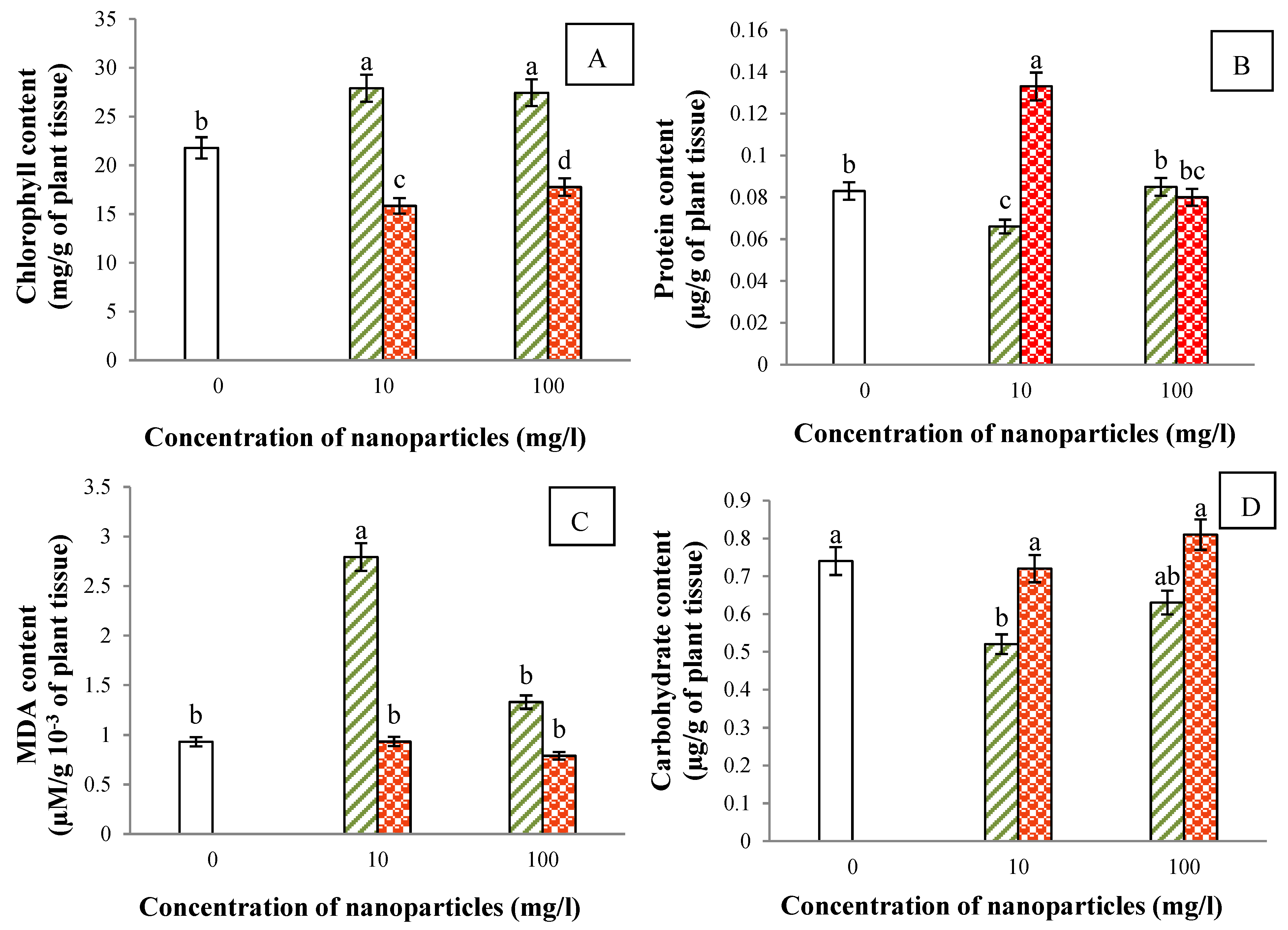
The nFe3O4 increased the total chlorophyll content of V. radiata plants, the % increase being 28 at 10 mg/L and 26 at 100 mg/L nFe3O4 after 14 days of treatment, but nZnO had an opposite effect on the chlorophyll content in V. radiata plants (Figure 2A). Chlorophyll content of nZnO-treated plants decreased significantly compared to both control and nFe3O4-treated plants. In comparison to the control, a decrease of 27% at 10 mg/L and of 18% at 100 mg/L of nZnO was observed. The significant increase in chlorophyll content by nFe3O4 may be attributed to the role of iron in the biosynthesis of chlorophyll [46]. Iron helps in the conversion of coproporphyrinogen to protoporphyrin and protoporphyrin to protochlorophyllide-monomethyl ester [46,47], a precursor of chlorophyll. However, as in the case of nZnO, a significant decrease in the chlorophyll content might be due to the replacement of the central metal atom of chlorophyll (Mg2+) by Zn2+, which has resulted in inhibition of chlorophyll synthesis, leading to reduced photosynthetic efficiency [48]. Mukherjee et al., (2016) [49] suggested a similar reason for nZnO toxicity in green pea plants.
The stimulatory effect of nFe3O4 on the chlorophyll contents in watermelon at a 50 mg/L concentration [50], in Zingiber officinale at 0.1 mg/L [20] and in Ocimum basilicum at 1, 2 and 3 mg/L concentrations [34], and the reduction in chlorophyll content by nZnO in Bacopa monniera and Lolium perenne at a 200 mg/kg concentration [51,52] and in green pea at 125, 250 and 500 mg/L concentrations [32] are in agreement with the results obtained in the present work.

3.5. Protein Content

No appreciable difference in the protein contents of 14-day-old V. radiata plants at a 100 mg/L concentration of both the nanoparticles was observed (Figure 2B). However, at lower concentrations (10 mg/L), the effects varied. The nFe3O4 decreased the protein content by about 20% whereas nZnO increased the protein content significantly by about 60%. Since Zn is an important cofactor required for activity and regulation of many enzymes involved in transcription, translation and signal transduction, elevated levels of Zn ions released from nZnO might have triggered the synthesis of Zn-requiring proteins. Kisan et al., (2015) [53] also observed increased protein levels in spinach by nZnO at 1.0 mg/L and proposed that Zn enhanced cation exchange in roots of plants due to which absorption of essential nutrients such as nitrogen increased and which was responsible for higher protein contents. Patra et al., (2013) [54] also reported that protein content was increased by nZnO at 0.5, 1.0, 2.0 and 4.0 mg/L concentrations in mung bean. The inhibitory effect of nFe3O4 at a lower concentration may be explained in terms of increased production of ROS, which causes damage to cell constituents, particularly proteins and amino acid residues through oxidation [55,56]. However, when exposed to higher concentrations of either zinc or iron nanoparticles, the V. radiata plants were able to maintain internal homeostasis and did not reflect any change in their protein contents vis à vis the control plants, probably due to well-known mechanisms of excess metal exclusion and sequestration into the vacuoles.

3.6. Gaseous Exchange, Chlorophyll Fluorescence and Carbohydrate Content

The nFe3O4 at both 10 and 100 mg/L concentrations positively influenced the photosynthetic rate and chlorophyll fluorescence. The enhancement of the photosynthetic rate was about 11 and 9% and the Fv/Fm ratio increased by about 17 and 15% at 10 and 100 mg/L of nFe3O4, respectively. Contrarily, nZnO decreased both the photosynthetic rate and efficiency of PS II in a concentration-dependent manner, though the changes were not statistically significant. Nevertheless, both the nanoparticles increased the stomatal conductance and transpiration rate in 14-day-old V. radiata plants compared to the control plants, and out of the two concentrations of nanoparticles used, the increase was more pronounced at 10 mg/L (Table 3).
Increased flux of CO2 and water vapours as depicted by increased stomatal conductance and transpiration rate, respectively, coupled with increased efficiency of PS II and higher chlorophyll contents moderately increased the photosynthetic rate of nFe3O4-treated V. radiata plants, and hence, their growth. In nZnO-treated plants a gentle decrease in photosynthetic rate despite an increase in stomatal conductance and transpiration rate might be ascribed to the decreased chlorophyll levels as well as to the reduced efficiency of PS II. Consistent results reported by other researchers include significant increase in photosynthetic parameters in soybean after spraying the citrate-coated Fe2O3 nanoparticles at a 500 mg/L concentration [57] and about 50% inhibition of the gas exchange parameters in Arabidopsis by ZnO nanoparticles at 300 mg/L [58].
Although chlorophyll content and photosynthetic rate are directly correlated with carbohydrate content, somewhat contrasting results were obtained in the present study. The nFe3O4-treated V. radiata plants were found to have lower carbohydrates despite having higher chlorophyll contents and marginally better photosynthetic rates compared to the control plants, where the % decrease was 30% and 16% at 10 and 100 mg/L of nFe3O4 concentrations, respectively (Figure 2D). Conversely, nZnO treatment did not affect carbohydrate content at 10 mg/L and increased it slightly by 9% at a 100 mg/L concentration, though the chlorophyll contents and photosynthetic rates of the treated plants were less in comparison to the control plants. Hence, in the present study a decrease in carbohydrate contents of the shoots under nFe3O4 might be correlated with the increased transport of sugars to roots and other younger parts to support their growth, whereas comparable or higher carbohydrate content vis à vis the control in the presence of nZnO showed that perhaps the sugars accumulated in shoots due to disturbed efflux of carbohydrates to roots, which might also be one of the reasons for decreased growth and biomass of V. radiata at a 100 mg/L nZnO concentration. A detailed review of the changes in accumulation and translocation of sugars under abiotic stress was provided by Krasavina et al., (2014) [59].

3.7. Lipid Peroxidation

Lipid peroxidation is related to the amount of MDA in plant cells which is formed due to the peroxidation of membrane lipids. As shown in Figure 2C, the nFe3O4 increased the lipid peroxidation in 14-day-old V. radiata plants by 200 and 43% at 10 and 100 mg/L concentrations, respectively, whereas no appreciable change in MDA contents was observed with nZnO at a 10 mg/L concentration and slightly decreased levels of MDA were noted at 100 mg/L of nZnO. Similar results were reported by Souza et al., (2019) [60], where the authors showed that nFe3O4 at 30, 40 and 50 mg/L concentrations increased the lipid peroxidation in Lemna minor and by Burman et al., (2013) [61], where a decrease in the lipid peroxidation in nZnO (at 1.5 and 10 mg/L)-treated chickpeas was observed. The nFe3O4-induced damage to the membrane may be linked to enhanced production of superoxide radicals. Unlike Zn, Fe is a redox-active metal and participates directly in ROS producing Haber–Weiss and Fenton reactions. Additionally, it was reported that iron accentuates NADPH oxidase-mediated ROS production in a dose-dependent manner [62]. Conversely, Zn reduces the activity of NADPH oxidase and, therefore, suppresses superoxide production [63] which could be responsible for decreased lipid peroxidation in nZnO-treated V. radiata plants. However, the less damaging effect at the higher nFe3O4 concentration in the present study suggests that perhaps excess of nFe3O4 was excluded from the cytoplasm and sequestered in the vacuole and, thus, could not exert the toxic effects proportional to its concentration. Isolation of iron into the vacuole is a major tolerance mechanism in plants during iron overload to protect the cell from oxidative stress [64]. Furthermore, the growth of a plant is also a manifestation of a complex interplay between different metabolic networks. In the present study, the nFe3O4-treated plants grew well even when confronted with enhanced ROS by effectively raising the activities of antioxidant enzymes (discussed under Section 3.8) and their photosynthetic efficiency. Overall, the results demonstrate that nFe3O4 did generate oxidative stress in V. radiata, but the plants were able to neutralise it to a great extent. The growth of nZnO-treated plants did not significantly differ from the growth of control plants despite their reduced chlorophyll contents and photosynthetic capacity, perhaps because the loss was compensated by the reduced production of ROS, thereby helping the plants to divert their resources towards maintaining growth rather than fighting off the stress.

3.8. Activities of Antioxidant Enzymes

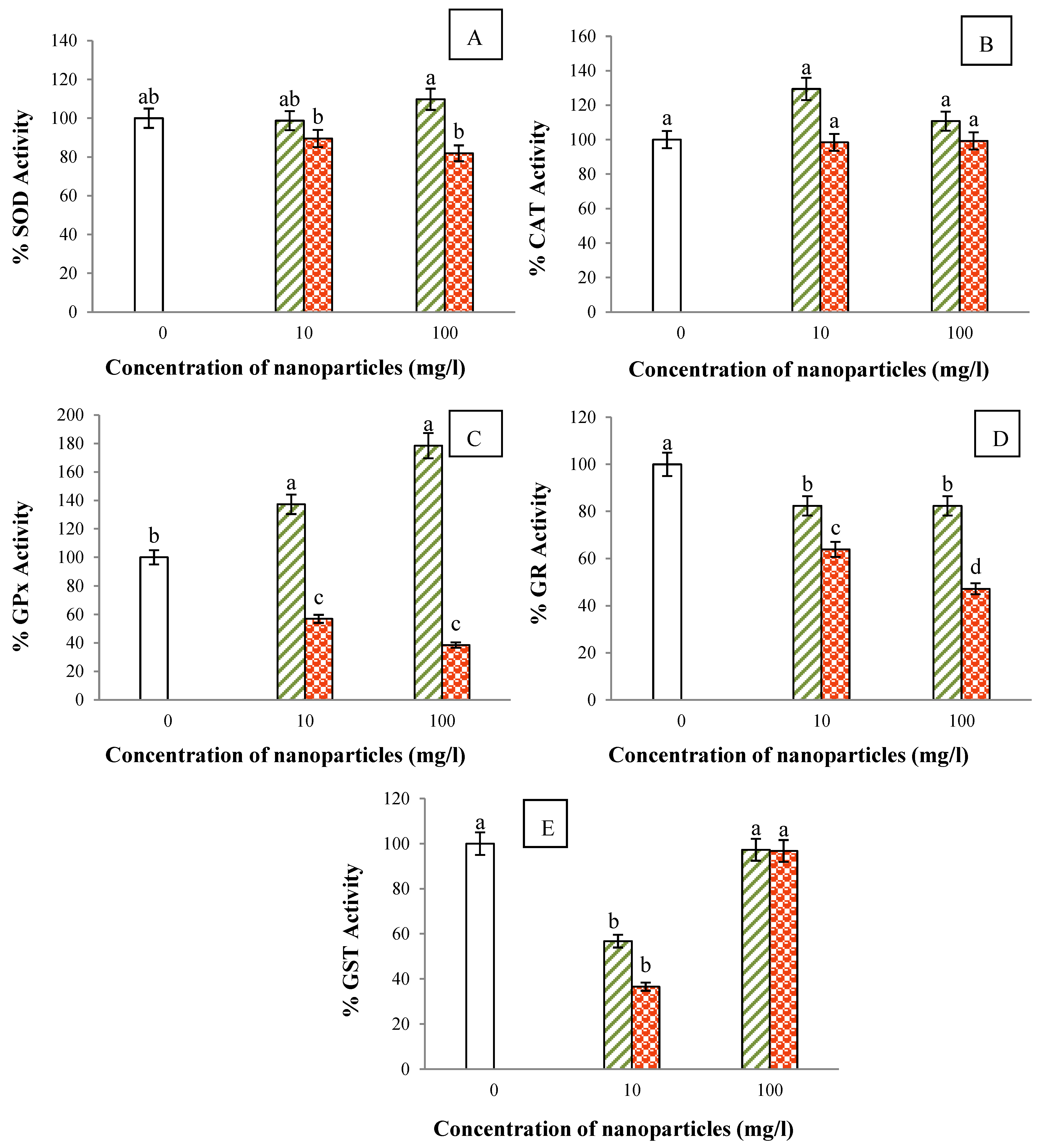
Activities of five different antioxidant enzymes, viz. SOD, CAT, GPx, GR and GST, in nanoparticle-treated V. radiata plants after 14 days of treatment were recorded (Figure 3A–E) and results were compared to the activities of antioxidant enzymes in control plants. At a lower concentration (10 mg/L), nFe3O4 had no effect on SOD activity but increased the activities of CAT and GPx by 30 and 37%, respectively, and decreased the activities of GR and GST by about 18 and 43%, respectively. However, at a higher concentration (100 mg/L) of nFe3O4, slight stimulation of SOD and CAT (10% each) and a strong stimulation (78%) of GPx was recorded. The nZnO-treated V. radiata plants exhibited either no change or decreased activities of antioxidant enzymes compared to the control plants. The % decrease for SOD, GPx, GR and GST was 10, 43, 36 and 43%, respectively, at 10 mg/L of nZnO, whereas at 100 mg/L the activities of SOD, GPx, GR and GST reduced by 18, 62, 53 and 3%, respectively. The activity of CAT was nearly the same in both nZnO-treated and untreated plants. Overall, changes in the activities of antioxidant enzymes when correlated with the results of protein estimation and lipid peroxidation obtained in the present study denoted that nZnO-treated V. radiata plants produced less ROS compared to the control and nFe3O4-treated plants. Overproduction of ROS by Fe via stimulation of NADPH oxidase or participation in biological redox reactions such as Haber–Weiss and Fenton reactions demands adjustments in the activities of antioxidant enzymes for survival and may be responsible for increased activities of SOD, CAT and GPx in the nFe3O4-treated plants.
Besides this, inside a cell, the ROS are also produced during photosynthesis by partial oxidation of water by an oxygen-evolving complex [65] and due to auto-oxidizable properties of Fe–S centres, ferredoxin and reduced thioredoxin. Thylakoid membrane pigments also form strong oxidants by interacting with O2 [66,67]. Decreased chlorophyll might have directly reduced the rate of ROS production, and hence, the activities of antioxidant enzymes in nZnO-treated plants. Increased activities of SOD, CAT and GPx may also be attributed to higher chlorophyll contents, and hence, increased ROS production in nFe3O4-treated V. radiata plants. The main function of SOD, CAT and GPx is to maintain H2O2 homeostasis in a cell, where SOD neutralises superoxide radicals and singlet oxygen by converting them to H2O2, whereas H2O2, which itself is a source of hydroxyl radicals, is removed by the combined action of CAT and GPx. Since GPx and GST both compete for the same substrate, i.e., glutathione, a decrease in GST activity in nFe3O4-treated plants might be a compensatory mechanism to balance for the increased GPx activity. Nanomaterials have been shown to boost antioxidant enzyme activity in a concentration-dependent manner, resulting in lower ROS levels and improved growth and photosynthesis. Neutralisation of singlet oxygen and H2O2 prevents the formation of more toxic radicals such as hydroxyl anions, thereby stabilizing the membrane and cellular functioning. SOD acts as the first line of defence against toxic radicals, removing superoxide from cells and reducing damage to metabolic pathways such as photosynthesis [68].
The results obtained in the present study are also in consonance with the already published findings. As reported by Xiao et al., (2019) [69], exposure of Citrus maxima to nZnO at 250, 500 and 1000 mg/L concentrations decreased the SOD activity but did not affect CAT and peroxidase activities. Similar results were obtained for SOD activity in chickpea at 1.5 and 10 ppm nZnO concentrations [61] and for CAT and peroxidase in green pea at 125, 250 and 500 mg/kg nZnO concentrations [32]. Stimulation of CAT, GPx and SOD activities in wheat plants by nFe3O4 at 15 and 20 mg/L concentrations was reported by Iannone et al., (2016) [70]. Li et al., (2013) [50] observed that in watermelon, SOD activity did not change at 2 mg/L but increased in 20 and 50 mg/L of nFe3O4, whereas CAT activity increased in all the three concentrations used.

4. Conclusions

V. radiata plants were raised hydroponically in a well-aerated nutrient solution spiked with two different concentrations (10 and 100 mg/L) of nFe3O4 and nZnO, separately. The nFe3O4-treated plants grew better than the nZnO-treated and control plants, which may be linked to higher chlorophyll contents along with the enhanced photosynthetic rate and efficiency. The 14-day-old nZnO-treated V. radiata plants had no appreciable difference in growth and considerably lower chlorophyll contents compared to the control plants; however, the reductions in CO2 assimilation rate and chlorophyll fluorescence were not significant. The disagreement of photosynthetic ability with the carbohydrate contents suggests that both the nanoparticles may influence the removal and translocation of sugars from the source organs.
An analysis of oxidative stress markers reflect reduced production of ROS by nZnO compared to control and nFe3O4-treated plants. The V. radiata plants treated with nFe3O4 experienced more oxidative stress, and suffered protein loss and membrane damage, but dealt with the stress efficiently without compromising the growth by increasing the activities of SOD, CAT and GPx. However, more detailed molecular, biochemical and genetic studies are needed to understand the species-specific intricacies involved in adjusting to the presence of nanoparticles. Overall, nFe3O4 at both 10 and 100 mg/L concentrations supported the growth of V. radiata and had a positive influence on the photosynthetic parameters, whereas no significant advantage or disadvantage of nZnO at either concentration was observed. Further, nFe3O4 may be suitably employed for various nanobiotechnological applications concerning this plant, such as for the fortification of V. radiata with Fe and improvements in the efficiency of Fe fertilisers.

Author Contributions

All authors contributed to the study conception and design. Material preparation, data collection and analysis were performed by N.R., K.K., P.B. and P.S.; J.Y., V. and R. assisted with data validation and paper revision. The first draft of the manuscript was written by N.R. and K.K.; V.H. edited, corrected and finalised the manuscript. The overall work was supervised by V.H. All authors have read and agreed to the published version of the manuscript.

Funding

This study utilised the facilities generated out of the Department of Science and Technology (DST), Ministry of Science and Technology, Government of India grant under its “Fund for Improvement of Science & Technology Infrastructure (FIST)” scheme (No. SR/FST/LS1-529/2012(C)). Financial support from Haryana State Council for Science, Innovation and Technology (HSCSIT, End st. No. HSCSIT/R&D/2020/474) is also acknowledged. Poonam B. is thankful to HRDG, Council of Scientific and Industrial Research (CSIR File No. 09/382(0219)/2019-EMR-I) for financial assistance. J.Y. and R. are also thankful to HRDG, Council of Scientific and Industrial Research (CSIR File No. 09/382(0218)/2019-EMR-I and 09/382(0217)/2019-EMR-I, respectively) for financial assistance.

Institutional Review Board Statement

This article does not contain any studies with human participants or animals performed by any of the authors.

Informed Consent Statement

Not applicable.

Data Availability Statement

All data generated during study are included in the manuscript.

Conflicts of Interest

The authors have no conflict of interest to declare.

References

  1. Rizwan, M.; Ali, S.; Qayyum, M.F.; Ok, Y.S.; Adrees, M.; Ibrahim, Z.M.; Farid, M.; Abbas, F. Effect of metal and metal oxide nanoparticles on growth and physiology of globally important food crops: A critical review. J. Hazard. Mater. 2017, 322, 2–16. [Google Scholar] [CrossRef] [PubMed]
  2. Teske, S.S.; Detweiler, C.S. The biomechanisms of metal and metal oxide nanoparticles interactions with cells. Int. J. Environ. Res. Public Health 2015, 12, 1112–1134. [Google Scholar] [CrossRef] [PubMed] [Green Version]
  3. Raliya, R.; Biswas, P.; Tarafdar, J.C. TiO2 nanoparticle biosynthesis and its physiological effect on mung bean (Vigna radiata L.). Biotechnol. Rep. 2015, 5, 22–26. [Google Scholar] [CrossRef] [PubMed] [Green Version]
  4. Anand, K.V.; Anugraga, A.R.; Kannan, M.; Singaravelu, G.; Govindraju, K. Bio-engineered magnesium oxide nanoparticles as nano-priming agent for enhancing seed germination and seedling vigour in green gram (Vigna radiata L.). Mater. Lett. 2020, 271, 127792. [Google Scholar] [CrossRef]
  5. Ramesh, R.; Catherine, G.; Sundaram, S.J.; Khan, F.L.A.; Kaviyarasu, K. Synthesis of Mn3O4 nano complex using aqueous extract of Helianthus annuus seed cake and its effect on biological growth of Vigna radiata. Mater. Today Proc. 2020, 36, 184–191. [Google Scholar] [CrossRef]
  6. Najafi, S.; Jamei, R. Effect of silver nanoparticles and Pb(NO3)2 on the yield and chemical composition of Mung bean (Vigna radiata). J. Stress Physiol. Biochem. 2014, 10, 316–325. [Google Scholar]
  7. Prakash, M.; Gopalakrishnan, N.; Deung-Hyun, K.; Ill, M.C. Copper oxide nanoparticle toxicity in mung bean (Vigna radiata L.) seedlings: Physiological and molecular level responses of in vitro grown plants. Acta Physiol. Plant. 2014, 36, 2947–2958. [Google Scholar]
  8. Jahagirdar, A.S.; Shende, S.; Gade, A.; Rai, M. Bioinspired synthesis of copper nanoparticles and its efficacy on seed viability and seedling growth in mung bean (Vigna radiata L.). Curr. Nanosci. 2020, 16, 246–252. [Google Scholar] [CrossRef]
  9. Piccino, F.; Gottschalk, F.; Seeger, S.; Nowack, B. Industrial production quantities and uses of ten engineered nanomaterials in Europe and the world. J. Nanoparticle Res. 2012, 14, 1109–1120. [Google Scholar] [CrossRef] [Green Version]
  10. Pramod, M.; Dhoke, S.K.; Khanna, A.S.; Tarafdar, J.C. Effect of nano-ZnO on growth of mung bean (Vigna radiata) and chickpea (Cicer arietinum) seedlings using plant agar method. Appl. Biol. Res. 2011, 13, 54–61. [Google Scholar]
  11. Rani, P.; Kaur, G.; Rao, K.V.; Singh, J.; Rawat, M. Impact of green synthesized metal oxide nanoparticles on seed germination and seedling growth of Vigna radiata (Mung Bean) and Cajanus cajan (Red Gram). J. Inorg. Organomet. Polym. Mater. 2020, 30, 4053–4062. [Google Scholar] [CrossRef]
  12. Amooaghaie, R.; Norouzi, M.; Saeri, M. Impact of zinc and zinc oxide nanoparticles on the physiological and biochemical processes in tomato and wheat. Can. Sci. Publ. Bot. 2016, 95, 441–455. [Google Scholar] [CrossRef]
  13. Sun, Y.; Jing, R.; Zheng, F.; Zhang, S.; Jiao, W.; Wang, F. Evaluating phytotoxicity of bare and starch-stabilized zero-valent iron nanoparticles in mung bean. Chemosphere 2019, 236, 124336. [Google Scholar] [CrossRef] [PubMed]
  14. Sun, Y.; Wang, W.; Zheng, F.; Zhang, S.; Wang, F.; Liu, S. Phytotoxicity of iron-based materials in mung bean: Seed germination tests. Chemosphere 2020, 251, 12643. [Google Scholar] [CrossRef] [PubMed]
  15. Mahawar, H.; Prasanna, R.; Kaur, S.; Thapa, S.; Kanchan, A.; Singh, R.; Kaushik, S.C.; Singh, S.; Nain, L. Deciphering the mode of interactions of nanoparticles with mung bean (Vigna radiata L.). Isr. J. Plant Sci. 2018, 65, 74–82. [Google Scholar] [CrossRef]
  16. Khare, T.; Kumar, V.; Kishor, P.B.K. Na+ and Cl- ions show additive effects under NaCl stress on induction of oxidative stress and the responsive antioxidative defense in rice. Protoplasma 2014, 252, 1149–1165. [Google Scholar] [CrossRef]
  17. Kumar, V.; Shriram, V.; Hoque, T.S.; Hasan, M.M.; Burritt, D.J.; Hossain, M.A. Glycinebetaine-mediated abiotic oxidative-stress tolerance in plants: Physiological and biochemical mechanisms. In Stress Signaling in Plants: Genomics and Proteomics Perspective; Sarwat, M., Ed.; Springer International: Cham, Switzerland, 2017; Volume 2, pp. 111–133. [Google Scholar]
  18. Maity, D.; Aggarwal, D.C. Synthesis of iron oxide nanoparticles under oxidizing environment and their stabilization in aqueous media. J. Magn. Magn. Mater. 2007, 308, 46–55. [Google Scholar] [CrossRef]
  19. Moghaddam, A.B.; Nazari, T.; Badraghi, J.; Kazemzad, M. Synthesis of ZnO nanoparticles and electrodeposition of polypyrrole/ZnO nanocomposite film. Int. J. Electrochem. Sci. 2009, 4, 247–257. [Google Scholar]
  20. Siva, G.V.; Benita, L.F.J. Iron oxide nanoparticles promote agronomic traits of ginger (Zingiber officinale Rosc.). Int. J. Adv. Res. Biol. Sci. 2016, 3, 230–237. [Google Scholar]
  21. Dubois, M.; Gilles, K.A.; Hamilton, J.K.; Rebers, P.A.; Smith, F. Colorimetric Method for Determination of Sugars and Related Substances. Anal. Chem. 1956, 28, 350–356. [Google Scholar] [CrossRef]
  22. Lowry, O.H.; Rosebrough, N.J.; Farr, A.L.; Randall, R.J. Protein measurement with the folin phenol reagent. J. Biol. Chem. 1951, 193, 265–275. [Google Scholar] [CrossRef]
  23. Heath, R.L.; Packer, L. Photoperoxidation in isolated chloroplasts, I. Kinetics and stoichiometry of fatty acid peroxidation. Arch. Biochem. Biophys. 1968, 125, 189–198. [Google Scholar] [CrossRef]
  24. Beauchamp, C.; Fridovich, I. Superoxide dismutase: Improved assays and an assay applicable to acrylamide gels. Anal. Biochem. 1971, 44, 276–287. [Google Scholar] [CrossRef]
  25. Aebi, H.E. Catalase. In Methods of Enzymatic Analysis; Bergmeyer, H.U., Ed.; Verlag Chemie: Weinhem, Germany, 1983; Volume 3, pp. 273–286. [Google Scholar]
  26. David, M.; Richard, J.S. Glutathione reductase. In Methods of Enzymatic Analysis; Bergmeyer, H.U., Ed.; Academic Press: New York, NY, USA, 1983; Volume 3, pp. 258–265. [Google Scholar]
  27. Rani, P.; Unni, M.K.; Karthikeyan, J. Evaluation of antioxidant properties of berries. Indian J. Clin. Biochem. 2004, 19, 103–110. [Google Scholar] [CrossRef] [PubMed] [Green Version]
  28. Habig, W.H.; Pabst, M.J.; Jocoby, W.B. Glutathione-S-transferase: The first enzymatic step in mercapturic acid formation. J. Biol. Chem. 1974, 249, 7310–7339. [Google Scholar]
  29. Lori, N.; Eydokia, P.; Vassilis, Z.; Anna, P.; Eleana, H.; Christos, N.P. Magnetic nanoparticles in medical diagnostic applications: Synthesis, characterization and proteins conjugation. Curr. Nanosci. 2016, 12, 455–468. [Google Scholar]
  30. Deepa, R.; Muthuraj, D.; Kumar, E.; Veeraputhiran, V. Microwave assisted synthesis of zinc oxide nanoparticles and its antimicrobial efficiency. J. Nanosci. Technol. 2019, 5, 640–641. [Google Scholar]
  31. Bandyopadhyay, S.; Plascencia-Villa, G.; Mukherjee, A.; Rico, C.M.; Jose-Yacaman, M.; Peralta-Videa, J.R.; Gardea-Torresdey, J.L. Comparative phytotoxicity of ZnO NPs, bulk ZnO, and ionic zinc onto the alfalfa plants symbiotically associated with Sinorhizobium meliloti in soil. Sci. Total Environ. 2015, 515, 60–69. [Google Scholar] [CrossRef]
  32. Mukherjee, A.; Peralta-Videa, J.R.; Bandyopadhyay, S.; Rico, C.M.; Zhao, L.; Gardea-Torresdey, J.L. Physiological effects of nanoparticulate ZnO in green peas (Pisum sativum L.) cultivated in soil. Metallomics 2014, 6, 132–138. [Google Scholar] [CrossRef]
  33. Rui, M.; Ma, C.; Hao, Y.; Guo, J.; Rui, Y.; Tang, X.; Zhu, S. Iron oxide nanoparticles as a potential iron fertilizer for peanut (Arachis hypogaea). Front. Plant Sci. 2016, 7, 815. [Google Scholar] [CrossRef] [Green Version]
  34. Elfeky, S.A.; Mohammed, M.A.; Khater, M.S.; Osman, Y.A.; Elsherbini, E. Effect of magnetite nano-fertilizer on growth and yield of Ocimumbasilicum L. Int. J. Indig. Med. Plants 2013, 46, 1286–1293. [Google Scholar]
  35. Pérez-de-Luque, A. Interaction of nanomaterials with plants: What do we need for real applications in agriculture? Front. Environ. Sci. 2017, 5, 12. [Google Scholar] [CrossRef] [Green Version]
  36. Lin, D.; Xing, B. Phytotoxicity of nanoparticles: Inhibition of seed germination and root growth. Environ. Pollut. 2007, 150, 243–250. [Google Scholar] [CrossRef] [PubMed]
  37. Wang, H.; Kou, X.; Pei, Z.; Xiao, J.Q.; Shan, X.; Xing, X. Physiological role of magnetite (Fe3O4) nanoparticles on perennial ryegrass (Lolium perenne L.) and pumpkin (Cucurbita mixta) plants. Nanotoxicology 2011, 5, 30–42. [Google Scholar]
  38. Plaksenkova, I.; Jermalonoka, M.; Bankovska, L.; Gavarane, I.; Gerbreders, V.; Sledevskis, J.; Kokina, I. Effects of Fe3O4 nanoparticles stress on the growth and development of rocket Eruca sativa. J. Nanomater. 2019, 2019, 2678247. [Google Scholar] [CrossRef] [Green Version]
  39. Pawar, V.A.; Ambekar, J.D.; Kale, B.B.; Apte, S.K.; Laware, S.L. Response in chickpea (Cicer arietinum L.) seedling growth to seed priming with iron oxide nanoparticles. Int. J. Biosci. 2019, 14, 82–91. [Google Scholar]
  40. Boonyanitipong, P.; Kositsup, B.; Kumar, P.; Baruah, S.; Dutta, J. Toxicity of ZnO and TiO2 nanoparticles on germinating rice seed Oryza sativa L. Int. J. Biosci. Biochem. Bioinform. 2011, 1, 282–285. [Google Scholar] [CrossRef] [Green Version]
  41. Lee, S.; Chung, H.; Kim, S.; Lee, I. The genotoxic effect of ZnO and CuO nanoparticles on early growth of buckwheat, Fagopyrum esculentum. Water Air Soil Pollut. 2013, 224, 1668. [Google Scholar] [CrossRef]
  42. Kim, J.H.; Lee, Y.; Kim, E.J.; Gu, S.; Sohn, E.J.; Seo, Y.S.; Chang, Y.S. Exposure of iron nanoparticles to Arabidopsis thaliana enhances root elongation by triggering cell wall loosening. Environ. Sci. Technol. 2014, 48, 3477–3485. [Google Scholar] [CrossRef]
  43. Pokhrel, L.R.; Dubey, B. Evaluation of developmental responses of two crop plants exposed to silver and zinc oxide nanoparticles. Sci. Total Environ. 2013, 452, 321–332. [Google Scholar] [CrossRef]
  44. Singh, A.; Singh, N.B.; Afzal, S.; Singh, T.; Hussain, I. Zinc oxide nanoparticles: A review of their biological synthesis, antimicrobial activity, uptake, translocation and biotransformation in plants. J. Mater. Sci. 2018, 53, 185. [Google Scholar] [CrossRef]
  45. Wang, X.; Yang, X.; Chen, S.; Li, Q.; Wang, W.; Hou, C.; Gao, X.; Wang, L.; Wang, S. Zinc oxide nanoparticles affect biomass accumulation and photosynthesis in Arabidopsis. Front. Plant Sci. 2016, 6, 12–43. [Google Scholar] [CrossRef] [PubMed] [Green Version]
  46. Hsu, W.; Miller, G.W. Copro-porphyrinogenase in tobacco (Nicotiana tabacum L.). Biochem. J. 1969, 117, 215–220. [Google Scholar] [CrossRef] [PubMed] [Green Version]
  47. Spiller, S.C.; Castelfranco, A.; Castelfranco, P. Effects of iron and oxygen on chlorophyll biosynthesis. I. In vivo observations of iron and oxygen-deficient plants. Plant Physiol. 1982, 69, 107–111. [Google Scholar] [CrossRef] [PubMed] [Green Version]
  48. Kupper, H.; Kupper, F.C.; Spiller, M. Environmental relevance of heavy metal-substituted chlorophylls using the example of water plants. J. Exp. Bot. 1996, 47, 259–266. [Google Scholar] [CrossRef] [Green Version]
  49. Mukherjee, A.; Sun, Y.; Morelius, E.; Tamez, C.; Bandyopadhyay, S.; Niu, G.; Gardea-Torresdey, J.L. Differential toxicity of bare and hybrid ZnO nanoparticles in green pea (Pisum sativum L.): A life cycle study. Front. Plant Sci. 2016, 6, 12–42. [Google Scholar] [CrossRef] [Green Version]
  50. Li, J.; Chang, P.R.; Huang, J.; Wang, Y.; Yuan, H.; Ren, H. Physiological effects of magnetic iron oxide nanoparticles towards watermelon. J. Nanosci. Nanotechnol. 2013, 13, 5561–5567. [Google Scholar] [CrossRef]
  51. Ali, G.; Srivastava, P.S.; Iqbal, M. Influence of cadmium and zinc on growth and photosynthesis of Bacopa monniera cultivated in vitro. Biol. Plant. 2000, 43, 599–601. [Google Scholar] [CrossRef]
  52. Bonnet, M. Effects of zinc and influence of Acremonium lolii on growth parameters, chlorophyll a fluorescence and antioxidant enzyme activities of ryegrass (Lolium perenne L. cv Apollo). J. Exp. Bot. 2000, 51, 945–953. [Google Scholar]
  53. Kisan, B.; Shruthi, H.; Sharanagouda, H.; Revanappa, S.B.; Pramod, N.K. Effect of nano-zinc oxide on the leaf physical and nutritional quality of spinach. Agrotechnology 2015, 5, 135. [Google Scholar]
  54. Patra, P.; Choudhury, S.R.; Mandal, S.; Basu, A.; Goswami, A.; Gogoi, R.; Gopal, M. Effect of sulfur and ZnO nanoparticles on stress physiology and plant (Vigna radiata) nutrition. In Advanced Nanomaterials and Nanotechnology; Springer: Berlin/Heidelberg, Germany, 2013; pp. 301–309. [Google Scholar]
  55. Bona, E.; Marsano, F.; Cavaletto, M.; Berta, G. Proteomic characterization of copper stress response in Cannabis sativa roots. Proteomics 2007, 7, 1121–1130. [Google Scholar] [CrossRef] [PubMed]
  56. Villiers, F.; Ducruix, C.; Hugouvieux, V.; Jarno, N.; Ezan, E.; Garin, J.; Junot, C.; Bourguignon, J. Investigating the plant response to Cd exposure by proteomic and metabolomic approaches. Proteomics 2011, 11, 1650–1663. [Google Scholar] [CrossRef] [PubMed]
  57. Alidoust, D.; Isoda, A. Effect of γFe2O3 nanoparticles on photosynthetic characteristic of soybean (Glycine max (L.) Merr.): Foliar spray versus soil amendment. Acta Physiol. Plant. 2013, 35, 3365–3375. [Google Scholar] [CrossRef]
  58. Wang, X.P.; Li, Q.Q.; Pei, Z.M.; Wang, S.C. Effects of zinc oxide nanoparticles on the growth, photosynthetic traits and antioxidative enzymes in tomato plants. Biol. Plantarium 2018, 62, 801–808. [Google Scholar] [CrossRef]
  59. Krasavina, M.S.; Burmistrova, N.A.; Raldugina, G.N. Chapter 11—The Role of Carbohydrates in Plant Resistance to Abiotic Stresses. In Emerging Technologies and Management of Crop Stress Tolerance; Parviz, A., Saiema, R., Eds.; Academic Press: New York, NY, USA, 2014; pp. 229–270. [Google Scholar]
  60. Souza, L.R.R.; Bernardes, L.E.; Barbetta, M.F.S.; Veiga, M.A.M.S. Iron oxide nanoparticle phytotoxicity to the aquatic plant Lemna minor: Effect on reactive oxygen species (ROS) production and chlorophyll a/chlorophyll b ratio. Environ. Sci. Pollut. Res. 2019, 26, 24121–24131. [Google Scholar] [CrossRef]
  61. Burman, U.; Saini, M.; Kumar, P. Effect of zinc oxide nanoparticles on growth and antioxidant system of chickpea seedlings. Toxicol. Environ. Chem. 2013, 95, 605–612. [Google Scholar] [CrossRef]
  62. Yauger, Y.J.; Bermudez, S.; Moritz, K.E.; Glaser, E.; Stoica, B.; Byrnes, K.R. Iron accentuated reactive oxygen species release by NADPH oxidase in activated microglia contributes to oxidative stress in vitro. J. Neuro Inflamm. 2019, 16, 41. [Google Scholar] [CrossRef]
  63. Chvapil, M. Zinc and NADPH-oxidation-dependent lipid peroxidation. In Oxygen Free Radicals and Tissue Damage; CIBA Foundation Symposium (New Series) No. 65; Excerpta Medica: Amsterdam, The Netherlands, 1979; pp. 163–166. [Google Scholar]
  64. Morrissey, J.; Guerinot, M.L. Iron uptake and transport in plants: The good, the bad, and the ionome. Chem. Rev. 2009, 109, 4553–4567. [Google Scholar] [CrossRef] [Green Version]
  65. Fine, P.L.; Frasch, W.D. The oxygen-evolving complex requires chloride to prevent hydrogen peroxide formation. Biochemistry 1992, 31, 12204–12210. [Google Scholar] [CrossRef]
  66. Dat, J.; Vandenabeele, S.; Vranova, E.; Van, M.M.; Inze, D.; Breusegem, F. Dual action of the active oxygen species during plant stress responses. Cell. Mol. Life Sci. 2000, 57, 779–795. [Google Scholar] [CrossRef]
  67. Foyer, C.; Noctor, G. Oxygen processing in photosynthesis: Regulation and signaling. New Phytol. 2000, 146, 359–388. [Google Scholar] [CrossRef] [Green Version]
  68. Ighodaro, O.M.; Akinloye, O.A. First line defense antioxidants-superoxide dismutase (SOD), catalase (CAT) and glutathione peroxidase (GPX): Their fundamental role in the entire antioxidant defense grid. Alex. J. Med. 2018, 54, 287–293. [Google Scholar]
  69. Xiao, L.; Wang, S.; Yang, D.; Zou, Z.; Li, J. Physiological effects of MgO and ZnO nanoparticles on the Citrus maxima. J. Wuhan Univ. Technol. (Mater. Sci.) Ed. 2019, 34, 243–253. [Google Scholar] [CrossRef]
  70. Iannone, M.F.; Groppa, M.D.; Sousa, M.E.; Raap, M.B.F.; Benavides, M.P. Impact of magnetite iron oxide nanoparticles on wheat (Triticum aestivum L.) development: Evaluation of oxidative damage. Environ. Exp. Bot. 2016, 131, 77–88. [Google Scholar] [CrossRef]
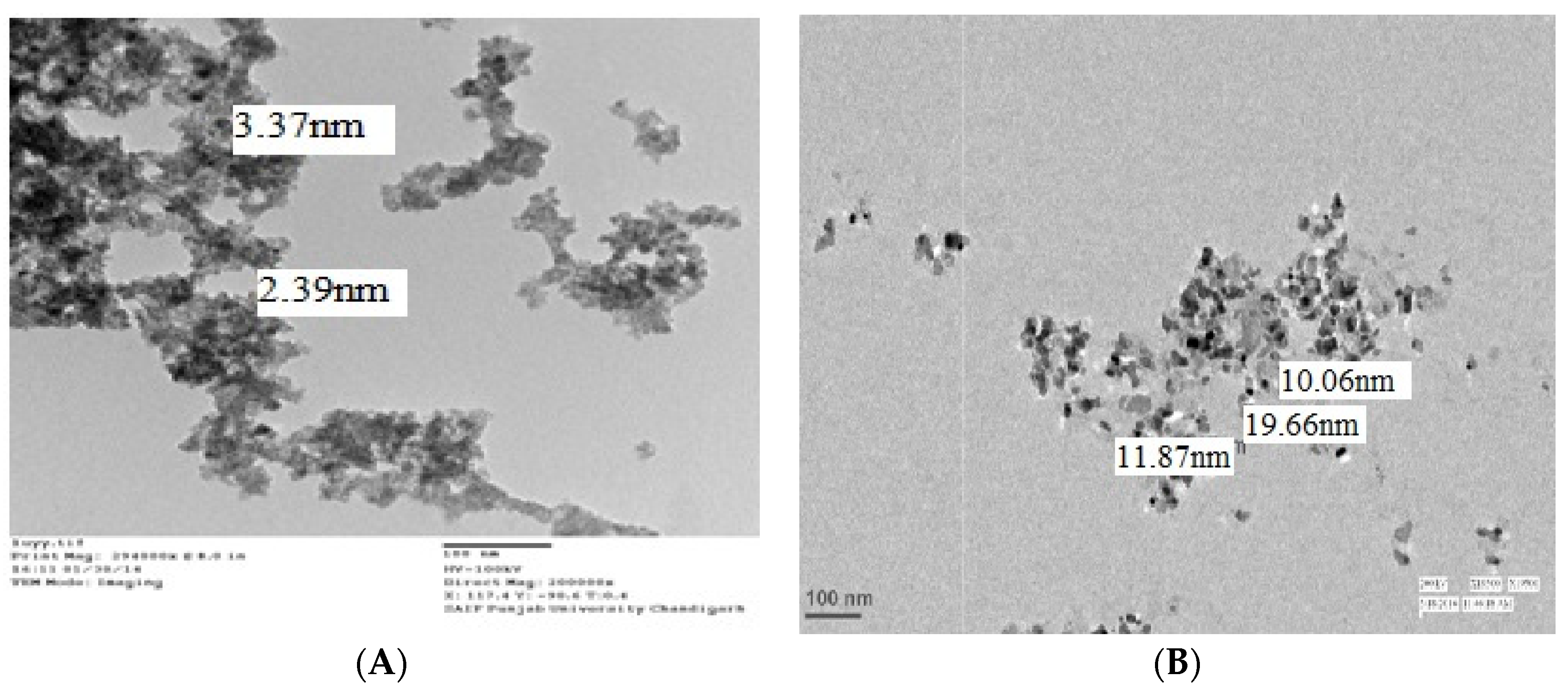
Figure 1. Characterisation of nanoparticles: TEM, XRD and FTIR analysis of nFe3O4 (A,C,E, respectively) and nZnO (B,D,F, respectively).
Sustainability 14 08251 g001aSustainability 14 08251 g001b
Scheme 1. A summarised depiction of the nFe3O4 (1–4 nm)- and nZnO (10–20 nm)-induced changes in V. radiata.
Sustainability 14 08251 sch001
Figure 2. Bar graph showing the contents of chlorophyll (A), protein (B), MDA (C) and carbohydrates (D) in nFe3O4 ( Sustainability 14 08251 i001)- and nZnO ( Sustainability 14 08251 i002)-treated and untreated14-day-old V. radiata plants. The values are mean of five different replicates, error bars represent standard error and different letters indicate significant differences at p < 0.05.
Sustainability 14 08251 g002
Figure 3. Activities of antioxidant enzymes: SOD (A), CAT (B), GPx (C), GR (D) and GST (E) as measured in 14-day-old V. radiata seedlings grown in nFe3O4 ( Sustainability 14 08251 i003)- and nZnO ( Sustainability 14 08251 i004)-spiked Hoagland solution. Column with label “0” on the x-axis represents control. The values are mean of five different replicates, error bars represent standard error and different letters indicate significant differences at p < 0.05.
Sustainability 14 08251 g003
Table 1. Elemental content in V. radiata shoots after growing the plants for 14 days in nanoparticle-spiked Hoagland solution.
Concentration of Nanoparticles (mg/L)Elemental Content in Shoot (mg/kg) Mean ± S.E.
ZnFe
Control0.26 ± 0.01 c3.81 ± 0.01 b
103.0 ± 0.01 b3.82 ± 0.01 b
10090.1 ± 0.45 a86.4 ± 0.16 a
Data are presented as mean ± S.E. (n = 5). Values followed by different letters indicate significant difference at p < 0.05.
Table 2. The nFe3O4- and nZnO-induced changes in 14-day-old V. radiata seedlings as measured by comparing shoot length, root length, and fresh and dry weight of treated plants with the control plants.
S. No.Growth ParametersConcentration of Nanoparticles (mg/L)
ControlFe3O4ZnO
01010010100
1.Shoot length (cm)22.46 ± 1.98 b25.37 ± 0.92 b28.88 ± 0.90 a23.18 ± 1.92 b22.24 ± 2.18 b
2.Root length (cm)17.46 ± 1.99 a19.17 ± 0.95 a22.25 ± 1.06 a18.94 ± 2.8 a16.84 ± 2.05 a
3.Plant biomass (g)Fresh weight0.61 ± 0.25 c0.83 ± 0.15 b0.97 ± 0.28 a0.70 ± 0.09 c0.57 ± 0.15 d
Dry weight0.049 ± 0.02 a0.080 ± 0.01 a0.087 ± 0.03 a0.058 ± 0.01 a0.045 ± 0.03 a
Data are presented as mean ± S.E. (n = 5). Values followed by different letters indicate significant difference at p < 0.05.
Table 3. Effect of nFe3O4 and nZnO on photosynthetic rate, stomatal conductance, transpiration rate and chlorophyll fluorescence in 14-days-old V. radiata plants as compared to the control plants.
S. No.Parameters TestedConcentration of Nanoparticles (mg/L)
ControlFe3O4ZnO
01010010100
1.Photosynthetic rate (µmol CO2 m−2s−1)4.77 ± 0.17 a5.283 ± 0.41 a5.203 ± 0.44 a4.373 ± 0.81 a4.283 ± 0.41 a
2.Stomatal conductance
(µmol CO2 m−2s−1)
0.056 ± 0.006 b0.173 ± 0.040 a0.113 ± 0.021 ab0.116 ± 0.015 ab0.116 ± 0.015 ab
3.Transpiration rate (mmol H2O m−2s−1)1.01 ± 0.197 b2.53 ± 0.153 a2.1 ± 0.137 a2.67 ± 0.397 a2.15 ± 0.555 a
4.Chlorophyll fluorescence (Fv/Fm)0.581 ± 0.026 b0.678 ± 0.020 a0.668 ± 0.012 a0.558 ± 0.011 b0.521 ± 0.013 b
Data are presented as mean ± S.E. (n = 5). Values followed by different letters indicate significant difference at p < 0.05.
Publisher’s Note: MDPI stays neutral with regard to jurisdictional claims in published maps and institutional affiliations.