Article

Photosynthetic Characteristics of Macroalgae Ulva fasciata and Sargassum thunbergii in the Daya Bay of the South China Sea, with Special Reference to the Effects of Light Quality

1
Key Laboratory of Tropical Marine Bio-Resources and Ecology, South China Sea Institute of Oceanology, Chinese Academy of Sciences, Guangzhou 510530, China
2
Southern Marine Science and Engineering Guangdong Laboratory (Guangzhou), Guangzhou 511458, China
3
Key Laboratory of Science and Technology on Operational Oceanography, South China Sea Institute of Oceanology, Chinese Academy of Sciences, Guangzhou 510301, China
4
University of Chinese Academy of Sciences, Beijing 100049, China
5
National and Local Joint Engineering Research Center of Ecological Treatment Technology for Urban Water Pollution, Wenzhou University, Wenzhou 325035, China
*
Author to whom correspondence should be addressed.
These authors contributed equally to this work.
Sustainability 2022, 14(13), 8063; https://doi.org/10.3390/su14138063
Submission received: 2 June 2022 / Revised: 28 June 2022 / Accepted: 29 June 2022 / Published: 1 July 2022
(This article belongs to the Section Sustainable Oceans)

Abstract

:
The changes in underwater light in field usually occur not only in intensity but in spectrum, affecting the photophysiology of marine photoautotrophs. In this study, we comparably examined the photosynthesis of two dominating macroalgae in the Daya Bay, Chlorophyta Ulva fasciata and Phaeophyta Sargassum thunbergii, under white light, as well as under red, green and blue light. The results showed that the net photosynthetic O2 evolution rate (Pn) of U. fasciata under field light increased from 25.2 ± 3.06 to 168 ± 1.2 µmol O2 g FW−1 h−1 from dawn to noon, then decreased to 42.4 ± 0.20 µmol O2 g FW−1 h−1 at dusk. The Pn of S. thunbergii exhibited a similar diel change pattern, but was over 50% lower than that of U. fasciata. The maximal photosynthetic rate (Pmax) of U. fasciata derived from the photosynthesis vs. irradiance curve under white light (i.e., 148 ± 15.8 µmol O2 g FW−1 h−1) was ~30% higher than that under blue light, while the Pmax of S. thunbergii under white light (i.e., 39.2 ± 3.44 µmol O2 g FW−1 h−1) was over 50% lower than that under red, green and blue light. Furthermore, the daily primary production (PP) of U. fasciata was ~20% higher under white than blue light, while that of S. thunbergii was 34% lower, indicating the varied light spectral compositions influence algal photosynthetic ability and thus their primary production in field, and such an influence is species-specific.

1. Introduction

Marine macroalgae, including Chlorophyta, Phaeophyta and Rhodophyta, commonly inhabit the littoral zone to a depth with sufficient light to drive photosynthesis in the worldwide coastal regions [1,2]. They support about two-thirds of the autotrophic biomass in the world oceans [3] and play a vital role in marine ecosystems by providing high trophic levels via refuge and herbivory or detrital food chains [1,4], contributing to the surplus nutrients’ removal from surroundings [5,6] and large amounts of organic carbon burial [3,7]. Many macroalgae also provide people with foods [5,8], biofuels [9], medicines [10] and industrial products [11].
In nature, macroalgae productivity, growth and distribution are governed by a complex of environmental variables, among which light is particularly important, as it energizes photosynthesis to produce organic matter [2]. Light intensity, which generally varies with incident solar radiation, water depth, etc. [12,13,14], serves to energize the photosynthesis of macroalgae, thus regulating the growth and primary productivity [15,16,17]. Apart from the light intensity, the light spectral composition, which varies mainly due to the inherent optical properties of water body and the presence of dissolved and particle matters therein [12,13,14], also influences algal photophysiology and thus their growth, metabolism and productivity [11,18,19,20,21,22]. Effects of different light wavebands vary, such as in the photosynthetic oxygen evolution in red alga Griffithsia monilis [18] and brown alga Laminaria sp. [19]. Blue light inhibits the growth of green alga Codium tomentosum, as compared to red or white light [20], and even shorter wavebands of light reduce the nutrient uptakes of Gracilaria lemaneiformis, as well as the activity of its associated enzymes [23]. On the other hand, macroalgae can also adaptively cope with varying light environments through regulating their morphological, physiological and molecular traits [16,20,21,22]. For instance, in the deep coastal water, where the light is lower but with richer green wavelength, red algae such as Plocamium cartilagineum have additional phycobilisome for light-harvesting complexes [24,25], while brown algae Coilodesme californica and Laminaria sp. display more rapid rises in light absorption toward the blue-green due to fucoxanthin [19]. The diverse photosynthetic pigments within Ulva and Sargassum genera also indicate that different physiological solutions have evolved to deal with at least one photobiological problem [1,26]. Considering the increasing changes in the light quantity and quality due to anthrophonic activities in the worldwide coastal regions [12], it is of general interest to characterize how macroalgae photosynthetically respond to light intensity and spectral composition.
Daya Bay, which sustains a high standing stock of fish and benthic animals, as well as rich biodiversity [27], is geographically located in the northern South China Sea (Figure 1A). The Daya Bay is a semi-enclosed bay with an irregularly semidiurnal tide, and covers an area of ~600 km2 with depths of 5 to 18 m and an annual mean temperature of ~22 °C [28,29,30]. Since the 1980s, Daya Bay and its adjacent areas have experienced fast industrial development, causing its ecosystem to be seriously deteriorated [31,32]. To understand such an ecological effect, many studies have been conducted, examining the physical and chemical variables and planktonic features in the Daya Bay [31,32,33]; however, few studies have concerned macroalgae [24,34,35], although there are more than 200 macroalgae species living in this bay [34], hindering an in-depth understanding of the causes for ecological deteriorations therein. Moreover, eutrophication together with climate change are altering the intensity and spectrum of underwater light in worldwide coastal waters [12,13], with no exception for the Daya Bay. In the present study, therefore, we aim to clarify (i) the photosynthetic characteristics of two dominating macroalgae species in field, Ulva fasciata (Chlorophyta) and Sargassum thunbergii (Phaeophyta), and (ii) how they photosynthetically respond to different spectral compositions (i.e., red, green, blue and white light), to detect the effects of light quality on their photosynthesis. Probing photophysiological responses to light quality may also be helpful for the precise estimation of macroalgal primary production in field, considering that the light quality often varies greatly with water depth.

2. Materials and Methods

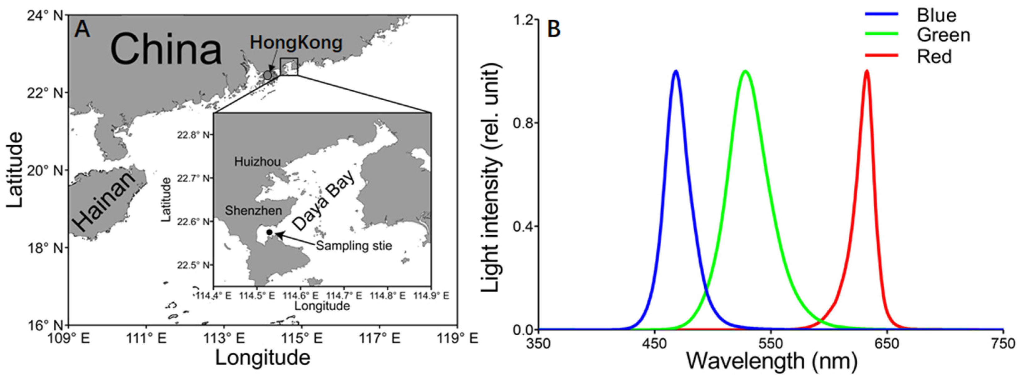
2.1. Experimental Protocol

On 27 and 28 July 2021, an in situ experiment was conducted on a fish raft with Chlorophyta Ulva fasciata and Phaeophyta Sargassum thunbergii in the Daya Bay (114°31′ E, 22°44′ N), northern South China Sea (Figure 1A). In field, these two macroalgae species co-inhabit at ~0.5 m depth around a 500 m offshore fish raft, and are also the dominating species on a rocky seabed of the Daya Bay.
During the experimental period, the photosynthetic O2 evolution and consumption, and the chlorophyll fluorescence of U. fasciata and S. thunbergii were tracked with 2 h intervals, as well as the field physical–chemical factors. Meanwhile, the net photosynthetic O2 evolution rate vs. irradiance (P vs. E) curves were measured under red, green, blue and white (Red + Green + White) light to probe the light quality-induced effects. Additionally, extra algal thalli were collected and transported to the laboratory in a chilly, dark carrying bucket to measure light absorption of pigment extraction, as described below.

2.2. Diel Photosynthetic O2 Evolution and Dark Respiration

Every 2 h, the thalli of U. fasciata and S. thunbergii were collected from the field condition; we gently removed the surface moisture with tissue paper and weighted 0.1–0.2 g for photosynthetic O2 evolution measurement. Then, the weighted thallus was cut into 2–3 cm and transferred into the 15 mL photosynthetic chamber of oxygen electrode equipment (YZQ-201A, Yizongqi Technology Co. Ltd., Beijing, China). After 30 min of dark acclimation, the increase rate of dissolved oxygen concentration in the chamber (μmol O2 L−1 h−1) was monitored under field temperature and light conditions. This YZQ-201A instrument is equipped with a light source and can provide light intensities of 0 to 2400 µmol photons m−2 s−1, and it can also maintain the temperature within the chamber with a cooler. It has been used successfully in the past to measure photosynthesis in a number of macroalgae [36]. The algal photosynthetic O2 evolution rate (Pn) was calculated by normalizing the O2 increase rate (μmol O2 L−1 h−1) to algal fresh weight density (g FW L−1), and expressed as μmol O2 g FW−1 h−1. At the same time, the algal O2 consumption rate in the dark (Rd) was measured, calculated and expressed as μmol O2 g FW−1 h−1. Three independent photosynthetic or respiration rates were measured for each algal species at each time point.

2.3. Diel Chlorophyll Fluorescence

Every 2 h, 2–3 cm of the thallus of U. fasciata and S. thunbergii was cut off from the uniform mother thallus that grew at same water depth, and was dark-acclimated for 15 min. We then measured the chlorophyll fluorescence using a portable fluorometer (AquaPen-C AP-C 100, Photon Systems Instruments, Prague, Czech Republic). The maximum photochemical quantum yield (FV/FM) of Photosystem II (PSII) was calculated using the maximal fluorescence (FM) measured under a saturation light pulse (3000 μmol photons m−2 s−1, 0.6 s) and minimum fluorescence (FO) measured under a weak modulated measuring light [37] as:
F V F M = F M F O F M
After this, the actinic light was activated to the field light level for 5 min, and then the maximal fluorescence (FM’) and minimum fluorescence (Ft) were measured under the saturation light and actinic light, respectively, to obtain the effective PSII photochemical quantum yield (ΦPSII) [37] as:
Φ PSII = F M F t F M
Then, the relative electron transport rate (rETR) was roughly estimated [38] as:
rETR =   Φ PSII × 0.5 × PAR
where 0.5 indicates the absorbed light energy being equally allocated to PS II and PS I.
We performed a single measurement for chlorophyll fluorescence for each algal species at each time point to practically coordinate the limited time for measuring all the physical–chemical and photosynthetic O2 evolution parameters.

2.4. P vs. E Curves under Different Light Qualities

To probe the effects of light quality on photosynthesis, the P vs. E curves of U. fasciata and S. thunbergii were measured at ~10:00 a.m. of each experimental day, under red, green, blue and white light, and at 9 levels of each light quality (i.e., 0, 50, 100, 200, 400, 800, 1000, 1200 and 1500 μmol photons m−2 s−1). The net photosynthetic O2 evolution rate (Pn) at each light level was measured with the YZQ-201A photosynthetic instrument that is equipped with three qualities of light sources (Figure 1B). Three independent P vs. E curves were measured for each algal species. The Rd obtained before and after the P vs. E curve measurements was compared and we found no significant difference, indicating that the effects of cutting damage and light exposure are limited. Additionally, the gross photosynthetic O2 evolution rate (Pg) was calculated by summing Pn and Rd.
Photosynthetic parameters, the P vs. E curve-derived light utilization efficiency (α, slope), saturation irradiance (EK, μmol photons m−2 s−1) and maximum photosynthetic O2 evolution rate (Pmax, μmol O2 g FW−1 h−1) were calculated [39] as:
P g =   P max ×   tan h ( α × I P max ) + Rd
E K = ( P max + R d ) / α
To mimic the effect of light quality on daily primary production, the photosynthetic evoluted O2 was converted to fixed C with PQ of 1.5 [40], and the daily primary production (PP, mg C m−3 d−1) was obtained through integrating the fixed C at different light levels of each light quality throughout a day [17,36] as:
PP = dawn dusk [ Pmax   × tanh ( α   × E ( t ) Pmax ) + Rd ]

2.5. Environmental Factors and Pigment Light Absorption

Every 2 h, the photosynthetically active radiation (PAR) received by macroalgae was monitored with a PAR sensor (US-SQS/L, ULM-500, Walz, Germany) at sampling depth. Meanwhile, the temperature and salinity were measured with the multi-parameter water quality monitor Sonde (YSI 6600, Yellow Springs Instruments, Yellow Springs, OH, USA).
To measure light absorption spectra of pigments, approximately 0.10 g of the collected fresh thalli was weighted, extracted with 10 mL absolute methanol and ground with quartz sands (HF-24, Hefan Instrument Co., Ltd., Shanghai, China). After extracting overnight at 4 °C in the dark, the extraction was centrifuged at 5000× g for 10 min (4 °C); then, the optical absorption spectrum of supernatant was scanned from 350 to 750 nm with a spectrophotometer (UV-1800, Shimadzu, Kyoto, Japan).

2.6. Data Analysis

Mean and standard deviations (mean ± sd) are presented in figures. One-way ANOVA with Bonferroni’s post hoc test (Prism 5, Graphpad Software Inc., San Diego, CA, USA) was used to detect the significant differences among different light qualities or between macroalgae species, and the paired t-test (Prism 5, Graphpad Software Inc., San Diego, CA, USA) was used to detect significant differences between photosynthetic rates of two species within diel changes in field or across series light intensities of four light qualities, with a confidence level of 0.05.

3. Results

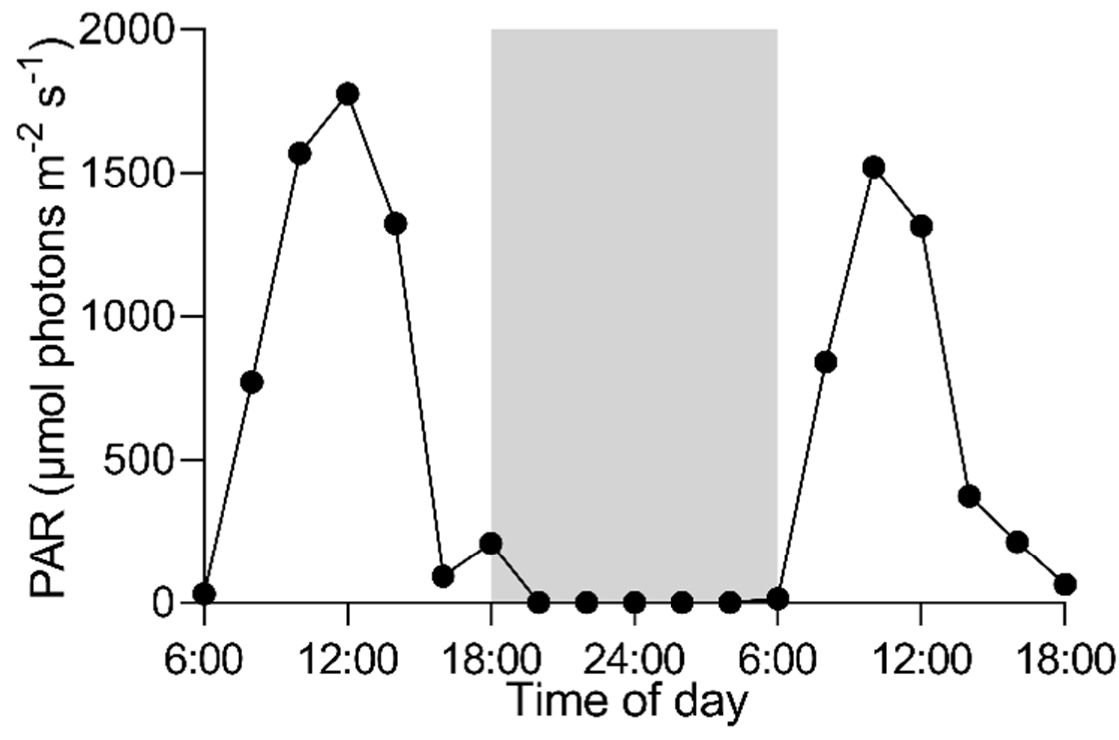
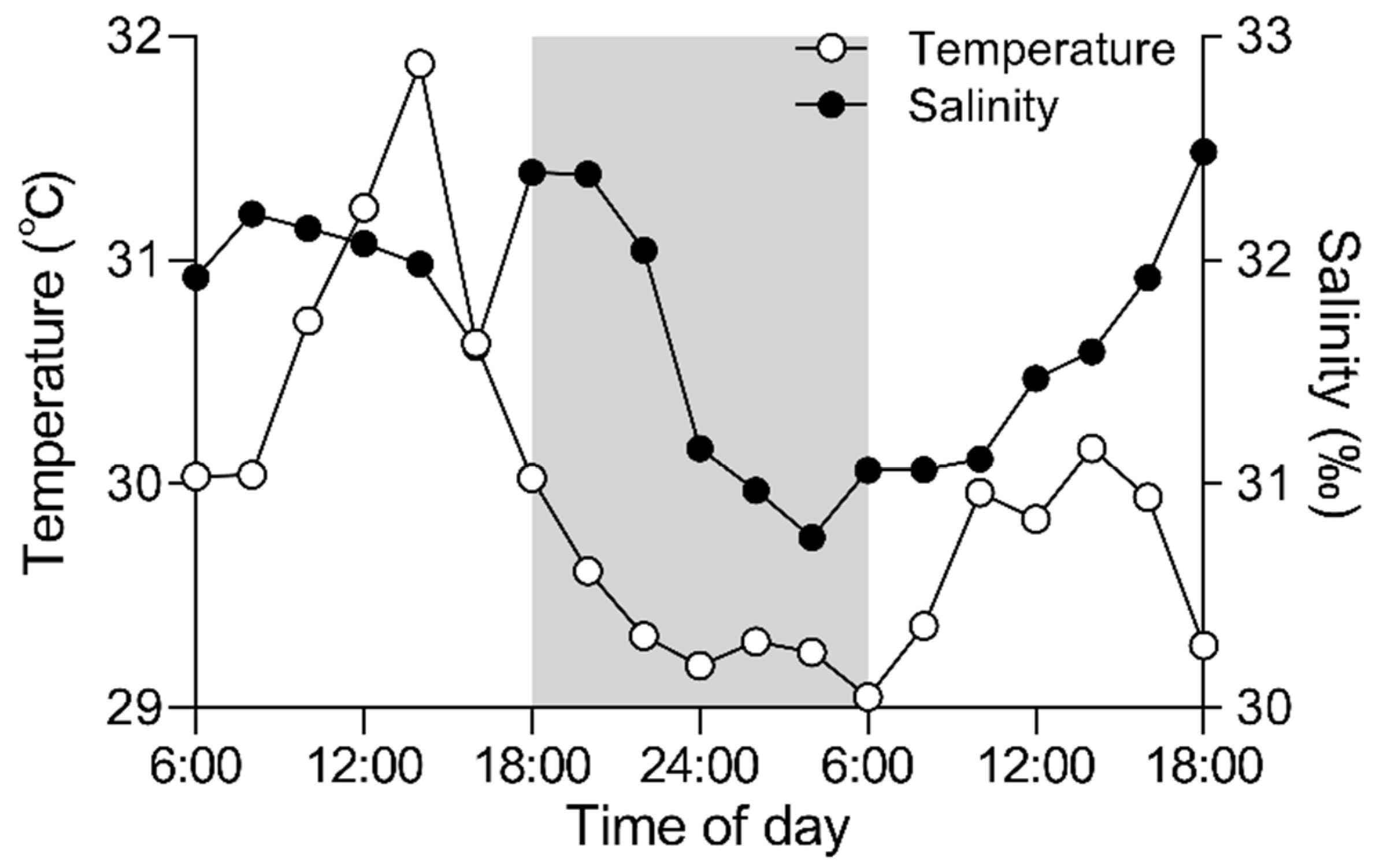
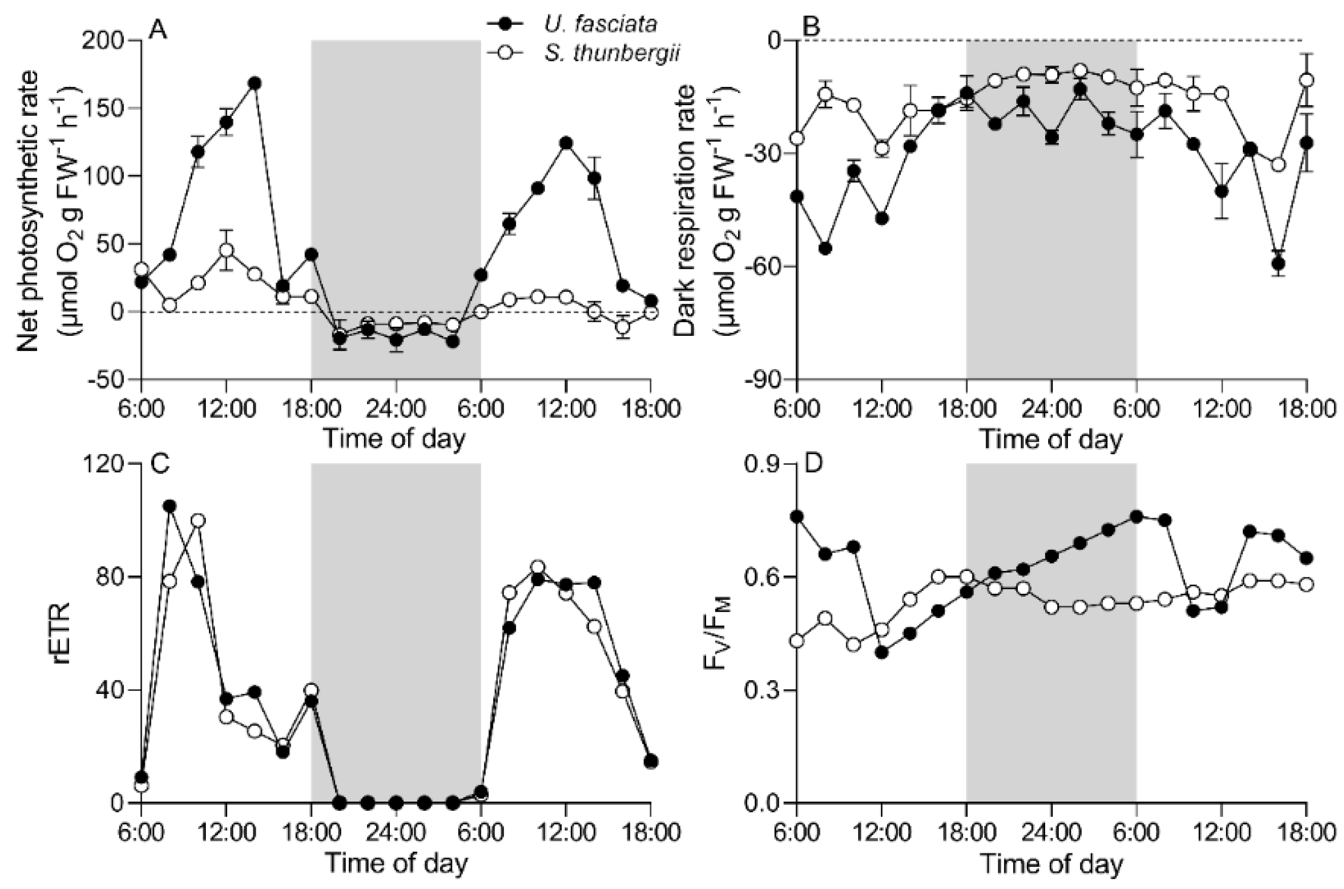
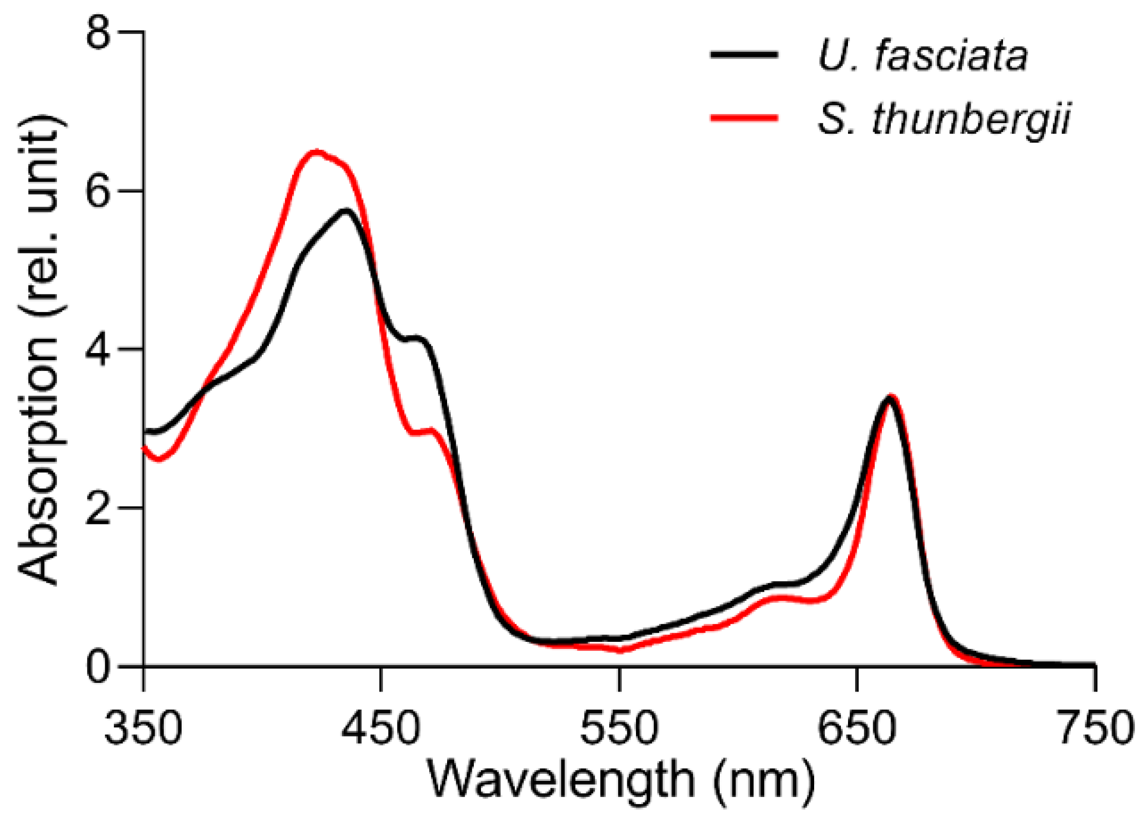
3.1. Daily Field Environmental Changes

On experimental days (27–28 July), the highest solar PAR at sampling depth reached the maximal value of ~1800 μmol photons m−2 s−1 at noon (Figure 2). The seawater temperature varied from 29.05 °C to 31.88 °C, with the lowest and highest values at 6:00 a.m. and 14:00 p.m., respectively; the salinity varied from 30.76‰ to 32.38‰ (Figure 3).

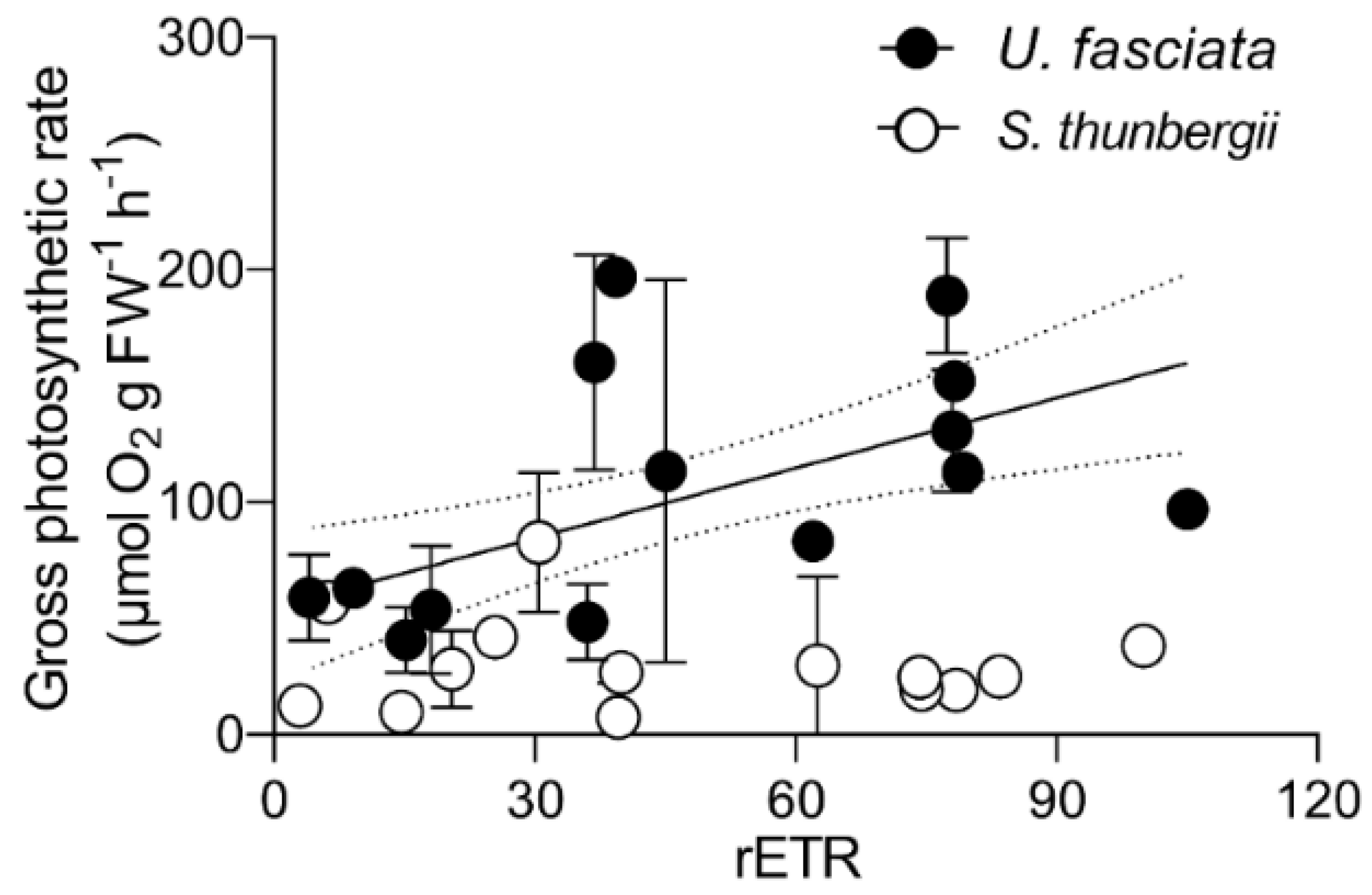
3.2. Daily Photosynthesis and Respiration Changes

In field, the net photosynthetic O2 evolution rate (Pn) of U. fasciata increased from 25.2 ± 3.06 to 168 ± 1.2 μmol O2 g FW−1 h−1 from dawn to noon, then decreased to 42.4 ± 0.20 μmol O2 g FW−1 h−1 at dusk (Figure 4A). The Pn of S. thunbergii ranged from 4.96 ± 4.77 to 45.3 ± 14.7 μmol O2 g FW−1 h−1 in the daytime, much lower than that of U. fasciata (e.g., ~ 68% lower at 12:00 a.m.). The dark respiration rate (Rd) of U. fasciata was significantly higher than that of S. thunbergii (28.6 ± 13.0 vs. 15.8 ± 7.93 μmol O2 g FW−1 h−1) (paired t-test, t = 5.71, p < 0.001), and the Rd in both macroalgae species showed more scatter during the day than at night (Figure 4B). Coinciding with the diel change in Pn, the relative electron transfer rate (rETR) of PSII of U. fasciata increased from dawn to noon, then decreased to dusk, and a similar diel pattern of the rETR also occurred in S. thunbergii (Figure 4C). Moreover, maximum PSII photochemical quantum yield (FV/FM) of U. fasciata, an indicator of photosynthetic potential, decreased from 0.76 to 0.40 from dawn to noon, then gradually increased to 0.73 the next morning; the FV/FM of S. thunbergii increased from 0.43 to 0.60 from dawn to dusk, but decreased to 0.52 the next morning (Figure 4D).

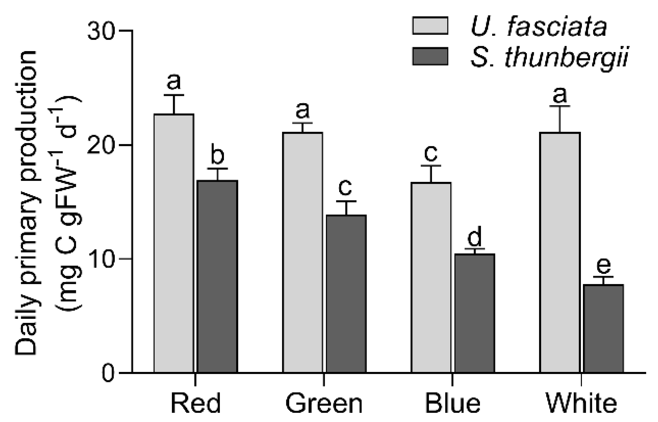
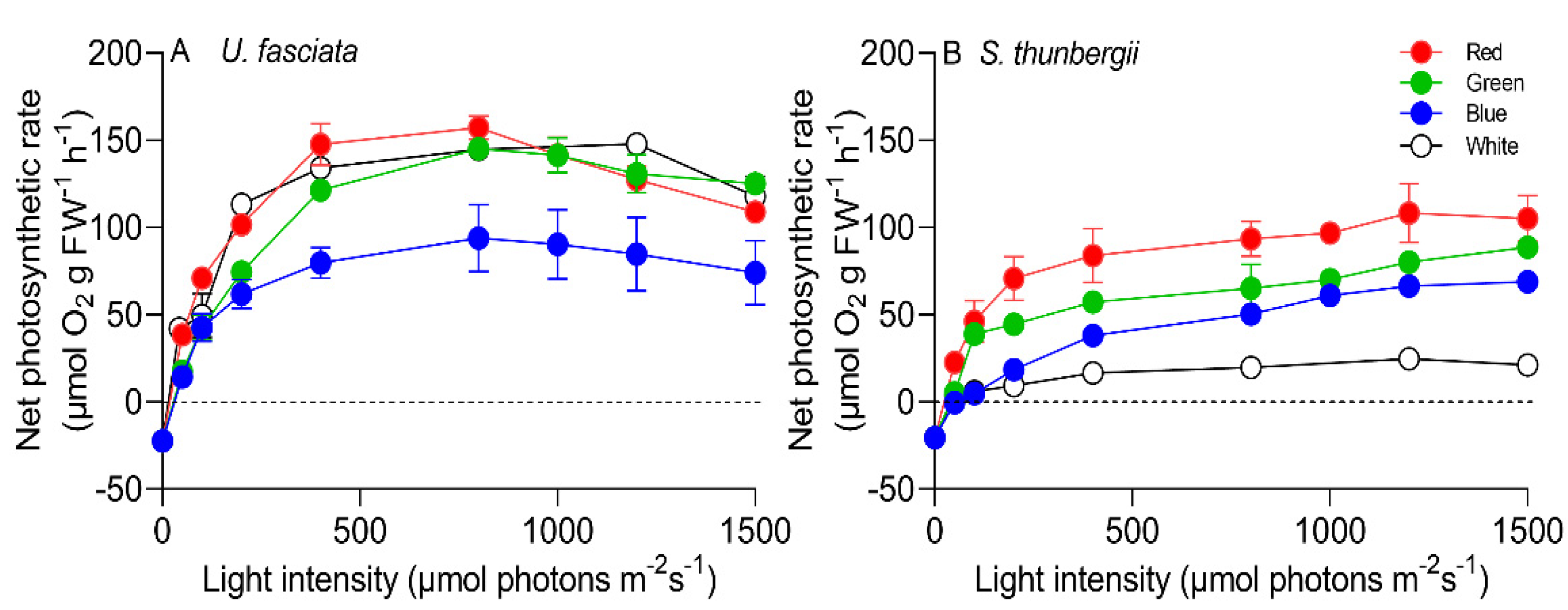
3.3. Photosynthetic Characteristics under Different Light Qualities

The P vs. E characteristics of U. fasciata and S. thunbergii under red, blue, green and white light are shown in Figure 5. In general, the Pn of U. fasciata displayed little or no photoinhibition under less than 800 µmol photons m−2 s−1 white light, but was reduced under higher light (Figure 5A). Red and green light insignificantly affected the Pn of U. fasciata across series light intensities, as compared to white light, but blue light significantly reduced the Pn (paired t-test, t = −5.58, p < 0.01). In S. thunbergii, the Pn showed little or no photoinhibition, even under 1500 µmol photons m−2 s−1 white light; however, it was significantly enhanced by red, green and blue light (paired t-test, red, t = 2.67, p < 0.05; green, t = 5.02, p < 0.01; blue, t = 6.73, p < 0.001) (Figure 5B). Moreover, the P vs. E curve-derived maximum photosynthetic O2 evolution rate (Pmax) of U. fasciata under white light was 148 ± 15.8 μmol O2 g FW−1 h−1, ~30% higher than that under blue light, while the saturation irradiance (EK) was lower (Table 1). Light utilization efficiency (α) showed an insignificant difference compared to red, green or blue light (Table 1). In S. thunbergii, however, the Pmax under white light was 39.2 ± 3.44 μmol O2 g FW−1 h−1, lower than that under red, green and blue light. The EK was lower under white light than blue light, while the α was higher (Table 1).
The daily primary production (PP) estimated from the P vs. E curves of U. fasciata and S. thunbergii also differed under red, green, blue and white light (Figure 6). The PP of U. fasciata was 21.2 ± 2.25 mg C g FW−1 d−1 under white light, and was about 20% higher under blue light. In S. thunbergii, however, the PP under white light (i.e., 7.78 ± 0.68 mg C g FW−1 d−1) was approximately 120%, 80% and 30% lower than that under red, green and blue light, respectively.

4. Discussion

Most previous studies on the effect of light on the physiology of macroalgae involved manipulation of the integrated photosynthetically active radiation (e.g., [16,17,24]), but few studies referred to the physiological responses to spectral composition, although pioneering studies over 70 years ago showed that different pigmentations of red, green and brown algae affected the absorption and action spectra across the PAR spectrum [19]. In this study, we showed that U. fasciata had higher photosynthetic capacity that displayed larger diel changes, as compared to S. thunbergii. Blue light reduced the photosynthetic capacity of U. fasciata, but simulated that of S. thunbergii compared to white light (Figure 5, Table 1), indicating that the spectral composition of available light would alter the photophysiology of macroalgae and thus the primary production in field condition, and such an effect is species-specific.
Light regime often shapes the photosynthetic behaviors of macroalgae on the circadian scale [20,24], as well as the seasonal scale [1]. Consistently, both U. fasciata and S. thunbergii presented a clearly diel change pattern in photosynthetic O2 productivity (Figure 4A) and PSII activity (Figure 4D), although the varying extent differed between these two macroalgae species. Such diel variation has been well documented [e.g., 20,24], and explained as the light energy can not only drive photosynthesis but also harm the photosynthetic apparatus if over its optical level, thus causing photoinhibition. Moreover, the photosynthetic ability of U. fasciata varied more with varying light intensities within a day, as compared to S. thunbergii (Figure 4), indicating that U. fasciata is more light-sensitive. This might be caused by the lower “package effect” of the thin, sheet-like thallus of U. fasciata enabling this species to have a higher ratio of surface area to volume [1,17,41,42]. The Ulva species has also been reported to excrete polysaccharides outside the cells and form a film on the thalli surface, especially under stressful light [43], which may have protected them from the high light at noon (Figure 2) and helped to maintain a high photosynthetic rate (Figure 4A). The higher photosynthetic ability of U. fasciata than S. thunbergii might also be attributed to its higher cellular protein contents [21,24], because proteins are the main components of all kinds of key enzymes involved in the photosynthesis of macroalgae [28,44]. On the other hand, the thin sheet-like macroalgae species are known to capture more light per unit biomass than the thick-branched ones through developing a larger photosynthetic surface per unit of fixed C [1,17]; therefore, the sheet-like U. fasciata have higher efficiency in collecting light for photosynthesis than the branched S. thunbergii, and thus higher photosynthetic capacity (Figure 4A). More interestingly, a positive correlation between gross photosynthetic O2 productivity (Pn plus Rd) and PSII relative electron transfer rate (rETR) appeared in U. fasciata, but not in S. thunbergii (Figure 7), which may caution against using rETR as an indicator for photosynthetic capacity [24,28], but the mechanisms yielding this species-specific difference need to be explored further.
Apart from light intensity, the underwater light spectral composition also varies with depth, due to the water optical properties and dissolved or particle matters therein [12,13,14]. Such variation differently altered the photophysiological traits of U. fasciata and S. thunbergii (Figure 5, Table 1), for which the evolved species-specific diversities of photosynthetic pigments and thus the varied light absorption between them (Figure 8) may be attributable [19,24,26]. For instance, the Ulva genus is known to contain Chl a/b as the main light-harvesting pigments for photosynthesis, while the Sargassum genus contains additional Chl c and fucoxanthin as auxiliary channels to obtain light sources [41,45]. This may have impacted the species-specific efficiency in collecting light of given spectra (Figure 8) [19], which may have led to the varied photosynthetic abilities among different light qualities (Figure 5). Such a phenomenon also occurred in red alga G. monilis [18], green alga C. tomentosum [20] and brown algae C. californica and Laminaria sp. [19]. Moreover, the pigment composition was also suggested to determine the depth where the macroalgae can survive in field [3,46], although the subsequent tests failed to support it under low light status, because the total pigment amount was consistently observed to be much more important in regulating algal growth than their qualitative compositions [41]. In view of ecology, however, the divergent pigment composition allows an efficient utilization of field light energy owing to the partitioning of the light spectrum, thus favoring the species co-existence within a certain community [47]. Our results supported this, as well, considering that the U. fasciata and S. thunbergii, whose spectral absorptions of methanol extracts (Figure 8) reflect their individual preferences of different light spectra and thus different primary productivity (Figure 6), inhabit the Daya Bay together.

5. Conclusions

In this study, we found that both field photosynthetic O2 evolution rate and dark O2 consumption rate of U. fasciata were higher than those of S. thunbergii, and the photosynthetic rate of U. fasciata under white light was higher than that under blue light, and the reverse occurred in S. thunbergii. Moreover, the light spectral quality-induced variation in the daily primary production was as high as 20% and 120% in U. fasciata and S. thunbergii, respectively. Our results, together with others [21,22,28,41], demonstrate that a note of caution on light quality should be put to the studies on macroalgal photophysiology and primary productivity, considering that the light quantity and quality are changing with depth and raising eutrophication in worldwide coastal regions.

Author Contributions

Conceptualization and methodology, M.W., Z.W. and G.L.; investigation, M.W., Z.W., G.M. and G.L.; writing—original draft preparation, M.W., Z.W. and G.L.; writing—review and editing, X.X., Z.M., Y.T. and G.L. All authors have read and agreed to the published version of the manuscript.

Funding

This research was funded by the National Natural Science Foundation (41890853), the National Key Research and Development Program of China (2019YFA0606704), the Special Foundation for National Science and Technology Basic Research Program of China (2018FY100104) and the Natural Science Foundation of Guangdong Province (2022A1515011461).

Institutional Review Board Statement

Not applicable.

Informed Consent Statement

Not applicable.

Data Availability Statement

The data presented in this study are available from the first (M.W and Z.W.) and corresponding (G.L.) authors, upon reasonable request.

Conflicts of Interest

The authors declare no conflict of interest.

References

  1. Hurd, C.L.; Harrison, P.J.; Bischof, K.; Lobban, C.S. Seaweed Ecology and Physiology; Cambridge University Press: Cambridge, UK, 2014. [Google Scholar]
  2. Ji, Y.; Gao, K. Effects of climate change factors on marine macroalgae: A review. Adv. Mar. Biol. 2021, 88, 91–135. [Google Scholar] [PubMed]
  3. Smith, S.V. Marine macrophytes as a global carbon sink. Nature 1981, 211, 838–840. [Google Scholar] [CrossRef] [PubMed] [Green Version]
  4. Ware, C.; Dijkstra, J.A.; Mello, K.; Stevens, A.; O’Brien, B.; Ikedo, W. A novel three-dimensional analysis of functional architecture that describes the properties of macroalgae as a refuge. Mar. Ecol. Progr. Ser. 2019, 608, 93–103. [Google Scholar] [CrossRef] [Green Version]
  5. Yang, Y.; Chai, Z.; Wang, Q.; Chen, W.; He, Z.; Jiang, S. Cultivation of seaweed Gracilaria in Chinese coastal waters and its contribution to environmental improvements. Algal Res. 2015, 9, 236–244. [Google Scholar] [CrossRef]
  6. Zollmann, M.; Rubinsky, B.; Liberzon, A.; Golberg, A. Multi-scale modeling of intensive macroalgae cultivation and marine nitrogen sequestration. Commun. Biol. 2021, 4, 848. [Google Scholar] [CrossRef]
  7. Krause-Jensen, D.; Duarte, C.M. Substantial role of macroalgae in marine carbon sequestration. Nat. Geosci. 2016, 9, 737–742. [Google Scholar] [CrossRef]
  8. Yang, L.; Lu, Q.; Brodie, J. A review of the bladed Bangiales (Rhodophyta) in China: History, culture, and taxonomy. Eur. J. Phycol. 2017, 52, 251–263. [Google Scholar] [CrossRef]
  9. Gao, G.; Burgess, G.; Wu, M.; Wang, S.; Gao, K. Using macroalgae as biofuel: Current opportunities and challenges. Bot. Mar. 2020, 63, 355–371. [Google Scholar] [CrossRef]
  10. Falkenberg, M.; Nakano, E.; Zambotti-Villela, L.; Zatelli, G.A.; Philippus, A.C.; Imamura, K.B.; Velasquez, A.M.A.; Freitas, R.F.; Tallarico, L.F.; Colepicolo, P.; et al. Bioactive compounds against neglected diseases isolated from macroalgae: A review. J. Appl. Phycol. 2019, 31, 797–823. [Google Scholar] [CrossRef] [Green Version]
  11. Messyasz, B.; Michalak, I.; Łęska, B.; Schroeder, G.; Górka, B.; Korzeniowska, K.; Lipok, J.; Wieczorek, P.; Rój, E.; Wilk, R.; et al. Valuable natural products from marine and freshwater macroalgae obtained from supercritical fluid extracts. J. Appl. Phycol. 2018, 30, 591–603. [Google Scholar] [CrossRef]
  12. Tait, L.W.; Hawes, I.; Schiel, D.R. Shining light on benthic macroalgae mechanisms of complementarity in layered macroalgal assemblages. PLoS ONE 2014, 9, e114146. [Google Scholar] [CrossRef] [PubMed] [Green Version]
  13. Gao, K.; Li, G.; Helbling, E.W.; Villafañe, E.V. Variability of UVR effects on photosynthesis of summer phytoplankton assemblages from a tropical coastal area of the South China Sea. Photochem. Photobiol. 2007, 83, 802–809. [Google Scholar] [CrossRef] [PubMed]
  14. Kirk, J.T.O. Light and Photosynthesis in Aquatic Ecosystems; Cambridge University Press: Cambridge, UK, 1994. [Google Scholar]
  15. Xu, J.; Zhang, X.; Fu, Q.; Gao, G.; Gao, K. Water depth-dependent photosynthetic and growth rates of Gracilaria lemaneiformis, with special reference to effects of solar UV radiation. Aquaculture 2018, 484, 28–31. [Google Scholar] [CrossRef]
  16. Wu, H.; Gao, G.; Zhong, Z.; Li, X.; Xu, J. Physiological acclimation of the green tidal alga Ulva prolifera to a fast-changing environment. Mar. Environ. Res. 2018, 137, 1–7. [Google Scholar] [CrossRef] [PubMed]
  17. Li, G.; Mai, G.; Zhang, J.; Ni, G.; Shi, X.; Tan, Y.; Zou, D. Rising pCO2 interacts with algal density to reversely alter photosynthetic responses of Gracilaria lemaneiformis and Ulva conglobata. Algal Res. 2021, 54, 102231. [Google Scholar] [CrossRef]
  18. Larkum, A.W.D.; Weyrauch, S.K. Photosynthetic action spectra and light-harvesting in Griffithsia monilis (Rhodophyta). Photochem. Photobiol. 1977, 25, 65–72. [Google Scholar] [CrossRef]
  19. Haxo, F.T.; Blinks, L.R. Photosynthetic action spectra of marine algae. J. Gen. Physiol. 1950, 33, 389–442. [Google Scholar] [CrossRef] [Green Version]
  20. Marques, R.; Cruz, S.; Calado, R.; Lillebø, A.; Abreu, H.; Pereira, R.; Pitarma, B.; da Silva, J.M.; Cartaxana, P. Effects of photoperiod and light spectra on growth and pigment composition of the green macroalga Codium tomentosum. J. Appl. Phycol. 2021, 33, 471–480. [Google Scholar] [CrossRef]
  21. Lüning, K.; Dring, M.J. Action spectra and spectral quantum yield of photosynthesis in marine macroalgae with thin and thick thalli. Mar. Biol. 1985, 87, 119–129. [Google Scholar] [CrossRef]
  22. Talarico, L.; Maranzana, G. Light and adaptive responses in red macroalgae: An overview. J. Photochem. Photobiol. B Biol. 2000, 56, 1–11. [Google Scholar] [CrossRef]
  23. Xu, Z.; Gao, K. NH4+ enrichment and UV radiation interact to affect the photosynthesis and nitrogen uptake of Gracilaria lemaneiformis (Rhodophyta). Mar. Pollut. Bull. 2012, 64, 99–105. [Google Scholar] [CrossRef] [PubMed]
  24. Shi, X.; Zou, D.; Hu, S.; Mai, G.; Ma, Z.; Li, G. Photosynthetic characteristics of three cohabitated macroalgae in the Daya Bay, and their responses to temperature rise. Plants 2021, 10, 2441. [Google Scholar] [CrossRef] [PubMed]
  25. Scarabel, L.; Camuccio, P.; Foltran, A.; Maranzana, G.; Talarico, L.; Rascio, N.; Andreoli, C. Thalli of Plocamium cartilagineum (L.) Dixon (Gigartinales, Rhodophyta) growing at two different depths. Gior. Bot. It. 1996, 130, 690–693. [Google Scholar]
  26. Dring, M.J. Blue light effects in marine macroalgae. In Blue Light Effects in Biological Systems; Serger, H., Ed.; Springer: Berlin, Germany, 1984; pp. 509–516. [Google Scholar]
  27. Huang, H.; Lin, Q.; Lin, Y.; Jia, X.; Li, C.; Wang, W. Spatial-temporal variation of large macrobenthic animals in cage culture sea area in Daya Bay. Chin. Environ. Sci. 2005, 25, 412–416, (In Chinese with English abstract). [Google Scholar]
  28. Xu, G.; Liu, J.; Song, X.; Tan, M.; Ren, H.; Li, D.; Tan, Y.; Huang, L.; Li, G. Diel rhythm in photosynthetic performance of phytoplankton assemblages is predicted to be light-dependent from in situ and mesocosm chlorophyll fluorescence. J. Coast. Res. 2020, 104, 445–454. [Google Scholar] [CrossRef]
  29. Yu, J.; Tang, D.; Wang, S.; Lian, J.; Wang, Y. Changes of water temperature and harmful algal bloom in the Daya Bay in the northern South China Sea. Mar. Sci. Bull. 2007, 9, 25–33. [Google Scholar]
  30. Xu, G. Environments and Resources of Daya Bay; Anhui Press of Science and Technology: Hefei, China, 1989. (In Chinese) [Google Scholar]
  31. Wang, Y.; Lou, Z.; Sun, C.; Sun, S. Ecological environment changes in Daya Bay, China, from 1982 to 2004. Mar. Pollut. Bull. 2008, 56, 1871–1879. [Google Scholar] [CrossRef]
  32. Li, T.; Liu, S.; Huang, L.; Huang, H.; Lian, J.; Yan, Y.; Lin, S.-J. Diatom to dinoflagellate shift in the summer phytoplankton community in a bay impacted by nuclear power plant thermal effluent. Mar. Ecol. Progr. Ser. 2011, 424, 75–85. [Google Scholar] [CrossRef]
  33. Mai, G.; Song, X.; Xia, X.; Ma, Z.; Tan, Y.; Li, G. Photosynthetic characteristics of smaller and larger cell size-fractioned phytoplankton assemblies in the Daya Bay, northern South China Sea. Microorganisms 2022, 10, 16. [Google Scholar] [CrossRef]
  34. Yang, Y. Coastal Environmental Bioremediation and Seaweed Resource Utilization; Science Press: Beijing, China, 2016; pp. 1–364. (In Chinese) [Google Scholar]
  35. Luo, D.; Chen, X.; Li, T.; Yi, J. Production of TM satellite image distribution map of macroalgae in Daya Bay. Remote Sens. Inf. 1990, 4, 30–32, (In Chinese with English abstract). [Google Scholar]
  36. Li, G.; Wan, M.; Shi, X.; Qin, G.; Mai, G.; Huang, L.; Tan, Y.; Zou, D. Comparative study on photophysiology of four macroalgae from the Zhongsha Atoll, with special reference to the effects of temperature rise. J. Trop. Oceanogr. 2022, 43, 101–110, (In Chinese with English abstract). [Google Scholar]
  37. Genty, B.; Briantais, J.M.; Baker, N.R. The relationship between the quantum yield of photosynthetic electron transport and quenching of chlorophyll fluorescence. Biochim. Biophys. Acta 1989, 990, 87–92. [Google Scholar] [CrossRef]
  38. Platt, T.; Gallegos, C.L.; Harrison, W.G. Photoinhibition of photosynthesis in natural assemblages of marine phytoplankton. J. Mar. Res. 1980, 38, 687–701. [Google Scholar]
  39. Henley, W.J. Measurement and interpretation of photosynthetic light-response curves in algae in the context of photoinhibition and diel changes. J. Phycol. 1993, 29, 729–739. [Google Scholar] [CrossRef]
  40. Williams, P.J.L.; Rainea, R.C.T.; Bryan, J.R. Agreement between the 14C and oxygen methods of measuring phytoplankton production: Reassessment of the photosynthetic quotient. Oceanol. Acta 1979, 2, 411–416. [Google Scholar]
  41. Enríquez, S.; Agustí, S.; Duarte, C.M. Light absorption by marine macrophytes. Oecologia 1994, 98, 121–129. [Google Scholar] [CrossRef]
  42. Dudgeon, S.R.; Johnson, A.S. Thick vs. thin: Thallus morphology and tissue mechanics influence differential drag and dislodgement of two co-dominant seaweeds. J. Exp. Mar. Biol. Ecol. 1992, 165, 23–43. [Google Scholar] [CrossRef]
  43. Bischof, K.; Peralta, G.; Kräbs, G.; van de Poll, W.H.; Pérez-Lloréns, J.L.; Breeman, A.M. Effects of solar UV-B radiation on canopy structure of Ulva communities from Southern Spain. J. Exp. Bot. 2002, 53, 2411–2421. [Google Scholar] [CrossRef] [Green Version]
  44. Vitlin Gruber, A.; Feiz, L. Rubisco assembly in the chloroplast. Front. Mol. Biosci. 2018, 5, 24. [Google Scholar] [CrossRef]
  45. Chao-Rodríguez, Y.; Domínguez-Gómez, J.A.; Sánchez-Carnero, N.; Rodríguez-Pérez, D. A comparison of spectral macroalgae taxa separability methods using an extensive spectral library. Algal Res. 2017, 26, 463–473. [Google Scholar] [CrossRef]
  46. Engelman, T.W. Farbe und Assimilation. Bot. Zeit. 1883, 41, 1–13, 17–29. [Google Scholar]
  47. Stomp, M.; Huisman, J.; de Jongh, F.; Veraart, A.J.; Gerla, D.; Rijkeboer, M.; Ibelings, B.W.; Wollenzien, U.I.A.; Stal, L.J. Adaptive divergence in pigment composition promotes phytoplankton biodiversity. Nature 2004, 432, 104–107. [Google Scholar] [CrossRef] [PubMed]
Figure 1. (A) Map of experimental site in the Daya Bay, northern South China Sea; (B) the spectra of red, green and blue light sources used in this experiment.
Sustainability 14 08063 g001
Figure 2. Daily changes in the macroalgae-exposed solar PAR irradiation (µmol photons m−2 s−1) in field condition. Gray shadow indicates nighttime.
Sustainability 14 08063 g002
Figure 3. Daily changes in field temperature (°C) and salinity (‰) at sampling site. Gray shadow indicates nighttime.
Sustainability 14 08063 g003
Figure 4. Net photosynthetic oxygen evolution rate (A, µmol O2 g FW−1 h−1) and dark respiration (B, µmol O2 g FW−1 h−1), and relative electron transfer rate (C, rETR) and maximum photochemical quantum yield (D, FV/FM) of Photosystem II (PS II) of U. fasciata and S. thunbergii in field throughout the experimental days. Gray shadows indicate nighttime.
Sustainability 14 08063 g004
Figure 5. Net photosynthetic O2 evolution rate (Pn, µmol O2 g FW−1 h−1) vs. irradiance (µmol photons m−2 s−1) curves of (A) U. fasciata and (B) S. thunbergii under red, green, blue and white light.
Sustainability 14 08063 g005
Figure 6. Daily primary production (PP, mg C g FW−1 d−1) of U. fasciata and S. thunbergii under red, green, blue and white light. Different letters above the bars indicate the significant difference (p < 0.05).
Sustainability 14 08063 g006
Figure 7. Gross photosynthetic O2 evolution rate (µmol O2 g FW−1 h−1) as a function of relative electron transfer rate (rETR) of U. fasciata and S. thunbergii.
Sustainability 14 08063 g007
Figure 8. Optical density of methanol extracts from the thalli of U. fasciata and S. thunbergii. The absorption was normalized to OD680.
Sustainability 14 08063 g008
Table 1. The photosynthetic O2 evolution rate vs. irradiance curve-derived light utilization efficiency (α, slope), saturation irradiance (EK, µmol photons m−2 s−1) and maximal photosynthetic rate (Pmax, µmol O2 g FW−1 h−1) of U. fasciata and S. thunbergii under blue, green, red and white light.
SpeciesParametersRedGreenBlueWhite
U. fasciataα0.98 ± 0.18 a0.58 ± 0.06 b0.66 ± 0.15 b0.79 ± 0.20 ab
EK73.1 ± 11.5 a123 ± 8.53 b243 ± 11.3 c91.9 ± 15.9 a
Pmax153 ± 11.2 a153 ± 5.65 a104 ± 8.86 b148 ± 15.8 a
S. thunbergiiα0.64 ± 0.10 a0.45 ± 0.10 a0.15 ± 0.02 b0.20 ± 0.04 b
EK82.0 ± 8.95 a91.9 ± 14.2 a244 ± 19.6 b91.9 ± 13.6 a
Pmax113 ± 6.84 a89.3 ± 7.58 b78.3 ± 3.24 c39.2 ± 3.44 d
* Different letters next to numbers indicate the significant difference among different light qualities (p < 0.05).
Publisher’s Note: MDPI stays neutral with regard to jurisdictional claims in published maps and institutional affiliations.

