
Adsorption Studies of Arsenic(V) by CuO Nanoparticles Synthesized by Phyllanthus emblica Leaf-Extract-Fueled Solution Combustion Synthesis

 ,
,  ,
,  ,
,  and
and
Abstract
1. Introduction
2. Materials and Methods
2.1. Green Synthesis of CuO NPs
2.2. Characterization of CuO NPs
2.3. Adsorption Experiments
3. Results and Discussion
3.1. Characterization of CuO NPs
3.1.1. UV-Vis Spectral Analysis
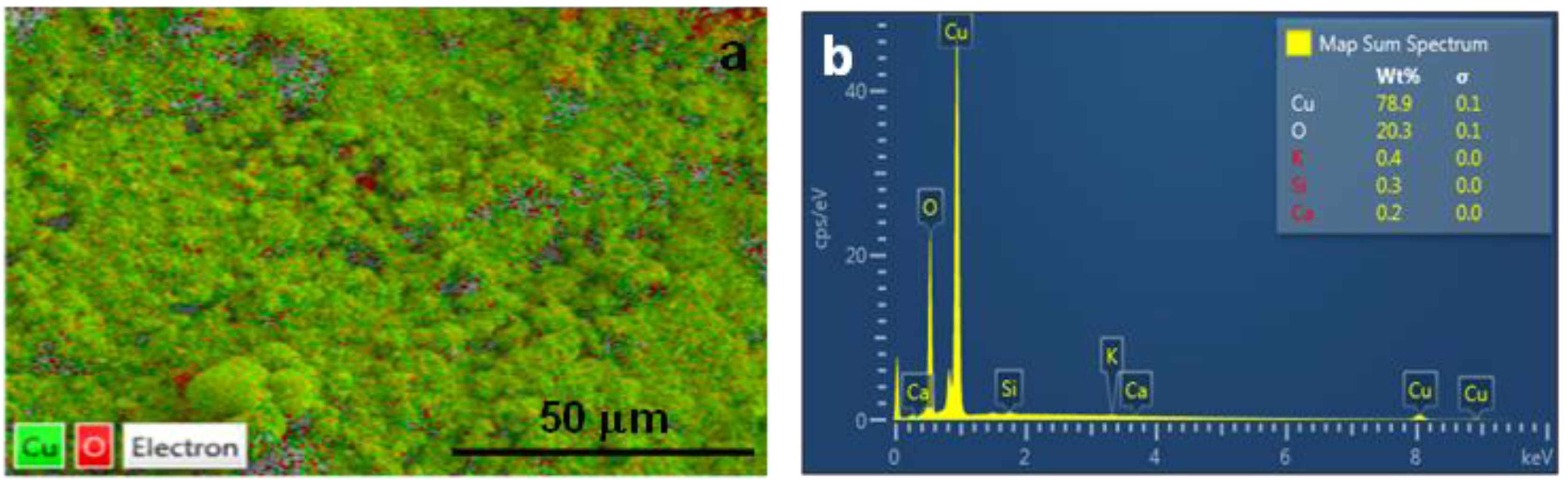
3.1.2. SEM and EDX Analysis
3.1.3. XPS Analysis
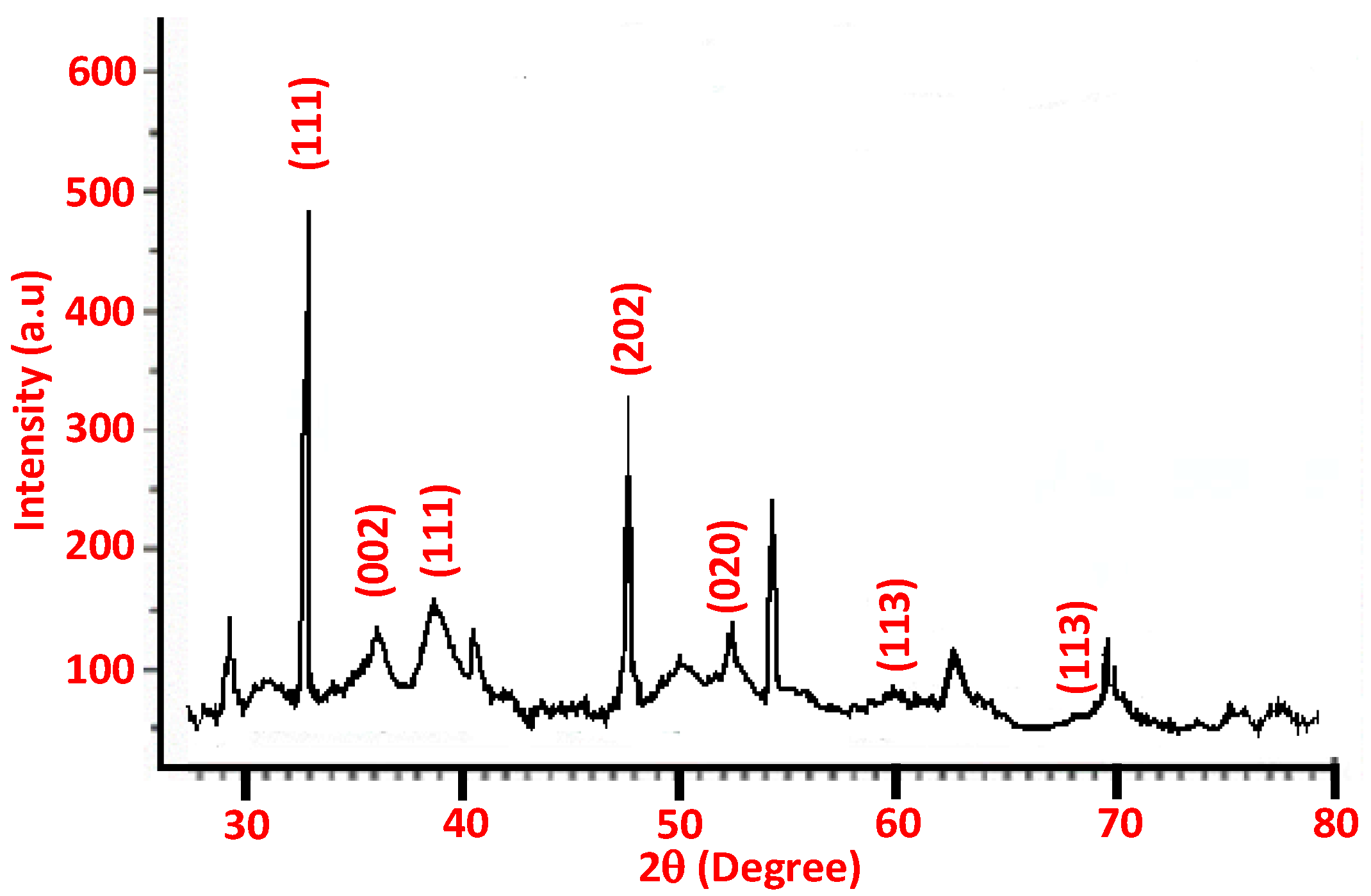
3.1.4. XRD Analysis
3.1.5. Size/Size Distribution and Zeta Potential Analysis
3.2. Arsenic Adsorption Results
3.2.1. Effect of Nanoparticles Dosage and Initial As(V) Concentration
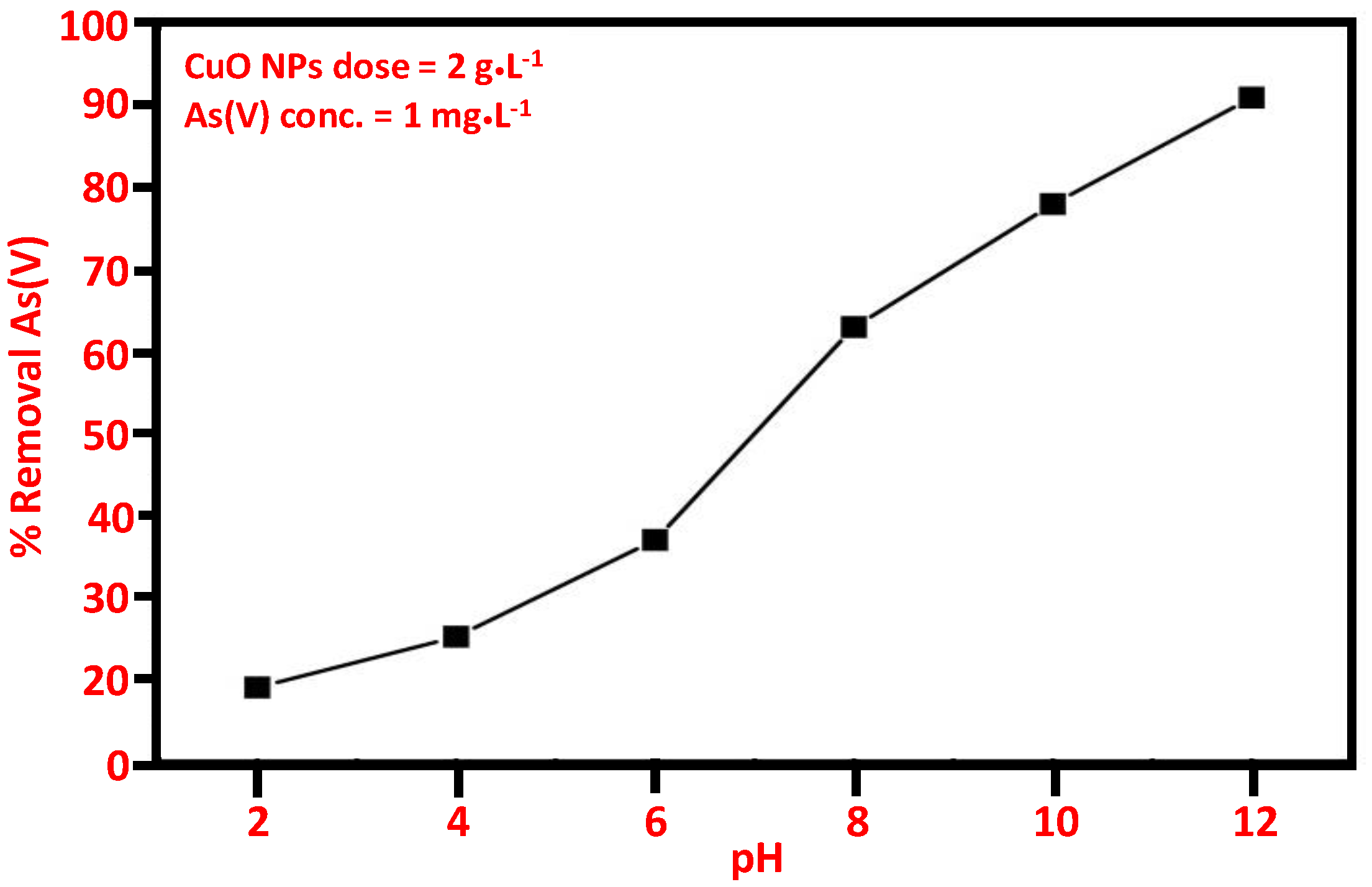
3.2.2. Effect of pH on As(V) Removal
3.2.3. Effect of Contact Time on As(V) Removal
3.2.4. Adsorption Isotherms
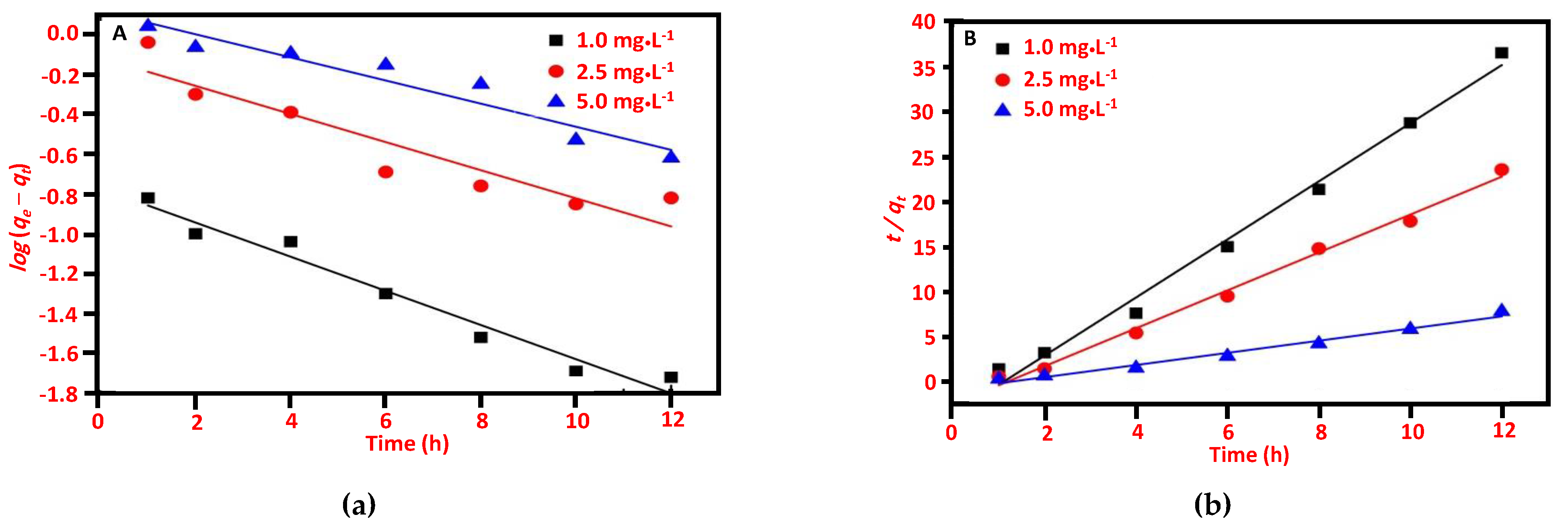
3.2.5. Adsorption Kinetics
4. Conclusions
Author Contributions
Funding
Institutional Review Board Statement
Informed Consent Statement
Data Availability Statement
Acknowledgments
Conflicts of Interest
References
- Anawar, H.M.; Akai, J.; Komaki, K.; Terao, H.; Yoshioka, T.; Ishizuka, T.; Safiullah, S.; Kato, K. Geochemical occurrence of arsenic in groundwater of Bangladesh: Sources and mobilization processes. J. Geochem. Explor. 2003, 77, 109–131. [Google Scholar] [CrossRef]
- Flora, S.J.S. 1-Arsenic: Chemistry, occurrence, and exposure. In Handbook of Arsenic Toxicology; Flora, S.J.S., Ed.; Academic Press: Oxford, UK, 2015; pp. 1–49. [Google Scholar]
- Malana, M.A.; Khosa, M.A. Groundwater pollution with special focus on arsenic, Dera Ghazi Khan-Pakistan. J. Saudi Chem. Soc. 2011, 15, 39–47. [Google Scholar] [CrossRef]
- Ghosh, J.; Sil, P.C. 8-Mechanism for arsenic-induced toxic effects. In Handbook of Arsenic Toxicology; Flora, S.J.S., Ed.; Academic Press: Oxford, UK, 2015; pp. 203–231. [Google Scholar]
- Karim, M.M. Arsenic in groundwater and health problems in Bangladesh. Water Res. 2000, 34, 304–310. [Google Scholar] [CrossRef]
- Mohan, D.; Pittman, C.U., Jr. Arsenic removal from water/wastewater using adsorbents—A critical review. J. Hazard. Mater. 2007, 142, 1–53. [Google Scholar] [CrossRef]
- Han, B.; Runnells, T.; Zimbron, J.; Wickramasinghe, R. Arsenic removal from drinking water by flocculation and microfiltration. Desalination 2002, 145, 293–298. [Google Scholar] [CrossRef]
- Janin, A.; Zaviska, F.; Drogui, P.; Blais, J.-F.; Mercier, G. Selective recovery of metals in leachate from chromated copper arsenate treated wastes using electrochemical technology and chemical precipitation. Hydrometallurgy 2009, 96, 318–326. [Google Scholar] [CrossRef]
- Pous, N.; Casentini, B.; Rossetti, S.; Fazi, S.; Puig, S.; Aulenta, F. Anaerobic arsenite oxidation with an electrode serving as the sole electron acceptor: A novel approach to the bioremediation of arsenic-polluted groundwater. J. Hazard. Mater. 2015, 283, 617–622. [Google Scholar] [CrossRef]
- Mafu, L.D.; Mamba, B.B.; Msagati, T.A. Synthesis and characterization of ion imprinted polymeric adsorbents for the selective recognition and removal of arsenic and selenium in wastewater samples. J. Saudi Chem. Soc. 2016, 20, 594–605. [Google Scholar] [CrossRef]
- Amy, G.L.; Chen, H.-W.; Dinzo, A.; Brandhuber, P. Adsorbent Treatment Technologies for Arsenic Removal; Awwa Research Foundation and American Water Works Association: Mumbai, India, 2005. [Google Scholar]
- Kelly, K.L.; Coronado, E.; Zhao, L.L.; Schatz, G.C. The Optical Properties of Metal Nanoparticles: The Influence of Size, Shape, and Dielectric Environment. J. Phys. Chem. B 2003, 107, 668–677. [Google Scholar] [CrossRef]
- Cuenya, B.R. Synthesis and catalytic properties of metal nanoparticles: Size, shape, support, composition, and oxidation state effects. Thin Solid Films 2010, 518, 3127–3150. [Google Scholar] [CrossRef]
- Hua, M.; Zhang, S.; Pan, B.; Zhang, W.; Lv, L.; Zhang, Q. Heavy metal removal from water/wastewater by nanosized metal oxides: A review. J. Hazard. Mater. 2012, 211, 317–331. [Google Scholar] [CrossRef]
- Andjelkovic, I.; Stankovic, D.; Jovic, M.; Markovic, M.; Krstic, J.; Manojlovic, D.; Roglic, G. Microwave-hydrothermal synthesis of TiO2 and zirconium doped TiO2 adsorbents for removal of As (III) and As (V). J. Saudi Chem. Soc. 2015, 19, 429–435. [Google Scholar] [CrossRef]
- Pillewan, P.; Mukherjee, S.; Roychowdhury, T.; Das, S.; Bansiwal, A.; Rayalu, S. Removal of As(III) and As(V) from water by copper oxide incorporated mesoporous alumina. J. Hazard Mater. 2011, 186, 367–375. [Google Scholar] [CrossRef]
- Zhang, G.; Ren, Z.; Zhang, X.; Chen, J. Nanostructured iron(III)-copper(II) binary oxide: A novel adsorbent for enhanced arsenic removal from aqueous solutions. Water. Res. 2013, 47, 4022–4031. [Google Scholar] [CrossRef]
- Adil, S.F.; Assal, M.E.; Khan, M.; Al-Warthan, A.; Siddiqui, M.R.H.; Liz-Marzán, L.M. Biogenic synthesis of metallic nanoparticles and prospects toward green chemistry. Dalton Trans. 2015, 44, 9709–9717. [Google Scholar] [CrossRef]
- Shaik, M.; Albalawi, G.; Khan, S.; Khan, M.; Adil, S.; Kuniyil, M.; Al-Warthan, A.; Siddiqui, M.; Alkhathlan, H.; Khan, M. “Miswak” based green synthesis of silver nanoparticles: Evaluation and comparison of their microbicidal activities with the chemical synthesis. Molecules 2016, 21, 1478. [Google Scholar] [CrossRef]
- Saif, S.; Tahir, A.; Asim, T.; Chen, Y. Plant mediated green synthesis of CuO nanoparticles: Comparison of toxicity of engineered and plant mediated CuO nanoparticles towards Daphnia magna. Nanomaterials 2016, 6, 205. [Google Scholar] [CrossRef]
- Sutradhar, P.; Saha, M.; Maiti, D. Microwave synthesis of copper oxide nanoparticles using tea leaf and coffee powder extracts and its antibacterial activity. J. Nanostruct. Chem. 2014, 4, 86. [Google Scholar] [CrossRef]
- Kumar, B.; Smita, K.; Cumbal, L.; Debut, A.; Angulo, Y. Biofabrication of copper oxide nanoparticles using Andean blackberry (Rubus glaucus Benth.) fruit and leaf. J. Saudi Chem. Soc. 2017, 21, S475–S480. [Google Scholar] [CrossRef]
- Harne, S.; Sharma, A.; Dhaygude, M.; Joglekar, S.; Kodam, K.; Hudlikar, M. Novel route for rapid biosynthesis of copper nanoparticles using aqueous extract of Calotropis procera L. latex and their cytotoxicity on tumor cells. Colloids Surf. B Biointerfaces 2012, 95, 284–288. [Google Scholar] [CrossRef]
- Kiruba Daniel, S.C.G.; Vinothini, G.; Subramanian, N.; Nehru, K.; Sivakumar, M. Biosynthesis of Cu, ZVI, and Ag nanoparticles using Dodonaea viscosa extract for antibacterial activity against human pathogens. J. Nanoparticle Res. 2012, 15, 1–10. [Google Scholar] [CrossRef]
- Kumar, P.P.N.V.; Shameem, U.; Kollu, P.; Kalyani, R.L.; Pammi, S.V.N. Green Synthesis of Copper Oxide Nanoparticles Using Aloe vera Leaf Extract and Its Antibacterial Activity Against Fish Bacterial Pathogens. BioNanoScience 2015, 5, 135–139. [Google Scholar] [CrossRef]
- Suramwar, N.V.; Thakare, S.R.; Khaty, N.T. One pot synthesis of copper nanoparticles at room temperature and its catalytic activity. Arab. J. Chem. 2012, 9, S1807–S1812. [Google Scholar] [CrossRef]
- Manjari, G.; Saran, S.; Arun, T.; Rao, A.V.B.; Devipriya, S.P. Catalytic and recyclability properties of phytogenic copper oxide nanoparticles derived from Aglaia elaeagnoidea flower extract. J. Saudi Chem. Soc. 2017, 21, 610–618. [Google Scholar] [CrossRef]
- Tamuly, C.; Hazarika, M.; Das, J.; Bordoloi, M.; Borah, D.J.; Das, M.R. Bio-derived CuO nanoparticles for the photocatalytic treatment of dyes. Mater. Lett. 2014, 123, 202–205. [Google Scholar] [CrossRef]
- Sankar, R.; Manikandan, P.; Malarvizhi, V.; Fathima, T.; Shivashangari, K.S.; Ravikumar, V. Green synthesis of colloidal copper oxide nanoparticles using Carica papaya and its application in photocatalytic dye degradation. Spectrochim Acta A Mol. Biomol. Spectrosc. 2014, 121, 746–750. [Google Scholar] [CrossRef]
- Abbasi, A.M.; Khan, M.A.; Ahmad, M.; Zafar, M. Medicinal Plant Biodiversity of Lesser Himalayas-Pakistan; Springer Science & Business Media: Berlin, Germany, 2011. [Google Scholar]
- Jansen, P.; Cardon, D. Plant Resources of Tropical Africa 3. Dyes and Tannins; PROTA Foundation Netherland: Wageningen, The Netherlands, 2005. [Google Scholar]
- Khan, M.; Khan, M.; Kuniyil, M.; Adil, S.F.; Al-Warthan, A.; Alkhathlan, H.Z.; Tremel, W.; Tahir, M.N.; Siddiqui, M.R.H. Biogenic synthesis of palladium nanoparticles using Pulicaria glutinosa extract and their catalytic activity towards the Suzuki coupling reaction. Dalton Trans. 2014, 43, 9026–9031. [Google Scholar] [CrossRef]
- Khan, M.; Khan, S.T.; Khan, M.; Adil, S.F.; Musarrat, J.; Al-Khedhairy, A.A.; Al-Warthan, A.; Siddiqui, M.R.H.; Alkhathlan, H.Z. Antibacterial properties of silver nanoparticles synthesized using Pulicaria glutinosa plant extract as a green bioreductant. Int. J. Nanomed. 2014, 9, 3551. [Google Scholar]
- Khan, M.; Kuniyil, M.; Shaik, M.R.; Khan, M.; Adil, S.F.; Al-Warthan, A.; Alkhathlan, H.Z.; Tremel, W.; Tahir, M.N.; Siddiqui, M.R.H. Plant extract mediated eco-friendly synthesis of Pd@ graphene nanocatalyst: An efficient and reusable catalyst for the Suzuki-Miyaura coupling. Catalysts 2017, 7, 20. [Google Scholar] [CrossRef]
- Saif, S.; Tahir, A.; Asim, T.; Chen, Y.; Adil, S.F. Polymeric Nanocomposites of Iron–Oxide Nanoparticles (IONPs) Synthesized Using Terminalia chebula Leaf Extract for Enhanced Adsorption of Arsenic (V) from Water. Colloids Interfaces 2019, 3, 17. [Google Scholar] [CrossRef]
- Asimuddin, M.; Shaik, M.R.; Fathima, N.; Afreen, M.S.; Adil, S.F.; Siddiqui, R.H.; Jamil, K.; Khan, M. Study of Antibacterial Properties of Ziziphus mauritiana based Green Synthesized Silver Nanoparticles against Various Bacterial Strains. Sustainability 2020, 12, 1484. [Google Scholar] [CrossRef]
- Hossain, M.; Kecsenovity, E.; Varga, A.; Molnár, M.; Janáky, C.; Rajeshwar, K. Solution combustion synthesis of complex oxide semiconductors. Int. J. Self-Propagating High-Temp. Synth. 2018, 27, 129–140. [Google Scholar] [CrossRef]
- Lenka, R.; Mahata, T.; Sinha, P.; Tyagi, A. Combustion synthesis of gadolinia-doped ceria using glycine and urea fuels. J. Alloys Compd. 2008, 466, 326–329. [Google Scholar] [CrossRef]
- Dagher, S.; Haik, Y.; Ayesh, A.I.; Tit, N. Synthesis and optical properties of colloidal CuO nanoparticles. J. Lumin. 2014, 151, 149–154. [Google Scholar] [CrossRef]
- Rajgovind, S.G.; Gupta, D.; Jasuja, N.; Joshi, S. Pterocarpus marsupium derived phyto-synthesis of copper oxide nanoparticles and their antimicrobial activities. J. Microb. Biochem. Technol. 2015, 7, 140–144. [Google Scholar]
- Clayton, K.N.; Salameh, J.W.; Wereley, S.T.; Kinzer-Ursem, T.L. Physical characterization of nanoparticle size and surface modification using particle scattering diffusometry. Biomicrofluidics 2016, 10, 054107. [Google Scholar] [CrossRef] [PubMed]
- Grabinski, C.M. Nanoparticle Deposition and Dosimetry for In Vitro Toxicology; Case Western Reserve University: Cleveland, HO, USA, 2015. [Google Scholar]
- Sousa, V.S.; Teixeira, M.R. Aggregation kinetics and surface charge of CuO nanoparticles: The influence of pH, ionic strength and humic acids. Environ. Chem. 2013, 10, 313–322. [Google Scholar] [CrossRef]
- Martinson, C.A.; Reddy, K. Adsorption of arsenic (III) and arsenic (V) by cupric oxide nanoparticles. J. Colloid Interface Sci. 2009, 336, 406–411. [Google Scholar] [CrossRef]
- Goswami, A.; Raul, P.; Purkait, M. Arsenic adsorption using copper (II) oxide nanoparticles. Chem. Eng. Res. Des. 2012, 90, 1387–1396. [Google Scholar] [CrossRef]
- Cao, A.-m.; Monnell, J.D.; Matranga, C.; Wu, J.-m.; Cao, L.-l.; Gao, D. Hierarchical nanostructured copper oxide and its application in arsenic removal. J. Phys. Chem. C 2007, 111, 18624–18628. [Google Scholar] [CrossRef]
- Kumar, I.; Ranjan, P.; Quaff, A.R. Cost-effective synthesis and characterization of CuO NPs as a nanosize adsorbent for as (III) remediation in synthetic arsenic-contaminated water. J. Environ. Health Sci. Eng. 2020, 18, 1131–1140. [Google Scholar] [CrossRef] [PubMed]
- Singh, D.K.; Verma, D.K.; Singh, Y.; Hasan, S.H. Preparation of CuO nanoparticles using Tamarindus indica pulp extract for removal of As (III): Optimization of adsorption process by ANN-GA. J. Environ. Chem. Eng. 2017, 5, 1302–1318. [Google Scholar] [CrossRef]


| Adsorbents | Langmuir Model | Freundlich Model | ||||
|---|---|---|---|---|---|---|
| qm (mg/g) | KL (L/mg) | R2 | n | Kf (mg/g) | R2 | |
| CuO NPs | 1.17 | 0.19 | 0.876 | 1.58 | 0.18 | 0.999 |
| Adsorbents | Initial As (V) (mg/L) | Pseudo-First-Order Model | Pseudo-Second-Order Model | ||||
|---|---|---|---|---|---|---|---|
| K1 (min−1) | qe (mg·g−1) | R2 | K2 (mg·min·g−1) | qe (mg·g−1) | R2 | ||
| CuO NPs | 1 | 0.19 | 0.16 | 0.96 | 1.33 | 0.30 | 0.99 |
| 2.5 | 0.16 | 0.75 | 0.87 | 1.02 | 0.47 | 0.99 | |
| 5 | 0.11 | 1.25 | 0.91 | 0.81 | 1.1 | 0.99 | |
Publisher’s Note: MDPI stays neutral with regard to jurisdictional claims in published maps and institutional affiliations. |
© 2021 by the authors. Licensee MDPI, Basel, Switzerland. This article is an open access article distributed under the terms and conditions of the Creative Commons Attribution (CC BY) license (http://creativecommons.org/licenses/by/4.0/).
Share and Cite
Saif, S.; Adil, S.F.; Khan, M.; Hatshan, M.R.; Khan, M.; Bashir, F. Adsorption Studies of Arsenic(V) by CuO Nanoparticles Synthesized by Phyllanthus emblica Leaf-Extract-Fueled Solution Combustion Synthesis. Sustainability 2021, 13, 2017. https://doi.org/10.3390/su13042017
Saif S, Adil SF, Khan M, Hatshan MR, Khan M, Bashir F. Adsorption Studies of Arsenic(V) by CuO Nanoparticles Synthesized by Phyllanthus emblica Leaf-Extract-Fueled Solution Combustion Synthesis. Sustainability. 2021; 13(4):2017. https://doi.org/10.3390/su13042017
Chicago/Turabian StyleSaif, Sadia, Syed F. Adil, Mujeeb Khan, Mohammad Rafe Hatshan, Merajuddin Khan, and Farzana Bashir. 2021. "Adsorption Studies of Arsenic(V) by CuO Nanoparticles Synthesized by Phyllanthus emblica Leaf-Extract-Fueled Solution Combustion Synthesis" Sustainability 13, no. 4: 2017. https://doi.org/10.3390/su13042017
APA StyleSaif, S., Adil, S. F., Khan, M., Hatshan, M. R., Khan, M., & Bashir, F. (2021). Adsorption Studies of Arsenic(V) by CuO Nanoparticles Synthesized by Phyllanthus emblica Leaf-Extract-Fueled Solution Combustion Synthesis. Sustainability, 13(4), 2017. https://doi.org/10.3390/su13042017