Article

Evaluation of the Potential Role of Bacillus altitudinis MT422188 in Nickel Bioremediation from Contaminated Industrial Effluents

1
Department of Molecular Biology, Virtual University of Pakistan, M.A Jinnah Campus, Lahore 54000, Pakistan
2
Institute of Molecular Biology and Biotechnology, The University of Lahore, Defense Road Campus, Lahore 54000, Pakistan
3
Department of Chemistry, Lahore University of Management Sciences (LUMS), Lahore 54000, Pakistan
4
Department of Pharmacy, COMSATS University Islamabad, Lahore Campus, Lahore 54000, Pakistan
*
Author to whom correspondence should be addressed.
Sustainability 2021, 13(13), 7353; https://doi.org/10.3390/su13137353
Submission received: 7 June 2021 / Revised: 28 June 2021 / Accepted: 28 June 2021 / Published: 30 June 2021
(This article belongs to the Special Issue Soil Heavy Metal Pollution, Remediation, and Risk Assessment)

Abstract

:
The incessant pervasiveness of heavy metals in the environment is one of the precursory factors of pollution. This research study was endeavored upon to investigate the bioremediation potential of a nickel (Ni)-resistant bacterial isolate, identified as Bacillus altitudinis MT422188, whose optimum growth parameters were demonstrated at pH 7, temperature 32 °C, and 1 mM phosphate. Minimal Inhibitory Concentration (MIC) and EC50 for Ni were observed to be 20 and 11.5 mM, respectively, whereas the cross heavy-metal resistance was discerned as Cu2+ (25 mM) > Zn2+ (15 mM) > Cr6+ (10 mM) > Pb2+ (5 mM) > Co2+ (8 mM) > Cd2+ (3 mM) > Hg2+ (0 mM). Ni biosorption studies by live and heat-killed bacterial cells were suggestive of Ni uptake being facilitated by an ATP-independent efflux system. A pilot-scale study displayed the effective removal of Ni (70 mg/L and 85 mg/L) at 4- and 8-day intervals, respectively. Moreover, chemotaxis and motility assays indicated the role of Ni as a chemoattractant for bacterial cells. The presence of Ni reduced the GR (0.001 ± 0.003 Ug−1FW), POX (0.001 ± 0.001 Ug−1FW), and SOD (0.091 ± 0.003 Ug−1FW) activity, whereas Sodium dodecyl sulphate—Polyacrylamide gel electrophoresis (SDS-PAGE) revealed the presence of metallothionein (60 kDa). Kinetic and isotherm studies suggested a pseudo second-order and Freundlich model to be better fitted for our study. The thermodynamic parameters (∆H° = 3.0436 kJ/mol, ∆S° = 0.0224 kJ/mol/K) suggested the process to be endothermic, spontaneous, and favorable in nature. FTIR analysis elucidated the interaction of hydroxyl and carboxyl groups with Ni. Scanning Electron Microscopy (SEM) and Energy Dispersive X-Ray Spectroscopy (EDS) demonstrated changes in the morphological and elemental composition of the bacterial cells, which affirmed their interaction with Ni during biosorption. In summary, our study concludes the efficient role of Bacillus altitudinis MT422188 in removing Ni from polluted industrial effluents.

1. Introduction

Many natural and anthropogenic processes have introduced heavy metals into the environment, thus aggravating the problem of pollution, which poses serious threats to the sensitive and balanced ecological systems of the Earth [1]. The continuous emission of heavy metals ensures their persistence in the environment, due to which they do not tend to deteriorate or break down over time, thus rendering them hazardous for the environment and human health in exceedingly high concentrations [2,3]. The release of industrial wastewater leads to the interaction of contaminants with various types of living beings, as it has been used for the irrigation of plants and crops in many underdeveloped countries of the world [4,5].
Nickel (Ni) is a member of group X of the periodic table and is the 24th most copious element found on Earth. It is one of the elements that cannot be broken down naturally, thereby contributing to the increasing risk of environmental pollution, endangering the ecological systems and living beings globally [6]. In natural conditions, it is found in soils in minute concentrations, but the majority of Ni navigates its way into the greater part of the environment through anthropogenic processes such as smelting, usage in fertilizers, pesticides, sludges, and wastewaters [7]. In terms of toxicity, Ni can be termed as hazardous for human health, as it can damage the central nervous system and result in respiratory disorders [8]. Furthermore, the International Agency for Research on Cancer (IARC) has characterized Ni and its related compounds as group 1 and 2B carcinogens, respectively. The use of Ni in various industries such as electroplating, metallurgical and food processing, coins, steel, and alloys, as well as its usage as a catalyst and for the production of Ni-Cd batteries, allow it to inevitably end up in aquatic environments due to its release in the form of industrial waste sans any treatment [9]. Like many other heavy metals, Ni can be toxic in high concentrations. In drinking water, the permissible limit of Ni is 0.02 mg/L, whereas the allowable limit of Ni in industrial waste is reported to be 1.0 mg/L [10,11].
High concentrations of Ni have been reported to be removed by various physical and chemical methods, but biological methods have demonstrated more success at efficient removal. The bioaccumulation of Ni by hyperaccumulating plants has been reported [12]. Bioremediation has been regarded as a cheap, efficient, and environmentally friendly method for the removal of heavy metals from contaminated environments. Under intense conditions, many living organisms have evolved to survive in high concentrations of heavy metals. Microorganisms such as bacteria have adapted the requirement of Ni for essential processes, thus developing resistance mechanisms that can aid in combating high concentrations of Ni inside the bacterial cell as well as the surrounding environment. These bacterial species whose living and non-living cells can also act as potential agents of biosorption for heavy-metal ions represent an efficient and cost-effective process, with little to no detrimental effects to the environment. Moreover, the negative charge on the bacterial cell wall facilitates its interaction with various heavy-metal ions, including Ni, thus resulting in the efficacious transition of these ions through the cell membrane [13].
This research study was designed to isolate and characterize an Ni-resistant bacterial isolate from contaminated industrial effluents and to investigate the biosorption potential, as well as its antioxidative, resistance, motility, chemotactic, and physico-chemical patterns with and without Ni.

2. Materials and Methods

2.1. Site Sampling and Isolation of Bacteria

The sampling of effluents was performed in the industrial area of Kala Shah Kaku, in the province of Punjab, where many steel, chemical, and textile industries are found. Samples were collected from several discharge points of different steel industries. Bottles were filled with 500 mL of wastewater, prior to which the bottles were rinsed with nitric acid (dilute). The samples, which were collected twice throughout the course of the study, were kept at 4 °C to regulate the temperature while transporting them to the microbiology laboratory for further analyses. The physicochemical features of the samples were determined according to standard protocol. The samples then proceeded to the isolation of Ni-resistant bacterial species on Luria-Bertani (LB) agar, augmented with 0.1 mM of Ni at 37 °C. Standard microbiological methods were followed for the isolation of bacteria, after which the morphological features of the selected bacterial isolate were observed [14].

2.2. Determination of MIC and Cross Heavy-Metal Resistance Pattern

The minimum inhibitory concentration (MIC) assay was performed using minimal salt agar medium [15] augmented with varying concentrations of Ni (0.1–30 mM). The cross heavy-metal resistance pattern was investigated via the test-tube dilation method, with different concentrations (0.1–30 mM) of several heavy-metal salts (CuSO4·5H2O, K2Cr2O7, CdCl2·H2O, HgCl2, PbCl2, ZnCl2·6H2O, CoCl2·6H2O) added to the salt medium, to which 1 mM phosphate and 4 g/L sodium succinate were added previously [16]. Results were observed in the form of optical density at 578 nm after incubating for 48 h at 37 °C.

2.3. Molecular Characterization of Selected Bacterial Isolate

The selected bacterial isolate on the basis of MIC was observed for its morphological and biochemical features, after which it was identified at the molecular level through 16 s rRNA sequencing. The obtained sequence was subjected to Basic Local Alignment Search Tool (BLAST) and submitted to the National Center for Biotechnology Information (NCBI).

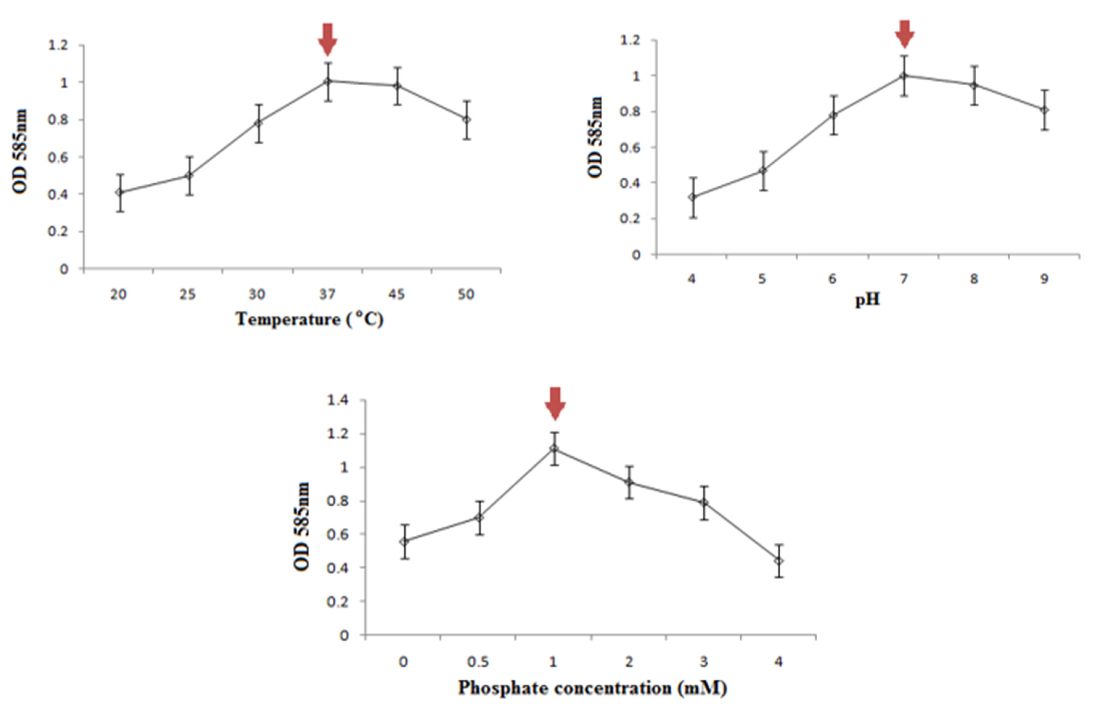
2.4. Determination of Optimum Growth Conditions

The growth features of the bacterial isolate at optimum pH, temperature, and phosphate concentration were determined by the method of Shamim and Rehman [17]. A total of 1% fresh bacterial culture was inoculated in minimal salt broth at varying pH (4–9), temperature (10, 25, 37, 45, 50 °C), and phosphate concentrations (0.1, 0.5, 1, 1.5, 2 mM), followed by incubation for 24 h. The results were noted by taking the optical density at 578 nm.

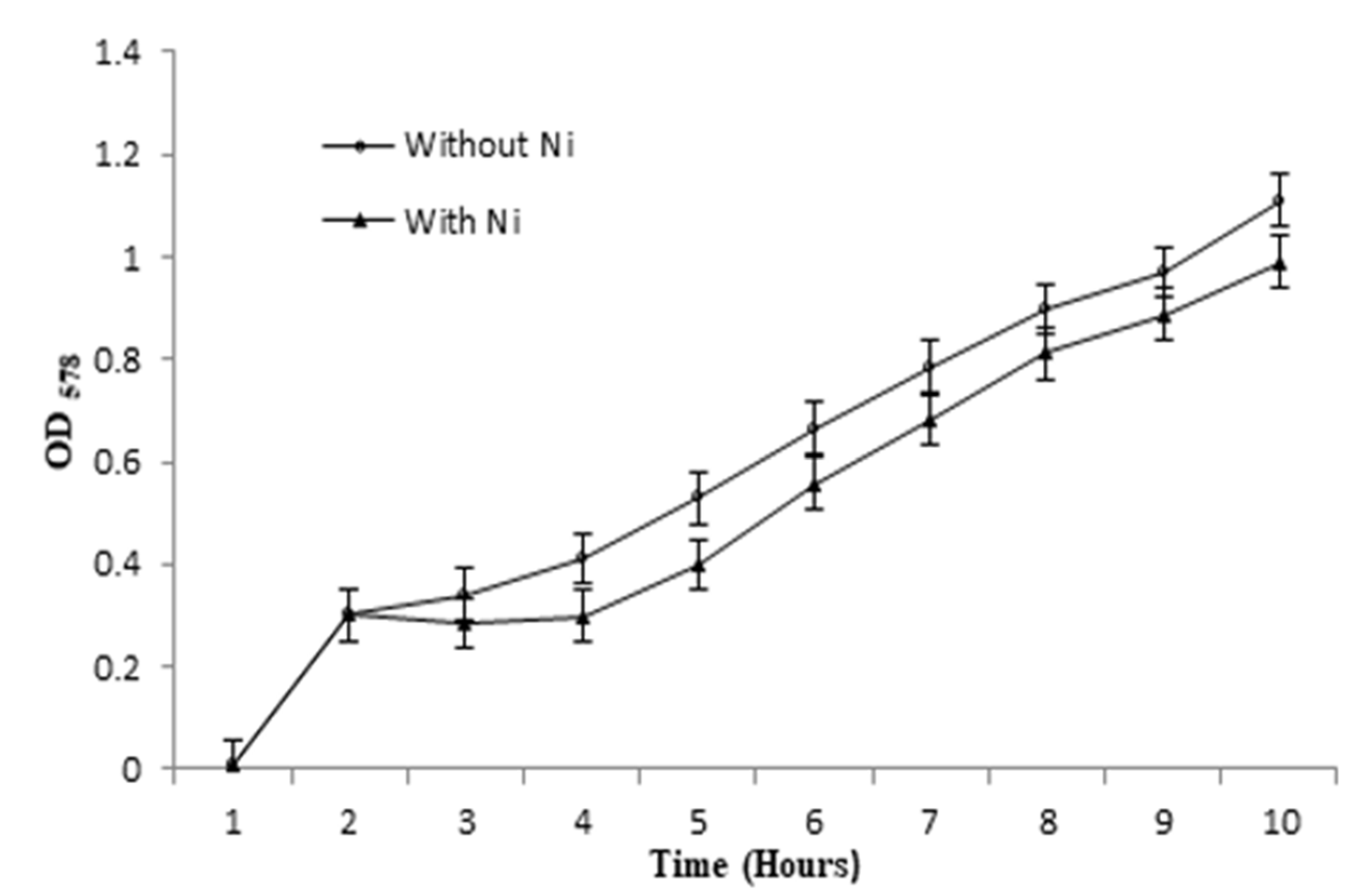
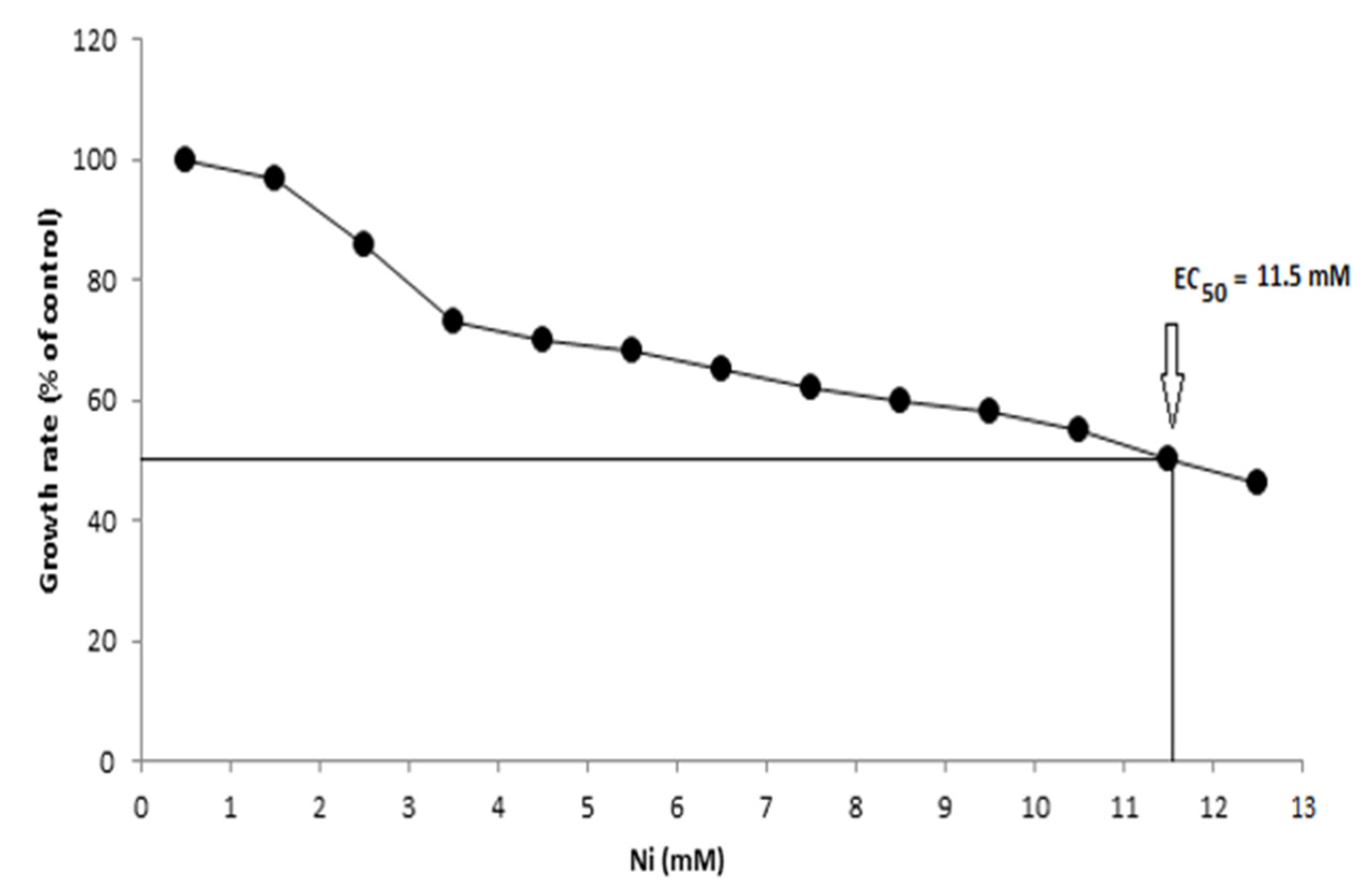
2.5. Evaluation of Bacterial Growth and EC50 in Presence of Ni

For this experiment, 1% fresh bacterial culture was added to sterile minimal salt broth to achieve the log phase of bacterial growth (OD578 = 0.3–0.4), after which the experimental setup was supplemented with Ni (0.1 mM). Following an interval of 30 min, the optical density was recorded at 578 nm every 60 min for up to 9 h. The observed readings were then compared with the control setup and were plotted in graphical form [18]. In a similar manner, the experimental and control groups were set up for the determination of EC50. The experimental groups were supplemented with different concentrations of Ni (0, 0.25, 0.5, 0.75, 1.0, 1.25, 1.5, 1.75, 2. 2.5, 3, 3.5, 4 mM), followed by the absorbance obtained at regular intervals (OD = 578 nm). The results were plotted in graphical form [19].

2.6. Ni Biosorption Experiments

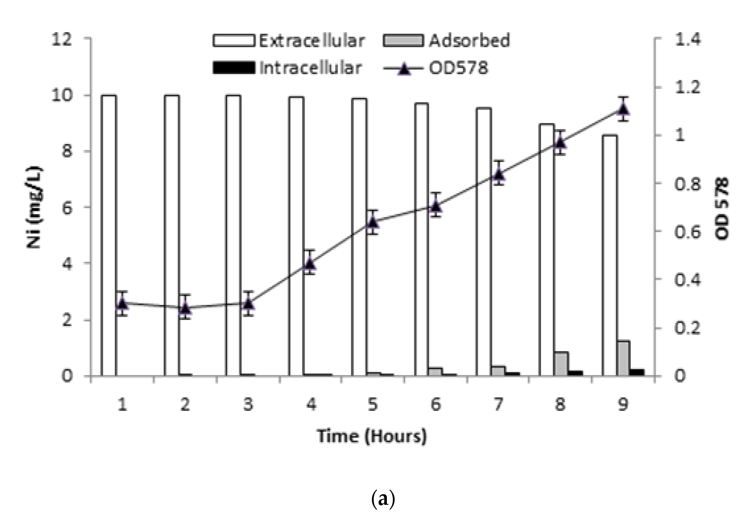
2.6.1. Determination of Ni Uptake

The metal uptake analysis was performed by the method of Shamim et al. [18], whereby 1% bacterial culture was inoculated in sterile minimal salt medium previously augmented with 1 mM phosphate and 4 g/L sodium succinate. The flasks were incubated at 37 °C until OD578 reached 0.3–0.4, after which 10 mgL−1 Ni were added to the experimental setup. After 30 min, 10 mL samples were harvested and the optical density (578 nm) was also recorded. This was termed as the first hour sample and reading, and the samples were harvested consecutively after 0, 1, 2, 3, 4, 5, 6, 7, 8, and 9 h. The pellet in the first falcon tube was then washed thrice with Milli-Q water (10 mL) and EDTA (0.1 mM), and centrifuged again at 12,000 rpm to obtain a pellet. The water was collected in a new falcon tube. The harvested pellets were then acid digested with the help of 0.2 N nitric acid (HNO3) and incubated at 37 °C. The previously centrifuged supernatant samples, the water samples, and the pellet samples were then made ready for atomic absorption spectrometry (AAS) analysis at 228.8 nm the next day, prior to which the standards for the instrument were run to generate a calibration curve. The subsequent results were tabulated and graphs were plotted.

2.6.2. Ni Uptake in Presence of Inhibitors

This experiment was carried out likewise the previous method, along with the addition of two metabolic inhibitors, DNP (1 mM) and DCCD (100 µM), prior to the supplementation of Ni in the medium at the log phase of the bacterial cells. The rest of the procedure was carried out as before [18].

2.6.3. Ni Removal by Killed Bacterial Cells

The determination of metal uptake by killed bacterial cells was carried out by the method of Shamim et al. [18]. A total of 1% of bacterial culture was inoculated in experimental and control setups of sterile minimal salt broth medium, after which the logarithmic bacterial cells were killed via heat inactivation. Ni (10 mgL−1) was added to the killed cells, which were then incubated for 30 min. The rest of the procedure was followed according to the previous experimental setups.

2.6.4. Pilot-Scale Study for Ni Removal

The pilot-scale study was carried out by following the method of Shamim and Rehman [17]. The evaluation of Ni removal efficacy of the bacterial isolate from industrial effluents was carried out in three plastic containers, with the first being filled with tap water (10 L), and the other two with industrial effluents (10 L) (30 °C, pH 6.2, dissolved oxygen content 0.0134 ± 0.02 g L−1, Ni 1.5 ± 0.01 g L−1). In the first two containers, fresh bacterial culture (1%) was added, following the addition of Ni in all containers. The samples from the containers placed at room temperature were collected after 4 and 8 days and were harvested at 15,000 rpm. The acid-digested supernatants were used to determine the Ni removal efficacy of the bacterial cells.

2.7. Profiling of Antioxidative Enzymes

For the evaluation of antioxidative enzymes, the preparation of the enzyme extract was performed by inoculating 1% bacterial culture in sterile minimal salt broth medium, which was incubated for 24 h at 37 °C, followed by the addition of Ni (0.1 mM) and incubation for another 24 h. Post 48 h of incubation, a sample (50 mL) was harvested for 10 min at 12,000 rpm. The supernatant was discarded and the pellet was homogenized in extraction buffer (5 mL) under cold conditions [20]. The solution was then homogenized at 12,000 rpm for 10 min. The supernatant was transferred to a new falcon tube labeled as the enzyme extract, which was used for the estimation of various antioxidative enzymes such as superoxide dismutase (SOD) [21], ascorbate peroxidase (APOX) [22], catalase (CAT) [23], peroxidase (POX) [24], and glutathione reductase (GR) [25].

2.8. Profiling of Proteins and Metallothioneins

The estimation of protein was carried out by plotting a curve of various concentrations of protein (1–30 µg) against a standard (bovine serum albumin). The samples for non-elucidated and elucidated protein content were prepared using Bradford reagent and the results were observed by taking the absorbance of samples (OD595nm) [26].
For the profiling of intracellular proteins (metallothioneins), the bacterial culture was inoculated in minimal salt broth medium in the presence and absence of Ni (0.1 mM) stress. Following an incubation of 24 h, the cell pellets were harvested at 15,000 rpm for 10 min. The pellets were homogenized and mixed in 1X gel-loading buffer, after which they were placed on a thermomixer for 5 min at 95 °C. Experimental and control samples were then resolved by SDS-PAGE using resolving gel (12%) and stacking gel (5%). After the subsequent stain and de-stain process, the gel was visualized and the bands were compared to the standard protein ladder [27].

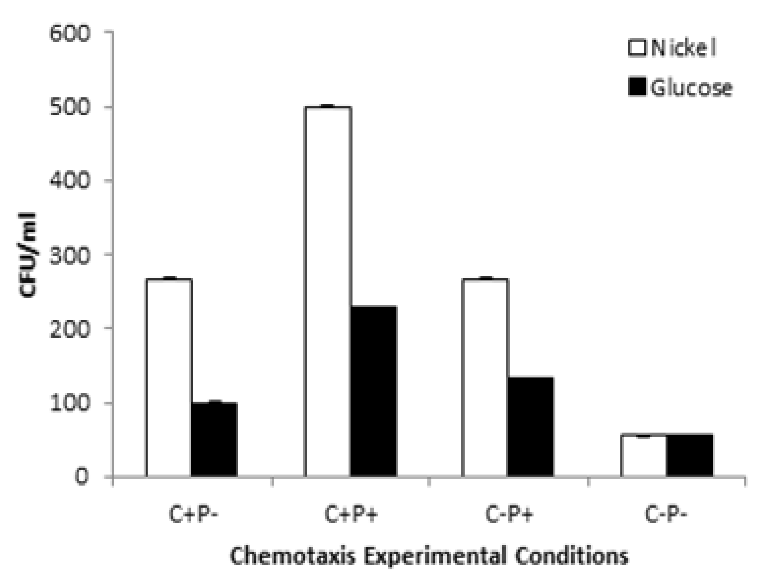
2.9. Determination of Motility and Chemotactic Behavior

The evaluation of motility patterns (swim, swarm, twitch) of the bacterial cells in the presence and absence of Ni was conducted by the method of Murray et al. [28]. Likewise, the chemotactic behavior of the bacterial cells with and without Ni was elucidated using the capillary chemotaxis method [29].

2.10. Isothermic, Thermodynamics, and Adsorption Kinetic Studies

The isotherm studies to study the adsorption mechanism of the bacterial cells were performed using Langmuir and Freundlich isotherm models [30,31]. The Langmuir model was studied via the following non-linear (1) and linear (2) equations, respectively:
q e = q m a x b C e 1 + b C e
C e q e = C e q m a x + 1   b q m a x
where Ce = concentration of metal ions in solution at equilibrium (mM),
qe = amount of metal ions adsorbed at equilibrium (mM/g),
qmax = Langmuir constant (mM/g),
b = adsorption constant.
The Freundlich model was studied via the following non-linear (3) and linear (4) equations, respectively [32]:
q e = k f C e 1 / n          
The linear form of the equation is:
ln q e = ln k f + 1 n     ln C e                
where Kf and n = Freundlich constants.
The biosorption kinetic studies were evaluated using Lagergren’s pseudo first-order and pseudo second-order kinetic models [33], where the linear equations of the pseudo first-order (5) and pseudo second-order (6) [34,35] are as follows:
ln ( q e q t ) = ln q e k 1 t
t q t = 1 k 2 ( q e 2 ) + t q e
where
qe and qt = amount of metals ions adsorbed at equilibrium,
t = time,
k1 and k2 = first-order and second-order rate constant, respectively.
The thermodynamic studies were carried out using the Gibbs free energy change (∆G°), enthalpy change (∆H°), and entropy change (∆S°) parameters with the following Equations (7) and (8):
∆G° = ∆H° − T∆S°
∆G° = −RTlnKD
where
R = universal gas constant,
T = temperature in Kelvin (K),
KD = value calculated from qe/ Ce.

2.11. Fourier Transform Infrared Spectroscopy (FTIR) Analysis of Bacterial Cells

FTIR analysis was utilized to obtain IR spectra for bacterial cells with and without Ni stress. For sample preparation, minimal broth medium was inoculated with 1% of bacterial culture with (experimental) and without (control) Ni (0.1 mM). The samples were incubated for 24 h at 37 °C, after which they were harvested and the resultant pellet was homogenized in deionized water (500 µL). FTIR (Bruker) analysis was then performed in the mid-IRF region of 4000–500 cm−1 [36].

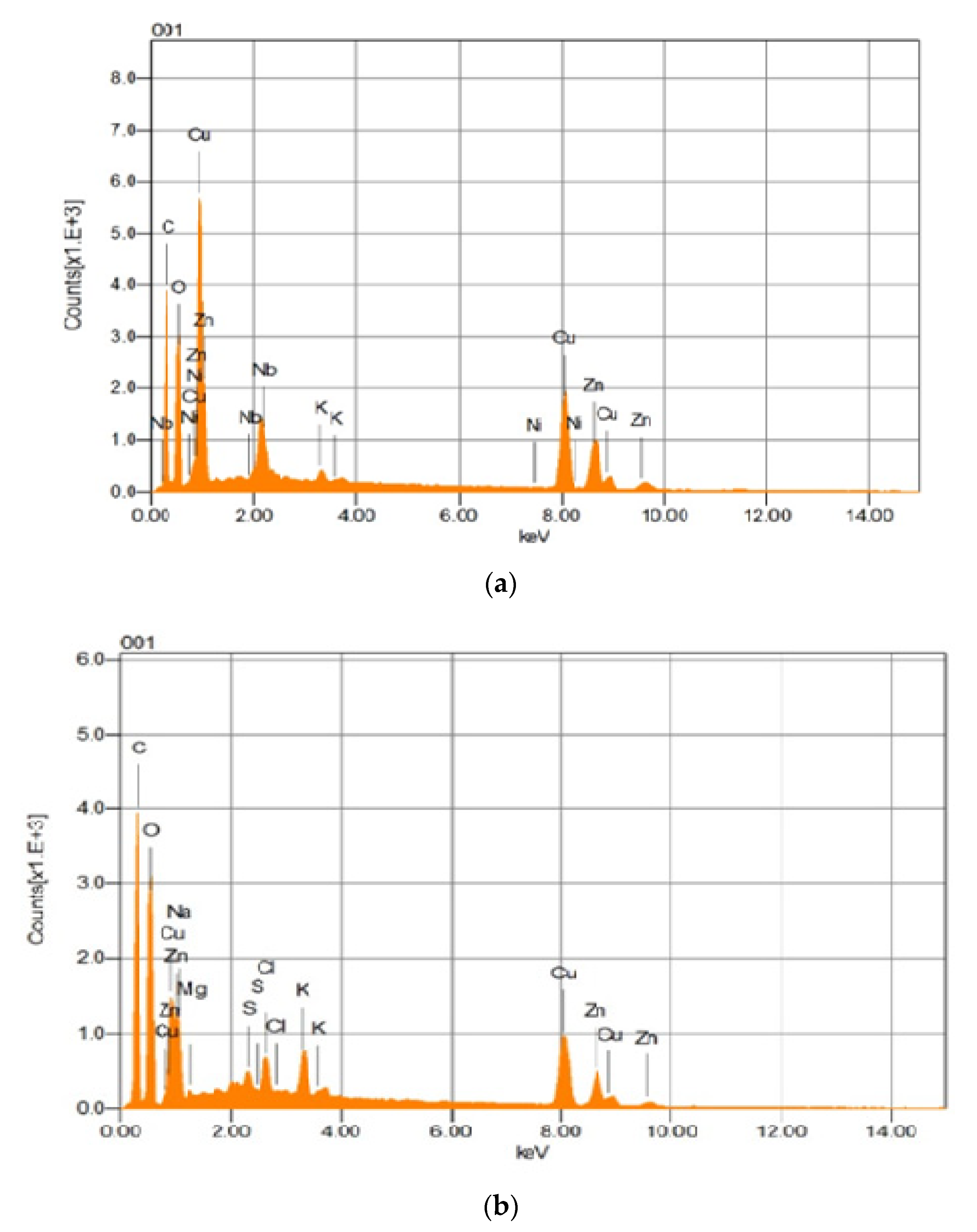
2.12. Scanning Electron Microscopy (SEM) and Energy Dispersive Spectroscopy (EDS) Analysis

The SEM and EDS analyses were performed to study the pattern of Ni biosorption by bacterial cells. For both of the analyses, the method by Khan et al. [37] was adopted. Prepared samples were evaluated on SEM (JSM-IT-100, JEOL, Japan) coupled with EDS, under several magnifications.

2.13. Statistical Analysis

The experiments were carried out thrice to avoid error. All experimental setups were run with specific control setups under suitable and similar conditions. During result compilation, cumulative average and standard error was calculated using IBM SPSS (V. 24.0).

3. Results

3.1. Enumeration of Ni-Resistant Bacterial Isolates

A total of 35 bacterial isolates were isolated from the collected samples, which were then subjected to an MIC assay for the selection of the most resistant isolate.

3.2. Minimum Inhibitory Concentration (MIC) and Cross Heavy-Metal Resistance

Among the bacterial isolates, one isolate (ZBM-05) resisted Ni up to a concentration of 20 mM, and the cross heavy-metal resistance pattern was found to be Cu2+ (25 mM) > Zn2+ (15 mM) > Cr6+ (10 mM) > Pb2+ (5 mM) > Co2+ (8 mM) > Cd2+ (3 mM) > Hg2+ (0 mM).

3.3. Selection and Molecular Identification of Bacterial Isolate

The most resistant bacterial isolate was selected on the basis of the MIC and cross heavy-metal resistance, which was then identified via 16s rRNA ribotyping. The isolate was identified as Bacillus altitudinis, and the accession number acquired from NCBI for the strain was MT422188.

3.4. Optimum Growth Conditions

The optimum growth was found to be at 32 °C, pH 7, and 1 mM phosphate for the bacterial cells, as depicted in Figure 1.

3.5. Bacterial Growth and EC50

The growth curve demonstrated that the bacterial cells entered the log phase when they were introduced in the medium. The presence of Ni was observed to act as a stress for the bacterial cells, as observed from the overall reduction of the growth, as shown in Figure 2. The EC50 of the bacterial cells was demonstrated to be 11.5 mM, which means that at this concentration about 50% of the cells were dead and 50% of the cells were live and functional, performing cellular functions, as shown in Figure 3.

3.6. Ni Biosorption Experiments

3.6.1. Ni Uptake by Live Bacterial Cells

The bacterial cells entered log phase in the third hour, after which a steady increase in the bacterial cells was observed. The cells also started to uptake Ni from the aqueous medium, which was evident from the decrease in Ni. The intracellular accumulation and adsorption also increased in the eight and ninth hours, where the concentration of Ni was shown to be less, indicating the accumulation of Ni inside the bacterial cells (Figure 4a).

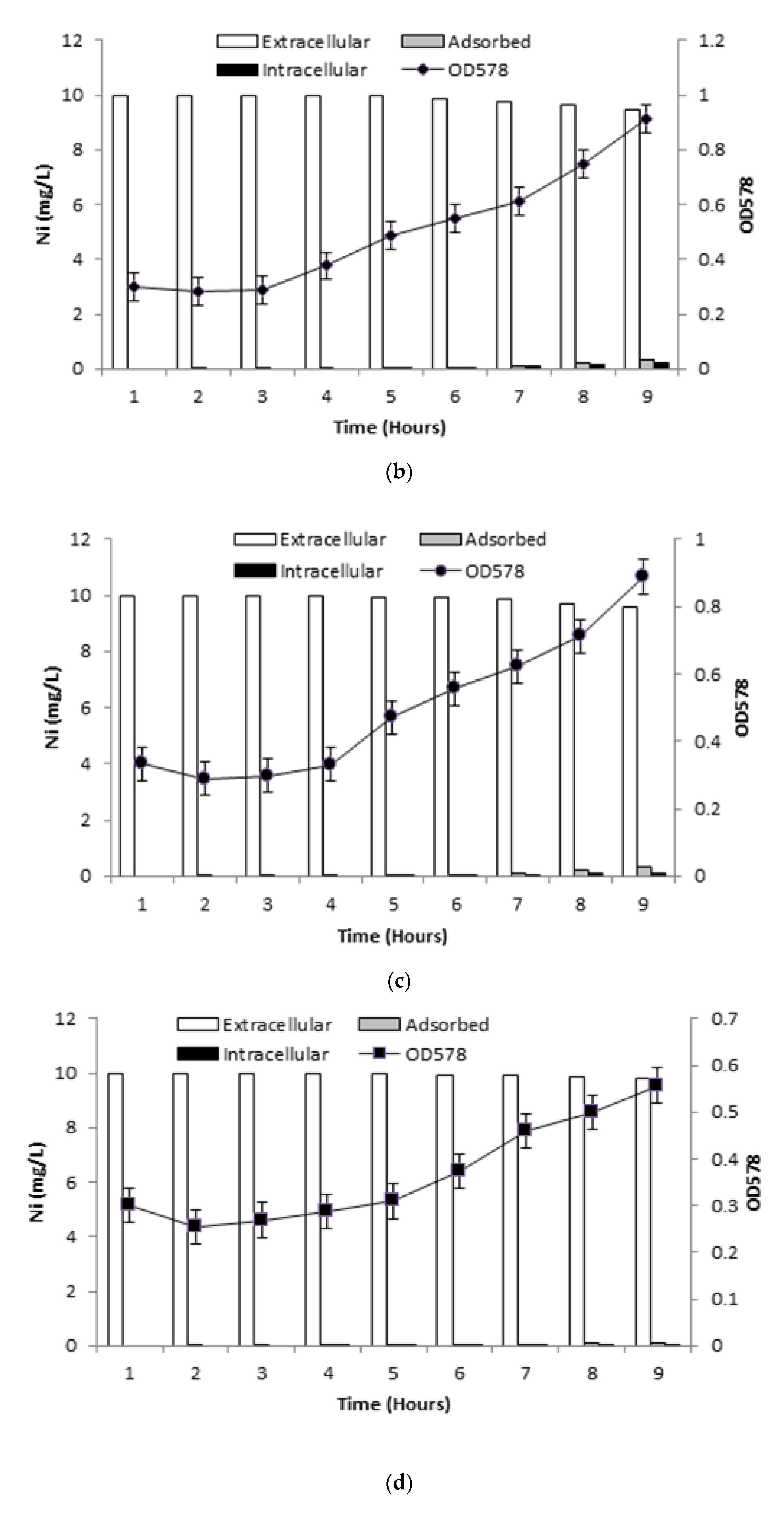
3.6.2. Ni Uptake in Presence of Inhibitors

The presence of DNP (inhibitor 1) reduced the overall growth of the bacterial cells, along with the presence of Ni in the aqueous medium. The growth of cells started to increase after the fourth hour, and kept steadily increasing until the ninth hour. The uptake of Ni from the medium was observed to be substantial from the sixth hour onwards, which also resulted in an increase in the adsorption and intracellular accumulation from the sixth to the ninth hour (Figure 4b). DCCD (inhibitor 2) had a somewhat similar effect on the growth of bacterial cells as DNP. The growth of bacterial cells was seen to drastically increase in the ninth hour, with uptake of Ni from the aqueous medium starting from the seventh hour onwards. The intracellular accumulation and adsorption mechanism was also observed at the same hour, as depicted by Figure 4c. In the final inhibitor setup, the growth of bacterial cells decreased in the presence of three stress inducers used in combination (Ni, DNP, DCCD). The bacterial cells entered the log phase, but the growth was not as pronounced as in the other experimental setups. The presence of the inhibitors did not stop the cells from removing Ni from medium, as observed from the decrease of Ni in the ninth hour. The intracellular accumulation and adsorption of Ni was also observed minimally in the last two hours, as shown in Figure 4d.

3.6.3. Ni Removal by Killed Bacterial Cells

The killed bacterial cells failed to grow, as they did not reach the lag or log phase. The dead cells did not uptake any Ni from the aqueous medium, although the availability of bacterial cells (biomass) enabled the adsorption of Ni ions onto the bacterial cell surface (Figure 5).

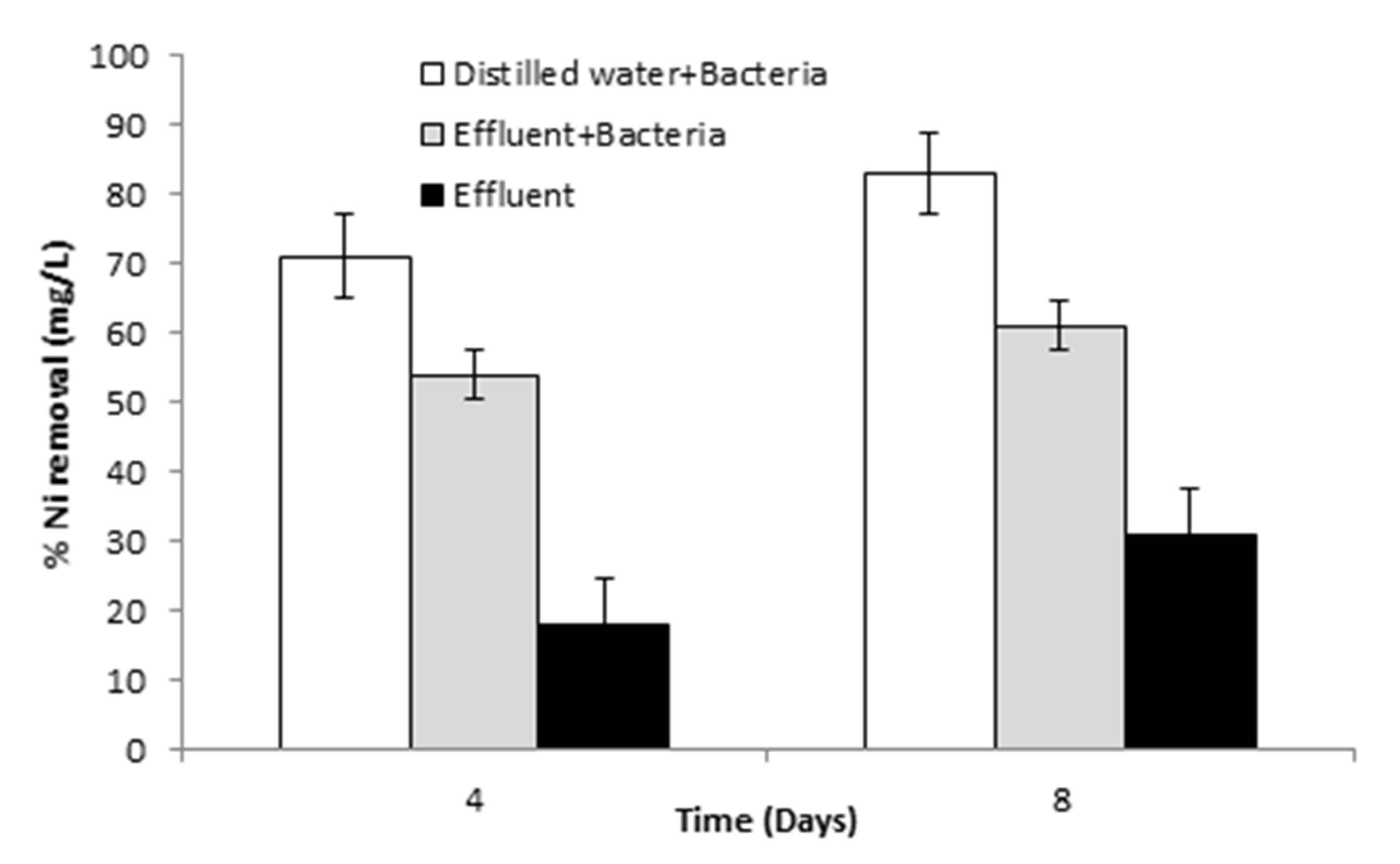
3.6.4. Pilot-Scale Study for Ni Removal

In the pilot-scale study, B. altitudinis was observed to remove 70 and 85 mg/L after a time of 4 and 8 days, respectively, which was greater than the amount of Ni removed from other experimental setups, thus establishing its Ni bioremediation efficacy, as depicted in Figure 6.

3.7. Profiling of Antioxidative Enzymes

The evaluation of antioxidative enzymes demonstrated the reduced activity of GR (0.001 ± 0.003 Ug−1FW), POX (0.001 ± 0.001 Ug−1FW), and SOD (0.091 ± 0.003 Ug−1FW) activity in the presence of Ni in B. altitudinis cells. The presence of Ni failed to induce APOX and CAT activities in measurable quantities, as shown in Table 1.

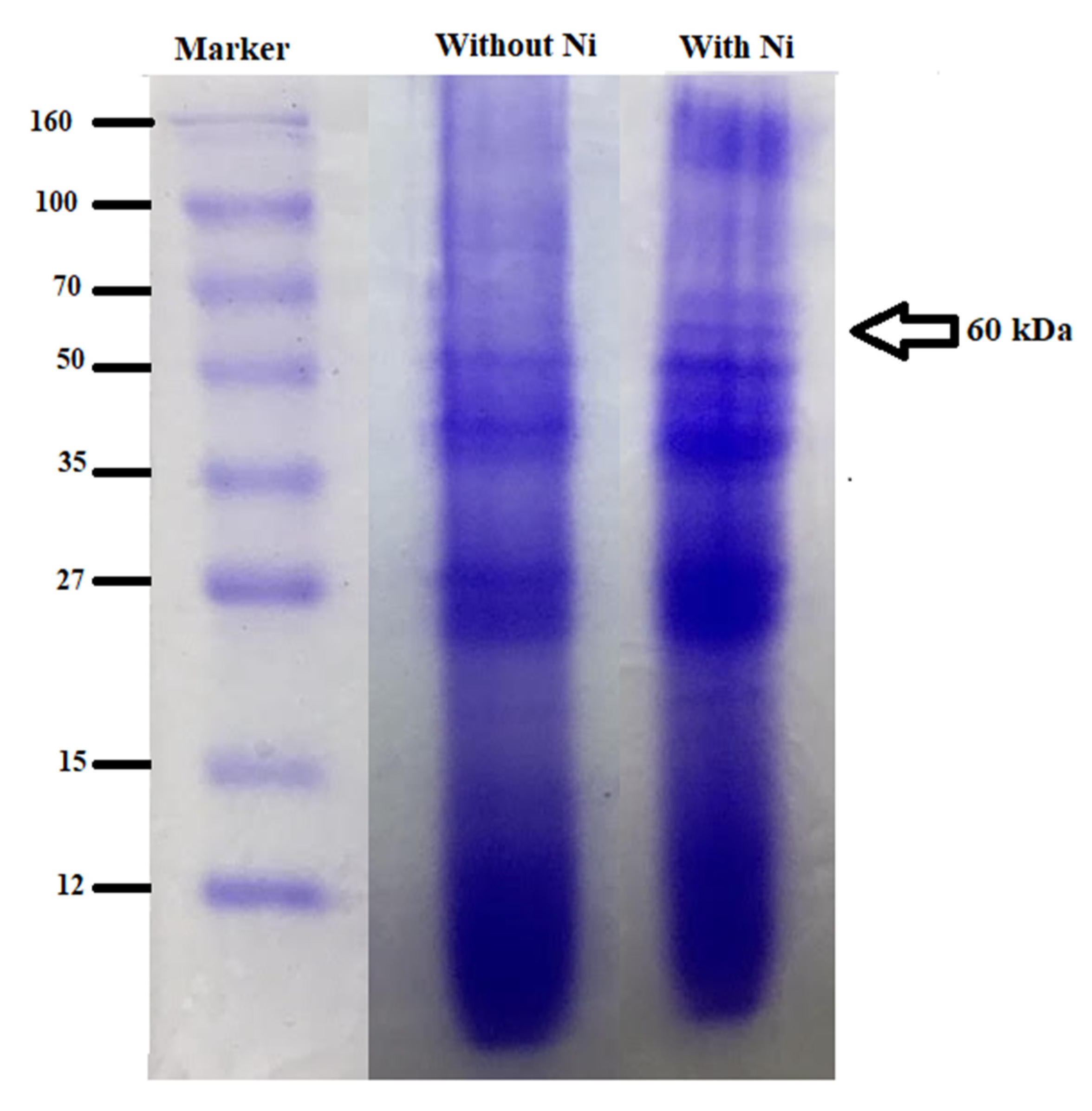
3.8. Profiling of Metallothioneins

The results of SDS-PAGE demonstrated bands of various sizes in the control (50, 38, 30, 19 kDa), whereas in the experimental sample, a band of 60 kDa in the presence of Ni indicated the induction of metallothionein in the presence of metal ions in the bacterial cell, as depicted by Figure 7.

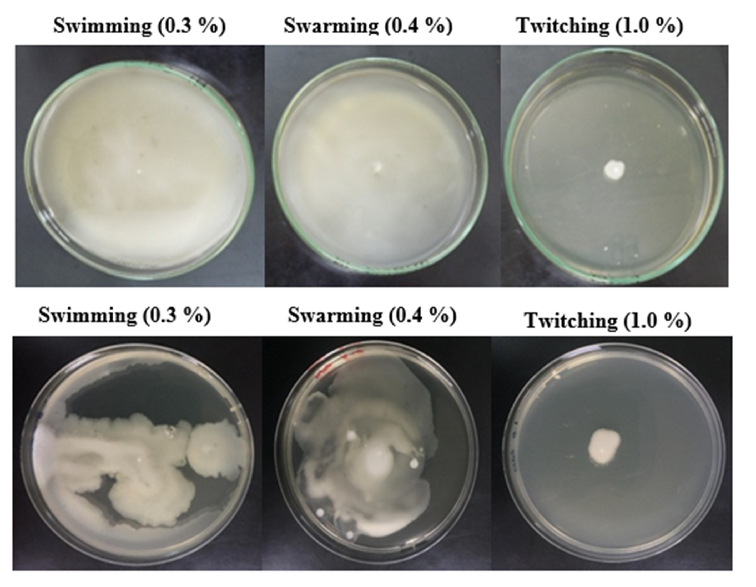
3.9. Pattern of Motility and Chemotactic Behavior

The swimming, swarming, and twitching pattern of motility was observed in B. altitudinis cells in 0.3, 0.5, and 1% agar medium, which demonstrated remarkable motile behavior in the presence of Ni (Figure 8). The capillary chemotaxis experiment demonstrated the role of Ni as a chemoattractant for B. altitudinis, resulting in positive motility towards Ni in the medium (Figure 9).

3.10. Isothermic, Thermodynamic, and Adsorption Kinetic Studies

For the isotherm models, the graphs were plotted for both Langmuir and Freundlich isotherm models, respectively. The values for the Langmuir and Freundlich constants as well as the R2 values are comprehensively provided in Table 2. For the Langmuir and Freundlich models, the R2 values were observed to be in the range of 0.90–0.98 and 0.900–0.999, respectively, which therefore demonstrated the compatibility of the Freundlich model with our study.
The pseudo first and second order of kinetics and their constants are shown in Table 3. The coefficient values (qexp and qcal) of the first order were compared but were not compatible with each other. In the case of the second order, compatibility was positively observed between the two coefficients. Moreover, the comparison of the R2 values of both orders demonstrated that pseudo second-order kinetics favored our study.
The thermodynamic parameters of our study demonstrated the Gibbs free energy (∆G°) for various temperatures, whereas the enthalpy change (∆H°) and entropy change (∆S°) were found to be 3.0436 KJ/mol and 0.0224 kJ/mol/K, respectively, as shown in Table 4.

3.11. Fourier Transform Infrared Spectroscopy (FTIR) Analysis

The FTIR analysis revealed the characteristic peaks of hydroxyl and carboxyl functional groups playing a role in the biosorption of Ni by B. altitudinis. The peak shifts were determined to be due to the interaction of Ni with bacterial cells (Table 5).

3.12. SEM and EDS Analyses

The SEM and EDS analyses demonstrated the positive physical and chemical changes in B. altitudinis cells, which were indicative of their biosorption and features in bacterial cells against Ni, as shown by Figure 10a,b and Figure 11a,b.

4. Discussion

Heavy metals are major contributors to pollution, as they are toxic substances that tend to accumulate over time in valuable resources around the world [38]. This research study was conducted to determine the efficacy and potential role of an Ni-resistant bacterial isolate in Ni bioremediation. The bacterial isolate, named ZBM-5, was selected on the basis of its MIC and cross heavy-metal resistance pattern. The isolate was identified as Bacillus altitudinis MT422188, and was used to remove Ni from aqueous media. The findings from our research are in agreement with the findings of Alboghobeish et al. [39], who in their work also reported the presence of Ni-resistant bacteria from heavy-metal contaminated wastewaters. Bacteria are dependent upon optimal temperatures, pH, and other parameters for their growth, which can decline or cease if they are allowed to grow at other temperatures. The growth conditions were examined, which demonstrated the optimum temperature, pH, and phosphate concentration of B. altitudinis to be 32 °C, 7, and 1 mM, respectively (Figure 1). The work of Zhenggang et al. [40] described the optimum temperature of Bacillus cereus to be 35 °C, which was determined over a range of other examined temperatures. Furthermore, another study reported the optimum temperature of another bacterial species, Sphaerotilus natans, to be 35 °C [41], demonstrating that most bacterial species are mesophilic in nature, functioning over a moderate range of temperatures that can be easily applied for optimum growth.
In this study, the MIC and the multiple metal resistance pattern of the bacteria were examined. The concentration that inhibited the growth of the bacterial isolate even after the desired incubation was termed “the effective MIC.” In a similar manner, the cross heavy-metal resistance pattern was checked for different heavy metals when their increasing concentrations were applied in solution. The concentration that failed to facilitate the growth of bacteria after the incubation period was termed “the inhibiting concentration of the respective metal.” The MIC of B. altitudinis was observed to be 20 mM, whereas the multiple metal resistant behavior of the different metals was observed to be Cu2+> Zn2+> Cr6+> Pb2+> Co2+> Cd2+> Hg2+. In their study, Afzal et al. [42] also studied Ni-resistant bacterial isolates collected from wastewater samples from industrial effluents. The MIC of Ni was observed to be 8 mM in their study. Another study, conducted by Chowdhary et al. [43], stated the MIC of Ni resistant bacterial isolate to be 24 mM, which is in agreement with our results. The results are also similar to the results of Alboghobeish et al. [39], who reported the MBC of the Ni-resistant Klebsiella oxytoca species to be 24 mM. In agreement with previously reported works, another study reported the zone of inhibition of Ni-resistant bacteria to be 24 mM after it was treated and examined in a well-diffusion assay [44]. These results could establish the basis of resistance in bacterial species against Ni, whether chromosomal or plasmid mediated. This resistance is basically an adaptive mechanism that bacteria take up in order to ensure survival under stress conditions and high metal toxicity. The determination of EC50 in the case of Ni was 11.5 mM (Figure 3), which means that at this concentration 50% of the bacterial cells were dead and 50% of the bacterial cells were live and functional, taking part in the cellular processes and responding to the metal toxicity in the environment. In the research findings by Speir et al. [45], the EC20 value was examined for Zn, which was shown to be 330 mg kg−1, in order to determine the metal toxicity in soil. The growth of bacteria was observed to be less in the presence of Ni in our research work, indicating the role of Ni as a stress for bacterial cells, though the growth followed the same pattern as seen in the absence of Ni (Figure 2). The findings by Afzal et al. [42] also reported a decrease in the bacterial growth in the presence of Ni, which demonstrates that the bacterial growth declined when it was compared with the growth pattern of the control (without stress).
The elevated concentration of pollutants such as heavy metals results in their contact with the surrounding microorganisms of the environment. Exposure to bacteria results in their interaction with bacterial cell walls, which leads to the production of harmful compounds and metabolites. The ingress of heavy metals inside bacterial cells is regulated by uptake systems that are specific to certain species of bacteria. These uptake systems are reported to be of two types, the first being a fast and unspecified system that occurs mostly across an osmotic gradient and is ATP independent, whereas the second uptake system is comparatively slower, specified, and dependent upon ATP. The entry of Ni is regulated by the ABC transporter family [46]. In this research work, the uptake mechanism of Ni by B. altitudinis was investigated. The reduced growth of cells in the presence of Ni indicated that the bacterial cells were in a stressful environment in the presence of Ni (Figure 4a). The presence of Ni and DNP demonstrated the uptake of Ni from the medium by the bacterial cells in the sixth hour, which also resulted in the intracellular accumulation and adsorption of Ni in the presence of DNP (Figure 4b). The second inhibitor, DCCD, had a similar effect on the uptake of Ni by bacterial cells, with almost identical uptake patterns (Figure 4c). The effect of Ni and both inhibitors combined demonstrated the growth pattern of the bacterial cells to be not as pronounced as compared to other experimental setups (Figure 4d), although the presence of both inhibitors did not cease the uptake of Ni from the medium, thereby indicating the role of an unspecified efflux system that is not dependent upon ATP. The uptake of Ni by killed bacterial cells was also investigated in the study, where it was observed that the killed cells were not able to uptake Ni from the medium, but adsorption was reported due to the availability of biosorbent sites that adsorb Ni from the environment without the aid of a specific uptake system (Figure 5). Previous researchers have also reported on the biosorption ability of bacterial killed biomass [47,48]. The pilot-scale study was also conducted to determine the Ni removal efficacy of B. altitudinis, indicating it to be an effective agent of Ni bioremediation, as it was able to remove remarkable quantities of Ni (Figure 6), which was also reported by Rivas-Castillo et al. [49].
The motility of microorganisms is one of the main features that are essential for their survival. The requirement of nutrients and the search for them is enabled by the motile nature of the microorganisms. The same can be said about their escape from their surroundings if they encounter any stress in their surroundings, where their motility enables them to move away. This is achieved with the help of external features of microorganisms such as flagella and pili. The microorganisms, bacteria in particular, attain the help of various motility patterns in order to survive in various stressful conditions in their surroundings. The motion that is adapted by bacteria to move towards favorable conditions and vice versa is known as the phenomenon of chemotaxis. The migration of bacteria is directed under a chemical gradient, where they move away or towards it under conditions for their endurance and survival. The regulation of movement patterns in bacterial cells tends to cause a motion toward attractants or repellents in an environment. This stimulus in the environment is detected by bacterial chemoreceptors that aid in transmitting the signal of motion to the signal transduction system of the bacteria, after which the bacteria opt for a suitable movement pattern under stimulating conditions. The flagellar movements are controlled by the regulatory protein CheY. CheY-P, the phosphorylated form of CheY, binds to the flagellar motor, which leads to tumbling motions, whereas its non-phosphorylated form leads to running motions. The other regulatory proteins, CheA and CheW, act as a complex and a linker between the two proteins, respectively [50]. In this study, the mechanisms of motility and chemotaxis were investigated in a series of experiments. The presence of Ni demonstrated remarkable swimming, swarming, and twitching behavior in bacterial cells, which indicates that at a given concentration Ni acted as chemoattractant for the bacterial cells and they preferred to move freely in its presence (Figure 8). The chemotaxis experiments demonstrated that B. altitudinis demonstrated movement towards Ni when it was present in capillary tubes, as well as in the pond, resulting in more CFU/mL (Figure 9). The positive chemotaxis for Ni affirmed that B. altitudinis was a metal lover (Figure 9).
The presence of metallothioneins (MTs) in living beings is the basis of the important role they serve in many biological functions, as these proteins are reported to be highly conserved in living organisms, making them unchanged through many generations. The major function that MTs serve can be that of metal detoxification and conferring metal resistance among living organisms and bacteria. The MTs present in bacteria are majorly attributed to conferring resistance against various heavy metals, as demonstrated by the findings of many studies [18,37,51]. This work was also used to determine the presence of MTs in B. altitudinis with and without Ni stress (Figure 7). In agreement with our study, various other studies have also reported on the induction of MTs under heavy stress over the years [52,53]. The expression of antioxidative enzymes was also evaluated in this research study to gain better insight into the enzymes responsible for combating the generation of ROS in the system. The activity of GR, POX, and SOD enzymes was observed to decrease in the presence of Ni, whereas the activities of APOX and CAT were not observed in a quantifiable range, neither with nor without Ni stress (Table 1). Tammam et al. [54] reported an increase in SOD enzyme activity and a decline in GR enzyme activity under Ni stress.
The interactions between Ni ions and bacterial cells, resulting in the subsequent adsorption, was evaluated using isotherm models, pseudo-order kinetics, and thermodynamics (Table 2, Table 3 and Table 4). The isotherms of adsorption are the primary prerequisites in the design and development of adsorption systems [55]. Although several isotherm equations have been reported for studying adsorption models, Langmuir and Freundlich remain two of the most commonly used isotherms, where the former is of the assumption that adsorption occurs at particular sites homogenously (monolayer) inside the adsorbent, and the latter is suggestive of heterogenous (multilayer) systems. In both models, the R2 values were reported to be above 0.92, indicating a good mathematical fit (Table 2). However, the greater coefficient values of the Freundlich model suggest it to be a better model fit for our study. Similar findings in Bacillus sp. were reported by Cai et al. [56]. The thermodynamic parameters were also investigated in order to evaluate the feasibility of the process. The Gibbs free energy (∆G°) was observed to be negative, which indicates that the overall process of adsorption is feasible and spontaneous. Moreover, positive values of enthalpy change (∆H°) and entropy change (∆S°) demonstrated that the process was endothermic and favorable in nature. The results of our study are in agreement with Asare et al. [57].
The FTIR analysis was performed for the evaluation of bacterial functional groups associated with the adsorption of Ni ions by B. altitudinis (Table 5). The FTIR spectra demonstrated broad peaks at 3279.39 cm−1 and 1637.22 cm−1, where no Ni stress was added, which corresponded to the presence hydroxyl and carboxyl groups, respectively. The hydroxyl group demonstrated C-H stretching, due to which broad peaks were formed, whereas the carboxyl group demonstrated C=O stretching bonds in asymmetric and symmetric vibrations. In the presence of Ni, these peaks were observed to be shifted to 3272.11 cm−1 and 1636.97 cm−1, respectively. The shift in the two peaks under Ni stress was observed to be due to the interaction and binding of Ni ions with the bacterial cells. The findings of Khan et al. [37] are in agreement with our study, where the involvement of various functional groups of bacteria were observed in biosorption by bacteria. The use of techniques like SEM and EDS aided in determining the biosorption and interaction of Ni ions with the bacterial cells at a microscopic level. The SEM images (Figure 10a,b) demonstrated the shape and structure of the bacterial cells before and after Ni biosorption. The size of the bacterial cells was observed to be changed after the biosorption of Ni ions, as the cells were observed to be bigger and swollen in size, indicating intracellular accumulation of Ni. Similarly, a morphological as well as elemental alteration of the bacterial cells after Ni biosorption was observed in the EDS images (Figure 11a,b), where the characteristic peak of Ni was only found after Ni biosorption. Our results were found to be similar to the findings of Rahman et al. [58].

5. Conclusions

This study was performed for the isolation and characterization of Ni-resistant bacteria which were isolated from Ni-polluted industrial effluents. The bacterial isolate was identified as Bacillus altitudinis, which was studied for its bioremediation potential against Ni. This study aimed to shed light through various experiments on the biosorption potential of Ni by B. altitudinis, proving it to be an efficient biosorbent. Further progress in investigating the molecular mechanisms of the bacterium in removing Ni from polluted industrial effluents can be helpful in gaining a better insight into the resistance and efflux systems that are responsible for bacterial resistance against Ni. Chemotaxis and motility assays demonstrated Ni to be a chemoattractant for B. altitudinis, thereby facilitating the movement of bacterial cells towards Ni in a stressful environment. The presence of Ni inducted a metallothionein of 60 kDa, whereas the FTIR analysis revealed the interaction of hydroxyl and carboxyl groups with Ni, with similar features observed in SEM and EDS. Equilibrium, kinetic, and thermodynamic studies depicted the adsorption of Ni onto bacterial cells to be in support of the Freundlich isotherm model, pseudo second-order kinetics, and endothermic, spontaneous, and feasible reaction, respectively. Therefore, these properties were helpful in elucidating the efficient role of B. altitudinis in Ni biosorption.

Author Contributions

Conceptualization and supervision, S.S.; investigation, Z.B. and M.K.; methodology, S.S.; writing—original draft, Z.B., M.K., and S.S.; writing—review and editing, S.S, software and analyses, G.A.C. and G.M. All authors have read and agreed to the published version of the manuscript.

Funding

This research was fully funded by the Higher Education Commission (HEC), Pakistan, as Project No. 21-1120/SRGP/R&D/HEC/2016.

Institutional Review Board Statement

Not applicable.

Informed Consent Statement

Not applicable.

Data Availability Statement

All the data have been added to this article.

Acknowledgments

The authors are grateful to the Higher Education Commission (HEC), Pakistan, as this project was fully funded by the HEC as Project No. 21-1120/SRGP/R&D/HEC/2016.

Conflicts of Interest

The authors declare no conflict of interest.

References

  1. Hao, X.; Zhu, J.; Rensing, C.; Liu, Y.; Gao, S.; Chen, W.; Huang, Q.; Liu, Y.-R. Recent advances in exploring the heavy metal(loid) resistant microbiome. Comput. Struct. Biotechnol. J. 2021, 19, 94–109. [Google Scholar] [CrossRef]
  2. Dash, S.; Borah, S.S.; Kalamdhad, A.S. Heavy metal pollution and potential ecological risk assessment for surficial sediments of Deepor Beel, India. Ecol. Indic. 2021, 122, 107265. [Google Scholar] [CrossRef]
  3. Kumar, A.; Cabral-Pinto, M.; Kumar, A.; Kumar, M.; Dinis, P.A. Estimation of Risk to the Eco-Environment and Human Health of Using Heavy Metals in the Uttarakhand Himalaya, India. Appl. Sci. 2020, 10, 7078. [Google Scholar] [CrossRef]
  4. Cabral Pinto, M.M.S.; Ferreira da Silva, E.A. Heavy metals of Santiago Island (Cape Verde) top soils: Estimated Background Value maps and environmental risk assessment. J. Afr. Earth Sci. 2015, 101, 162–176. [Google Scholar] [CrossRef]
  5. Silva, M.M.V.G.; Cabral-Pinto, M.M.S.; Dinis, P. Geochemistry of subtropical arenosols from Kui-to region (Angola). Urbanization effects and environmental implications. J. Afr. Earth Sci. 2021, 183, 104307. [Google Scholar] [CrossRef]
  6. Malea, P.; Mylona, Z.; Panteris, E.; Kevrekidis, D.P.; Kevrekidis, T. Nickel uptake kinetics and its structural and physiological impacts in the seagrass Halophila stipulacea. Ecotoxicol. Environ. Saf. 2021, 208, 111386. [Google Scholar] [CrossRef]
  7. Sharma, A.; Bhagat, M.; Urfan, M.; Ahmed, B.; Langer, A.; Ali, V.; Vyas, D.; Yadav, N.S.; Hakla, H.R.; Sharma, S.; et al. Nickel excess affects phenology and reproductive attributes of Asterella wallichiana and Plagiochasma appendiculatum growing in natural habitats. Sci. Rep. 2021, 11, 1–14. [Google Scholar] [CrossRef]
  8. Zhong, M.-S.; Jiang, L. Refining health risk assessment by incorporating site-specific background concentration and bioaccessibility data of Nickel in soil. Sci. Total Environ. 2017, 581-582, 866–873. [Google Scholar] [CrossRef] [PubMed]
  9. Genchi, G.; Carocci, A.; Lauria, G.; Sinicropi, M.S.; Catalano, A. Nickel: Human Health and Environmental Toxicology. Int. J. Environ. Res. Public Health 2020, 17, 679. [Google Scholar] [CrossRef] [Green Version]
  10. Mardiyono; Sajidan; Masykuri, M.; Setyono, P. Bioremediation of nickel heavy metals in electroplating industrial liquid waste with Bacillus subtilis. In Proceedings of The International Conference on Science and Applied Science (ICSAS) 2019; AIP Publishing: Melville, NY, USA, 2019; Volume 2202, p. 020084. [Google Scholar]
  11. Kumar, A.; Balouch, A.; Pathan, A.A.; Abdullah; Jagirani, M.S.; Mahar, A.M.; Zubair, M.; Laghari, B. Remediation of Nickel ion from wastewater by applying various techniques: A review. Acta Chem. Malays. 2019, 3, 1–15. [Google Scholar] [CrossRef]
  12. Van der Ent, A.; Callahan, D.L.; Noller, B.N.; Mesjasz-Przybylowicz, J.; Przybyłowicz, W.J.; Barnabas, A.; Harris, H.H. Nickel biopathways in tropical nickel hyperaccumulating trees from Sabah (Malaysia). Sci. Rep. 2017, 7, srep41861. [Google Scholar] [CrossRef] [PubMed]
  13. Aryal, M. A comprehensive study on the bacterial biosorption of heavy metals: Materials, performances, mechanisms, and mathematical modellings. Rev. Chem. Eng. 2019, 20190016. [Google Scholar] [CrossRef]
  14. Cheesbrough, M. Biochemical tests to identify bacteria. In District Laboratory Practice in Tropical Countries-Part 2; Cambridge University Press: Cambridge, UK, 2001; pp. 63–70. [Google Scholar]
  15. Pattanapipitpaisal, P.; Brown, N.L.; Macaskie, L.E. Chromate reduction and 16S rRNA identification of bacteria isolated from a Cr(VI)-contaminated site. Appl. Microbiol. Biotechnol. 2001, 57, 257–261. [Google Scholar] [CrossRef] [PubMed]
  16. Shamim, S. Comparative Analysis of Metal Resistance, Accumulation and Antioxidant Enzymes in Cupriavidus metallidurans CH34 and Pseudomonas putida mt2 during Cadmium Stress. Ph.D. Thesis, University of the Punjab, Punjab, Pakistan, 2014. [Google Scholar]
  17. Shamim, S.; Rehman, A. Cadmium resistance and accumulation potential of Klebsiella pneumoniae strain CBL-1 isolated from industrial wastewater. Pak. J. Zool. 2012, 44, 203–208. [Google Scholar]
  18. Shamim, S.; Rehman, A.; Qazi, M.H. Cadmium-Resistance Mechanism in the Bacteria Cupriavidus metallidurans CH34 and Pseudomonas putida mt2. Arch. Environ. Contam. Toxicol. 2014, 67, 149–157. [Google Scholar] [CrossRef]
  19. Pepi, M.; Heipieper, H.J.; Fischer, J.; Ruta, M.; Volterrani, M.; Focardi, S.E. Membrane fatty acids adaptive profile in the simultaneous presence of arsenic and toluene in Bacillus sp. ORAs2 and Pseudomonas sp. ORAs5 strains. Extremophiles 2008, 12, 343–349. [Google Scholar] [CrossRef]
  20. Israr, M.; Sahi, S.V.; Jain, J. Cadmium Accumulation and Antioxidative Responses in the Sesbania drummondii Callus. Arch. Environ. Contam. Toxicol. 2005, 50, 121–127. [Google Scholar] [CrossRef]
  21. Beauchamp, C.; Fridovich, I. Superoxide dismutase: Improved assays and an assay applicable to acrylamide gels. Anal. Biochem. 1971, 44, 276–287. [Google Scholar] [CrossRef]
  22. Nakano, Y.; Asada, K. Purification of Ascorbate Peroxidase in Spinach Chloroplasts; Its Inactivation in Ascorbate-Depleted Medium and Reactivation by Monodehydroascorbate Radical. Plant Cell Physiol. 1987, 28, 131–140. [Google Scholar] [CrossRef]
  23. Luck, F.H. Catalase. In Methods of Enzymatic Analysis; Bergmeyer, H.U., Ed.; Academic Press: New York, NY, USA, 1965; pp. 885–894. [Google Scholar]
  24. Reuveni, R.; Shimoni, M.; Karchi, Z.; Kuch, J. Peroxidase activity as a biochemical marker for resistance of muskmelon Cucumis melo to Pseudopernospora cubensis. Phytopathology 1992, 82, 749–753. [Google Scholar] [CrossRef]
  25. Rao, M.V.; Paliyath, G.; Ormrod, D.P. Ultraviolet-B- and Ozone-Induced Biochemical Changes in Antioxidant Enzymes of Arabidopsis thaliana. Plant Physiol. 1996, 110, 125–136. [Google Scholar] [CrossRef] [Green Version]
  26. Bradford, M.M. A rapid and sensitive method for the quantitation of microgram quantities of protein utilizing the principle of protein-Dye binding. Anal. Biochem. 1976, 72, 248–254. [Google Scholar] [CrossRef]
  27. Laemmli, U.K. Cleavage of structural proteins during the assembly of the head of bacteriophage T4. Nature 1970, 227, 680–685. [Google Scholar] [CrossRef] [PubMed]
  28. Murray, T.S.; Ledizet, M.; Kazmierczak, B.I. Swarming motility, secretion of type 3 effectors and biofilm formation phenotypes exhibited within a large cohort of Pseudomonas aeruginosa clinical isolates. J. Med Microbiol. 2010, 59, 511–520. [Google Scholar] [CrossRef] [PubMed]
  29. Adler, J. A Method for Measuring Chemotaxis and Use of the Method to Determine Optimum Conditions for Chemotaxis by Escherichia coli. J. Gen. Microbiol. 1973, 74, 77–91. [Google Scholar] [CrossRef] [Green Version]
  30. Volesky, B. Biosorption process simulation tools. Hydrometallurgy 2003, 71, 179–190. [Google Scholar] [CrossRef]
  31. Wierzba, S. Biosorption of lead(II), zinc(II) and nickel(II) from industrial wastewater by Stenotrophomonas maltophilia and Bacillus subtilis. Pol. J. Chem. Technol. 2015, 17, 79–87. [Google Scholar] [CrossRef] [Green Version]
  32. Joo, J.-H.; Hassan, S.; Oh, S.-E. Comparative study of biosorption of Zn2+ by Pseudomonas aeruginosa and Bacillus cereus. Int. Biodeterior. Biodegradation 2010, 64, 734–741. [Google Scholar] [CrossRef]
  33. Lagergren, S. Zur theorie der sogenannten adsorption gel€oster stoffe, Kungliga Svenska Veten-skapsakademiens. Handlingar 1898, 24, 1–39. [Google Scholar]
  34. Aksu, Z. Equilibrium and kinetic modelling of cadmium (II) biosorption by C. vulgaris in a batch system: Effect of temperature. Sep. Purif. Technol. 2001, 21, 285–294. [Google Scholar] [CrossRef]
  35. Ho, Y.S.; McKay, G. Batch Sorber Design Using Equilibrium and Contact Time Data for the Removal of Lead. Water Air Soil Pollut. 2000, 124, 141–153. [Google Scholar] [CrossRef]
  36. Deokar, A.R.; Lin, L.-Y.; Chang, C.-C.; Ling, Y.-C. Single-walled carbon nanotube coated antibacterial paper: Preparation and mechanistic study. J. Mater. Chem. B 2013, 1, 2639–2646. [Google Scholar] [CrossRef]
  37. Khan, Z.; Rehman, A.; Hussain, S.Z.; Nisar, M.A.; Zulfiqar, S.; Shakoori, A.R. Cadmium resistance and uptake by bacterium, Salmonella enterica 43C, isolated from industrial effluent. AMB Express 2016, 6, 54–69. [Google Scholar] [CrossRef] [PubMed] [Green Version]
  38. Fonge, B.A.; Larissa, M.T.; Egbe, A.M.; Afanga, Y.A.; Fru, N.G.; Ngole-Jeme, V.M. An assessment of heavy metal exposure risk associated with consumption of cabbage and carrot grown in a tropical Savannah region. Sustain. Environ. 2021, 7, 7. [Google Scholar] [CrossRef]
  39. Alboghobeish, H.; Tahmourespour, A.; Doudi, M. The study of Nickel Resistant Bacteria (NiRB) isolated from wastewaters polluted with different industrial sources. J. Environ. Health Sci. Eng. 2014, 12, 44. [Google Scholar] [CrossRef] [Green Version]
  40. Zhenggang, X.; Yi, D.; Huimin, H.; Liang, W.; Yunlin, Z.; Guiyan, Y. Biosorption Characteristics of Mn (II) by Bacillus cereus Strain HM-5 Isolated from Soil Contaminated by Manganese Ore. Pol. J. Environ. Stud. 2018, 28, 463–472. [Google Scholar] [CrossRef] [Green Version]
  41. Ashokkumar, P.; Loashini, V.M.; Bhavya, V. Effect of pH, temperature and biomass on biosorption of heavy metals by Sphaerotilus natans. Int. J. Microbiol. Mycol. 2017, 6, 32–38. [Google Scholar]
  42. Afzal, A.M.; Rasool, M.H.; Waseem, M.; Aslam, B. Assessment of heavy metal tolerance and biosorptive potential of Klebsiella variicola isolated from industrial effluents. AMB Express 2017, 7, 1–9. [Google Scholar] [CrossRef]
  43. Chowdhary, P.; More, N.; Raj, A.; Bharagava, R.N. Characterization and identification of bacterial pathogens from treated tannery wastewater. Microbiol. Res. Int. 2017, 5, 30–36. [Google Scholar] [CrossRef]
  44. Nirbhavane, H.M.; Bagde, U.S. Study of lead and nickel resistance mechanism in Enterobacter species. Indian J. Microbiol. Res. 2020, 5, 229–235. [Google Scholar] [CrossRef]
  45. Speir, T.W.; Van Schaik, A.P.; Hunter, L.C.; Ryburn, J.L.; Percival, H.J. Attempts to derive EC50 values for heavy metals from land-applied Cu-, Ni-, and Zn-spiked sewage sludge. Soil Biol. Biochem. 2007, 39, 539–549. [Google Scholar] [CrossRef]
  46. Denic, M.; Turlin, E.; Michel, V.; Fischer, F.; Khorasani-Motlagh, M.; Zamble, D.; Vinella, D.; de Reuse, H. A novel mode of control of nickel uptake by a multifunctional metallochaperone. PLoS Pathog. 2021, 17, e1009193. [Google Scholar] [CrossRef] [PubMed]
  47. Al-Gheethi, A.A.; Mohamed, R.M.; Jais, N.M.; Efaq, A.N.; Halid, A.A.; Wurochekke, A.A.; Amir-Hashim, M.K. Influence of pathogenic bacterial activity on growth of Scenedesmus sp. and removal of nutrients from public market wastewater. J. Water Health 2017, 15, 741–756. [Google Scholar] [CrossRef] [Green Version]
  48. Saad, A.M.; Saad, M.M.; Ibrahim, N.A.; El-Hadedy, D.; Ibrahim, E.I.; El-Din, A.Z.K.; Hassan, H.M. Evaluation of Aspergillus tamarii NRC 3 biomass as a biosorbent for removal and recovery of heavy metals from contaminated aqueous solutions. Bull. Natl. Res. Cent. 2019, 43, 10. [Google Scholar] [CrossRef] [Green Version]
  49. Rivas-Castillo, A.M.; Mejía-Escobedo, Y.; Rojas-Avelizapa, N.G. Study of Bacillus megaterium potential application for high metal content residues biotreatment. Open J. Bacteriol. 2018, 2, 004–008. [Google Scholar] [CrossRef] [Green Version]
  50. Karmakar, R. State of the art of bacterial chemotaxis. J. Basic Microbiol. 2021, 61, 366–379. [Google Scholar] [CrossRef]
  51. Sharma, J.; Shamim, K.; Dubey, S.K.; Meena, R.M. Metallothionein assisted periplasmic lead sequestration as lead sulfite by Providencia vermicola strain SJ2A. Sci. Total Environ. 2017, 579, 359–365. [Google Scholar] [CrossRef]
  52. Ilyas, S.; Rehman, A. Metal resistance and uptake by Trichosporon asahii and Pichia kudriavzevii isolated from industrial effluents. Arch. Environ. Prot. 2018, 44, 77–84. [Google Scholar] [CrossRef]
  53. Mulik, A.; Bhola, J.; Bhadekar, R. Protein profiling and antioxidant enzyme activity of cadmium and lead tolerant Kocuria sp. BRI 36. Int. J. Appl. Eng. Res. 2018, 13, 8357–8363. [Google Scholar]
  54. Tammam, A.; Badr, R.; Abou-Zeid, H.; Hassan, Y.; Bader, A. Nickel and ozone stresses induce differential growth, antioxidant activity and mRNA transcription in Oryza sativa cultivars. J. Plant Interact. 2019, 14, 87–101. [Google Scholar] [CrossRef] [Green Version]
  55. Chen, X.; Tian, Z.; Cheng, H.; Xu, G.; Zhou, H. Adsorption process and mechanism of heavy metal ions by different components of cells, using yeast (Pichia pastoris) and Cu2+ as biosorption models. RSC Adv. 2021, 11, 17080–17091. [Google Scholar] [CrossRef]
  56. Cai, Y.; Li, X.; Liu, D.; Xu, C.; Ai, Y.; Sun, X.; Zhang, M.; Gao, Y.; Zhang, Y.; Yang, T.; et al. A Novel Pb-Resistant Bacillus subtilis Bacterium Isolate for Co-Biosorption of Hazardous Sb (III) and Pb (II): Thermodynamics and Application Strategy. Int. J. Environ. Res. Public Health 2018, 15, 702. [Google Scholar] [CrossRef] [PubMed] [Green Version]
  57. Asare, E.A.; Dartey, E.; Sarpong, K.; Effah-Yeboah, E.; Amissah-Reynolds, P.K.; Tagoe, S.; Balali, G.I. Adsorption Isotherm, Kinetic and Thermodynamic Modelling of Bacillus subtilis ATCC13952 Mediated Adsorption of Arsenic in Groundwaters of Selected Gold Mining Communities in the Wassa West Municipality of the Western Region of Ghana. Am. J. Anal. Chem. 2021, 12, 121–161. [Google Scholar] [CrossRef]
  58. Rahman, Z.; Thomas, L.; Singh, V.P. Biosorption of heavy metals by a lead (Pb) resistant bacterium, Staphylococcus hominis strain AMB-2. J. Basic Microbiol. 2019, 59, 477–486. [Google Scholar] [CrossRef] [PubMed]
Figure 1. Growth parameters of B. altitudinis at varying temperature, pH, and phosphate concentrations. The optimal growth feature is demonstrated by arrowhead (in red).
Sustainability 13 07353 g001
Figure 2. Growth curve of B. altitudinis with and without Ni.
Sustainability 13 07353 g002
Figure 3. EC50 of B. altitudinis in the presence of Ni.
Sustainability 13 07353 g003
Figure 4. AAS graphs of B. altitudinis in the presence of (a) Ni, (b) Ni and DNP, (c) Ni and DCCD, and (d) Ni, DNP, and DCCD.
Sustainability 13 07353 g004aSustainability 13 07353 g004b
Figure 5. AAS graph of dead B. altitudinis bacterial cells in the presence of Ni.
Sustainability 13 07353 g005
Figure 6. Pilot-scale study of B. altitudinis for Ni removal after 4 and 8 days, respectively.
Sustainability 13 07353 g006
Figure 7. Metallothionein profiling of B. altitudinis with and without Ni. The marker is the protein standard with known molecular weight. MT is depicted by the pointed arrowhead.
Sustainability 13 07353 g007
Figure 8. Motility patterns (swimming, swarming, twitching) of B. altitudinis.
Sustainability 13 07353 g008
Figure 9. Chemotaxis behavior of B. altitudinis cells in four different experimental setups, where C+P- = Ni in the capillary, but not in the pond; C+P+ = Ni in both the capillary and pond; C-P+ = Ni in the pond, but not in the capillary; and C-P- = Ni in neither the capillary nor the pond.
Sustainability 13 07353 g009
Figure 10. SEM images (a) in the presence of Ni and (b) in the absence of Ni.
Sustainability 13 07353 g010
Figure 11. EDS images (a) in the presence of Ni and (b) in the absence of Ni.
Sustainability 13 07353 g011
Table 1. Activity of antioxidative enzymes in the presence and absence of Ni.
Experimental SetupGR
(Ug−1FW)
POX
(Ug−1FW)
SOD
(Ug−1FW)
APOX
(Ug−1FW)
CAT
(Ug−1FW)
Experiment
B. altitudinis with Ni0.001 ± 0.0030.001 ± 0.0010.091 ± 0.003--
B. altitudinis without Ni0.004 ± 0.0020.091 ± 0.0040.160 ± 0.005--
Table 2. Langmuir and Freundlich isotherm models for adsorption of Ni by B. altitudinis.
ExperimentsLangmuir IsothermFreundlich Isotherm
qmaxbR2nKfR2
Live cells + Ni5.9550.004380.90421.8141.5660.9999
Live cells + Ni + DNP1.62650.0030.98011.7831.550.9999
Live cells + Ni + DCCD2.91290.00150.9111.78151.55060.9999
Live cells + Ni + DNP + DCCD1.35940.01140.9031.76181.54090.9999
Table 3. Pseudo first-order and second-order kinetics for adsorption of Ni by B. altitudinis.
ExperimentsPseudo First Order Pseudo Second Order
qexpk1qcalR2k2qcalR2
Live cells + Ni9.340.04972.6390.99971.30210.950.9903
Live cells + Ni + DNP8.640.05022.550.98850.21410.4160.9844
Live cells + Ni + DCCD8.780.5052.5680.99990.247610.4160.9856
Live cells + Ni + DNP + DCCD8.340.27662.5890.93570.184110.38420.9884
Dead cells + Ni2.090.11211.3760.95810.289810.5040.9896
Table 4. Thermodynamic parameters for adsorption of Ni by B. altitudinis.
∆G° (kJ/mol)
Thermodynamic Parameters∆H° (kJ/mol)∆S°(kJ/mol/K)10 °C
(283.15 K)
25 °C
(298.15 K)
37 °C
(310.15 K)
45 °C
(318.15 K)
Values3.04360.0224–9.3962–9.67012–10.0085–4.1655
Table 5. FTIR spectra of B. altitudinis cells in the presence and absence of Ni.
FTIR SpectraB. altitudinis without NiB. altitudinis with NiFunctional Group
Region (cm−1)1637.221636.77Hydroxyl (–OH)
3279.393272.97Carboxyl (–C=O)
Publisher’s Note: MDPI stays neutral with regard to jurisdictional claims in published maps and institutional affiliations.

