Review

Interaction between Biofilm Formation, Surface Material and Cleanability Considering Different Materials Used in Pig Facilities—An Overview

1
Department of Soil and Agri-Food Engineering, Faculty of Agriculture and Food Sciences, Laval University, 2425, Rue de l’Agriculture, Quebec City, QC G1V 0A6, Canada
2
Research and Development Institute for the Agri-Environment, 2700, Rue Einstein, Quebec City, QC G1P 3W8, Canada
*
Author to whom correspondence should be addressed.
Sustainability 2021, 13(11), 5836; https://doi.org/10.3390/su13115836
Submission received: 8 April 2021 / Revised: 17 May 2021 / Accepted: 19 May 2021 / Published: 22 May 2021
(This article belongs to the Special Issue Agricultural Engineering for Sustainable Agriculture)

Abstract

:
Sometimes the contamination in pig facilities can persist even after the washing and disinfection procedure. Some factors could influence this persistence, such as bacteria type, biofilm formation, material type and washing parameters. Therefore, this review summarizes how the type of surface can influence bacteria colonization and how the washing procedure can impact sanitary aspects, considering the different materials used in pig facilities. Studies have shown that biofilm formation on the surface of different materials is a complex system influenced by environmental conditions and the characteristics of each material’s surface and group of bacteria. These parameters, along with the washing parameters, are the main factors having an impact on the removal or persistence of biofilm in pig facilities even after the cleaning and disinfection processes. Some options are available for proper removal of biofilms, such as chemical treatments (i.e., detergent application), the use of hot water (which is indicated for some materials) and a longer washing time.

1. Introduction

Nowadays, beef, pork and chicken are the main sources of animal protein consumed on a global scale. Typically, as for many livestock production systems, pigs are raised under controlled environments (i.e., closed facilities) and high animal density. This combination is one of the main reasons leading to massive deaths of animals when a herd is stricken by a disease. The economic consequences can be disastrous, as they also lead to slower animal growth as well as to higher costs associated with the treatment and management of sick animals [1,2].
Several pathogens can cause swine diseases worldwide, such as bacteria including Streptococcus ssp., Salmonella spp., Campylobacter spp. and Escherichia ssp. [3,4,5] or viruses including Coronaviridae spp. and Kobuvirus spp. [2,6]. The most common diseases in pigs caused by bacteria are respiratory and enteric infections. The most prominent bacteria causing these infections are Mycoplasma hyopneumoniae, with secondary bacterial bronchopneumonia, which is responsible for the main cases of pneumonia, and Escherichia coli, resulting in neonatal and post-weaning diarrhea in pigs [7]. Many of these microorganisms can also cause diseases in humans because they are often transmitted through contaminated food [5].
An example of a severe outbreak of diarrhea and deaths in pigs was noticed in 2019 in China, caused by Escherichia coli, which resulted in huge economic losses. These strains of bacteria demonstrated resistance to several antibiotics, such as cefalexin, cefazolin, amikacin, gentamicin, penicillin, kanamycin, ampicillin, piperacillin, minocycline, tetracycline, doxycycline, streptomycin, lincomycin, vancomycin and erythromycin [8]. In addition, Porcine epidemic diarrhea virus (PEDV) is another disease reported worldwide, which causes diarrhea and piglet deaths, consequently resulting in economic loss. It was first reported in the U.K. in 1971, and later in other countries in Europe, Asia, the USA and, in 2014, in Canada. The virus entry in Canadian herds may have occurred through contaminated food [6,9]. In 2019, when the African swine fever (ASF) outbreaks occurred mainly in China, more than 1 million pigs had been culled by 23 April 2019, which resulted in a decline of 18.8% of the hog inventory and 21% of the breeding sows of the country [10]. The ASF virus showed great capacity to spread on a global scale, affecting several countries in Europe, Africa and Asia [11]. The main factor that allowed the virus to spread to other continents was the informal shipment of infected pork products [12].
Different drugs can be found on the market to treat these diseases, and the most widely used are antibiotics; in the swine industry, penicillin and tetracyclines are the most common. However, with the case of an indiscriminate antibiotics use to prevent and control diseases, it could cause continuous antibiotics exposure to microorganisms. Consequently, this could trigger a substantial selection pressure resulting in the emergence of resistant strains to these agents (i.e., antimicrobial resistance—AMR) [13]. For example, the strains of E. coli [8] and methicillin-resistant Staphylococcus aureus (MRSA) CC398 [14] have been reported in the literature as highly resistant strains to antibiotics. In addition, for certain diseases, such as ASF, there are no vaccines or treatments available. Therefore, the only measure to contain and cease contamination is prevention and control, such as the implementation of appropriate surveillance and strict sanitary measures [11,12].
Since pig facilities present a high microbial charge, control measures such as washing procedures are used to prevent most of the direct and indirect contamination between animals. However, pig producers do not always pay enough attention to the efficiency of the washing procedures.
Bacteria are able to resist and attach on surfaces due to the formation of a complex structure known as biofilm [15], making it difficult to achieve proper washing. Consequently, contamination can persist. In addition, some pathogens can survive for days and even for months in the environment as well as on the surface of material such as plastics, metals, objects and, fabrics [4,16,17,18].
The means used by microorganisms to survive and persist will depend on the type of surface/material. In addition, washing procedure efficiency can be influenced by material type. However, as previously mentioned, until now not much attention has been given to investigating the influence of the washing procedure on the persistence of microorganisms on the materials’ surface livestock facilities. Therefore, the main goal of this review was to study the interaction between biofilm, surface material and cleanability on sanitation, while considering different materials usually employed in pig facilities. To achieve this, two sub-topics were investigated: (i) the influence of the material surface on bacterial colonization and (ii) the impact of the washing procedures on the sanitary conditions of the material, i.e., capability of removing the biofilm on the surface material.
This review is divided in three parts, the first introduces the main materials used in pig facilities. The second presents the bacteria colonization process and how different surfaces can influence the colonization process and even the bacteria’s persistence. In the last part, this review explores the impact of washing procedures on the sanitary conditions of the materials.
To conduct this review, the principle of content analysis was used [19]. This method focuses on accurately making valid inferences on the collected data with the aim of disclosing central aspects of previous studies. The search, selection and collection of the data used peer-reviewed articles, pertinent books and technical reports available on Science Direct and Engineering Village databases.

2. Material Types in Pig Facilities

In industrialized countries, pig production is realized in different types of facilities, typically organized according to the animals’ life stage [20,21]: (i) gestation; (ii) farrowing; (iii) nursery and (iv) grow-finish. In breeding and gestation buildings, sows are bred and kept individually in stalls or in group pen for the gestation period (16–20 weeks). Farrowing facilities house lactating sows and their offspring until the piglets are weaned (3–4 weeks of age). The weaned piglets are kept in nursery barns for about 6–9 weeks in pens comprising 20–30 animals. In grow-finish barns, pigs are kept in pens containing 30 to 50 animals until they reach their market weight (16–18 weeks).
Each pig facility needs different types of construction materials for the building itself and for the equipment and components (crates, partition, feeders, etc.). The choice of material used inside the building depends on its purpose (e.g., wall, floor, crates) and particularly on whether or not the animals are in contact with it [22]. Firstly, the material must have physical, mechanical and thermal properties that meet the standard requirements for their use, such as floor, pen partitions, walls or other [23]. The surface must be easily cleanable and must not get damaged by the cleaning methods used [22,23,24]. Moreover, the material should not cause injury to the animals [25]. Table 1 presents the main materials used for each pig facility type, with a focus on those materials that are in direct contact with the animals or that undergo cleaning during the washing process.
The materials can be separated into four main groups: cement-based materials, plastic, metals and engineered wood materials [26]. Cement-based materials such as concrete, cement or brick with different qualities are used in large scale for walls, plain floors, slatted floors or even partitions in the pig facilities. Plastics are used for slatted floors, doors, feeders, partitions, walls and ceilings.

3. Influence of the Surface Type in the Bacteria Colonization

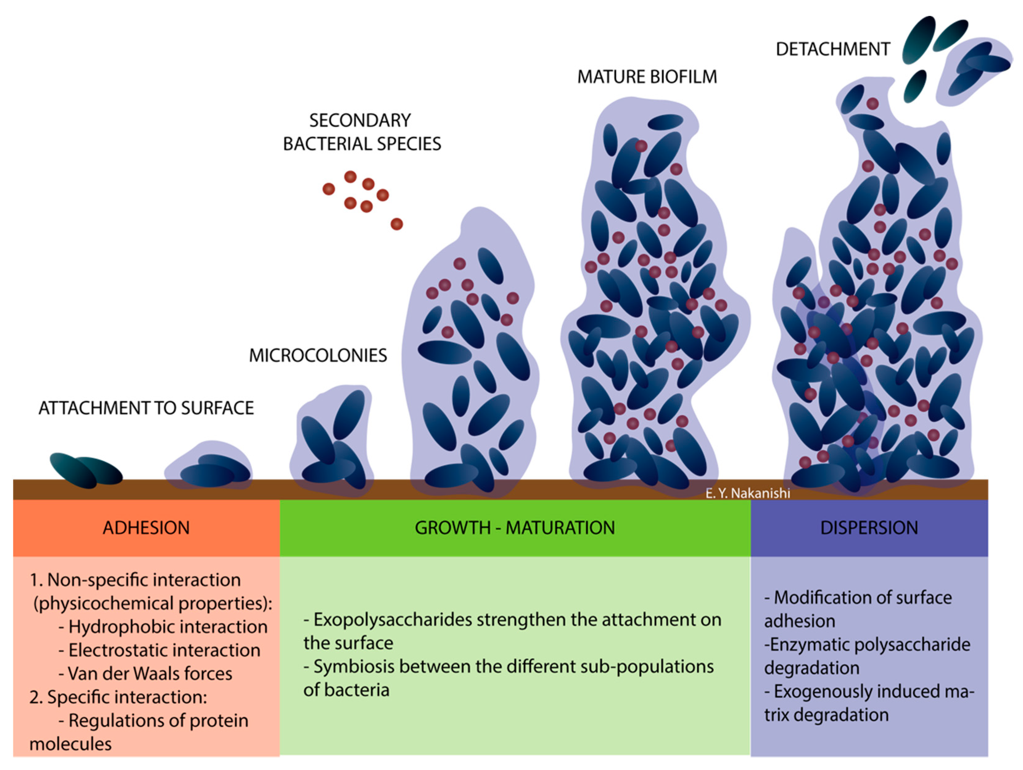
3.1. Biofilm and Formation Phases

The characterization of the material’s surface helps to provide a better understanding of how microorganisms interact and colonize in different materials, i.e., bio-receptivity of the material [27]. Bacterial colonization on a surface is a complex process that depends on the interplay of three main factors: (i) the material surface characteristics, such as specific surface roughness, surface porosity or topography, chemical composition and wettability or surface-free energy (SFE); (ii) the bacterial properties, i.e., surface charge, surface energy, shape and size, appendages, adhesins, etc. and (iii) the environmental conditions, such as the flow conditions around the surface (temperature, humidity, viscosity, hydrodynamics, pH, surface tension, ionic strength and dielectric properties) [22,27,28,29,30,31].
The pathogenicity of some bacteria, such as S. epidermidis, is attributed to its ability to adhere to surfaces, form a biofilm and remain on the surface [30]. The adhesion of micro-organisms to surfaces is an important parameter for assessing cleanability and thus minimizing transmission to animals in pig facilities [22]. The biofilms provide structural stability and protection from stressful conditions. The biofilms are composed of complex microbial structures, derived from extracellular polymeric substance (EPS). EPSs are formed mainly of proteins and carbohydrates. Biofilm formation depends mainly on microorganism type and density, temperature, pH, nutrient availability and type of materials [32].
To the best of our knowledge, no research has linked biofilm stages to ongoing infections in pig facilities. However, the phases and factors leading to biofilm formation are presented below. These do not explain any ongoing infection in pig facilities, but they describe at which stage the material plays a role in the bacterial attachment and the complexity of the biofilm that allow bacteria to remain on the surface even after cleaning and disinfection processes.
Figure 1 shows a schematic model of the phases and factors leading to biofilm formation. The first step in the process of bacterial adhesion to a biotic or abiotic surface consists of the initial attraction of the cells to the surface. The initial attraction involves locating, approaching, and sensing the proximity of the surface, followed by attachment [30]. This first step of bacteria attachment is reversible primarily through so-called non-specific interactions associated with physicochemical properties [33]. This early stage of biofilm formation is closely related to the properties of materials, which means that surface roughness, surface-free energy (SFE) and chemical composition have a large role in bacteria attachment.
The physical properties of the attachment process are a function of the distance and SFE. The main forces in play are Van der Waals attraction forces, the effect of surface electrostatic charge, hydrophobic interactions, Brownian motion and gravitational forces. Chemical interactions are linked to the surface-associated chemical gradients (chemotaxis) and adhesion sites’ surface-bound chemoattractants (“haptotaxis”). These gradients are formed from the presence of various chemical stimuli or the degradation of certain surface components [28,29,30,34,35].
Bacterial attachment makes adhesion possible in the second step of biofilm formation. Bacteria attach themselves irreversibly by specific interactions that involve distinct recognition, such as the regulation of protein molecule binding on the interacting surfaces [33,35,36]. This suggests a firmer adhesion of the bacteria to a surface by the selective-bridging function of bacterial surface structures, which include lipopolysaccharides, capsules, fimbriae, or pili and slime [30,37].
After the adhesion process, the biofilm is formed, i.e., the bacteria multiply rapidly and achieve a high cell density and grow in structures called microcolonies. These microcolonies produce one or more exopolysaccharides self-secreted by bacteria, helping to strengthen the attachment to the surface [33,38]. The biofilm maturation allows the transport of nutrients and metabolic waste, oxygenation, communication between the different sub-populations of bacteria within the biofilm and the synchronization of their behavior by the signal molecule exchanges [33]. In addition, mature biofilms can provide extra protection for the bacteria against environmental stressors, such as desiccation or UV rays, disinfectant or sanitizing agents as well as antibiotics [39,40,41]. This is possible due to the exopolymer matrix that prevents the diffusion of harmful chemical and environmental influences inside the biofilm [39].
However, many factors such as cellular density, the lack of nutrients, oxygen, extracellular signaling molecules, and environmental conditions can incite the detachment and dispersion process. The bacteria or specific subpopulation detached will spread to new locations [30,33,42,43]. An extensive review of the biofilm dispersion process by Rumbaugh and Sauer [43] considered the main mechanisms to be: (i) modification of surface adhesions; (ii) enzymatic polysaccharide degradation and (iii) exogenously induced matrix degradation.

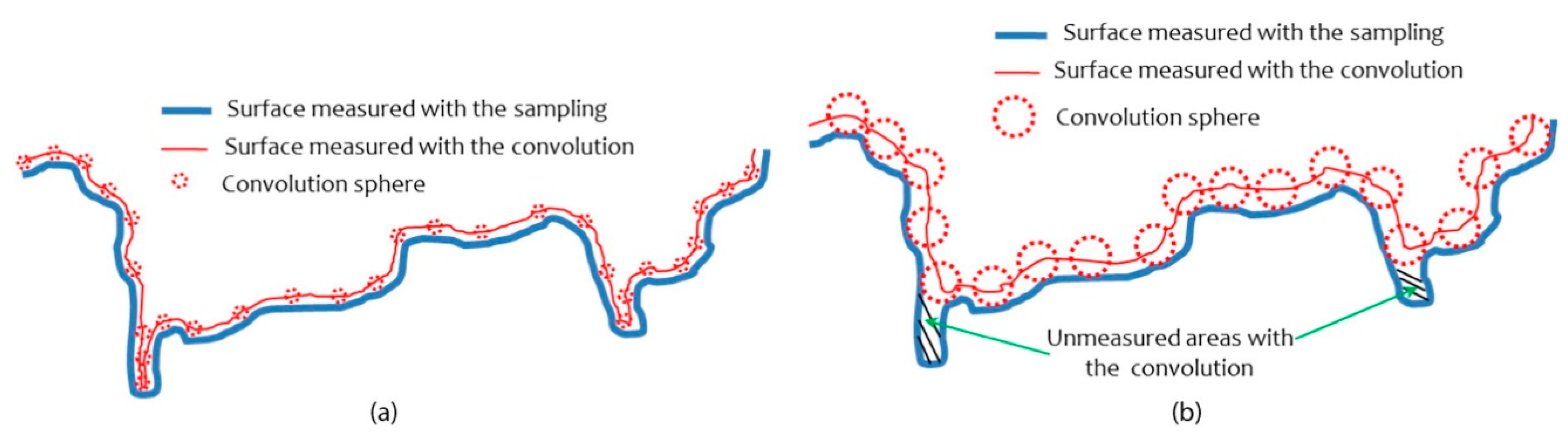
3.2. Surface Roughness Properties

3.2.1. Importance of Surface Roughness in Pig Facilities

In pig facilities, surface roughness is an important selection criterion for materials, mainly for floors. Rough floors are more abrasive to the feet and knees and thus can cause injuries to nursing pigs. Materials with high porosity or roughness can also retain moisture and manure, which will subsequently facilitate bacteria growth and make cleaning more difficult [24]. Materials with smooth surfaces can also cause hoof and leg injuries, as they can be slippery [23,24]. However, smooth surfaces drain and dry more rapidly, facilitating cleaning and disinfection.
Therefore, surface roughness significantly impacts bacterial attachment and subsequent biofilm formation in pig facilities. The pits, grooves and irregularities of rougher surfaces provide protection to the bacteria from removal forces, i.e., cleaning processes, and provide a larger global area available for colonization [28]. In fact, attachment can be established more easily on rough materials where bacteria are sheltered [36].

3.2.2. Surface Roughness of Materials

Different techniques have been used to analyze specific surface roughness and surface porosity, such as scanning electron microscopy (SEM), atomic force microscopy (AFM), lateral force microscopy (LFM), optical microscopy, coherence scanning interferometry (CSI) and confocal microscopy. The standard roughness parameters can be established by bidimensional (R) or tridimensional (S) analysis. The roughness parameters most used are the arithmetic mean of roughness (Ra) and the mean square deviation of roughness (Rq) [44,45]. Ra is defined as the average deviation of the profile in relation to its mean line and Rq is considered as an amplitude parameter that describes the variation of the surface topography in relation to an average plane [46,47]. Therefore, in order to take into account the location and the spacing between peaks and valleys, other roughness parameters can be used, such as the mean peak height (Rpm), the mean valley depth (Rvm), the mean peak-to-valley height (Rz), the ten points height (Rz) and the maximum valley depth (Rv) [45].
Table 2 presents the roughness parameter values obtained in the literature for the four main groups of materials found within a pig facility (cement-based materials, plastic, metals and engineered wood materials). Cement-based materials, in general, present a complex structure with a high surface roughness. Engineered wood materials, like cement, have a high surface roughness mainly due to the anatomic characteristic of the wood fibers [48,49,50]. Consequently, ordinary and prefabricated concrete as well as plywood and particleboard are more difficult to clean and keep in adequate sanitary condition.
In the case of the HDPE-coated steel slats found in pig facilities, the surface roughness is higher compared to other plastic materials. This comes from the thermal spray coating used in the production process. The coating is formed by the deposition of fine particles in a molten or semi-molten condition or even in fully solid state on the metal surface [51,52]. Overall, plastics and metals have a smoother roughness profile than the cement-based and engineered wood materials. Consequently, plastics and metals materials are easier to clean and maintain.

3.2.3. The Impact of Roughness on Bacteria Colonization

In the first step of biofilm formation, the manner in which bacteria locate, approach and sense the proximity of the surface or, in other words, how the bacteria view the surface, can be influenced by the roughness scale and the roughness geometry. The geometric roughness parameters provide the link between the microorganisms’ size and the roughness geometry [27].
Apedo et al. [27] studied the geometric roughness parameters of cement pastes with CSI analysis and the “window resizing” technique to calculate the sampling and the convolution. Sampling is the parameter related to the measuring tool, while the convolution is the parameter related to the roughness viewed by a bacterium of a given size, as shown in Figure 2. When the convolution sphere (probe radius, Rp) is ≤ 8, the surface measured with the sampling is the same for the convolution (Figure 2a), i.e., the perception of bacteria occurs on all surfaces. However, when the Rp > 8, the discrepancy between sampling and convolution becomes large enough because many parts of the sampled surface become inaccessible to the convolution sphere (Figure 2b). Furthermore, the surface viewed by the bacteria (convolution) is substantially smaller than the surface viewed by the measuring tool (sampling). Therefore, in these inaccessible parts, bacterial attachment is impossible.
Bacteria attachment factors (e.g., hydrodynamics, surface wettability, air entrapment, topography-induced cell ordering and segregation, physicochemical forces, cell membrane deformation and chemical gradient) could be influenced by the roughness scale, which includes the micrometric and nanometric scales. An extensive review on the influence of the roughness scale in the factors of attachment by Cheng et al. [29] considered for example that a surface with large pores presents a lower energy barrier for a bacterium to overcome in order to attach compared with the smaller pore surfaces, as the bacteria have a lower contact with the surface. In other words, there is an electrostatic and an acid–base interaction having lower forces against bacteria. Another example is the film’s conditioning and chemical gradients, which could affect attachment: (i) masking or changing surface properties; (ii) modifying the surface topography and (iii) providing sites for specific bacteria–surface interactions. Therefore, as presented by the authors, the film’s conditioning and chemical gradient factors have more effects on nanoscale roughness when compared to microscale.

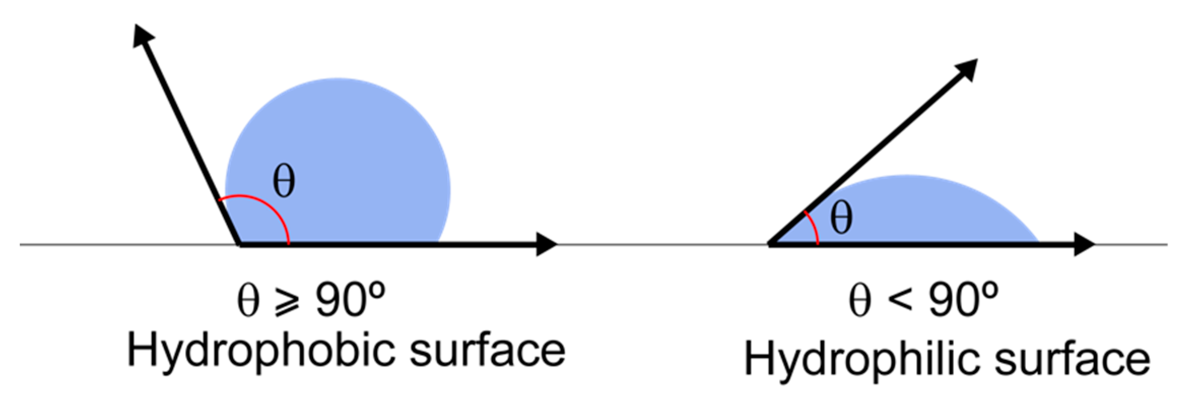
3.3. Surface-Free Energy Properties

3.3.1. Surface-Free Energy Materials

Materials with surface roughness in the nanoscale, such as some plastics and metals, present pits, grooves and irregularities inaccessible to the attachment of bacteria. For these types of surfaces, physical properties such as SFE can influence and improve bacterial attachment [28].
Surface-free energy has a definite impact on the wettability of materials. Surface-free energy is characterized by the interaction between the forces of cohesion and adhesion that determine whether or not wetting occurs [28]. This property is strongly dependent on the chemical composition of the surface and its roughness [57]. It can be established by contact angle measurement of the angle of the drop formed on the surface. Surface-free energy can be calculated by the three liquid phase methods based on the VanOss–Chaundhury–Good theory of wettability. The smaller the angle of the drop formed on the surface (less than 90°), the greater the wettability or SFE, i.e., it is considered a hydrophilic surface. On the other hand, an angle greater or equal to 90° represents a hydrophobic surface [62], as shown in Figure 3.
Cement-based materials present high surface-free energy, which means that cement is a hydrophilic material. Plastics exhibit different surface characteristics; for example, HPDE is considered a hydrophobic material, while PVC is considered a hydrophilic material [57,63]. Stainless surfaces are inherently hydrophilic with a nearly 90° contact angle [59] (Table 3).

3.3.2. Influence of SFE on Bacterial Colonization

Microbial surface hydrophobicity has been noted to be a dominant factor in influencing adhesion on surfaces [35]. Generally, hydrophobic surfaces, or surfaces with a low SFE, are preferred for attachment by a hydrophobic bacterium or a bacterium with a low SFE. However, bacteria with hydrophilic properties (high SFE) prefer to adhere to hydrophilic surfaces or surfaces with high SFE [28,38]. For example, Staphylococcus aureus presents a hydrophilic character, and thus this bacterium favors metal alloys (titanium-aluminum (6%) vanadium (4%)), over polymers (ultra-high molecular weight polyethylene) that present a more hydrophobic surface [64]. On the other hand, Mycobacterium avium is characterized by its outermost surface containing glycopeptidolipids and hydrophobic mycolic acids that give it a hydrophobic property [65]. Norton et al. [66] evaluated higher levels of M. avium biofilm on iron and galvanized pipe surfaces compared with copper or PVC surfaces.
According to Daffonchio et al. [67], bacteria are considered hydrophobic when presenting a contact angle greater than 45° and hydrophilic when presenting a contact angle less than 45°. Therefore, most bacteria shown in Table 4 have a contact angle lower than 45°, which means that they have a hydrophilic character, except Clostridium proteolyticum and Pseudomonas aeruginosa that present a hydrophobic character (CA > 45°). Considering the materials used in pig facilities (Table 3), these bacteria with a hydrophilic property will prefer to colonize first on the cement and engineered wood materials and then on polyvinyl chloride (PVC), stainless steel and high-density polyethylene (HDPE).
Moreover, the intensity of adhesion forces and the accumulation velocity in the colonization process are influenced by bacterial hydrophobicity [31,35]. Harimawan et al. [35] explored the nature of the adhesion mechanisms of two Gram-negative bacteria (Pseudomonas aeruginosa and Massilia timoniae) and one Gram-positive bacteria (Bacillus subtilis) on stainless steel. The authors observed that the Gram-negative bacteria presented higher adhesion forces compared with the Gram-positive bacteria. The main reason for this is because P. aeruginosa (CA = 47.9°) and M. timonae (CA = 39.6°) have a higher hydrophobicity than B. subtilis (CA = 31.1°), as their contact angles (P. aeruginosa and M.timonae) are closer to the hydrophobicity of the stainless steel, with a contact angle of 86.8°. Further, in a study carried out by Mueller et al. [31], the authors demonstrated that P. aeruginosa is more hydrophobic than P. fluorescens, and consequently the process of cellular accumulation of P. aeruginosa was 5 times faster than P. fluorescens on stainless steel, copper, silicon and glass materials. It shows that when comparing bacteria with different hydrophobicity on the same surface, the cell accumulation is faster when the bacteria’s hydrophobicity is closer to the one of the surfaces, which is P. aeruginosa in this case.
Even though a difference exists between the hydrophobicity of the surface and of the bacteria, the colonization process can still occur [71]. In this case, the non-specific chemical interaction with the material surface is an important mechanism for bacterial adhesion. An example of this is the capacity of Staphylococcus epidermidis, a bacterium that has a hydrophilic property, to colonize on polyethylene, which is a hydrophobic surface. It is possible that because S. epidermidis produces the protein adhesin (e.g., exopolysaccharide) it can adhere to a hydrophobic surface [64,72]. In addition, the non-specific chemical interaction is influenced by the chemical composition of the surface material. Therefore, the level of corrosion or decomposition of the material can influence adhesion and biofilm formation [66]. The materials used in pig facilities are exposed to several different conditions, for example, water from cleaning, drinking water, food residues, high humidity inside the building and chemical compounds from manure (i.e., ammonium and hydrogen sulfate), boosting corrosion and/or decomposition of the material [23].
As observed, the materials’ surface has a great impact on the bacterial colonization process. One method to prevent bacterial adhesion is changing the material surface characteristics [28]. The literature has suggested different methods for modifying the hydrophobicity of different types of materials, such as chemical change of hardened cement pastes (HCP) [73], mortar [53], low-density polyethylene (LDPE) [74], thermal change (termorretification) of plywood [61] and the addition of metal particles on plastics [56,57].

4. Impact of Washing Procedures on the Materials’ Sanitary Characteristics

4.1. Material Cleanability Evaluation Methods and Parameters

Although cleaning and disinfection processes are not able to totally eliminate the risk of disease, they can help minimize the negative impact of most endemic pigs’ infections [75]. Actually, proper cleaning and disinfection before stocking new pigs in a facility are very important for preventing the spread of infectious agents [76]. As mentioned before, bacteria could be sheltered in pits, grooves and irregularities of rougher surfaces [28] as well as inside the biofilm [15,39,40].
Many methods are available for evaluating the effect of cleaning and disinfection processes on materials’ surfaces, i.e., the presence or absence of bacteria or biofilm. These methods for evaluating cleanability are based on a measurement principle, which includes microbiological analysis [77], chemical analysis (i.e., chromatography and spectrometry) [78], biochemical analysis (i.e., ATP (adenosine triphosphate) bioluminescence) [76], physical analysis (i.e., colorimetry) [79,80], visual methods and radiochemical methods [80,81].
Luyckx et al. [77] evaluated the presence of some bacteria before and after the classical cleaning and disinfection protocol for nursery units. This protocol consisted of removing the manure with cold water, then after 24 h, the pen was soaked for 30 min with degreaser detergent. Subsequently, the pen was washed with high pressure (150 bar) cold water and finally, it was disinfected and kept empty for two weeks. With this washing protocol, the authors observed a 3.54 log CFU/sampling area reduction of Enterococcus spp. However, the presence of these bacteria was observed on floors and drinking nipples even after the protocol was used. For E. coli and fecal coliforms, reductions of 41% and 51% were observed, respectively; however, they were still found on floors, drinking nipples and feeding troughs after the washing protocol. Methicillin-resistant Staphylococcus aureus (MRSA) was reduced by 81% after the cleaning protocol, but drinking nipples were still the most contaminated by MRSA after disinfection. Therefore, according to Luyckx et al. [77], the area where bacteria still remain, even after the cleaning and disinfection protocols, are grid floors and drinking nipples.
Yi et al. [76] carried out the method using ATP bioluminescence in an empty pig farrowing unit. The evaluation was carried out before and after cleaning and at different times after disinfection (i.e., 1 h, 3 h, 6 h, 24 h, 48 h, 5 days and 7 days) in three locations: two on the polypropylene grid floor (center and corner) and one in feeders. The authors observed that the mean ATP bioluminescence levels of the floor corner and feeders measured after cleaning were not significantly different from those measured before cleaning. Moreover, the mean ATP bioluminescence levels increased 1 and 3 h after disinfection for floor corners and feeders, respectively. For the floor center, a significant decrease of the mean ATP bioluminescence levels occurred after cleaning and persisted up to 48 h after disinfection. After this time, the levels measured exceeded those measured immediately after cleaning.
The type of surface and biofilm are important elements of bacterial prevalence. For example, bacteria such as Haemophilus influenza, Bordetella pertussis, Proteus vulgaris and others can remain for days. On the other hand, bacteria such as Enterococcus spp., Staphylococcus aureus, Streptococcus, Acinetobacter spp., Escherichia coli, Klebsiella spp. and Pseudomonas aeruginosa can survive for months on dry surfaces [18]. Hurnik [82] describes some survival times of common pig pathogens in different environments:
  • Mycoplasma Hyopneumoniae: Up to 7 days in organic matter
  • Actinobacillus Pleuropneumoniae: Few days in organic matter
  • Pasteurella Multocida: 8 days in water or 6 days in liquid manure
  • Streptococcus suis: 25 days (9 °C) or 100 days (0 °C)
  • Salmonella ssp.: Years in manure, 115 days in water and 120 days in soil
  • Escherichia coli: 11 weeks in manure
In addition, some flaws in cleaning and disinfection processes are pointed out by Dias et al. [75], and they are related to the prevalence of bacteria even after cleaning and disinfecting. Those flaws are the incomplete removal of wastes before cleaning procedures, the lack of wall and ceiling disinfection and inadequate water quantity and pressure.

4.2. Effect of the Washing Procedure on Biofilm Destruction on Different Materials

No standardized washing procedure (i.e., following the same washing parameters and conditions) exists and, even when checking recommendations, the parameters are not standardized. Furthermore, each producer has his own washing procedure (parameters). Because many washing parameters are used, it raises the following question: Which are the proper measures to adequately remove the biofilm and eliminate bacteria?
Some washing parameters such as water jet pressure, water temperature, washing time and distance between the water jet and the material surface can be adjusted optimally to ensure that the biofilm is destroyed and that the cleaning process is more efficient. Although cleaning and disinfection processes are highly important in pig facilities, little attention is given to the relationship between the parameters influencing biofilm destruction and the cleanability of the surface materials in pig facilities. Moreover, from the perspective of the pig producers, there is a certain skepticism regarding the value of hygiene and disinfection.
Burfoot and Middleton [58] studied the impact of water pressure on the removal of biofilms of P. aeruginosa and S. aureus from stainless steel surfaces. A 15° flat nozzle with two nozzle pressures (2.5 and 9.5 MPa) and different distances between the nozzle and targeted surface (20 to 120 cm) was used to vary the impact pressure. The authors highlighted that the impact pressure did not have a significant effect on the removal of the biofilm. Furthermore, P. aeruginosa biofilm was the bacteria most difficult to remove.
The authors also investigated other cleaning parameters, such as water temperature (8 °C and 60 °C), washing time (5, 30 and 60 s), detergent and nozzle angle (45° and 90°) relative to the sample surface. The results showed that these parameters impacted the microbial reduction. The use of hot water resulted in a greater microbial reduction than cold water. When cold water was used, the microbial removal increased with the cleaning time, whereas the same trend was not observed with hot water. The application of detergent produced a 5.6-log10 microbial reduction for both hot and cold water at a pressure of 9.5 MPa. No clear relationship was found between the angle of the nozzle and microbial removal. However, it is important to highlight that this trend could be different with other surface materials.
Kymäläinen et al. [80] evaluated the impact of water temperature (10, 40 and 70 °C) on the surface cleanability of a cement-based material using colorimetric measurements. In this study, pig manure and synthetic pig manure were used to soil surfaces. The cleaning was carried out with high pressure (12 MPa), a passage speed of 0.9 m·s−1, a distance of 18 cm between the nozzle and the surface and a nozzle angle of 45° from the perpendicular. The results showed that cleanability was not influenced by the water temperatures used. However, according to Böhm [83], for concrete as well as for wood-based material surfaces, the optimum water temperature for cleaning is about 40 °C. On the other hand, for metal surfaces, higher water temperatures may provide better results, as long as the wash time is sufficient.
In addition, as mentioned by Hurnik [82], using hot water might reduce the washing time by about 22% when a presoak is not performed. However, there is no reduction on the washing time, between hot or cold water, when the presoak is used. An important point highlighted by the authors is that hot water is more efficient to realize the cleaning. However, it can create a fog that affects visualization during the cleaning procedure. Further, the application of a detergent can results in a reduction of 12% of the washing time.
When considering the type of flooring (fully or partial slatted), a pre-soak step decreases the washing time of partial slatted flooring but not of fully slatted flooring. One reason pre-soaking does not impact the washing time of fully slatted flooring is because there is no accumulation of manure like on partial slatted flooring [84]. However, there is no information about the impact pre-soaking has on biofilm removal.
Other parameters such as nozzle type can have important effects on cleaning processes. For example, Predicala et al. [84] observed that the use of a conventional nozzle (i.e., rotating nozzle) results in a 62% reduction of microbial ATP, while in other nozzles (Y-nozzle, water broom and 4-in nozzle) this reduction was 16–34% on concrete surfaces (floor). In this case, the effect of the impact force caused by a conventional nozzle allowed for a better cleaning on a rougher surface.
From these results, it appears that bacteria are still present even after the cleaning and disinfection processes. Therefore, the biofilm is not completely removed, bacterial transmission will continue and, incidentally, pigs may be infected. Consequently, further studies focused on washing parameters and their impact on biofilm removal on different surfaces are necessary for developing more efficient cleaning and disinfection processes and standards.

5. Conclusions

This review summarizes the main effects of surface material on bacterial colonization as well as the impacts of washing procedures on the sanitary factors related to the different materials used in pig facilities.
Firstly, biofilm formation on the surface of different types of materials is a complex system affected by the characteristics of each material’s surface, of each group of bacteria as well as the environmental conditions within the pig facility. The material properties (roughness and free-energy surface) play an important role in the early stage of bacterial attachment and consequently biofilm formation. Secondly, the use of common washing procedures does not allow the complete removal of the biofilm on surfaces.
In fact, the relationship between the surface material, bacteria characteristics and washing parameters can explain why some organisms are easily removed or why sometimes some bacteria persist in pig facilities even after the cleaning and disinfection process. The bacteria persistence comes from the fact that the biofilm is not removed completely; as a result, certain bacteria survive inside the biofilm and continue spreading diseases. This demonstrates the importance of not just removing bacteria but also removing the biofilm on the surface in order to prevent the spreading of diseases.
To increase biofilm removal, some parameters such as chemical treatment (i.e., detergent application), higher temperature (for some materials), or longer treatment/washing times have to be considered.
Therefore, this overview may serve to clarify the importance of the following three factors for improving sanitary conditions in pig facilities: biofilm, surface material and washing parameters.
However, it is important to emphasize here that more studies are required to better understand the relationships between the previously discussed factors. For example, studies should determine the best combination of washing parameters as a function of material type and bacteria that can be harmful to pigs. Since several studies conducted in pig facilities have shown that the feeders, drinkers and floors are the sites where bacteria persist the most even after the cleaning and disinfection process, future research should focus on metals, plastics and cement-based materials.

Author Contributions

Conceptualization, E.Y.N., J.H.P. and S.G.; methodology, formal analysis, investigation, E.Y.N.; writing—original draft preparation, E.Y.N.; writing—review and editing, E.Y.N., S.G., S.F. and J.H.P.; visualization, E.Y.N.; supervision, S.G. and S.F. All authors have read and agreed to the published version of the manuscript.

Funding

This work was funded through the Innov’Action Agroalimentaire program (IA119051), under the Canadian Partnership for Agriculture, an agreement between the governments of Canada and Quebec and MITACS Accelerate program (grant number IT16507).

Institutional Review Board Statement

Not applicable.

Informed Consent Statement

Not applicable.

Data Availability Statement

Not applicable.

Acknowledgments

The authors are also grateful to the framework agreement of collaboration between Université Laval (Chair in Educational Leadership on Sustainable Agricultural Buildings) and the Research and Development Institute for the Agri-Environment (IRDA).

Conflicts of Interest

The authors declare no conflict of interest.

References

  1. Les Éleveurs de porcs du Québec-Guide de Lavage, Désinfection et Séchage Des Porcheries. 2011. Available online: http://www.accesporcqc.ca/nsphp/portail/publications/pub_dl.php?dir=364&download=guidedeldsdesporcheries.pdf (accessed on 17 May 2021).
  2. Nantel-Fortier, N.; Lachapelle, V.; Letellier, A.; L’Homme, Y.; Brassard, J. Kobuvirus Shedding Dynamics in a Swine Production System and Their Association with Diarrhea. Vet. Microbiol. 2019, 235, 319–326. [Google Scholar] [CrossRef] [PubMed]
  3. Haas, B.; Grenier, D. Understanding the Virulence of Streptococcus Suis: A Veterinary, Medical, and Economic Challenge. Med. Mal. Infect. 2018, 48, 159–166. [Google Scholar] [CrossRef] [PubMed]
  4. Letellier, A.; Messier, S.; Paré, J.; Ménard, J.; Quessy, S. Distribution of Salmonella in Swine Herds in Quebec. Vet. Microbiol. 1999, 67, 299–306. [Google Scholar] [CrossRef]
  5. Smith, B.A.; Meadows, S.; Meyers, R.; Parmley, E.J.; Fazil, A. Seasonality and Zoonotic Foodborne Pathogens in Canada: Relationships between Climate and Campylobacter, E. Coli and Salmonella in Meat Products. Epidemiol. Infect. 2019, 147. [Google Scholar] [CrossRef] [PubMed] [Green Version]
  6. Perri, A.M.; Poljak, Z.; Dewey, C.; Harding, J.C.S.; O’Sullivan, T.L. Network Analyses Using Case-Control Data to Describe and Characterize the Initial 2014 Incursion of Porcine Epidemic Diarrhea (PED) in Canadian Swine Herds. Prev. Vet. Med. 2019, 162, 18–28. [Google Scholar] [CrossRef]
  7. Post, K.W. Overview of Bacteria. In Diseases of Swine; Zimmerman, J.J., Karriker, L.A., Ramirez, A., Schwartz, K.J., Stevenson, G.W., Zhang, J., Eds.; John Wiley & Sons: Hoboken, NJ, USA, 2019; pp. 743–748. [Google Scholar]
  8. Wang, Y.; Liao, J.; Mehmood, K.; Chang, Y.F.; Tang, Z.; Zhang, H. Escherichia Coli Isolated in Pigs, Guangdong, China: Emergence of Extreme Drug Resistance (XDR) Bacteria. J. Infect. 2020, 81, 318–356. [Google Scholar] [CrossRef] [PubMed]
  9. Costa, T.; Akdeniz, N. A Review of the Animal Disease Outbreaks and Biosecure Animal Mortality Composting Systems. Waste Manag. 2019, 90, 121–131. [Google Scholar] [CrossRef] [PubMed]
  10. FAO. Food Outlook–Biannual Report on Global Food Markets; FAO: Rome, Italy, 2019. [Google Scholar]
  11. Gallardo, C.; Fernández-Pinero, J.; Arias, M. African Swine Fever (ASF) Diagnosis, an Essential Tool in the Epidemiological Investigation. Virus Res. 2019, 271. [Google Scholar] [CrossRef]
  12. FAO. The Global Platform for African Swine Fever and Other Important Diseases of Swine; Animal Production and Health Report No. 4; FAO: Rome, Italy, 2014. [Google Scholar]
  13. Lekagul, A.; Tangcharoensathien, V.; Yeung, S. Patterns of Antibiotic Use in Global Pig Production: A Systematic Review. Vet. Anim. Sci. 2019, 7, 100058. [Google Scholar] [CrossRef]
  14. McCarthy, A.J.; Witney, A.A.; Gould, K.A.; Moodley, A.; Guardabassi, L.; Voss, A.; Denis, O.; Broens, E.M.; Hinds, J.; Lindsay, J.A. The Distribution of Mobile Genetic Elements (MGEs) in MRSA CC398 Is Associated with Both Host and Country. Genome Biol. Evol. 2011, 3, 1164–1174. [Google Scholar] [CrossRef] [PubMed] [Green Version]
  15. De Foy, C. Évaluation des Matériaux en Fonction de la Contamination Bactérienne de Surface, des Émissions d’odeurs et des Caractéristiques Physiques afin de Réduire la Dérive Sanitaire des Bâtiments Porcins. Master’s Thesis, Laval University, Québec City, QC, Canada, 2005. [Google Scholar]
  16. Colclasure, V.J.; Soderquist, T.J.; Lynch, T.; Schubert, N.; McCormick, D.S.; Urrutia, E.; Knickerbocker, C.; McCord, D.; Kavouras, J.H. Coliform Bacteria, Fabrics, and the Environment. Am. J. Infect. Control 2015, 43, 154–158. [Google Scholar] [CrossRef] [PubMed]
  17. Kim, Y.; Krishna, V.D.; Torremorell, M.; Goyal, S.M.; Cheeran, M.C.J. Stability of Porcine Epidemic Diarrhea Virus on Fomite Materials at Different Temperatures. Vet. Sci. 2018, 5, 21. [Google Scholar] [CrossRef] [Green Version]
  18. Kramer, A.; Schwebke, I.; Kampf, G. How Long Do Nosocomial Pathogens Persist on Inanimate Surfaces? A Systematic Review. BMC Infect. Dis. 2006, 6, 1–8. [Google Scholar] [CrossRef] [Green Version]
  19. Krippendorff, K. Content Analysis: An Introduction to Its Methodology, 2nd ed.; Sage Publications: Thousand Oaks, CA, USA, 2004; ISBN 0-7619-1544-3. [Google Scholar]
  20. Melmer, D.J.; O’Sullivan, T.L.; Poljak, Z. A Descriptive Analysis of Swine Movements in Ontario (Canada) as a Contributor to Disease Spread. Prev. Vet. Med. 2018, 159, 211–219. [Google Scholar] [CrossRef]
  21. USDA. The United States Department of Agriculture-Foreign Animal Disease Preparedness and Response Plan; USDA: Washngton, DC, USA, 2011.
  22. Boon, C.R.; Wray, C. Building Design in Relation to the Control of Diseases of Intensively Housed Livestock. J. Agric. Eng. Res. 1989, 43, 149–161. [Google Scholar] [CrossRef]
  23. Gorman, C.; Turnbull, J.E. PLAN M-3003: Construction et Installations Techniques Des Porcheries; Canada Plan Service: Québec City, QC, Canada, 1988; Volume 1. [Google Scholar]
  24. MWPS-8 Midwest Plan Service. Swine Housin and Equipment Handbook, 4th ed.; Midwest Plan Service: Ames, IA, USA, 1983; ISBN 0-8937-054-8. [Google Scholar]
  25. AHDB. Agriculture and Horticulture Development Board-Finisher Pig Buildings Design and Build—A Blueprint for English Farms; BPEX: Warwickshire, UK, 2013; pp. 1–123. [Google Scholar]
  26. Pelletier, F.; Marquis, A.; Godbout, S.; Joncas, R. Gas and Odor Emiissions From Swine Building Material. Am. Soc. Agric. Eng. 2005, 48, 721–728. [Google Scholar] [CrossRef]
  27. Apedo, K.L.; Montgomery, P.; Serres, N.; Fond, C.; Feugeas, F. Geometrical Roughness Analysis of Cement Paste Surfaces Using Coherence Scanning Interferometry and Confocal Microscopy. Mater. Charact. 2016, 118, 212–224. [Google Scholar] [CrossRef] [Green Version]
  28. Brajkovic, D.; Antonijevic, D.; Milovanovic, P.; Kisic, D.; Zelic, K.; Djuric, M.; Rakocevic, Z. Surface Characterization of the Cement for Retention of Implant Supported Dental Prostheses: In Vitro Evaluation of Cement Roughness and Surface Free Energy. Appl. Surf. Sci. 2014, 311, 131–138. [Google Scholar] [CrossRef]
  29. Cheng, Y.; Feng, G.; Moraru, C.I. Micro-and Nanotopography Sensitive Bacterial Attachment Mechanisms: A Review. Front. Microbiol. 2019, 10, 191. [Google Scholar] [CrossRef] [PubMed] [Green Version]
  30. Katsikogianni, M.; Missirlis, Y.F. Concise Review of Mechanisms of Bacterial Adhesion to Biomaterials and of Techniques Used in Estimating Bacteria-Material Interactions. Eur. Cells Mater. 2004, 8, 37–57. [Google Scholar] [CrossRef]
  31. Mueller, R.F.; Characklis, W.G.; Jones, W.L.; Sears, J.T. Characterization of Initial Events in Bacterial Surface Colonization by Two Pseudomonas Species Using Image Analysis. Biotechnol. Bioeng. 1992, 39, 1161–1170. [Google Scholar] [CrossRef] [PubMed] [Green Version]
  32. Trinh, Q.T.; Bal Krishna, K.C.; Salih, A.; Listowski, A.; Sathasivan, A. Biofilm Growth on PVC and HDPE Pipes Impacts Chlorine Stability in the Recycled Water. J. Environ. Chem. Eng. 2020, 8, 104476. [Google Scholar] [CrossRef]
  33. Hathroubi, S. Rôle des Polysaccharides de Surface dans la Formation des Biofilms et rôle du Biofilm D’actinobacillus Pleuropneumoniae dans la Pathogénicité. Ph.D. Thesis, Université de Montréal, Montréal, QC, Canada, 2016. [Google Scholar]
  34. Busscher, H.J.; Norde, W.; van der Mei, H.C. Specific Molecular Recognition and Nonspecific Contributions to Bacterial Interaction Forces Downloaded From. Appl. Environ. Microbiol. 2008, 74, 2559–2564. [Google Scholar] [CrossRef] [PubMed] [Green Version]
  35. Harimawan, A.; Rajasekar, A.; Ting, Y.P. Bacteria Attachment to Surfaces-AFM Force Spectroscopy and Physicochemical Analyses. J. Colloid Interface Sci. 2011, 364, 213–218. [Google Scholar] [CrossRef] [PubMed]
  36. Gharechahi, M.; Moosavi, H.; Forghani, M. Effect of Surface Roughness and Materials Composition. J. Biomater. Nanobiotechnology 2012, 3, 541–546. [Google Scholar] [CrossRef] [Green Version]
  37. Berne, C.; Ducret, A.; Hardy, G.G.; Brun, Y.V. Adhesins Involved in Attachment to Abiotic Surfaces by Gram-Negative Bacteria. Microbiol. Spectr. 2015, 3. [Google Scholar] [CrossRef] [Green Version]
  38. Gupta, K.K.; Devi, D. Characteristics Investigation on Biofilm Formation and Biodegradation Activities of Pseudomonas Aeruginosa Strain ISJ14 Colonizing Low Density Polyethylene (LDPE) Surface. Heliyon 2020, 6, e04398. [Google Scholar] [CrossRef] [PubMed]
  39. Chen, B.; Abdallah, M.; Campistron, P.; Moulin, E.; Callens, D.; Khelissa, S.O.; Debreyne, P.; Chihib, N.E.; Delaplace, G. Detection of Biofilm Formation by Ultrasonic Coda Wave Interferometry. J. Food Eng. 2021, 290, 110219. [Google Scholar] [CrossRef]
  40. Li, G.; Wu, Y.; Li, Y.; Hong, Y.; Zhao, X.; Reyes, P.I.; Lu, Y. Early Stage Detection of Staphylococcus Epidermidis Biofilm Formation Using MgZnO Dual-Gate TFT Biosensor. Biosens. Bioelectron. 2020, 151, 111993. [Google Scholar] [CrossRef] [PubMed]
  41. Wagner, E.M.; Pracser, N.; Thalguter, S.; Fischel, K.; Rammer, N.; Pospíšilová, L.; Alispahic, M.; Wagner, M.; Rychli, K. Identification of Biofilm Hotspots in a Meat Processing Environment: Detection of Spoilage Bacteria in Multi-Species Biofilms. International. J. Food Microbiol. 2020, 328, 108668. [Google Scholar] [CrossRef] [PubMed]
  42. Darvishi, S.; Pick, H.; Oveisi, E.; Girault, H.H.; Lesch, A. Soft-Probe-Scanning Electrochemical Microscopy Reveals Electrochemical Surface Reactivity of E. Coli Biofilms. Sens. Actuators B Chem. 2021, 334, 129669. [Google Scholar] [CrossRef]
  43. Rumbaugh, K.P.; Sauer, K. Biofilm Dispersion. Nat. Rev. Microbiol. 2020, 18, 571–586. [Google Scholar] [CrossRef] [PubMed]
  44. Ficker, T.; Martišek, D. Digital Fracture Surfaces and Their Roughness Analysis: Applications to Cement-Based Materials. Cem. Concr. Res. 2012, 42, 827–833. [Google Scholar] [CrossRef]
  45. Santos, P.M.D.; Júlio, E.N.B.S. A State-of-the-Art Review on Roughness Quantification Methods for Concrete Surfaces. Constr. Build. Mater. 2013, 38, 912–923. [Google Scholar] [CrossRef]
  46. ISO. EN ISO 16610-21 Geometrical Product Specifications (GPS)—Filtration—Part 21: Linear Profile Filters: Gaussian Filters; ISO: Geneva, Switzerland, 2011. [Google Scholar]
  47. ISO. EN ISO 11562 Geometrical Product Specifications (GPS)—Surface Texture: Profile Method—Metrological Characteristics of Phase Correct Filters; ISO: Geneva, Switzerland, 1996. [Google Scholar]
  48. Kiliç, M.; Burdurlu, E.; Aslan, S.; Altun, S.; Tümerdem, Ö. The Effect of Surface Roughness on Tensile Strength of the Medium Density Fiberboard (MDF) Overlaid with Polyvinyl Chloride (PVC). Mater. Des. 2009, 30, 4580–4583. [Google Scholar] [CrossRef]
  49. Luo, B.; Zhang, J.; Bao, X.; Liu, H.; Li, L. The Effect of Granularity on Surface Roughness and Contact Angle in Wood Sanding Process. Meas. J. Int. Meas. Confed. 2020, 165, 108133. [Google Scholar] [CrossRef]
  50. Tabarsa, T.; Ashori, A.; Gholamzadeh, M. Evaluation of Surface Roughness and Mechanical Properties of Particleboard Panels Made from Bagasse. Compos. Part B 2011, 42, 1330–1335. [Google Scholar] [CrossRef]
  51. Liu, Y.; Shao, X.; Huang, J.; Li, H. Flame Sprayed Environmentally Friendly High Density Polyethylene (HDPE)–Capsaicin Composite Coatings for Marine Antifouling Applications. Mater. Lett. 2019, 238, 46–50. [Google Scholar] [CrossRef]
  52. Vuoristo, P. Thermal Spray Coating Processes. In Comprehensive Materials Processing; Hashmi, S., Batalha, G.F., Tyne, C.J.V., Yilbas, B., Eds.; Elsevier: Amsterdam, The Netherlands, 2014; Volume 4, pp. 229–276. ISBN 9780080965338. [Google Scholar]
  53. Barnat-Hunek, D.; Grzegorczyk-Frańczak, M.; Suchorab, Z. Surface Hydrophobisation of Mortars with Waste Aggregate by Nanopolymer Trietoxi-Isobutyl-Silane and Methyl Silicon Resin. Constr. Build. Mater. 2020, 264. [Google Scholar] [CrossRef]
  54. Szafraniec, M.; Barnat-Hunek, D.; Grzegorczyk-Frańczak, M.; Trochonowicz, M. Surface Modification of Lightweight Mortars by Nanopolymers to Improve Theirwater-Repellency and Durability. Materials 2020, 13, 1350. [Google Scholar] [CrossRef] [PubMed] [Green Version]
  55. Mezaroba, C.; Becker, D.; Nahorny, J.; Recco, A.A.C.; Fontana, L.C. Nano-Rugosidade Gerada Em Amostras de Polímero PEAD Através de Plasma RF de N2/O2. Matéria 2018, 23. [Google Scholar] [CrossRef]
  56. Hill, D.; Barron, A.R.; Alexander, S. Controlling the Wettability of Plastic by Thermally Embedding Coated Aluminium Oxide Nanoparticles into the Surface. J. Colloid Interface Sci. 2020, 567, 45–53. [Google Scholar] [CrossRef] [PubMed]
  57. Song, Z.; Lin, E.S.; Uddin, M.H.; Ong, J.W.; Abid, H.A.; Xiong, Z.; Li, D.; Liew, O.W.; Ng, T.W. Temporal Evolution of Wetting Transitions of Graphene Oxide Coated on Roughened Polyvinyl Chloride Surfaces. Mater. Today Commun. 2020, 25, 101650. [Google Scholar] [CrossRef]
  58. Burfoot, D.; Middleton, K. Effects of Operating Conditions of High Pressure Washing on the Removal of Biofilms from Stainless Steel Surfaces. J. Food Eng. 2009, 90, 350–357. [Google Scholar] [CrossRef]
  59. Song, J.W.; Zeng, D.L.; Fan, L.W. Temperature Dependence of Contact Angles of Water on a Stainless Steel Surface at Elevated Temperatures and Pressures: In Situ Characterization and Thermodynamic Analysis. J. Colloid Interface Sci. 2020, 561, 870–880. [Google Scholar] [CrossRef] [PubMed]
  60. Hiziroglu, S.; Jarusombuti, S.; Fueangvivat, V. Surface Characteristics of Wood Composites Manufactured in Thailand. Build. Environ. 2004, 39, 1359–1364. [Google Scholar] [CrossRef]
  61. Candan, Z.; Büyüksari, U.; Korkut, S.; Unsal, O.; Çakicier, N. Wettability and Surface Roughness of Thermally Modified Plywood Panels. Ind. Crops Prod. 2012, 36, 434–436. [Google Scholar] [CrossRef]
  62. Lazghab, M.; Saleh, K.; Pezron, I.; Guigon, P.; Komunjer, L. Wettability Assessment of Finely Divided Solids. Powder Technol. 2005, 157, 79–91. [Google Scholar] [CrossRef]
  63. Kisić, D.; Nenadović, M.; Barudžija, T.; Noga, P.; Vaňa, D.; Muška, M.; Rakočević, Z. Modification of Polyethylene’s Surface Properties by High Fluence Fe Implantation. Nucl. Instrum. Methods Phys. Res. Sect. B Beam Interact. Mater. At. 2020, 462, 143–153. [Google Scholar] [CrossRef]
  64. Barth, E.; Myrvik, Q.M.; Wagner, W.; Gristina, A.G. In Vitro and in Vivo Comparative Colonization of Staphylococcus Aureus and Staphylococcus Epidermidis on Orthopaedic Implant Materials. Biomaterials 1989, 10, 325–328. [Google Scholar] [CrossRef]
  65. Tatchou-Nyamsi-König, J.A.; Dague, E.; Mullet, M.; Duval, J.F.L.; Gaboriaud, F.; Block, J.C. Adhesion of Campylobacter Jejuni and Mycobacterium Avium onto Polyethylene Terephtalate (PET) Used for Bottled Waters. Water Res. 2008, 42, 4751–4760. [Google Scholar] [CrossRef] [PubMed]
  66. Norton, C.D.; LeChevallier, M.W.; Falkinham, J.O. Survival of Mycobacterium Avium in a Model Distribution System. Water Res. 2004, 38, 1457–1466. [Google Scholar] [CrossRef] [PubMed]
  67. Daffonchio, D.; Thaveesri, J.; Verstraete, W. Contact Angle Measurement and Cell Hydrophobicity of Granular Sludge from Upflow Anaerobic Sludge Bed Reactors. Appl. Environ. Microbiol. 1995, 61, 3676–3680. [Google Scholar] [CrossRef] [PubMed] [Green Version]
  68. De Pimentel-Filho, N.J.; de Martins, M.C.F.; Nogueira, G.B.; Mantovani, H.C.; Vanetti, M.C.D. Bovicin HC5 and Nisin Reduce Staphylococcus Aureus Adhesion to Polystyrene and Change the Hydrophobicity Profile and Gibbs Free Energy of Adhesion. Int. J. Food Microbiol. 2014, 190, 1–8. [Google Scholar] [CrossRef] [Green Version]
  69. Braga, P.C.; Reggio, S. Correlation between Reduction of Surface Hydrophobicity of S. Aureus and the Decrease in Its Adhesiveness Induced by Subinhibitory Concentrations of Brodimoprim. Pharmacol. Res. 1995, 32, 315–319. [Google Scholar] [CrossRef]
  70. Absolom, D.R.; Lamberti, F.V.; Policova, Z.; Zingg, W.; van Oss, C.J.; Wilhelm Neumann, A.A. Surface Thermodynamics of Bacterial Adhesion. Appl. Environ. Microbiol. 1983, 46, 90–97. [Google Scholar] [CrossRef] [PubMed] [Green Version]
  71. Cerca, N.; Pier, G.B.; Vilanova, M.; Oliveira, R.; Azeredo, J. Quantitative Analysis of Adhesion and Biofilm Formation on Hydrophilic and Hydrophobic Surfaces of Clinical Isolates of Staphylococcus Epidermidis. Res. Microbiol. 2005, 156, 506–514. [Google Scholar] [CrossRef] [PubMed] [Green Version]
  72. Timmerman, C.P.; Fleer, A.; Besnier, J.M.; de Graaf, L.; Cremers, F.; Verhoef, J. Characterization of a Proteinaceous Adhesin of Staphylococcus Epidermidis Which Mediates Attachment to Polystyrene. Infect. Immun. 1991, 59, 4187–4192. [Google Scholar] [CrossRef] [PubMed] [Green Version]
  73. Shen, C.; Zhu, Y.; Shi, W.; He, K.; Xiao, X.; Xu, X.; Shi, J.; Xu, G. Mechanically Stable Superhydrophobic Surface on Cement-Based Materials. Chem. Phys. 2020, 538, 110912. [Google Scholar] [CrossRef]
  74. Kondyurin, A.; Naseri, P.; Fisher, K.; McKenzie, D.R.; Bilek, M.M.M. Mechanisms for Surface Energy Changes Observed in Plasma Immersion Ion Implanted Polyethylene: The Roles of Free Radicals and Oxygen-Containing Groups. Polym. Degrad. Stab. 2009, 94, 638–646. [Google Scholar] [CrossRef]
  75. Dias, A.C.; Carraro, B.Z.; Dallanora, D.; Coser, F.J.; Machado, G.S.; Machado, I.P.; Pinheiro, R.; Rohr, S.A. Manual Brasileiro de Boas Práticas Agropecuárias Na Produção; ABCS: Brasília, Brazil, 2011. [Google Scholar]
  76. Yi, S.W.; Cho, A.; Kim, E.; Oh, S.I.; Roh, J.H.; Jung, Y.H.; Choe, C.; Yoo, J.G.; Do, Y.J. Evaluation of Adenosine Triphosphate Testing for On-Farm Cleanliness Monitoring Compared to Microbiological Testing in an Empty Pig Farrowing Unit. J. Anim. Sci. Technol. 2020, 62, 682–691. [Google Scholar] [CrossRef] [PubMed]
  77. Luyckx, K.; Millet, S.; van Weyenberg, S.; Herman, L.; Heyndrickx, M.; Dewulf, J.; de Reu, K. Comparison of Competitive Exclusion with Classical Cleaning and Disinfection on Bacterial Load in Pig Nursery Units. BMC Vet. Res. 2016, 12, 1–10. [Google Scholar] [CrossRef] [PubMed] [Green Version]
  78. Boyd, R.D.; Verran, J.; Hall, K.E.; Underhill, C.; Hibbert, S.; West, R. Cleanability of Stainless Steel as Determined by X-Ray Photoelectron Spectroscopy. Appl. Surf. Sci. 2001, 172, 135–143. [Google Scholar] [CrossRef]
  79. Kuisma, R.; Kymäläinen, H.-R.; Hellstedt, M.; Jauhiainen, P.; Määttä, J.; Sjöberg, A.-M. Properties and Cleanability of New and Traditional Surface Materials in Cattle Barns-a Field Study. Agric. Food Sci. 2008, 17, 227–239. [Google Scholar] [CrossRef] [Green Version]
  80. Kymäläinen, H.-R.; Määttä, J.; Puumala, M.; Kaustell, K.O.; Mattila, T.; Joutsen, B.-L.; Kuisma, R.; Hurme, K.-R.; Uusi-Rauva, A.; Sjöberg, A.-M. A Laboratory Study of the Effect of Coating on Cleanability of Concrete Flooring for Use in Piggeries. Biosyst. Eng. 2008, 99, 88–98. [Google Scholar] [CrossRef]
  81. Määttä, J.; Kymäläinen, H.R.; Sjöberg, A.M. Application of Radiochemical Determination Methods in Cleanability Research of Building Materials. J. Environ. Radioact. 2011, 102, 649–658. [Google Scholar] [CrossRef] [PubMed]
  82. Hurnik, D. Investigation into Optimal Washing and Disinfection Techniques for Pig Pens. In Proceedings of the London Swine Conference-Production at the Leading Edge, London, ON, Canada, 6–7 April 2005; pp. 135–138. [Google Scholar]
  83. Böhm, R. Disinfection and Hygiene in the Veterinary Field and Disinfection of Animal Houses and Transport Vehicles. Int. Biodeterior. Biodegrad. 1998, 41, 217–224. [Google Scholar] [CrossRef]
  84. Predicala, B.Z.; Alvarado, A.C. Alternatives for Animal Drinking and Barn Cleaning to Reduce Water Use in Swine Facilities. Can. Biosyst. Eng. 2014, 56, 5.7–5.15. [Google Scholar] [CrossRef]
Figure 1. Schematic model of the phases and factors involved in biofilm formation.
Sustainability 13 05836 g001
Figure 2. Measured surfaces using CSI (sampling and convolution): (a) Rp (probe radius) ≤ 8 (b) Rp > 8 [27].
Sustainability 13 05836 g002
Figure 3. Illustration of the angle measurement (θ) formed by a drop on hydrophobic and hydrophilic surfaces.
Sustainability 13 05836 g003
Table 1. Main indoor material according to its location, equipment or component composition for each type of swine facility.
Type of Facility Location/Equipment/ComponentMaterialReference
GestationGestation crateGalvanized steel[26]
Pens’ slatted floorPrecast concrete slats[23,24]
FarrowingSlatted floor in farrowing cratesPlastisol or high-density polyethylene (HDPE)[26]
Plastic-coated steel slats or plastic slotted flooring[24]
Rough expanded metal, plastic coated, fiberglass reinforced t-slats, welded wire, woven wire or cast-iron grid[23,26]
Central part of the slatted floor in farrowing cratesCast-iron grid[23,26]
Farrowing crates (partition)Galvanized steel[26]
Plywood, steel or concrete panels[23]
NurserySlatted floor in farrowing cratesPlastisol or high-density polyethylene (HDPE)[26]
Plastic-coated steel slats or plastic slotted flooring[24]
Rough expanded metal, plastic coated, molded plastics, perforated metal planks, fiberglass reinforced t-slats, flattened expanded metal, woven wire or cast-iron grid[23]
Grow-FinishWallsConcrete panels or blockwork or pre-stressed concrete panels[25]
Solid floorConcrete
Slatted floorPrecast concrete products (BS EN 12737:2004+A1:2007)[23,24,25]
DoorPolyvinyl chloride (PVC)[26]
Pen partitionPVC or galvanized steel
Plastic panel bolted to metal (stainless steel) posts or concrete blocks or concrete panel[25]
Steel fence or concrete (cast in place or prefabricated panel)[23]
High-density polyethylene (HDPE)[24]
FeederBlack plastic[25]
Table 2. Roughness parameters values of cement-based materials, plastics, metals and engineered wood materials.
GroupMaterialRa (µm)Rq (nm)Rp (µm)Rv (µm)Rz (µm)Reference
CementMortar4.32 -10.5 10.16 -[53]
Mortar + hydrophobization1.0–2.5-3.0–5.54.2–7.9-
Lightweight mortar 4.29 -10.3 10.6 -[54]
PlasticsHigh-density polyethylene (HDPE) 0.0224.2---[55]
Ethylene vinyl acetate (EVA)/poly-vinylidene dichloride film-13.86 ---[56]
Polyvinyl chloride (PVC)0.01727---[57]
HDPE-coated steel (by thermal spraying)0.22 ± 0.02----[51]
Metals316 Stainless0.268---[31,58]
Stainless steel 304-19---[59]
Engineered wood materialsParticleboard7.33–9.14---51–55[60]
Medium density fiberboard (MDF)2.57–3.81---27–34[48,60]
Plywood4.30–8.59----[61]
Ra—average roughness; Rq—mean square deviation of roughness; Rp—maximum peak height; Rv—maximum valley depth; Rz—mean peak-to-valley high.
Table 3. Contact angle and surface-free energy values of cement-based materials, plastics, metals and engineered wood materials.
GroupMaterialCA (°)SFE (mJ·m−2)Reference
Cement baseMortar39.7 a59.64[53]
Mortar + hydrophobization103–113 a15–21
Lightweight mortar12.1 a; 29.5 b81.1[54]
Lightweight mortar + hydrophobization38–107 a; 42–98 b18–70
PlasticsHigh-density polyethylene (HDPE)97 a; 46 b; 60 c37[63]
Ethylene vinyl acetate (EVA)/poly-vinylidene dichloride film88 a-[56]
Polyvinyl chloride (PVC)66 a-[57]
MetalsStainless steel 31686.826.9[31,35]
Stainless steel 30480–85 [59]
Engineered wood materialsPlywood33.8 a [61]
CA—contact angle with a distilled/deionized water, b diiodomethane and c ethylene glycol; SFE—surface-free energy.
Table 4. Contact angle and surface energy values of several bacteria.
ClassificationBacteriaCA (°)γBV (mN/m)Reference
Gram-positive aerobic to facultatively anaerobic cocciStaphylococcus aureus25.3 ± 2.9 a-[68]
18.5–26.4 b69.1 ± 0.6[69,70]
Staphylococcus epidermidis18–33 a-[71]
23.4 ± 0.5 b67.1 ± 0.3[70]
Enterococcus faecalis ATCC 605512.7 ± 1.1 b73[67]
Streptococcus thermophilus ST6928.0 ± 1.5 b64
Gram-positive aerobic to microaerophilic non-spore-forming bacilliListeria monocytogenes26.1 ± 1.2 b66.3 ± 0.6[70]
Gram-positive aerobic spore-forming bacilliBacillus subtilis31.1 ± 9.6 a-[35]
Bacillus cereus8.1 ± 0.8 b76[67]
Gram-positive anaerobic spore-forming bacilliClostridium proteolyticum DSM 3090T94.4 ± 0.9 b26
Gram-negative aerobic to facultatively anaerobic bacilliEscherichia coli16.7–22.2 b67.9–69.7[67,70]
Gram-negativePseudomonas aeruginosa47.9 ± 8.7 a-[35]
Massilia timonae39.6 ± 3.7 a-
CA—contact angle with a water, b saline water (0.1 M NaCl); γBV—surface energy.
Publisher’s Note: MDPI stays neutral with regard to jurisdictional claims in published maps and institutional affiliations.

