Article

Environmental Fate of Multistressors on Carpet Shell Clam Ruditapes decussatus: Carbon Nanoparticles and Temperature Variation

by
Lucia De Marchi
1,2,
Rui Jorge Miranda Rocha
3,
Andreia C.M. Rodrigues
3,
Amadeu M.V.M. Soares
3,
Carlo Pretti
1,4,
Federica Chiellini
5 and
Rosa Freitas
3,*
1
Interuniversity Consortium of Marine Biology of Leghorn “G. Bacci”, 57128 Livorno, Italy
2
Department of Biology, University of Pisa, Via Derna 1, 56126 Pisa, Italy
3
Centre for Environmental and Marine Studies (CESAM) & Department of Biology, University of Aveiro, 3810-193 Aveiro, Portugal
4
Department of Veterinary Sciences, University of Pisa, 56122 San Piero a Grado (PI), Italy
5
Department of Chemistry and Industrial Chemistry, University of Pisa, Udr INSTM Pisa, 56126 Pisa, Italy
*
Author to whom correspondence should be addressed.
Sustainability 2020, 12(12), 4939; https://doi.org/10.3390/su12124939
Submission received: 22 May 2020 / Revised: 12 June 2020 / Accepted: 12 June 2020 / Published: 17 June 2020

Abstract

:
Ruditapes decussatus is a native clam from the Southern Europe and Mediterranean area, relevant to the development of sustainable aquaculture in these regions. As sessile organisms, bivalves are likely to be exposed to chemical contaminations and environmental changes in the aquatic compartment and are widely used as bioindicator species. Carbon-based nanomaterials (CNTs) use is increasing and, consequently, concentrations of these contaminants in aquatic systems will rise. Therefore, it is imperative to assess the potential toxic effects of such compounds and the interactions with environmental factors such as water temperature. For this, we exposed R. decussatus clams to four different water temperatures (10, 15, 20 and 25 °C) in the presence or absence of CNTs for 96 h. Different parameters related with oxidative stress status, aerobic metabolism, energy reserves and neurotoxicity were evaluated. The relationship and differences among water temperatures and contamination were highlighted by principal coordinates analysis (PCO). CNTs exposure increased oxidative damage as protein carbonylation (PC) in exposed clams at 10 °C. Higher temperatures (25 °C) were responsible for the highest redox status (ratio between reduced and oxidized glutathione, GSH/GSSG) observed as well as neurotoxic effects (acetylcholinesterase—AChE activity). Antioxidant defenses were also modulated by the combination of CNTs exposure with water temperatures, with decrease of glutathione peroxidase (GR) activity at 15 °C and of glutathione S-transferases (GSTs) activity at 20 °C, when compared with unexposed clams. Clams energy reserves were not altered, probably due to the short exposure period. Overall, the combined effects of CNTs exposure and increasing water temperatures can impair R. decussatus cellular homeostasis inducing oxidative stress and damage.

1. Introduction

Population and industrialization increase have been a catalyst of global change, with worldwide aquatic ecosystems facing alterations caused by climate change related factors, due to the increasing amount of greenhouse gases in the atmosphere [1]. Accompanying such environmental changes, the marine ecosystems, and in particular their wildlife, have been greatly influenced by abiotic factors, including shifts in temperature regimes that have already shown to be responsible for the marine invertebrate geographic range expansion and retreat, mainly by influencing larval development and dispersion [2,3,4,5,6]. Nowadays, global warming and the frequency and duration of temperature extremes are becoming more common [7]. These events may be responsible for modifications in the structure and functioning of coastal systems, including decrease of species richness and abundance, as a result of thermal stress [4]. Bivalves constitute a well-known group of organisms in regards to the impacts of abiotic environmental changes on their health status, with consequences to their reproductive success and resilience to environmental changes. Available literature reported that the increase of water temperature generated an increase of bivalve’s metabolic rate, while decreasing temperatures decreased their metabolic capacity, affecting essential metabolic processes such as growth and reproduction [8]. Alterations on bivalve’s redox status were also demonstrated as a consequence of temperature rise, with changes on their oxidative status [9,10,11,12,13].
Human activities, such as industrial effluents, agriculture or urban waste management, have also been identified as environmental threats, with the associated water pollution representing a great menace to wildlife [14]. Among pollutants of increasing concern are carbon-based nanomaterials (NMs), such as carbon nanotubes (CNTs). Their ubiquity in the aquatic environment and the lack of knowledge concerning their potential chronic toxicity explain the concerns over these contaminants. Due to their properties, CNTs have been used worldwide in several applications, including field emission, conductive plastics, energy storage, molecular electronics, biomedical applications, air and water filtration, ceramic applications, solar collection, nanoporous filters or as catalyst supports [15]. Moreover, the grand challenge of a sustainable production and use of energy has focused research on the nanostructure of materials, particularly using CNTs [16]. However, several studies pointed out the need to assess the risks caused by these NMs, in particular when present in aquatic environments as a consequence of their wide range of applications. The effects of CNTs in natural aquatic systems depend on their ability to interact and aggregate, which is influenced by water properties such as the pH, ionic strength, dissolved organic matter, temperature and sunlight [17]. CNTs toxic effects to marine species, including bivalves, were already identified. In particular, studies conducted with bivalves already demonstrated that CNTs are responsible for changes in their filtration and respiration rates [18], decrease of settlement [19], immunological impairments, oxidative stress and DNA damage [20,21,22,23] and cytotoxicity [24,25].
The clam Ruditapes decussatus (Linnaeus, 1758) is widely distributed, from Atlantic Ocean through the Mediterranean Sea, as well as in the Red Sea [26]. Considering its ecology, this species is a burrowing siphonate bivalve that lives in sand and muddy-sand sediments of bays, estuaries and coastal lagoons [27]. This species is rated as one of the most popular bivalves with a high economic value in many European countries. In 2017, according to official FAO [28] statistics, there was a global production of 5363 tons, which represented a global value of 41115000 USD. In Portugal, this species is central to aquaculture’s revenue [29] and the main production areas of this species are the Ria de Aveiro and the Ria Formosa Lagoon, representing the 90% of the national production [30]. This species has been commonly used as bioindicator in monitoring studies [27,31,32,33,34] and under laboratory-controlled conditions, where the effect of metals [35,36,37,38,39,40,41], pharmaceuticals [42,43,44], agriculture and urban discharges [45,46] and NMs [47,48] were investigated.
Given the importance of fishing, harvesting and culturing of bivalves in coastal areas worldwide, it is imperative to assess ecologically relevant and realistic scenarios to inform decision-makers on their sustainable management, safety and conservation. However, in the environment, uncertainties exist when these biological resources are exposed to pollutants as well as climate changes related variables. Therefore, this study aimed to understand the influence of temperature on CNTs behavior and, consequently, toxicity induced in bivalves. To achieve this objective, a short-term experiment (96 h) was performed exposing the clam species R. decussatus to environmentally relevant concentrations of CNTs under different temperature levels, simulating temperature rise resulting from extreme weather events. Biochemical parameters related to clam’s oxidative stress, metabolism and neurotoxicity were evaluated.

2. Material and Methods

2.1. Contaminants

2.1.1. Description

Functionalized multi-walled carbon nanotubes (MWCNTs), by introducing polar groups such as carboxyl groups (MWCNTs–COOH: TNMC1 series, http://www.timesnano.com), were used in this study (see Table 1 for details on technical data).
CNTs were chosen considering: (I) higher stability in seawater that increased the potential availability of these materials for wildlife [49]; (II) commercial use and the production volume [50]. The concentration used in the present work (0.01 mg/L) was selected based on the predicted environmental concentrations (PECs) (0.001–1000 μg/L) of carbon NMs in aquatic systems [51]. MWCNTs–COOH were suspended in seawater and sonicated for few min using a Hz ultrasound bath (IKA Labortechnik IKASONIC U50) to promote the dispersion of the materials in the water medium.

2.1.2. Characterization

Three water samples (50 mL) per replicate were collected to measure the average size distribution by dynamic light scattering (DLS) and the polydispersity index (PDI) of CNTs suspended in artificial seawater at different exposure conditions and exposure times (T0: time zero, immediately after the dispersion of the materials in a water medium; T96: water samples collected after 96 h of exposure). Measurements were performed on 1000 μL of suspension in four samples per replicate (three replicates per condition), and five analyses per sample performed by DLS using a DelsaTM NanoC particle size analyzer (Beckman Coulter). Each analysis was carried out by performing 120 acquisitions. DLS analyses were repeated several times to ensure reproducible results. Size distributions were obtained by analyzing the autocorrelation functions through the Contin algorithm. The cumulant method was used to acquire data on the particle’s average hydrodynamic radii and on the PDI.

2.2. Sampling and Laboratory Conditions

R. decussatus individuals were collected at Ria Formosa (south of Portugal) and promptly sent in refrigerated conditions to our facilities at ECOMARE (University of Aveiro, Portugal). Clams were divided into four 250 L recirculated water systems (RAS), equipped with a filtration system and a UV-c unit (25 W UV-c, 6.000 μW s/cm2) and a temperature control unit (composed of a 300-W submergible heater and a ½ HP chiller, controlled by a thermostat), with similar salinity (35 ± 1), but different water temperatures (10, 15, 20 and 25 °C). This range of temperatures is representative of the seasonal fluctuations in water temperature to which clams are being subject in recent years at Ria Formosa [52], simulating, at the same time, temperature shifts as occurs during extreme weather events. R. decussatus individuals were let to acclimate to the different temperatures for 24 h. After this period, clams were moved to experimental RAS and divided into two treatments: (I) tanks at 10, 15, 20 and 25 °C water temperature in the absence of MWCNTs–COOH contamination; (II) tanks at 10, 15, 20 and 25 °C in the presence of MWCNTs–COOH (0.01 mg/L). After 96 h exposure, R. decussatus were individually sampled, the whole soft tissue removed from the shell, quickly frozen in liquid nitrogen and stored at −80 °C until further analyses.

2.3. Biologic Analyses

Soft tissue of each frozen organism (2 organisms per aquarium; 6 per condition) was homogenized with liquid nitrogen and divided into 0.5 g fresh weight (FW) aliquots and used to determine: energy reserves content (protein (PROT) content, glycogen (GLY) content); metabolic capacity (electron transport system (ETS) activity); cellular damage (lipid peroxidation (LPO) and protein carbonylation (PC) levels); redox balance (ratio between reduced (GSH) and oxidized (GSSG) glutathione content); antioxidant and biotransformation defense capacity (activity of catalase (CAT), glutathione peroxidase (GPx), glutathione reductase (GR), glutathione S-transferases (GSTs) enzymes) and neuro status (acetylcholinesterase (AChE) activity). Biochemical analyses were performed twice for each sample and parameter. All analyses and respective protocols for each biomarker are described in detail in De Marchi et al. [20,21].

2.4. Data Analyses

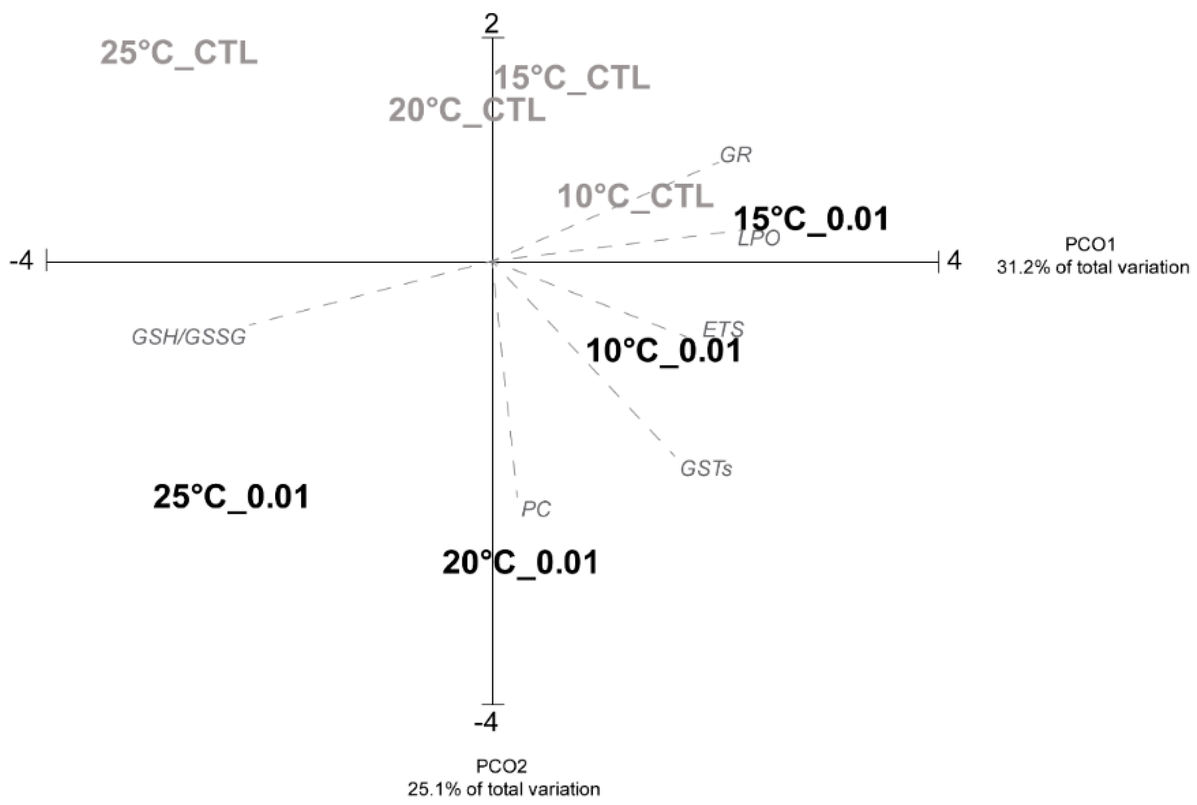
2.4.1. Principal Coordinates Analysis

A matrix gathering all markers responses (PROT and GLY contents; ETS activity, LPO and PC, levels, GSH/GSSG, CAT, GPx, GR, GSTs and AChE activities) per temperature, in the presence or absence of CNTs, was used to calculate a Euclidean distance similarity matrix which was simplified through the calculation of the distance among centroids matrix based on exposure condition (temperature and presence/absence CNTs). This matrix was then submitted to ordination analysis, performed by principal coordinates analysis (PCO). Pearson correlation vectors with correlation > 0.75 were superimposed on the PCO graph. The biomarkers that appeared in the graph (correlation > 0.75, ETS activity, LPO and PC levels, GSH/GSSG, GR and GSTs activities) were also represented as figures to better describe biochemical patterns; biomarkers with correlation < 0.75 (PROT and GLY contents, CAT, GPx and AChE activities) were presented in a Table.

2.4.2. Statistical Analysis

Biological analyses were submitted to hypothesis testing using the PERMANOVA (permutational multivariate analysis of variance) + add-on in PRIMER v6. The pseudo-F p-values in the PERMANOVA main tests were evaluated in terms of significance. When the main test revealed statistically significant differences (p < 0.05), pairwise comparisons were performed. The t-statistic in the pairwise comparisons was evaluated in terms of significance. Values lower than 0.05 were considered as significantly different. The null hypotheses tested were: (I) no significant impacts were observed due to CNTs concentrations, regardless the temperature; (II) no significant effects were observed due to temperature variations on the toxicity of the CNTs; (III) no significant effects were observed due to temperature variations on the sensitivity of organisms to the CNTs.

3. Results and Discussion

3.1. Characterization Analysis

Table 2 reports the results of the dynamic light scattering (DLS) characterization, used to detect the presence of macro/micro/nanosized and polydispersity index (PDI), a measure of the molecular weight distributions of MWCNTs–COOH particle aggregates in aqueous media at the concentration of 0.01 mg/L under different temperatures and time of exposures.
The results showed the presence of unstable micro-sized aggregates in all exposure periods. The mean size values of the suspended nanoparticles at the time 0 was similar to that recorded at the end of the experiment (T96) regardless of the different temperatures. However, comparing the aggregates of the materials among different temperature conditions, it was noticed a temperature-dependent increase of the mean NP-diameters with the largest aggregates under the highest temperature (25 °C), confirming the hypothesis that higher temperature caused faster aggregation due to decreased electrostatic repulsion and increased random Brownian motion and collision frequency [53].

3.2. Biological Analyses

As climate change alters sea surface temperatures, bivalve’s biochemical performance as well as physiology are altered. The biological consequences of temperature rise have been described—including changes in bivalve’s reproduction timing and success, growth, mortality and geographical distribution, with negative impacts on populations sustainability [54,55,56,57,58]. Challenges regarding bivalve’s sustainable management may become more pronounced in the presence of pollutants, with recent studies reporting higher impacts in bivalves exposed simultaneously to pollutants and temperature rise in comparison to the effects caused by each stressor acting alone [59,60].
A principal coordinate analysis (PCO) is represented in Figure 1, revealing the similarities in terms of biochemical responses among conditions (temperature levels and COOH–MWCNTs).
The PCO axis 1 explains 31.2% of the total variation, while PCO axis 2 explains 25.1%. PCO1 separated clams exposed to the highest temperature (both in the presence or absence of CNTs) in the negative side from the remaining conditions in the positive side of the axis. PCO2 separated contaminated clams (at temperatures 25, 20 and 10 °C) in the negative side from the uncontaminated clams at the positive side. Overall, these results evidence high correlations between ETS, GSTs and PC with contaminated clams under 10 and 20 °C, while LPO and GR showed high correlation with the uncontaminated individuals under the lowest temperature as well as with contaminated clams at 15 °C (Figure 1). Considering the ratio between GSH and GSSG, a correlation was detected with CNTs exposed specimens under 25 °C, due to the high redox status observed under this condition.
In terms of clams’ metabolic capacity (ETS) and energy reserves (PROT, GLY), the results obtained showed no significant differences between CNTs exposed and unexposed clams regardless the different tested temperatures except for control bivalves under 20 °C (GLY content) (Figure 2 and Table 3).
It has been demonstrated in the literature that the temperature plays an important role in clam’s biology, including alterations on clam’s energy reserves content [61]. Previous studies demonstrated that metabolism is temperature dependent: the increase of the metabolic rate is directly related to the increase of the water temperatures and vice versa [8]. Nevertheless, when the clams are subjected to temperature stress, they may exhibit defense strategies such as valve closure, gradually changing to an anaerobic mechanism to obtain their energy [8]. The results obtained suggest that the variation of the temperature did not affect the sensitivity of the clams when exposed to CNTs although the state of aggregation of the COOH–MWCNTs increased with the increasing of the temperatures (Table 2). It has also been shown that higher temperature can modify the physico-chemical characteristics of the nanoparticles causing the formation of large-size aggregates. In particular, high temperature corresponds to energy input into a particle suspension, which may lead to the disruption of interaction forces, increased random Brownian motion and changes of particle–particle collision [53]. This alteration can affect the mode of cellular uptake together with subsequent biological responses by the organisms [62]. However, the results obtained in the present study contrast with the information described above [8,62] showing no variation in terms of metabolic capacity and energy reserves content in contaminated organisms regardless of the temperature tested. These findings indicate that the used concentration (0.01 mg/L) was not high enough or the time of exposure was too short to result in metabolic depression or alteration of the energy expenditure. In particular, the lack of metabolic changes observed was associated with similar energy reserves content among temperatures with no differences between contaminated and uncontaminated clams. These results corroborate the low metabolic demand induced by both stressors, highlighting that GLY and PROT were not used by the organisms as energy source to activate defense mechanisms. Previous study, conducted with other bivalve species (mussel Perna viridis), showed similar results, with the maintenance of energy reserves regardless of the stress condition (cadmium (Cd) and copper (Cu) exposures), as a consequence of limited changes in ETS activity [19].
In terms of cellular damage (LPO), no significant differences were observed between contaminated and uncontaminated clams, regardless of the temperature tested; while clams exposed to the lowest temperature showed significantly higher PC levels in the CNTs treatment compared to control. Overall, these results indicated that the concentration tested was not high enough to induce cellular damage (Figure 3A,B). The present results are in agreement with previous studies conducted by Maria and Bebbiano [63]. These authors observed no changes in terms of LPO levels when the mussels Mytilus galloprovincialis were exposed to Cu at 5, 10 and 25 μg/L as a consequence of the low contaminant concentration. Using the same species, Munari et al. [64] showed that in both mussel gills and digestive gland, no significant changes in LPO levels were recorded after the exposure to diclofenac and pH variations. In terms of temperature effects, significantly lower LPO levels were observed at the highest tested temperature, both in contaminated and uncontaminated clams. These results may indicate that the organisms were able to avoid cellular damage due to compensatory mechanisms implemented by antioxidant systems. It has been already demonstrated that glutathione serves in numerous protective detoxification reactions and in the maintenance of cellular redox status [65]. Typically, as already reported by Regoli and Giuliani [66], exposures to xenobiotic concentrations may result in lower reduced glutathione (GSH) levels. Besides that, no changes in the GSH levels were observed under uncontaminated conditions [67]. In the present study, a significantly temperature-dependent increase of GSH/GSSG was observed, with the maximum value at the highest temperature (Figure 3C). However, no differences between contaminated and uncontaminated clams were registered. Furthermore, higher GSH content was observed at the highest temperature level, which agrees with lower LPO levels at this temperature. On the other hand, lower GSH/GSSG ratio (an indication of higher GSSG content), was observed at the lowest temperature representing a loss of redox balance at this condition, both in the presence or absence of CNTs. Lannig et al. [67], exposing Crassostrea virginica to 50 μg/L of Cd under 20, 24 and 28 °C, observed an increase in glutathione levels at the two highest temperatures (20 and 24 °C) permitting the oysters to maintain constant the ratio despite Cd-induced oxidative stress.
Nevertheless, a variety of antioxidants that serve to counterbalance the effect of oxidants, are not only composed by non-enzymatic categories such as GSH and GSSG, but also by enzymatic categories. Significantly higher GR activity was detected when the organisms were exposed to 0.01 mg/L under 15 °C, and then the activity drastically decreased at the two highest temperatures (20 and 25 °C), showing significant differences in comparison to all the other conditions. GR is a disulfide oxidoreductase, representing one of the most ancient proteins [68]. In invertebrates, the main role of GR is to participate in the glutathione antioxidant activity through its recycling process [69]. In details, GR catalyzes the reduction of GSSG to GSH and plays an essential central role in cell defense against reactive oxygen species (ROS). Higher activity in clams exposed to CNTs under the two lowest temperatures may suggest the early induction of the enzyme in response to the short-term experimental treatments to supply enough substrate (GSH) for the detoxification reactions. Analyzing the results obtained from clams submitted to temperature variations without CNTs, GR activity showed the lowest activity under 20 and 25 °C exposures (Figure 4A). Thus, the decrease of this enzyme’ activity seemed not directly related to the presence of CNTs but was dependent on the temperature tested, with lower activity at higher temperature, both in the presence or absence of CNTs (Figure 4A).
Invertebrates are poikilothermic ectotherms, with their body temperature being highly influenced by the environmental temperature [70]. Therefore, R. decussatus may have expressed a suite of responses and strategies (such as the alteration of their antioxidant systems) that acted to protect the cells against oxidative damage as also confirmed by the LPO and GSH/GSSG trends. The first line of antioxidant defense includes CAT and GPx enzymes [68]. Although the correlation observed between these antioxidant enzymes and the different conditions was lower than 75%, the enzymes activities were reported in Table 3. Both enzymes are involved in the detoxification process, which converts the hydrogen peroxide (H2O2) into H2O and O2 [66]. The obtained results demonstrated that changes in ROS levels did not result in significant differences in CAT activity among conditions, while GPx was implicated only when the clams were exposed to CNTs at 15, 20 and 25 °C, showing significantly higher activity compared to organisms exposed to CNTs at 10 °C (Table 3). These results suggested that the increase of the temperature, which developed the state of aggregation of the CNTs (Table 2), may rise the interaction with the organisms, causing the activation of GPx activity as a consequence of a major susceptibility of the clams to the test compounds. This hypothesis is in line with the finding from Handy et al. [71], which confirmed that CNTs stay initially in the aqueous phase and then they tend to aggregate and settle down, being benthic organisms particularly exposed to these contaminants. In a study conducted by Kuroda et al. [72], the authors observed different cytotoxicity and immune responses depending on the aggregation state of the CNTs. The aggregated CNTs were taken up in the phagosomes and although highly dispersed CNTs were scattered and incorporated in the cells, the amount of uptake was clearly less in comparison to the aggregate ones, which caused higher toxicity in the exposed organisms. Similarly, De Marchi et al. [20], exposing the clam Ruditapes philippinarum to COOH–MWCNTs for 28 days under different salinity levels, observed that larger CNTs aggregates caused greater toxic impacts in comparison to the results obtained with organisms exposed to smaller aggregates. GSTs are a group of enzymes involved in detoxication and conjugation of xenobiotics and in protecting against peroxidative damage [68]. Significant differences of GSTs activity were observed only in R. decussatus exposed to the contaminant, with the lowest activity under the highest temperature (Figure 4B). These results suggest an impairment of the conjugation activity with the xenobiotic as a consequence of a major susceptibility caused by high temperature. Similar GSTs trend was also described by Andrade et al. [9], showing an inhibition of GSTs activity when the mussels were contaminated with CNTs at increase temperature.
In the present study, the neurotoxic capacity of CNTs was also investigated under different temperatures, measuring the activity of the enzyme acetylcholinesterase (AChE) (Table 3). The inhibition of AChE by environmental contaminants including metals and nanomaterials has been investigated and reported. As an example, De Marchi et al. [21] investigated the effects of two different CNTs on R. philippinarum, showing AChE activity inhibition caused by both contaminants. In the present study, significantly lower neuro-activity was only observed when the clams were exposed to CNTs under 25 °C, confirming that the concentration used was not high enough to cause neurotoxicity in clams and only at the highest temperature and in the presence of CNTs clams showed inhibition of AChE. The alteration of AChE activity as a consequence of abiotic environmental factors has also been documented. In a study conducted by Bielen et al. [73], the authors examined the tolerance to different stress conditions (thermal stress and trace metal zinc pollution stress) in two similar freshwater bivalve species, the native Anodonta anatina and the invasive Sinanodonta woodiana. Focusing only to the results obtained after thermal stress acting alone (from 10 °C to 26 °C), A. anatina exhibited the highest AChE activity at 18 °C, while S. woodiana at 10 °C. The authors highlighted that, although the level of its inhibition is an established biomarker of neurotoxicity, on the other hand, enhanced ChE activity may indicate an inflammation process due to tissue injury [73].

4. Conclusions

The present study aimed to understand how temperature influence NPs behavior, changing their toxic effects towards R. decussatus. In general, similar biochemical responses were observed between exposed and not exposed organisms, revealing limited toxicity of the tested CNTs concentration. However, increased temperatures, such as the ones expected in the case of seasonal warming and/or global climate change, in combination with NPs exposure, may threaten clams’ populations especially on biotransformation activities and neuro status. Considering that the use of NPs is increasing exponentially among different fields together with climate change related factors, possible risks to the environment, especially on the aquatic environment, should be increasingly assessed. Overall, our results emphasizing the effects of warmer waters in the bivalve’s cellular homeostasis and metabolism, as well as their capability to respond the chemical contamination, that goes in line with previous studies reporting decreases in the nutritional quality of this species widely harvested and produced for human consumption, posing at risk the sustainability of this natural resource.

Author Contributions

Conceptualization, R.J.M.R. and R.F.; methodology, A.C.M.R. and L.D.M.; resources, A.M.V.M.S., C.P., F.C., R.F. and R.J.M.R.; data curation, A.C.M.R. and L.D.M.; writing—original draft preparation, L.D.M. and R.F.; writing—review and editing, R.J.M.R., R.F., L.D.M. and A.C.M.R.; supervision, R.J.M.R. and R.F.; project administration, A.M.V.M.S., R.F. and R.J.M.R.; funding acquisition, A.M.V.M.S., R.F., F.C., C.P. and R.J.M.R. All authors have read and agreed to the published version of the manuscript.

Funding

Rosa Freitas and Rui Rocha were funded by national funds (OE), through FCT—Fundação para a Ciência e a Tecnologia, I.P., in the scope of the framework contract foreseen in the numbers 4, 5 and 6 of the article 23, of the Decree-Law 57/2016, of 29 August, changed by Law 57/2017, of 19 July. This work was also financially supported by the project BISPECIAl: BIvalveS under Polluted Environment and ClImate chAnge PTDC/CTA-AMB/28425/2017 (POCI-01-0145-FEDER-028425) funded by FEDER, through COMPETE2020—Programa Operacional Competitividade e Internacionalização (POCI) and by national funds (OE), through FCT/MCTES; and by the Integrated Programme of SR&TD “Smart Valorization of Endogenous Marine Biological Resources under a Changing Climate” (reference Centro-01-0145-FEDER-000018), co-funded by Centro 2020 program, Portugal 2020, European Union, through the European Regional Development Fund. Bivalves and experimental systems were financed by the project BIODEPURA (MAR-02.01.01-FEAMP-0018), co-financed by the Operational Program Mar 2020, Portugal 2020 and by the European Maritime and Fisheries Fund (EMFF).

Acknowledgments

Lucia De Marchi was supported by an Assegno di Ricerca of Pisa University, Department of Biology. Thanks are due to FCT/MCTES for the financial support to CESAM (UIDB/50017/2020+UIDP/50017/2020).

Conflicts of Interest

The authors declare no conflicts of interest.

References

  1. IPCC. Climate Change 2014: Synthesis Report; IPCC: Geneva, Switzerland, 2014. [Google Scholar]
  2. De Montaudouin, X.; Bachelet, G. Experimental evidence of complex interactions between biotic and abiotic factors in the dynamics of an intertidal population of the bivalve Cerastoderma edule. Oceanol. Acta 1996, 19, 449–463. [Google Scholar]
  3. Gerwing, T.G.; Drolet, D.; Hamilton, D.J.; Barbeau, M.A. Relative Importance of Biotic and Abiotic Forces on the Composition and Dynamics of a Soft-Sediment Intertidal Community. PLoS ONE 2016, 11. [Google Scholar] [CrossRef] [PubMed] [Green Version]
  4. Grilo, T.F.; Cardoso, P.G.; Dolbeth, M.; Bordalo, M.D.; Pardal, M.A. Effects of extreme climate events on the macrobenthic communities’ structure and functioning of a temperate estuary. Mar. Pollut. Bull. 2011, 62, 303–311. [Google Scholar] [CrossRef] [PubMed]
  5. Jenewein, B.T. Determining the extent to which weather-related abiotic factors influence daily variation in early benthic phase mortality of intertidal marine invertebrates. Master’s Thesis, Thompson Rivers University, Kamloops, BC, Canada, June 2009. [Google Scholar]
  6. Woodin, S.A.; Wethey, D.S.; Dubois, S.F. Population structure and spread of the polychaete Diopatra biscayensis along the French Atlantic coast: Human-assisted transport by-passes larval dispersal. Mar. Environ. Res. 2014, 102, 110–121. [Google Scholar] [CrossRef] [Green Version]
  7. Bindoff, N.L.; Stott, P.A.; AchutaRao, K.M.; Allen, M.R.; Gillett, N.; Gutzler, D.; Hansingo, K.; Hegerl, G.; Hu, Y.; Jain, S.; et al. Chapter 10–Detection and attribution of climate change: From global to regional. In Climate Change 2013: The Physical Science Basis. IPCC Working Group I Contribution to AR5; Cambridge University Press: Cambridge, UK, 2013. [Google Scholar]
  8. Weber, K.; Sturmer, L.; Hoover, E.; Baker, S. The Role of Salinity in Hard Clam Aquaculture; University of Florida IFAS Extension: Gainesville, FL, USA, 2013; pp. 1–10. [Google Scholar]
  9. Andrade, M.; De Marchi, L.; Pretti, C.; Chiellini, F.; Morelli, A.; Figueira, E.; Rocha, R.J.M.; Soares, A.M.V.M.; Freitas, R. The impacts of warming on the toxicity of carbon nanotubes in mussels. Mar. Environ. Res. 2019, 145, 11–21. [Google Scholar] [CrossRef]
  10. Attig, H.; Kamel, N.; Sforzini, S.; Dagnino, A.; Jamel, J.; Boussetta, H.; Viarengo, A.; Banni, M. Effects of thermal stress and nickel exposure on biomarkers responses in Mytilus galloprovincialis (Lam). Mar. Environ. Res. 2014, 94, 65–71. [Google Scholar] [CrossRef]
  11. Ivanina, A.V.; Taylor, C.; Sokolova, I.M. Effects of elevated temperature and cadmium exposure on stress protein response in eastern oysters Crassostrea virginica (Gmelin). Aquat. Toxicol. 2009, 91, 245–254. [Google Scholar] [CrossRef]
  12. Matoo, O.B.; Ivanina, A.V.; Ullstad, C.; Beniash, E.; Sokolova, I.I. Interactive effects of elevated temperature and CO2 levels on metabolism and oxidative stress in two common marine bivalves (Crassostrea virginica and Mercenaria mercenaria). Comp. Biochem. Physiol. Mol. Integr. Physiol. 2013, 164, 545–553. [Google Scholar] [CrossRef]
  13. Verlecar, X.N.; Jena, K.B.; Chainy, G.B.N. Biochemical markers of oxidative stress in Perna viridis exposed to mercury and temperature. Chem. Biol. Interact. 2007, 167, 219–226. [Google Scholar] [CrossRef]
  14. Meijide, F.J.; Da Cuña, R.H.; Prieto, J.P.; Dorelle, L.S.; Babay, P.A.; Lo Nostro, F.L. Effects of waterborne exposure to the antidepressant fluoxetine on swimming, shoaling and anxiety behaviours of the mosquitofish Gambusia holbrooki. Ecotoxicol. Environ. Saf. 2018, 163, 646–655. [Google Scholar] [CrossRef]
  15. De Volder, M.F.L.; Tawfick, S.H.; Baughman, R.H.; Hart, A.J. Carbon nanotubes: Present and future commercial applications. Science 2013, 339, 535–539. [Google Scholar] [CrossRef] [PubMed] [Green Version]
  16. Centi, G.; Perathoner, S. Carbon Nanotubes for Sustainable Energy Applications. ChemSusChem 2011, 4, 913–925. [Google Scholar] [CrossRef] [PubMed]
  17. Freixa, A.; Acuña, V.; Sanchís, J.; Farré, M.; Barceló, D.; Sabater, S. Ecotoxicological effects of carbon based nanomaterials in aquatic organisms. Sci. Total Environ. 2018, 619–620, 328–337. [Google Scholar] [CrossRef] [PubMed]
  18. Mwangi, J.N.; Wang, N.; Ingersoll, C.G.; Hardesty, D.K.; Brunson, E.L.; Li, H.; Deng, B. Toxicity of carbon nanotubes to freshwater aquatic invertebrates. Environ. Toxicol. Chem. 2012, 31, 1823–1830. [Google Scholar] [CrossRef]
  19. Yeung, J.W.Y.; Zhou, G.J.; Leung, K.M.Y. Sub-lethal effects of cadmium and copper on RNA/DNA ratio and energy reserves in the green-lipped mussel Perna viridis. Ecotoxicol. Environ. Saf. 2016, 132, 59–67. [Google Scholar] [CrossRef]
  20. De Marchi, L.; Neto, V.; Pretti, C.; Figueira, E.; Chiellini, F.; Morelli, A.; Soares, A.M.V.M.; Freitas, R. Toxic effects of multi-walled carbon nanotubes on bivalves: Comparison between functionalized and nonfunctionalized nanoparticles. Sci. Total Environ. 2018, 622–623, 1532–1542. [Google Scholar] [CrossRef]
  21. De Marchi, L.; Neto, V.; Pretti, C.; Figueira, E.; Chiellini, F.; Morelli, A.; Soares, A.M.V.M.; Freitas, R. Effects of multi-walled carbon nanotube materials on Ruditapes philippinarum under climate change: The case of salinity shifts. Aquat. Toxicol. 2018, 199, 199–211. [Google Scholar] [CrossRef]
  22. Kádár, E.; Lowe, D.M.; Solé, M.; Fisher, A.S.; Jha, A.N.; Readman, J.W.; Hutchinson, T.H. Uptake and biological responses to nano-Fe versus soluble FeCl3 in excised mussel gills. Anal. Bioanal. Chem. 2010, 396, 657–666. [Google Scholar] [CrossRef]
  23. Rocha, T.L.; Gomes, T.; Sousa, V.S.; Mestre, N.C.; Bebianno, M.J. Ecotoxicological impact of engineered nanomaterials in bivalve molluscs: An overview. Mar. Environ. Res. 2015, 111, 74–88. [Google Scholar] [CrossRef]
  24. Canesi, L.; Fabbri, R.; Gallo, G.; Vallotto, D.; Marcomini, A.; Pojana, G. Biomarkers in Mytilus galloprovincialis exposed to suspensions of selected nanoparticles (Nano carbon black, C60 fullerene, Nano-TiO2, Nano-SiO2). Aquat. Toxicol. 2010, 100, 168–177. [Google Scholar] [CrossRef]
  25. Moore, M.N.; Readman, J.A.J.; Readman, J.W.; Lowe, D.M.; Frickers, P.E.; Beesley, A. Lysosomal cytotoxicity of carbon nanoparticles in cells of the molluscan immune system: An in vitro study. Nanotoxicology 2009, 3, 40–45. [Google Scholar] [CrossRef]
  26. Parache, A. La palourde. La Pêche Marit. 1982, 61, 496–506. [Google Scholar]
  27. Bebianno, M.J.; Géret, F.; Hoarau, P.; Serafim, M.A.; Coelho, M.R.; Gnassia-Barelli, M.; Roméo, M. Biomarkers in Ruditapes decussatus: A potential bioindicator species. Biomarkers 2004, 9, 305–330. [Google Scholar] [CrossRef] [PubMed]
  28. FAO. Aquaculture Production by Species and Country or Area. 2020. Available online: http://www.fao.org/fishery/static/Yearbook/YB2017_USBcard/root/aquaculture/b56.pdf (accessed on 13 June 2020).
  29. IPMA. Lista de Espécies. Available online: https://www.ipma.pt/pt/bivalves/docs/files/Lista_de_espxcies_em_14_04_2020.pdf (accessed on 9 June 2020).
  30. Matias, D.; Joaquim, S.; Matias, A.M.; Moura, P.; de Sousa, J.T.; Sobral, P.; Leitão, A. The reproductive cycle of the European clam Ruditapes decussatus (L. 1758) in two Portuguese populations: Implications for management and aquaculture programs. Aquaculture 2013, 406–407, 52–61. [Google Scholar] [CrossRef] [Green Version]
  31. Chalghmi, H.; Zrafi, I.; Gourves, P.Y.; Bourdineaud, J.P.; Saidane-Mosbahi, D. Combined effects of metal contamination and abiotic parameters on biomarker responses in clam: Ruditapes decussatus gills: An integrated approach in biomonitoring of Tunis lagoon. Environ. Sci. Process. Impacts 2016, 18, 895–907. [Google Scholar] [CrossRef] [PubMed]
  32. Cravo, A.; Pereira, C.; Gomes, T.; Cardoso, C.; Serafim, A.; Almeida, C.; Rocha, T.; Lopes, B.; Company, R.; Medeiros, A.; et al. A multibiomarker approach in the clam Ruditapes decussatus to assess the impact of pollution in the Ria Formosa lagoon, South Coast of Portugal. Mar. Environ. Res. 2012, 75, 23–34. [Google Scholar] [CrossRef]
  33. El-Shenawy, N.S. Heavy-metal and microbial depuration of the clam Ruditapes decussatus and its effect on bivalve behavior and physiology. Environ. Toxicol. 2004, 19, 143–153. [Google Scholar] [CrossRef]
  34. Hamza-Chaffai, A. Usefulness of Bioindicators and Biomarkers in Pollution Biomonitoring. Int. J. Biotechnol. Wellness Ind. 2014, 3, 19–26. [Google Scholar] [CrossRef]
  35. Caro, A.; Chereau, G.; Briant, N.; Roques, C.; Freydier, R.; Delpoux, S.; Escalas, A.; Elbaz-Poulichet, F. Contrasted responses of Ruditapes decussatus (filter and deposit feeding) and Loripes lacteus (symbiotic) exposed to polymetallic contamination (Port-Camargue, France). Sci. Total Environ. 2015, 505, 526–534. [Google Scholar] [CrossRef]
  36. Chalghmi, H.; Bourdineaud, J.P.; Haouas, Z.; Gourves, P.Y.; Zrafi, I.; Saidane-Mosbahi, D. Transcriptomic, Biochemical, and Histopathological Responses of the Clam Ruditapes decussatus from a Metal-Contaminated Tunis Lagoon. Arch. Environ. Contam. Toxicol. 2016, 70, 241–256. [Google Scholar] [CrossRef]
  37. Fathallah, S.; Medhioub, M.N.; Medhioub, A.; Kraiem, M.M. Toxicity of Hg, Cu and Zn on early developmental stages of the European clam (Ruditapes decussatus) with potential application in marine water quality assessment. Environ. Monit. Assess. 2010, 171, 661–669. [Google Scholar] [CrossRef]
  38. Figueira, E.; Cardoso, P.; Freitas, R. Ruditapes decussatus and Ruditapes philippinarum exposed to cadmium: Toxicological effects and bioaccumulation patterns. Comp. Biochem. Physiol. C Toxicol. Pharmacol. 2012, 156, 80–86. [Google Scholar] [CrossRef] [PubMed]
  39. Roméo, M.; Gnassia-Barelli, M. Metal distribution in different tissues and in subcellular fractions of the Mediterranean clam Ruditapes decussatus treated with cadmium, copper, or zinc. Comp. Biochem. Physiol. Part C Comp. 1995, 111, 457–463. [Google Scholar] [CrossRef]
  40. Serafim, A.; Bebianno, M.J. Effect of a polymetallic mixture on metal accumulation and metallothionein response in the clam Ruditapes decussatus. Aquat. Toxicol. 2010, 99, 370–378. [Google Scholar] [CrossRef] [PubMed]
  41. Velez, C.; Figueira, E.; Soares, A.M.V.M.; Freitas, R. The impacts of As accumulation under different pH levels: Comparing Ruditapes decussatus and Ruditapes philippinarum biochemical performance. Environ. Res. 2016, 151, 653–662. [Google Scholar] [CrossRef] [Green Version]
  42. Abdelhafidh, K.; Ali, M.; Hassen, K.; Badreddine, S.; Jaume, A.; Sandra, P.; Ethel, E.; Damià, B.; Hamouda, B.; Ezzeddine, M. Uptake and metabolism of carbamazepine (CBZ) by clam Ruditapes decussatus and its effects in biochemical responses. Xenobiotica 2018, 48, 727–733. [Google Scholar] [CrossRef]
  43. Costa, S.; Coppola, F.; Pretti, C.; Intorre, L.; Meucci, V.; Soares, A.M.V.M.; Freitas, R.; Solé, M. The influence of pre-exposure to climate change related factors on the response of two clam’s species to Diclofenac. Ecotoxicol. Environ. Saf. 2020, 189, 109899. [Google Scholar] [CrossRef]
  44. Sellami, B.; Aouani, I.; Maalaoui, A.; Dellali, M.; Aïssa, P.; Touil, S.; Sheehan, D.; Mahmoudi, E.; Hamouda, B. Effects of 2-(4-Methoxyphenyl)-5, 6-trimethylene-4H-1, 3, 2-oxathiaphosphorine-2-sulfide on biomarkers of Mediterranean clams Ruditapes decussatus. Ecotoxicol. Environ. Saf. 2015, 120, 263–269. [Google Scholar] [CrossRef]
  45. Campillo, J.A.; Sevilla, A.; Albentosa, M.; Bernal, C.; Lozano, A.B.; Cánovas, M.; León, V.M. Metabolomic responses in caged clams, Ruditapes decussatus, exposed to agricultural and urban inputs in a Mediterranean coastal lagoon (Mar Menor, SE Spain). Sci. Total Environ. 2015, 524–525, 136–147. [Google Scholar] [CrossRef]
  46. Kamel, N.; Jebali, J.; Banni, M.; Ben Khedher, S.; Chouba, L.; Boussetta, H. Biochemical responses and metals levels in Ruditapes decussatus after exposure to treated municipal effluents. Ecotoxicol. Environ. Saf. 2012, 82, 40–46. [Google Scholar] [CrossRef]
  47. Saidani, W.; Sellami, B.; Khazri, A.; Mezni, A.; Dellali, M.; Joubert, O.; Sheehan, D.; Beyrem, H. Metal accumulation, biochemical and behavioral responses on the Mediterranean clams Ruditapes decussatus exposed to two photocatalyst nanocomposites (TiO2 NPs and AuTiO2NPs). Aquat. Toxicol. 2019, 208, 71–79. [Google Scholar] [CrossRef] [PubMed]
  48. Sellami, B.; Bouzidi, I.; Saidani, W.; Mezni, A.; Sheehan, D.; Beyrem, H. Effects of Gold Nanoparticles on the Mediterranean Clams Ruditapes decussatus: Chemical and Biochemical Investigations. In Proceedings of the Euro-Mediterranean Conference for Environmental Integration, Sousse, Tunisia, 20–25 November 2017; Springer: Cham, Switzerland, 2018; pp. 577–580. [Google Scholar]
  49. Peng, X.; Jia, J.; Gong, X.; Luan, Z.; Fan, B. Aqueous stability of oxidized carbon nanotubes and the precipitation by salts. J. Hazard. Mater. 2009, 165, 1239–1242. [Google Scholar] [CrossRef] [PubMed]
  50. OECD. List of Manufactured Nanomaterials and List of Endpoints for Phase One of the Sponsorship Programme for the Testing of Manufactured Nanomaterials: Revision. In OECD Environment, Health and Safety Publications; Series on the Safety of Manufactured Nanomaterials; Organization for Economic Cooperation and Development: Paris, France, 2010. [Google Scholar]
  51. Zhang, X.; Zhou, Q.; Zou, W.; Hu, X. Molecular Mechanisms of Developmental Toxicity Induced by Graphene Oxide at Predicted Environmental Concentrations. Environ. Sci. Technol. 2017, 51, 7861–7871. [Google Scholar] [CrossRef]
  52. Cravo, A.; Rosa, A.; Jacob, J.; Correia, C. Dissolved oxygen dynamics in Ria Formosa Lagoon (South Portugal)—A real time monitoring station observatory. Mar. Chem. 2020, 223, 103806. [Google Scholar] [CrossRef]
  53. Wang, L.; Yang, X.; Wang, Q.; Zeng, Y.; Ding, L.; Jiang, W. Effects of ionic strength and temperature on the aggregation and deposition of multi-walled carbon nanotubes. J. Environ. Sci. China 2017, 51, 248–255. [Google Scholar] [CrossRef]
  54. Cheung, W.W.L.; Lam, V.W.Y.; Sarmiento, J.L.; Kearney, K.; Watson, R.; Pauly, D. Projecting global marine biodiversity impacts under climate change scenarios. Fish Fish. 2009, 10, 235–251. [Google Scholar] [CrossRef]
  55. Smolders, R.; Bervoets, L.; De Coen, W.; Blust, R. Cellular energy allocation in zebra mussels exposed along a pollution gradient: Linking cellular effects to higher levels of biological organization. Environ. Pollut. 2004, 129, 99–112. [Google Scholar] [CrossRef]
  56. Reid, G.K.; Gurney-Smith, H.J.; Marcogliese, D.J.; Knowler, D.; Benfey, T.; Garber, A.F.; Forster, I.; Chopin, T.; Brewer-Dalton, K.; Moccia, R.D.; et al. Climate change and aquaculture: Considering biological response and resources. Aquac. Environ. Interact. 2019, 11, 569–602. [Google Scholar] [CrossRef] [Green Version]
  57. Talmage, S.C.; Gobler, C.J. Effects of elevated temperature and carbon dioxide on the growth and survival of larvae and juveniles of three species of northwest Atlantic bivalves. PLoS ONE 2011, 6. [Google Scholar] [CrossRef]
  58. Velez, C.; Figueira, E.; Soares, A.M.V.M.; Freitas, R. Effects of seawater temperature increase on economically relevant native and introduced clam species. Mar. Environ. Res. 2017, 123, 62–70. [Google Scholar] [CrossRef]
  59. Freitas, R.; Coppola, F.; Henriques, B.; Wrona, F.; Figueira, E.; Pereira, E.; Soares, A.M.V.M. Does pre-exposure to warming conditions increase Mytilus galloprovincialis tolerance to Hg contamination? Comp. Biochem. Physiol. Part C Toxicol. Pharmacol. 2017, 203, 1–11. [Google Scholar] [CrossRef] [PubMed]
  60. Freitas, R.; Leite, C.; Pinto, J.; Costa, M.; Monteiro, R.; Henriques, B.; Di Martino, F.; Coppola, F.; Soares, A.M.V.M.; Solé, M.; et al. The influence of temperature and salinity on the impacts of lead in Mytilus galloprovincialis. Chemosphere 2019, 235, 403–412. [Google Scholar] [CrossRef] [PubMed] [Green Version]
  61. Anacleto, P.; Maulvault, A.L.; Bandarra, N.M.; Repolho, T.; Nunes, M.L.; Rosa, R.; Marques, A. Effect of warming on protein, glycogen and fatty acid content of native and invasive clams. Food Res. Int. 2014, 64, 439–445. [Google Scholar] [CrossRef] [PubMed]
  62. Hotze, E.M.; Phenrat, T.; Lowry, G.V. Nanoparticle aggregation: Challenges to understanding transport and reactivity in the environment. J. Environ. Q. 2010, 39, 1909–1924. [Google Scholar] [CrossRef] [Green Version]
  63. Maria, V.L.; Bebianno, M.J. Antioxidant and lipid peroxidation responses in Mytilus galloprovincialis exposed to mixtures of benzo(a)pyrene and copper. Comp. Biochem. Physiol. C Toxicol. Pharmacol. 2011, 154, 56–63. [Google Scholar] [CrossRef]
  64. Munari, M.; Matozzo, V.; Gagné, F.; Chemello, G.; Riedl, V.; Finos, L.; Pastore, P.; Badocco, D.; Marin, M.G. Does exposure to reduced pH and diclofenac induce oxidative stress in marine bivalves? A comparative study with the mussel Mytilus galloprovincialis and the clam Ruditapes philippinarum. Environ. Pollut. 2018, 240, 925–937. [Google Scholar] [CrossRef]
  65. Meister, A.; Anderson, M.E. Glutathione. Annu. Rev. Biochem. 1983, 52, 711–760. [Google Scholar] [CrossRef]
  66. Regoli, F.; Giuliani, M.E. Oxidative pathways of chemical toxicity and oxidative stress biomarkers in marine organisms. Mar. Environ. Res. 2014, 93, 106–117. [Google Scholar] [CrossRef]
  67. Lannig, G.; Flores, J.F.; Sokolova, I.M. Temperature-dependent stress response in oysters, Crassostrea virginica: Pollution reduces temperature tolerance in oysters. Aquat. Toxicol. 2006, 79, 278–287. [Google Scholar] [CrossRef] [Green Version]
  68. Felton, G.W. Oxidative Stress of Vertebrates and Invertebrates. In Oxidative Stress and Antioxidant Defenses in Biology; Springer: New York, NY, USA, 1995; pp. 356–434. [Google Scholar]
  69. Franco, J.L.; Trivella, D.B.B.; Trevisan, R.; Dinslaken, D.F.; Marques, M.R.F.; Bainy, A.C.D.; Dafre, A.L. Antioxidant status and stress proteins in the gills of the brown mussel Perna perna exposed to zinc. Chem. Biol. Interact. 2006, 160, 232–240. [Google Scholar] [CrossRef]
  70. Everatt, M.J.; Convey, P.; Bale, J.S.; Worland, M.R.; Hayward, S.A.L. Responses of invertebrates to temperature and water stress: A polar perspective. J. Therm. Biol. 2015, 54, 118–132. [Google Scholar] [CrossRef] [PubMed] [Green Version]
  71. Handy, R.D.; Von Der Kammer, F.; Lead, J.R.; Hassellöv, M.; Owen, R.; Crane, M. The ecotoxicology and chemistry of manufactured nanoparticles. Ecotoxicology 2008, 17, 287–314. [Google Scholar] [CrossRef] [PubMed]
  72. Kuroda, C.; Ueda, K.; Haniu, H.; Ishida, H.; Okano, S.; Takizawa, T.; Sobajima, A.; Kamanaka, T.; Yoshida, K.; Okamoto, M.; et al. Different aggregation and shape characteristics of carbon materials affect biological responses in RAW264 cells. Int. J. Nanomed. 2018, 13, 6079–6088. [Google Scholar] [CrossRef] [PubMed] [Green Version]
  73. Bielen, A.; Bošnjak, I.; Sepčić, K.; Jaklič, M.; Cvitanić, M.; Lušić, J.; Lajtner, J.; Simčič, T.; Hudina, S. Differences in tolerance to anthropogenic stress between invasive and native bivalves. Sci. Total Environ. 2016, 543, 449–459. [Google Scholar] [CrossRef]
Figure 1. Centroid ordination diagram based on clam’s biochemical parameters, measured for all the conditions (uncontaminated and contaminated clams, at different temperatures). Pearson correlation vectors are superimposed as supplementary variables. GR—glutathione reductase activity; LPO—lipid peroxidation levels; ETS—electron transport system activity; GSTs—glutathione S-transferases activity; PC—protein carbonylation levels; GSH/GSSG—ratio between reduced (GSH) and oxidized (GSSG) glutathione. Conditions identified with gray letters represent uncontaminated clams. Conditions identified with black letters represent contaminated clams.
Sustainability 12 04939 g001
Figure 2. Electron transport system activity (ETS), in Ruditapes decussatus exposed to different temperatures (10, 15, 20 and 25 °C), in the absence (gray bars) and presence (black bars) of carbon nanotubes. Results are means with standard deviation. Different letters represent significant differences (p < 0.05) among conditions (uppercase letters for uncontaminated clams; lowercase letters for contaminated clams). Similar letters represent no significant differences among conditions.
Sustainability 12 04939 g002
Figure 3. (A) Lipid peroxidation levels (LPO); (B) protein carbonylation levels (PC); (C) reduced (GSH) and oxidized (GSSG) glutathione ratio (GSH:GSSG ratio), in Ruditapes decussatus exposed to different temperatures (10, 15, 20 and 25 °C), in the absence (gray bars) and presence (black bars) of carbon nanotubes. Results are means with standard deviation. Different letters represent significant differences (p < 0.05) among conditions (uppercase letters for uncontaminated clams; lowercase letters for contaminated clams). Similar letters represent no significant differences among conditions. Asterisk (*) indicates significant differences (p < 0.05) between CNTs exposed and unexposed clams at each temperature.
Sustainability 12 04939 g003
Figure 4. (A) glutathione reductase activity (GR); (B) glutathione S-transferases activity (GSTs), in Ruditapes decussatus exposed to different temperatures (10, 15, 20 and 25 °C), in the absence (gray bars) and presence (black bars) of carbon nanotubes. Results are means with standard deviation. Different letters represent significant differences (p < 0.05) among conditions (uppercase letters for uncontaminated clams; lowercase letters for contaminated clams). Similar letters represent no significant differences among conditions. Asterisk (*) indicates significant differences (p < 0.05) between CNTs exposed and unexposed clams at each temperature.
Sustainability 12 04939 g004
Table 1. Characterization of the powder form of MWCNTs–COOH.
Technical Data
MWCNTs–COOHDiameter (nm)Length (um)Carbon Purity (%)Surface Area (m2/g)Amorphous Carbon (mol%)-COOH (wt%)
2–510–30984008–103.86
Table 2. Average size distribution (nm) and polydispersity index (PDI) of carboxylated MWCNTs–COOH suspensions analyzed in 0.10 mg/L exposure concentration at different exposure periods: T0: time zero, immediately after the dispersion of the materials in a water medium; T96: water samples collected after 96 h of exposure.
Temperature (°C)
10152025
T0T96T0T96T0T96T0T96
MWCNTs–COOHParticle size (nm)2307.12289.12564.32500.12601.22790.83989.14001.2
PDI0.980.971.010.911.131.160.260.33
Table 3. Protein content (PROT); glycogen content (GLY); superoxide dismutase activity (SOD); catalase activity (CAT); glutathione peroxidase activity (GPx); acetylcholinesterase activity (AChE) (mean ± standard deviation), in R. decussatus uncontaminated (CTL) and exposed to COOH–MWCNTs (Exp), both at different temperatures (10, 15, 20, 25 °C). Significant differences (p < 0.05) among different temperatures are represented with different letters (uppercase letters for CTL—uncontaminated clams; lowercase letters for Exp—contaminated clams). Significant differences (p < 0.05) between CTL and Exp, at each temperature, are represented with bold asterisks (*).
Temperature (°C)
10152025
Energy reserves contentPROT (mg/g)CTL80.99 ± 19.10 A93.68 ± 17.53 A91.23 ± 9.59 A89.12 ± 20.84 A
Exp89.24 ± 9.92 a102.87 ± 15.47 a78.46 ± 15.08 a81.40 ± 21.01 a
GLY
(mg/g)
CTL1.37 ± 0.36 A1.63 ± 0.30 A1.95 ± 0.52 B1.58 ± 0.21 A
Exp1.65 ± 0.41 a1.60 ± 0.49 a1.42 ± 0.50 a1.65 ± 0.11 a
Antioxidant defensesCAT
(U/g)
CTL9.25 ± 1.65 A10.83 ± 1.70 A9.92 ± 2.23 A11.10 ± 1.98 A
Exp8.87 ± 0.87 a10.58 ± 0.49 a10.28 ± 1.50 a9.93 ± 0.79 a
GPx
(U/g)
CTL0.05 ± 0.02 A0.05 ± 0.02 A*0.10 ± 0.02 A0.06 ± 0.02 A
Exp0.08 ± 0.02 a0.13 ± 0.01 b0.14 ± 0.01 b0.14 ± 0.02 b
Neuro statusAChE
(nmol/min/g)
CTL0.08 ± 0.02 A0.09 ± 0.01 A0.08 ± 0.02 A0.18 ± 0.05 B,*
Exp0.09 ± 0.02 a0.07 ± 0.01 a0.06 ± 0.01 a0.01 ± 0.00 b

