
An Unusual Cause of Inguinal Mass in a Patient with Urolithiasis: A Case Report of Deep (Aggressive) Angiomyxoma in a Male Patient

 ,
,  , , , and
Abstract
1. Introduction
2. Case Report
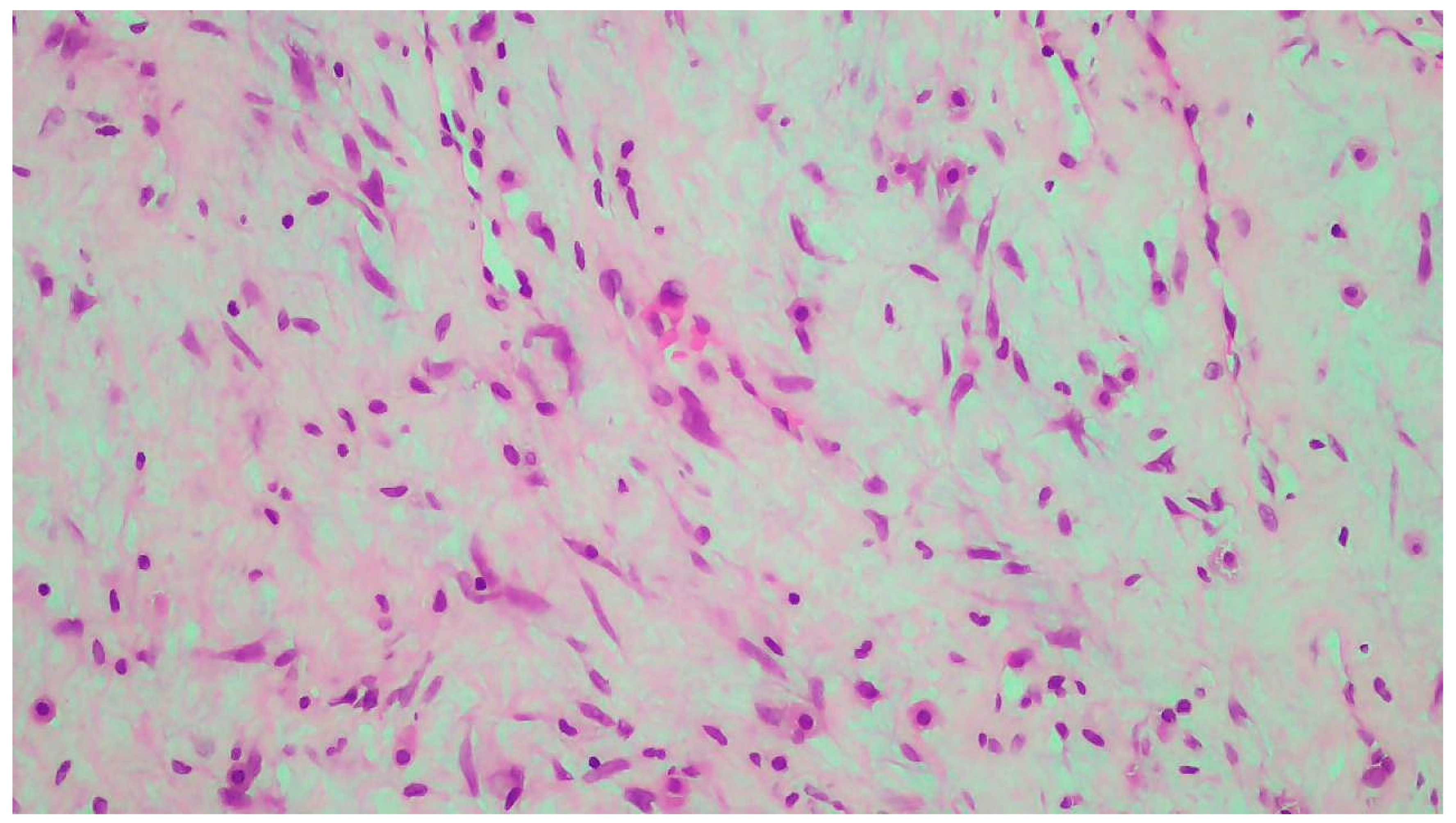
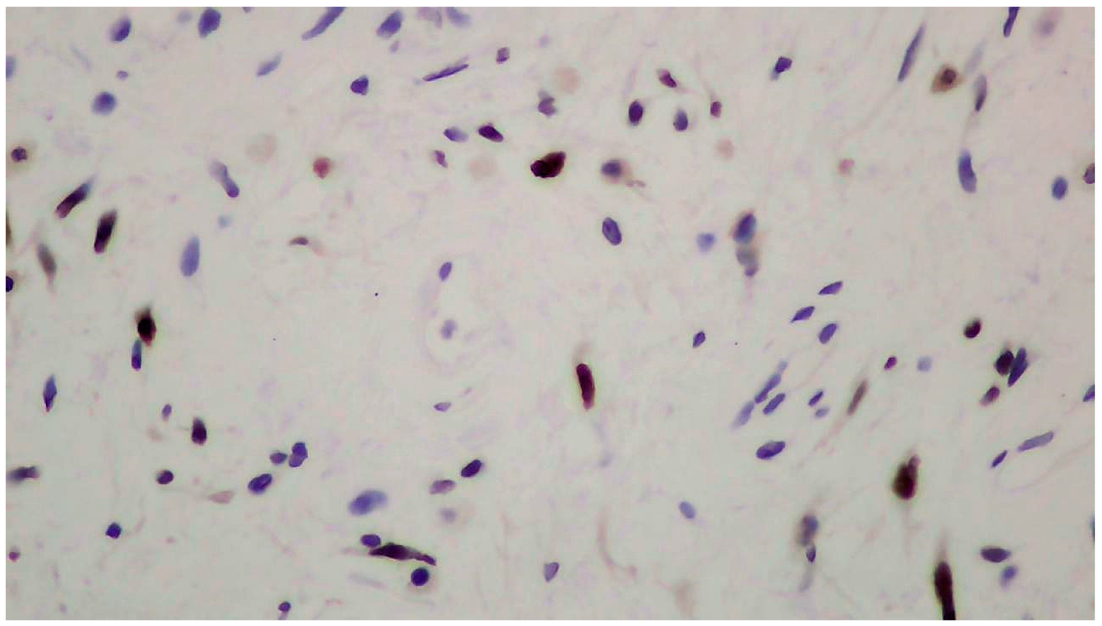
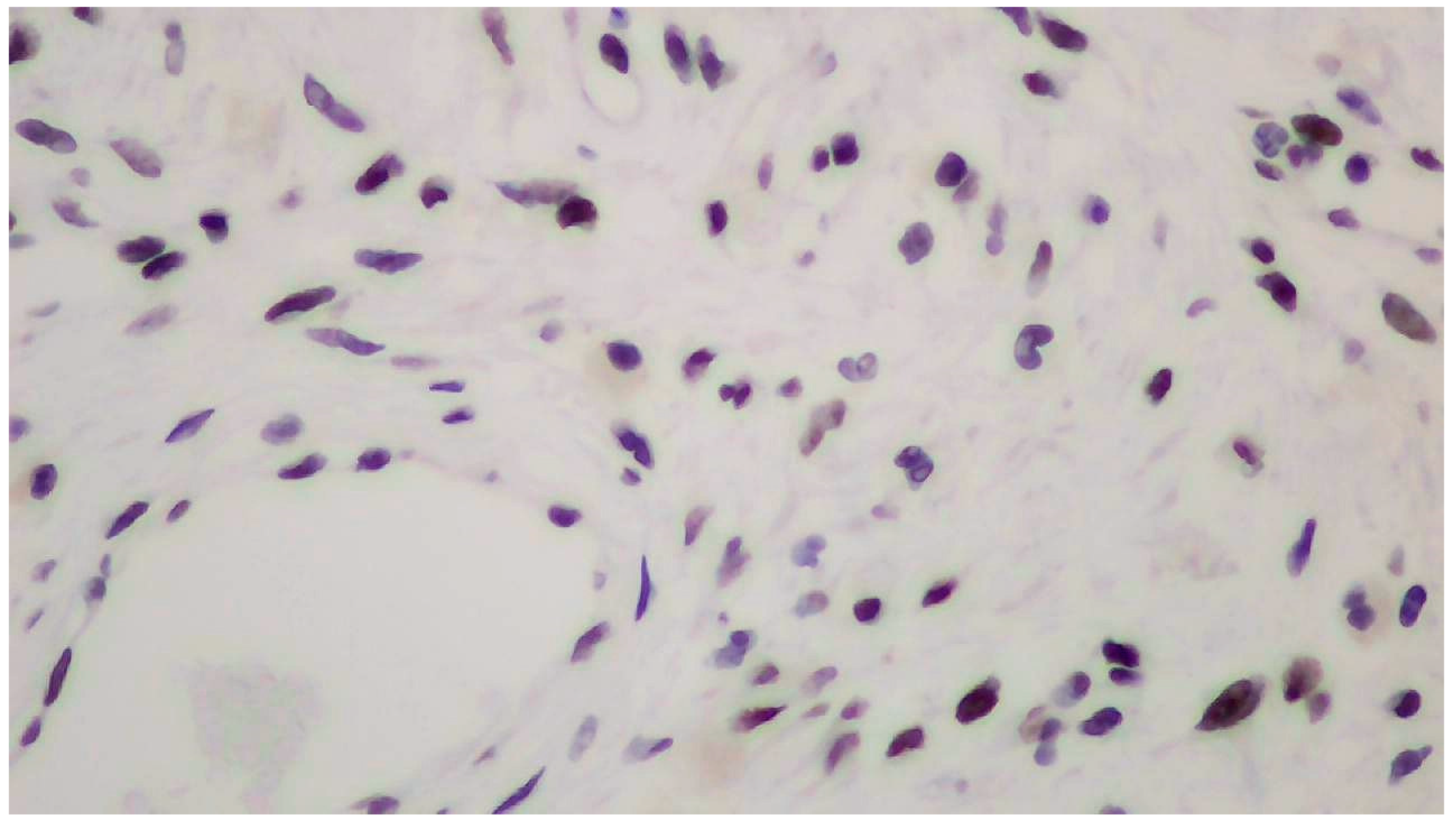
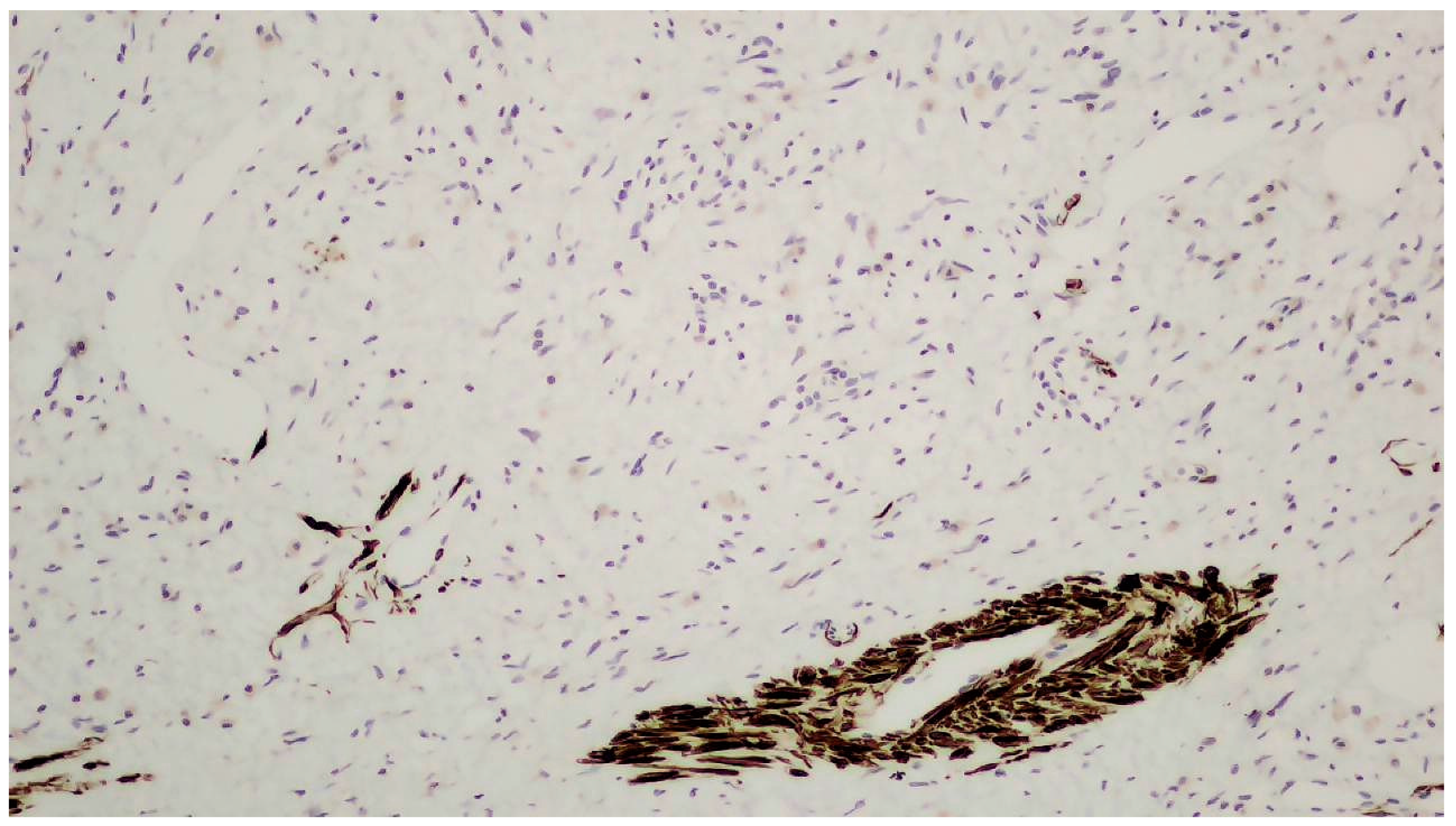
3. Discussion
4. Conclusions
Author Contributions
Funding
Institutional Review Board Statement
Informed Consent Statement
Data Availability Statement
Conflicts of Interest
References
- Celik, S.U.; Hesimov, I.; Kutlu, B.; Erkek, A.B. Aggressive Angiomyxoma: A Rare Tumor of Male Pelvic Cavity. Acta Med. Port. 2018, 31, 693–696. [Google Scholar] [CrossRef] [PubMed]
- Hisano, M.; Matsuura, T.; Kato, R.; Maekawa, S.; Kato, Y.; Kanehira, M.; Suzuki, M.; Nakayama, M.; Takata, R.; Obara, W. A case of male perineal aggressive angiomyxoma with expressions of female hormone receptors. IJU Case Rep. 2022, 5, 308–311. [Google Scholar] [CrossRef]
- Fetsch, J.F.; Laskin, W.B.; Lefkowitz, M.; Kindblom, L.G.; Meis-Kindblom, J.M. Aggressive angiomyxoma: A clinicopathologic study of 29 female patients. Cancer 1996, 78, 79–90. [Google Scholar] [CrossRef]
- Angelico, G.; Marletta, S.; Broggi, G.; Vigneri, P.; Vecchio, G.M.; Salvatorelli, L.; Magro, G. Practical Approach to the Diagnosis of the Vulvo-Vaginal Stromal Tumors: An Overview. Diagnostics 2022, 12, 357. [Google Scholar] [CrossRef] [PubMed]
- Cicogna, S.; Dellino, M.; Miano, S.T.; Magazzino, F.; Domenici, L.; Pignata, S.; Mangili, G.; Cormio, G. Aggressive Angiomyxoma of the Lower Female Genital Tract in Pregnancy: A Review of the MITO Rare Tumors Group. Cancers 2023, 15, 3403. [Google Scholar] [CrossRef]
- Hsieh, F.; Chuang, K.T.; Wu, Y.T.; Lin, C.H. Aggressive Angiomyxoma-Report of a Rare Male Buttock Lesion. Plast. Reconstr. Surg. Glob. Open 2018, 6, e1879. [Google Scholar] [CrossRef] [PubMed]
- Saha, K.; Sarkar, S.; Jash, D.; Chatterjee, S.; Saha, A.K. Aggressive angiomyxoma of greater omentum with pleural effusion in a young male. J. Cancer Res. Ther. 2014, 10, 371–373. [Google Scholar] [CrossRef] [PubMed]
- Chen, C.F.; Wang, T.Y.; Chen, M.; Lin, Y.C. Rare paratesticular aggressive angiomyxoma mimicking an epididymal tumor in an 82-year-old man: Case report. Open Med. 2021, 16, 973–977. [Google Scholar] [CrossRef]
- Sabbagh, A.J.; Arnaout, K.; Arnaout, A.Y.; Toutounji, B.; Ghabreau, L.; Ayoub, K.; Al-Hadid, I. Aggressive angiomayxoma in men: Case report and systematic review. Ann. Med. Surg. 2022, 79, 103880. [Google Scholar] [CrossRef]
- Gaunay, G.S.; Barazani, Y.; Kagen, A.C.; Stember, D.S. Aggressive angiomyxoma of the scrotum. Clin. Imaging 2013, 37, 1122–1124. [Google Scholar] [CrossRef] [PubMed]
- Idrees, M.T.; Hoch, B.L.; Wang, B.Y.; Unger, P.D. Aggressive angiomyxoma of male genital region. Report of 4 cases with immunohistochemical evaluation including hormone receptor status. Ann. Diagn. Pathol. 2006, 10, 197–204. [Google Scholar] [CrossRef] [PubMed]
- Murakami, S.; Ishiga, N.; Sho, T.; Sakai, K.; Fujii, Y.; Yamamoto, Y. Aggressive angiomyxoma occurring in the scrotum: Report of a case. Surg. Today 1996, 26, 289–291. [Google Scholar] [CrossRef]
- Djusad, S.; Sari, Y.M.; Tjahjadi, H. Deep (aggressive) angiomyxoma of the vagina misdiagnosed as Gartner cyst: A case report. Int. J. Surg. Case Rep. 2021, 83, 105948. [Google Scholar] [CrossRef] [PubMed]
- Nyandwi, L.; Idrissa, S.; Moustapha, H.; Ada, M.O.A.; Boubacar, E.; Boubacar, I.; Abdelfatteh, Z.; Amine, K.; Habibou, A. Aggressive angiomyxoma as a rare cause of scrotum enlargement in a 10-month-old boy: A case report. J. Med. Case Rep. 2022, 16, 293. [Google Scholar] [CrossRef] [PubMed]
- Chen, Y.; Wei, Y.; Chang, H.; Yu, C. Case report and literature review: Rare male aggressive angiomyxoma of the scrotum. Front. Surg. 2022, 9, 955655. [Google Scholar] [CrossRef] [PubMed]
- Carlinfante, G.; De Marco, L.; Mori, M.; Ferretti, S.; Crafa, P. Aggressive angiomyxoma of the spermatic cord. Two unusual cases occurring in childhood. Pathol. Res. Pract. 2001, 197, 139–144. [Google Scholar] [CrossRef]
- Chatzigrigoriadis, C.; Goulioumis, A.; Sperdouli, D.; Gyftopoulos, K. Embryological, anatomical and clinical considerations on pleuroperitoneal communication. Pleura Peritoneum 2023, 8, 101–111. [Google Scholar] [CrossRef]
- Zhang, Q.; Yan, H.L.; Lu, Q.; Luo, Y. Value of contrast-enhanced ultrasound in deep angiomyxoma using a biplane transrectal probe: A case report. World J. Gastroenterol. 2023, 29, 4214–4221. [Google Scholar] [CrossRef]
- Layfield, L.J.; Dodd, L.G. Fine-needle aspiration cytology findings in a case of aggressive angiomyxoma: A case report and review of the literature. Diagn. Cytopathol. 1997, 16, 425–429. [Google Scholar] [CrossRef]
- Kondo, T. Aggressive angiomyxoma in the inguinal region: A case report. J. Med. Case Rep. 2010, 4, 396. [Google Scholar] [CrossRef] [PubMed]

Disclaimer/Publisher’s Note: The statements, opinions and data contained in all publications are solely those of the individual author(s) and contributor(s) and not of MDPI and/or the editor(s). MDPI and/or the editor(s) disclaim responsibility for any injury to people or property resulting from any ideas, methods, instructions or products referred to in the content. |
© 2024 by the authors. Licensee MDPI, Basel, Switzerland. This article is an open access article distributed under the terms and conditions of the Creative Commons Attribution (CC BY) license (https://creativecommons.org/licenses/by/4.0/).
Share and Cite
Chatzigrigoriadis, C.; Tatanis, V.; Spinos, T.; Peteinaris, A.; Samaras, A.; Thanos, A.; Liatsikos, E.; Kallidonis, P. An Unusual Cause of Inguinal Mass in a Patient with Urolithiasis: A Case Report of Deep (Aggressive) Angiomyxoma in a Male Patient. Clin. Pract. 2024, 14, 2705-2712. https://doi.org/10.3390/clinpract14060213
Chatzigrigoriadis C, Tatanis V, Spinos T, Peteinaris A, Samaras A, Thanos A, Liatsikos E, Kallidonis P. An Unusual Cause of Inguinal Mass in a Patient with Urolithiasis: A Case Report of Deep (Aggressive) Angiomyxoma in a Male Patient. Clinics and Practice. 2024; 14(6):2705-2712. https://doi.org/10.3390/clinpract14060213
Chicago/Turabian StyleChatzigrigoriadis, Christodoulos, Vasileios Tatanis, Theodoros Spinos, Angelis Peteinaris, Angelos Samaras, Anastasios Thanos, Evangelos Liatsikos, and Panagiotis Kallidonis. 2024. "An Unusual Cause of Inguinal Mass in a Patient with Urolithiasis: A Case Report of Deep (Aggressive) Angiomyxoma in a Male Patient" Clinics and Practice 14, no. 6: 2705-2712. https://doi.org/10.3390/clinpract14060213
APA StyleChatzigrigoriadis, C., Tatanis, V., Spinos, T., Peteinaris, A., Samaras, A., Thanos, A., Liatsikos, E., & Kallidonis, P. (2024). An Unusual Cause of Inguinal Mass in a Patient with Urolithiasis: A Case Report of Deep (Aggressive) Angiomyxoma in a Male Patient. Clinics and Practice, 14(6), 2705-2712. https://doi.org/10.3390/clinpract14060213