Imaging Features of Alveolar Soft Part Sarcoma: Single Institution Experience and Literature Review
Abstract
:1. Introduction
1.1. General Characteristics and Epidemiology
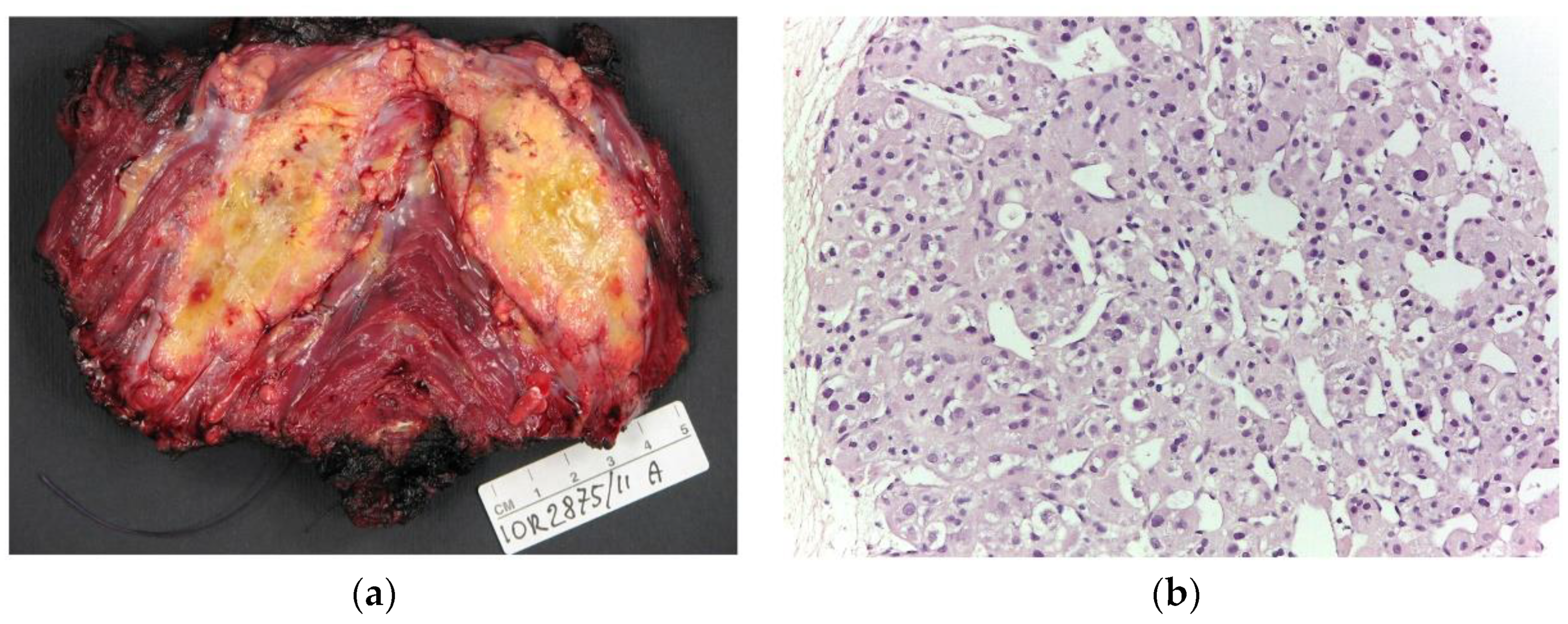
1.2. Anatomical Pathology and Genetic and Molecular Patterns
1.3. Imaging Features
2. Materials and Methods
2.1. Study Design
2.2. Clinical Data Collection
2.3. Imaging Analyses
2.3.1. Ultrasonography Analysis
2.3.2. MRI Studies and Analysis
2.3.3. CT Studies and Analysis
2.4. Statistical Analysis
2.5. Literature Review Search
3. Results
3.1. Clinical Data
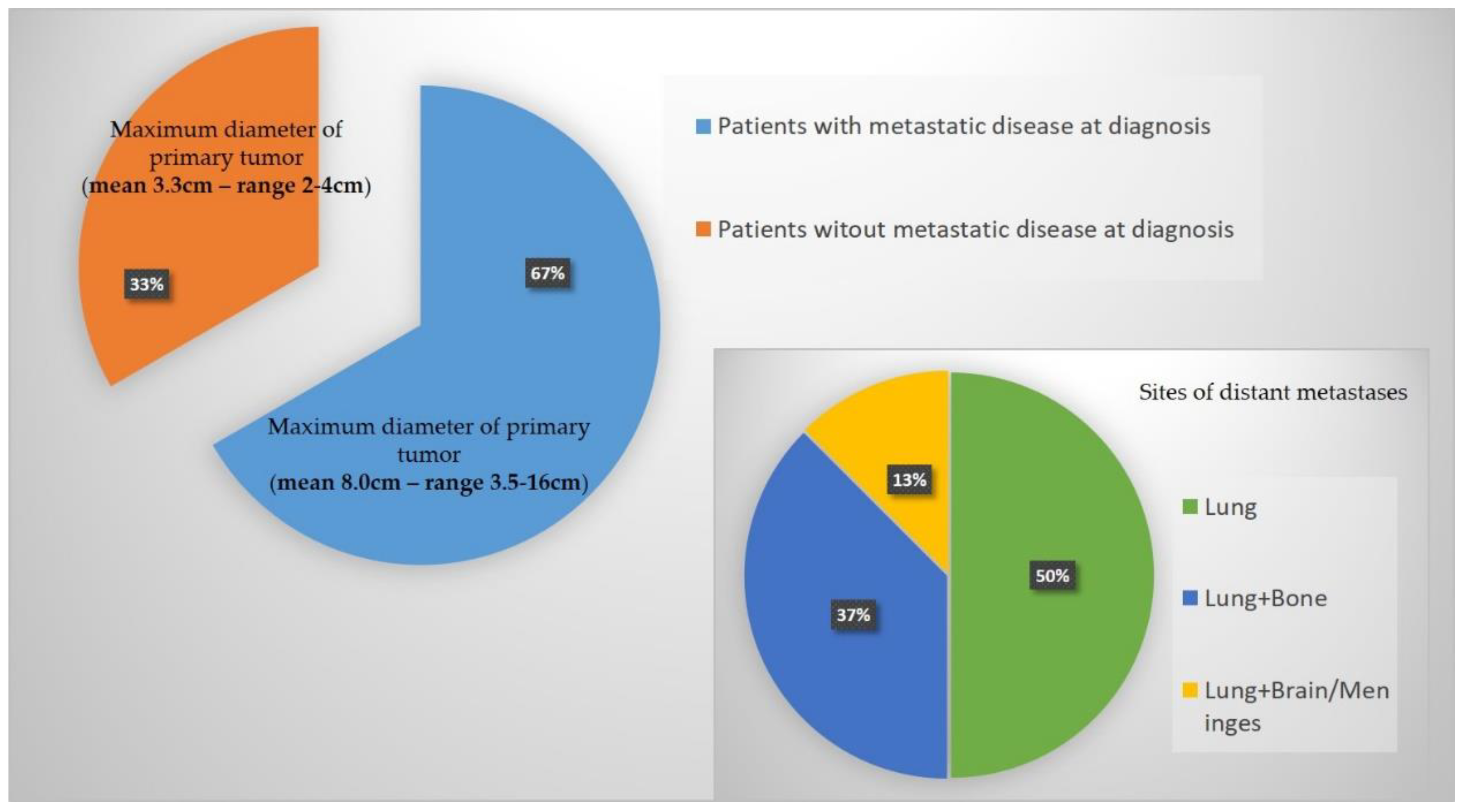
3.2. General Imaging Findings—Local and Distant Baseline Assessment
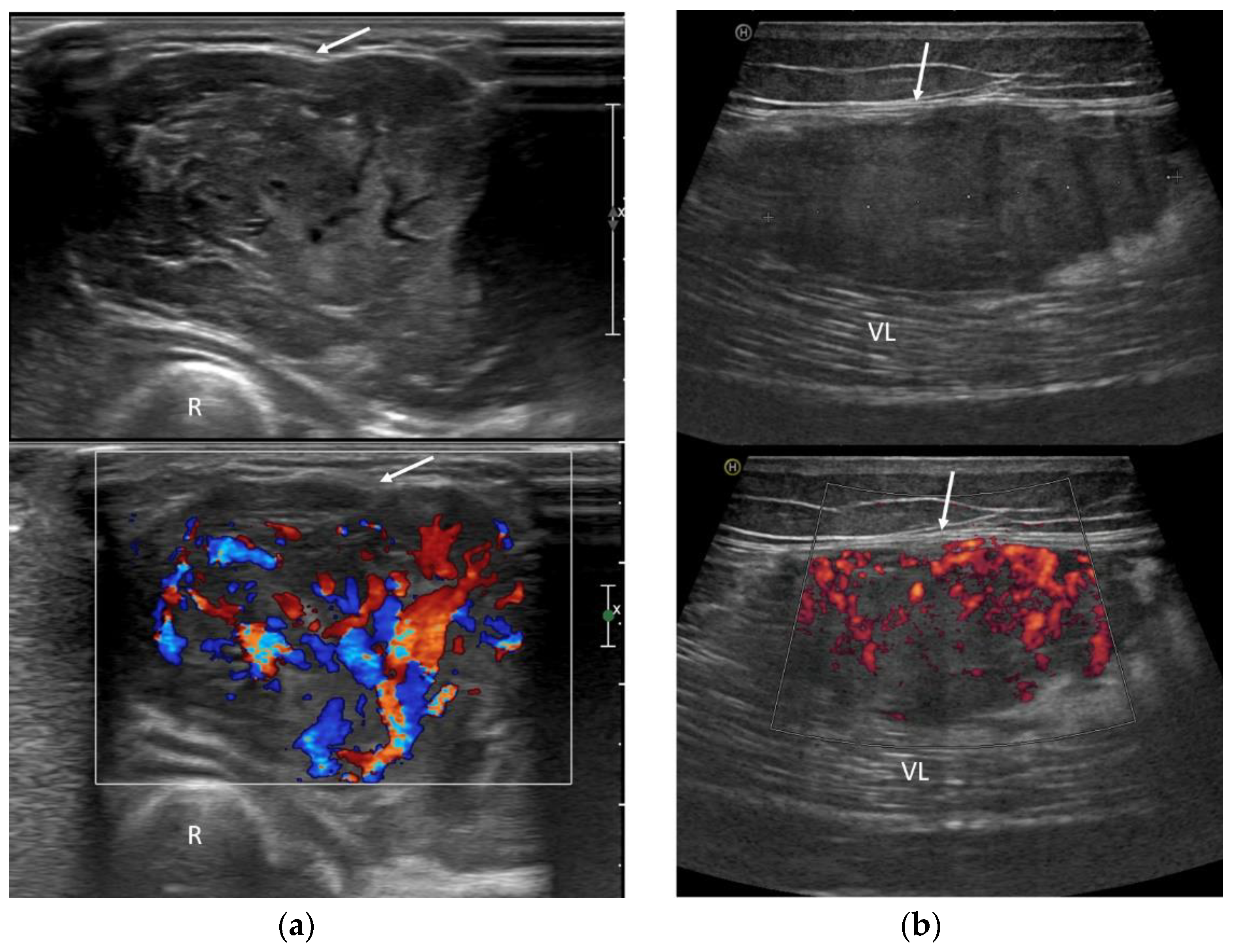
3.3. Ultrasound Features
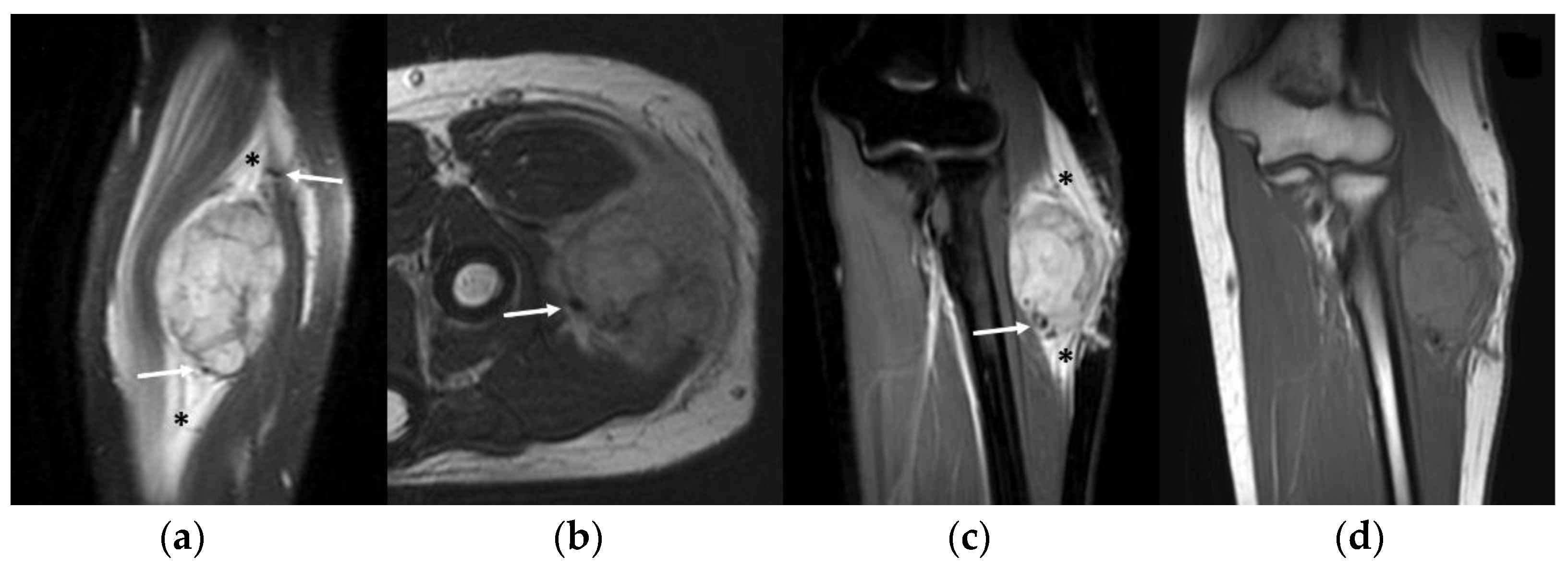
3.4. MRI Features
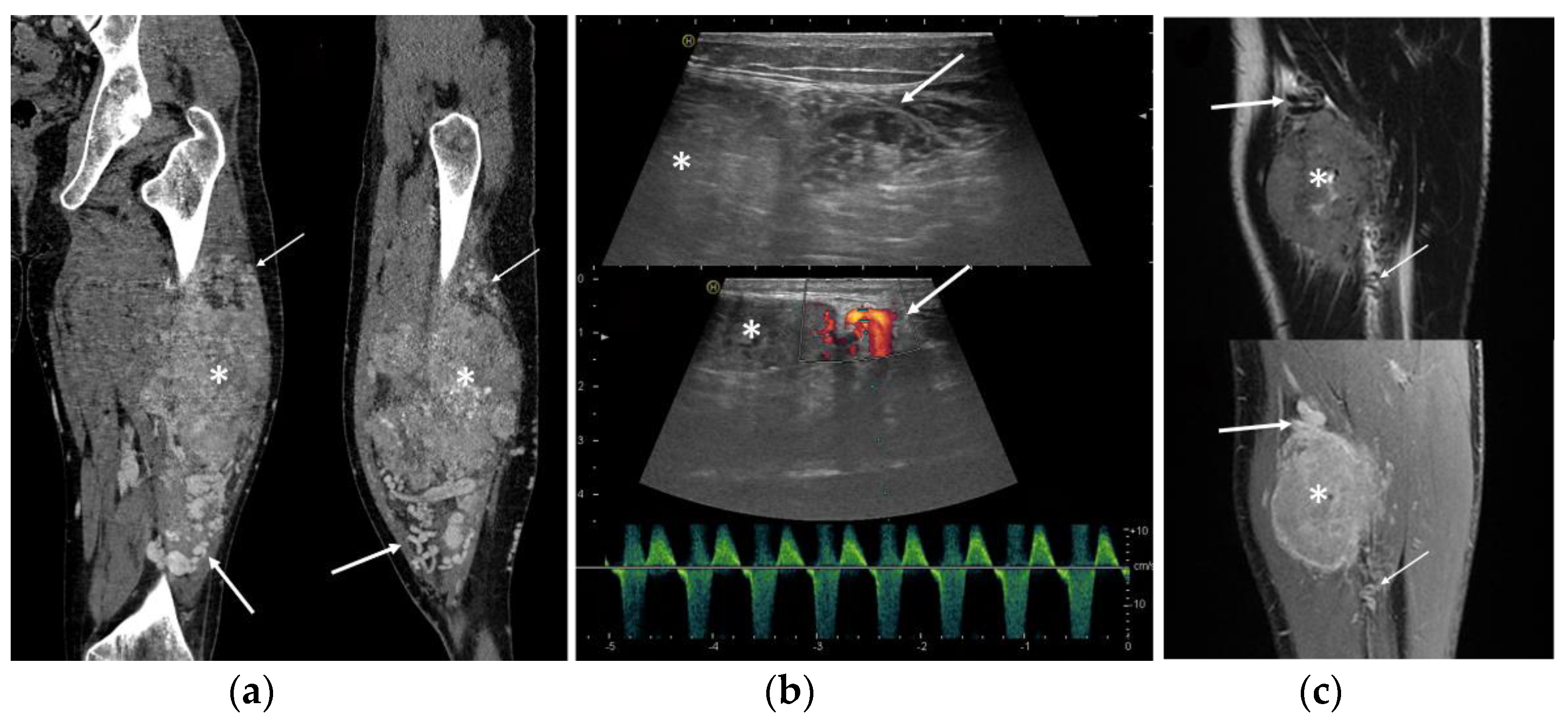
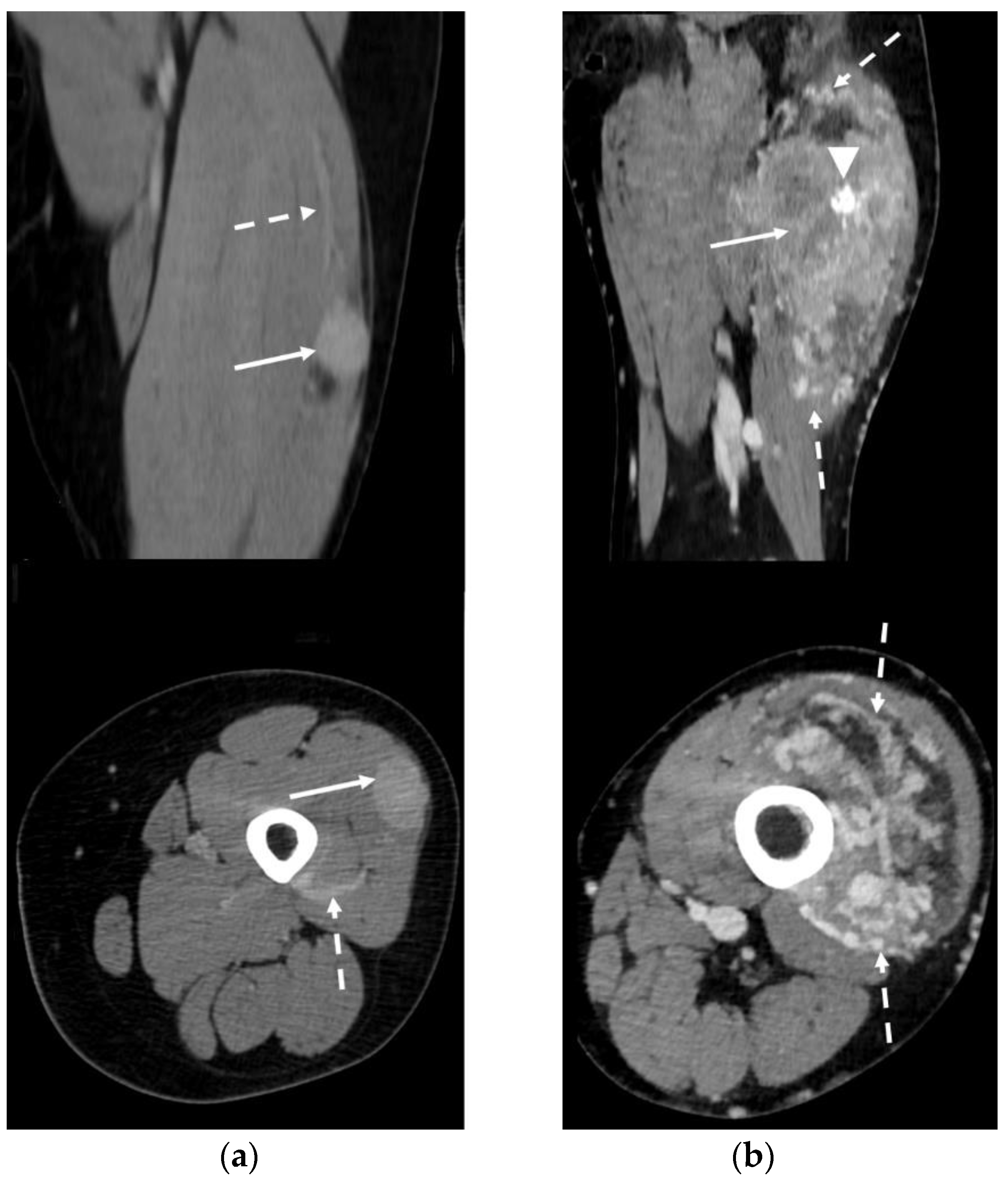
3.5. CT Features
3.6. Literature Review Results
4. Discussion
- Deep location
- Presence of peritumoral feeding vessels
- Inhomogeneous, mainly hypoechoic US pattern with strong internal vascularization at Color-Doppler evaluation
- Slight hyperintense MRI signal on T1-WI and a moderately inhomogeneous hyperintense signal on T2-WI
- MRI flow voids on fluid-sensitive sequences
- MRI peritumoral edema
- Slight low density and inhomogeneity on unenhanced CT
5. Conclusions
Author Contributions
Funding
Institutional Review Board Statement
Informed Consent Statement
Data Availability Statement
Conflicts of Interest
References
- Christopherson, W.M.; Foote, F.W., Jr.; Stewart, F.W. Alveolar soft-part sarcomas; structurally characteristic tumors of uncertain histogenesis. Cancer 1952, 5, 100–111. [Google Scholar] [CrossRef] [PubMed]
- Zhao, P.; Li, H.; Ren, H. Alveolar soft tissue sarcoma: A report of 50 cases at a single institution. Acta Chir. Belg. 2022, 123, 375–383. [Google Scholar] [CrossRef] [PubMed]
- Li, H.; Sun, J.; Ye, J.; Wu, J. Magnetic resonance imaging and computed tomography features of alveolar soft-part sarcoma in the right deltoid muscle: A case report. Oncol. Lett. 2016, 11, 2857–2860. [Google Scholar] [CrossRef]
- Hagerty, B.L.; Aversa, J.; Diggs, L.P.; Dominguez, D.A.; Ayabe, R.I.; Blakely, A.M.; Davis, J.L.; Luu, C.; Hernandez, J.M. Characterization of alveolar soft part sarcoma using a large national database. Surgery 2020, 168, 825–830. [Google Scholar] [CrossRef] [PubMed]
- Folpe, A.L.; Deyrup, A.T. Alveolar soft-part sarcoma: A review and update. J. Clin. Pathol. 2006, 59, 1127–1132. [Google Scholar] [CrossRef]
- Jaber, O.I.; Kirby, P.A. Alveolar Soft Part Sarcoma. Arch. Pathol. Lab. Med. 2015, 139, 1459–1462. [Google Scholar] [CrossRef] [PubMed]
- Paoluzzi, L.; Maki, R.G. Diagnosis, Prognosis, and Treatment of Alveolar Soft-Part Sarcoma: A Review. JAMA Oncol. 2019, 5, 254–260. [Google Scholar] [CrossRef]
- Ladanyi, M.; Antonescu, C.R.; Drobnjak, M.; Baren, A.; Lui, M.Y.; Golde, D.W.; Cordon-Cardo, C. The precrystalline cytoplasmic granules of alveolar soft part sarcoma contain monocarboxylate transporter 1 and CD147. Am. J. Pathol. 2002, 160, 1215–1221. [Google Scholar] [CrossRef]
- Song, L.; Zhang, Y.; Wang, Y.; Xia, Q.; Guo, D.; Cao, J.; Xin, X.; Cheng, H.; Liu, C.; Jia, X.; et al. Detection of various fusion genes by one-step RT-PCR and the association with clinicopathological features in 242 cases of soft tissue tumor. Front. Cell Dev. Biol. 2023, 11, 1214262. [Google Scholar] [CrossRef]
- Mukaihara, K.; Tanabe, Y.; Kubota, D.; Akaike, K.; Hayashi, T.; Mogushi, K.; Hosoya, M.; Sato, S.; Kobayashi, E.; Okubo, T.; et al. Cabozantinib and dastinib exert anti-tumor activity in alveolar soft part sarcoma. PLoS ONE 2017, 12, e0185321. [Google Scholar] [CrossRef]
- Kobos, R.; Nagai, M.; Tsuda, M.; Merl, M.Y.; Saito, T.; Laé, M.; Mo, Q.; Olshen, A.; Lianoglou, S.; Leslie, C.; et al. Combining integrated genomics and functional genomics to dissect the biology of a cancer-associated, aberrant transcription factor, the ASPSCR1-TFE3 fusion oncoprotein. J. Pathol. 2013, 229, 743–754. [Google Scholar] [CrossRef] [PubMed]
- Schöffski, P.; Wozniak, A.; Kasper, B.; Aamdal, S.; Leahy, M.; Rutkowski, P.; Bauer, S.; Gelderblom, H.; Italiano, A.; Lindner, L.; et al. Activity and safety of crizotinib in patients with alveolar soft part sarcoma with rearrangement of TFE3: European Organization for Research and Treatment of Cancer (EORTC) phase II trial 90101 “CREATE”. Ann. Oncol. 2018, 29, 758–765. [Google Scholar] [CrossRef] [PubMed]
- Goodwin, M.L.; Jin, H.; Straessler, K.; Smith-Fry, K.; Zhu, J.-F.; Monument, M.J.; Grossmann, A.; Randall, R.L.; Capecchi, M.R.; Jones, K.B. Modeling alveolar soft part sarcomagenesis in the mouse: A role for lactate in the tumor microenvironment. Cancer Cell 2014, 26, 851–862. [Google Scholar] [CrossRef]
- Tanaka, M.; Chuaychob, S.; Homme, M.; Yamazaki, Y.; Lyu, R.; Yamashita, K.; Ae, K.; Matsumoto, S.; Kumegawa, K.; Maruyama, R.; et al. ASPSCR1::TFE3 orchestrates the angiogenic program of alveolar soft part sarcoma. Nat. Commun. 2023, 14, 1957. [Google Scholar] [CrossRef]
- Fujiwara, T.; Kunisada, T.; Nakata, E.; Nishida, K.; Yanai, H.; Nakamura, T.; Tanaka, K.; Ozaki, T. Advances in treatment of alveolar soft part sarcoma: An updated review. Jpn. J. Clin. Oncol. 2023, hyad102. [Google Scholar] [CrossRef]
- Crombé, A.; Brisse, H.J.; Ledoux, P.; Haddag-Miliani, L.; Bouhamama, A.; Taieb, S.; Le Loarer, F.; Kind, M. Alveolar soft-part sarcoma: Can MRI help discriminating from other soft-tissue tumors? A study of the French sarcoma group. Eur. Radiol. 2019, 29, 3170–3182. [Google Scholar] [CrossRef] [PubMed]
- Tian, L.; Cui, C.-Y.; Lu, S.-Y.; Cai, P.-Q.; Xi, S.-Y.; Fan, W. Clinical presentation and CT/MRI findings of alveolar soft part sarcoma: A retrospective single-center analysis of 14 cases. Acta Radiol. 2016, 57, 475–480. [Google Scholar] [CrossRef]
- Viry, F.; Orbach, D.; Klijanienko, J.; Fréneaux, P.; Pierron, G.; Michon, J.; Neuenschwander, S.; Brisse, H.J. Alveolar soft part sarcoma-radiologic patterns in children and adolescents. Pediatr. Radiol. 2013, 43, 1174–1181. [Google Scholar] [CrossRef]
- Qiao, P.-F.; Shen, L.-H.; Gao, Y.; Mi, Y.-C.; Niu, G.-M. Alveolar soft part sarcoma: Clinicopathological analysis and imaging results. Oncol. Lett. 2015, 10, 2777–2780. [Google Scholar] [CrossRef]
- Cui, J.-F.; Chen, H.-S.; Hao, D.-P.; Liu, J.-H.; Hou, F.; Xu, W.-J. Magnetic Resonance Features and Characteristic Vascular Pattern of Alveolar Soft-Part Sarcoma. Oncol. Res. Treat. 2017, 40, 580–585. [Google Scholar] [CrossRef]
- Park, J.H.; Kang, C.H.; Kim, C.H.; Chae, I.J.; Park, J.H. Highly malignant soft tissue sarcoma of the extremity with a delayed diagnosis. World J. Surg. Oncol. 2010, 8, 84. [Google Scholar] [CrossRef] [PubMed]
- McCarville, M.B.; Muzzafar, S.; Kao, S.C.; Coffin, C.M.; Parham, D.M.; Anderson, J.R.; Spunt, S.L. Imaging features of alveolar soft-part sarcoma: A report from Children’s Oncology Group Study ARST0332. Am. J. Roentgenol. 2014, 203, 1345–1352. [Google Scholar] [CrossRef] [PubMed]
- Suh, J.-S.; Cho, J.; Lee, S.H.; Shin, K.-H.; Yang, W.I.; Lee, J.H.; Cho, J.-H.; Suh, K.J.; Lee, Y.J.; Ryu, K.N. Alveolar soft part sarcoma: MR and angiographic findings. Skelet. Radiol. 2000, 29, 680–689. [Google Scholar] [CrossRef] [PubMed]
- Li, X.; Ye, Z. Magnetic resonance imaging features of alveolar soft part sarcoma: Report of 14 cases. World J. Surg. Oncol. 2014, 12, 36. [Google Scholar] [CrossRef] [PubMed]
- Iwamoto, Y.; Morimoto, N.; Chuman, H.; Shinohara, N.; Sugioka, Y. The role of MR imaging in the diagnosis of alveolar soft part sarcoma: A report of 10 cases. Skelet. Radiol. 1995, 24, 267–270. [Google Scholar] [CrossRef] [PubMed]
- Li, W.; Zhang, S.M.; Fan, W.; Li, D.; Tian, H.M.; Che, D.M.; Yu, L.M.; Gao, S.M.; Liu, Y. Sonographic imaging features of alveolar soft part sarcoma: Case series and literature review. Medicine 2022, 101, e31905. [Google Scholar] [CrossRef]
- Gulati, M.; Mittal, A.; Barwad, A.; Pandey, R.; Rastogi, S.; Dhamija, E. Imaging and Pathological Features of Alveolar Soft Part Sarcoma: Analysis of 16 Patients. Indian J. Radiol. Imaging 2021, 31, 573–581. [Google Scholar] [CrossRef]
- Sood, S.; Baheti, A.D.; Shinagare, A.B.; Jagannathan, J.P.; Hornick, J.L.; Ramaiya, N.H.; Tirumani, S.H. Imaging features of primary and metastatic alveolar soft part sarcoma: Single institute experience in 25 patients. Br. J. Radiol. 2014, 87, 20130719. [Google Scholar] [CrossRef]
- Kim, H.S.; Lee, H.K.; Weon, Y.-C.; Kim, H.-J. Alveolar soft-part sarcoma of the head and neck: Clinical and imaging features in five cases. Am. J. Neuroradiol. 2005, 26, 1331–1335. [Google Scholar]
- De Pinieux, G.; Karanian, M.; Le Loarer, F.; Le Guellec, S.; Chabaud, S.; Terrier, P.; Bouvier, C.; Batistella, M.; Neuville, A.; Robin, Y.-M.; et al. Nationwide incidence of sarcomas and connective tissue tumors of intermediate malignancy over four years using an expert pathology review network. PLoS ONE 2021, 16, e0246958. [Google Scholar] [CrossRef]
- Stacchiotti, S.; Negri, T.; Zaffaroni, N.; Palassini, E.; Morosi, C.; Brich, S.; Conca, E.; Bozzi, F.; Cassinelli, G.; Gronchi, A.; et al. Sunitinib in advanced alveolar soft part sarcoma: Evidence of a direct antitumor effect. Ann. Oncol. 2011, 22, 1682–1690. [Google Scholar] [CrossRef]
- Wilky, B.A.; Trucco, M.M.; Subhawong, T.K.; Florou, V.; Park, W.; Kwon, D.; Wieder, E.D.; Kolonias, D.; Rosenberg, A.E.; Kerr, D.A.; et al. Axitinib plus pembrolizumab in patients with advanced sarcomas including alveolar soft-part sarcoma: A single-centre, single-arm, phase 2 trial. Lancet Oncol. 2019, 20, 837–848. [Google Scholar] [CrossRef] [PubMed]
- O’Sullivan Coyne, G.; Naqash, A.R.; Sankaran, H.; Chen, A.P. Advances in the management of alveolar soft part sarcoma. Curr. Probl. Cancer 2021, 45, 100775. [Google Scholar] [CrossRef] [PubMed]
- Spinnato, P. The Importance of Accurate Tumor Measurements and Staging in Oncologic Imaging: Impact on Patients’ Health. Acad. Radiol. 2021, 28, 767–768. [Google Scholar] [CrossRef]
- Scalas, G.; Parmeggiani, A.; Martella, C.; Tuzzato, G.; Bianchi, G.; Facchini, G.; Clinca, R.; Spinnato, P. Magnetic resonance imaging of soft tissue sarcoma: Features related to prognosis. Eur. J. Orthop. Surg. Traumatol. 2021, 31, 1567–1575. [Google Scholar] [CrossRef]
- Rimondi, E.; Mavrogenis, A.F.; Errani, C.; Calabrò, T.; Bazzocchi, A.; Facchini, G.; Donatiello, S.; Spinnato, P.; Vanel, D.; Albisinni, U.; et al. Biopsy is not necessary for the diagnosis of soft tissue hemangiomas. Radiol. Med. 2018, 123, 538–544. [Google Scholar] [CrossRef] [PubMed]
- Crombé, A.; Kind, M.; Fadli, D.; Miceli, M.; Linck, P.-A.; Bianchi, G.; Sambri, A.; Spinnato, P. Soft-tissue sarcoma in adults: Imaging appearances, pitfalls and diagnostic algorithms. Diagn. Interv. Imaging 2023, 104, 207–220. [Google Scholar] [CrossRef]
| Patient n° | Age, Sex | Symptoms | Lesion Location (Deep/Superficial) | Baseline Imaging Tools Available | Longest Diameter (cm) | Metastasis at Diagnosis (Sites) | Current Status | Survival (Months) |
|---|---|---|---|---|---|---|---|---|
| 1 | 24, M | Non-painful lump | Leg (deep) | US, MRI | 6 | Yes (lung) | AWD | 46 |
| 2 | 20, F | Non-painful lump | Popliteal fossa (deep) | MRI | 3.5 | No | NED | 144 |
| 3 | 16, F | Painful lump | Forearm (deep) | US, MRI | 4 | No | NED | 41 |
| 4 | 44, M | Painful lump | Thigh (deep) | MRI | 7 | Yes (lung, bone) | DOD | 135 |
| 5 | 66, F | Painful lump | Hip girdle (deep) | MRI, CT | 8 | Yes (lung, bone) | DOD | 24 |
| 6 | 26, F | Painful lump | Hip girdle (deep) | CT | 10 | Yes (lung, brain, meninges) | DOD | 39 |
| 7 | 20, M | Non-painful lump | Thigh (deep) | US, MRI | 8 | Yes (lung, bone) | DOD | 6 |
| 8 | 19, F | Non-painful lump | Leg (deep) | US, MRI | 3.5 | Yes (lung) | AWD | 6 |
| 9 | 25, F | Non-painful lump | Thigh (deep) | US, MRI, CT | 3.5 | No | NED | 32 |
| 10 | 35, M | Painful lump | Thigh (deep) | CT | 16 | Yes (lung) | DOD | 1 |
| 11 | 4, F | Painful lump | Arm (deep) | US, MRI | 2 | No | NED | 9 |
| 12 | 23, M | Non-painful lump | Thigh (deep) | US, MRI | 5.5 | Yes (Lung) | DOD | 165 |
| General Imaging Features | US | CE MRI | CE CT |
|---|---|---|---|
| Deep-seated location | Well-defined borders | Intra and peritumoral flow-voids | Low density on unenhanced scans |
| Peritumoral feeding vessels | Inhomogeneous hypoechoic pattern | Slightly high SI on T1w | - |
| Strong internal vascularization | Arteriosus Doppler sonographic pattern inside peritumoral vessels | High SI on T2w | - |
| First Author, Year, Reference Number | Number of Patients | Age (Years) | Average Longest Diameter | Metastatic Disease at Diagnosis | Baseline Imaging | Main Imaging Findings |
|---|---|---|---|---|---|---|
| Iwamoto, 1995, [25] | 10 | 11–40 | NA | 2/10 (20%) | MRI, Angiography | MRI: Slightly high SI on T1w, high SI on T2w. Flow voids. |
| Kim, 2005, [29] | 5 | 4–22 | 72 mm | 7/10 (70%) | CT, MRI, Angiography | CT: Strong CE. MRI: Slightly high SI on T1w, high SI on T2w. Flow voids. Angiography: Hypervascular lesion. |
| Park, 2010, [21] | 3 | NA | NA | 1/5 (20%) | MRI | MRI: Slightly high SI on T1w, high SI on T2w. Flow voids. |
| Viry, 2013, [18] | 6 | 7–17 | NA | 3/3 (100%) | CT, MRI | CT: Low density on unenhanced scans. MRI: Slightly high SI on T1w, high SI on T2w. Highly vascularized lesions. Intra-/peritumoral vessels with high flow (=flow voids). Central stellar necrotic component or central stellar highly vascular area (for >7 cm tumors). |
| Li, 2014, [16] | 14 | 27–54 | 98 mm | 4/5 (80%) | MRI | MRI: Slightly high SI on T1w, high SI on T2w. Flow voids. Inhomogeneous signal intensity. |
| Suh, 2014, [23] | 10 | 17–48 | NA | 8/14 (57.1%) | MRI, Angiography | MRI: Slightly high SI on T1w, high SI on T2w. Flow voids. Inhomogeneous SI. Angiography: Peritumoral vessels with arteriovenous shunts. |
| McCarville, 2014, [22] | 22 | 8–23 | 59 mm | 11/22 (50%) | CT, MRI | MRI: Slightly high SI on T1w, high SI on T2w. Flow voids. Nodular internal architecture, separated by thin hypodense bands. Intense or moderate CE with central necrosis. CT: Intra- and peritumoral vessels. Highly vascularized lesions on CE scan. |
| Qiao, 2015, [19] | 6 | 16–45 | 48 mm | Not reported | CT, MRI | CT: Highly vascularized on CE scan. Hypodense lesions on unenhanced scan. MRI: Hypointense on T1w, hyperintense on T2w. |
| Tian, 2016, [17] | 14 | 13–37 | 91 mm | Not reported | CT, MRI | CT: Low density on unenhanced scan. MRI: Slightly high SI on T1w, high SI on T2w. Highly vascularized lesions. Intra-/peritumoral vessels. Flow voids. |
| Sood, 2016, [28] | 25 | 18–40 | 102 mm | 18/25 /72%) | CT, MRI | CT: Low density on unenhanced scan. MRI: Slightly high SI on T1w, high SI on T2w. Highly vascularized lesions. Intra-/peritumoral vessels. Flow voids. |
| Cui, 2017, [20] | 12 | 21–34 | 68 mm | 5/12 (41.7%) | MRI | MRI: High signal intensity on T2w. Highly vascularized lesions. Flow voids. Central area of necrosis or hypervascularization. |
| Crombé, 2018, [16] | 25 | 7–53 | 66 mm | 14/25 (56%) | MRI | MRI: Slightly high SI on T1w, high SI on T2w. Infiltrative growth pattern. Deep location. Tubular feeding vessels. Flow voids (>5). Central area of necrosis. Absence of tail sign, absence of fibrotic signal. |
| Li, 2022, [24] | 3 | 23–30 | 81 mm | 2/3 (66.6%) | US, MRI, CT | US: Heterogeneous hypoechoic tissue. Well-defined margins. Intra- and peri-tubular vessels. Highly vascularized lesions on color-Doppler. MRI: Slightly high SI on T1w, high SI on T2w. Flow voids. CT: Slightly high density without significant bony destruction on CT. |
| Gulati, 2021, [27] | 16 | 3–72 | 83 mm | 14/16 (87.5%) | US, CT, MRI, PET | PET: SUV of >2.5. CT: Intense CE. MRI: Slightly high SI on T1w, high SI on T2w. Flow voids. Intense CE. US: Circumscribed lobulated homogeneously hypoechoic pattern. Multiple enlarged feeding vessels. |
Disclaimer/Publisher’s Note: The statements, opinions and data contained in all publications are solely those of the individual author(s) and contributor(s) and not of MDPI and/or the editor(s). MDPI and/or the editor(s) disclaim responsibility for any injury to people or property resulting from any ideas, methods, instructions or products referred to in the content. |
© 2023 by the authors. Licensee MDPI, Basel, Switzerland. This article is an open access article distributed under the terms and conditions of the Creative Commons Attribution (CC BY) license (https://creativecommons.org/licenses/by/4.0/).
Share and Cite
Spinnato, P.; Papalexis, N.; Colangeli, M.; Miceli, M.; Crombé, A.; Parmeggiani, A.; Palmerini, E.; Righi, A.; Bianchi, G. Imaging Features of Alveolar Soft Part Sarcoma: Single Institution Experience and Literature Review. Clin. Pract. 2023, 13, 1369-1382. https://doi.org/10.3390/clinpract13060123
Spinnato P, Papalexis N, Colangeli M, Miceli M, Crombé A, Parmeggiani A, Palmerini E, Righi A, Bianchi G. Imaging Features of Alveolar Soft Part Sarcoma: Single Institution Experience and Literature Review. Clinics and Practice. 2023; 13(6):1369-1382. https://doi.org/10.3390/clinpract13060123
Chicago/Turabian StyleSpinnato, Paolo, Nicolas Papalexis, Marco Colangeli, Marco Miceli, Amandine Crombé, Anna Parmeggiani, Emanuela Palmerini, Alberto Righi, and Giuseppe Bianchi. 2023. "Imaging Features of Alveolar Soft Part Sarcoma: Single Institution Experience and Literature Review" Clinics and Practice 13, no. 6: 1369-1382. https://doi.org/10.3390/clinpract13060123
APA StyleSpinnato, P., Papalexis, N., Colangeli, M., Miceli, M., Crombé, A., Parmeggiani, A., Palmerini, E., Righi, A., & Bianchi, G. (2023). Imaging Features of Alveolar Soft Part Sarcoma: Single Institution Experience and Literature Review. Clinics and Practice, 13(6), 1369-1382. https://doi.org/10.3390/clinpract13060123


