Next Article in Journal
The Practice of Teaching and Scientific Research on Cadaveric Material Remains Crucial for Medical Education
Previous Article in Journal
Evaluation of Abfraction Lesions Restored with Three Dental Materials: A Comparative Study
Previous Article in Special Issue
Contemporary Endodontic Approaches for Children
 
 
Retraction published on 25 December 2023, see Clin. Pract. 2024, 14(1), 31.
Review

RETRACTED: Traditional and Recent Root Canal Irrigation Methods and Their Effectiveness: A Review

by
Alfredo Iandolo
1,*,
Massimo Pisano
1,
Alessio Buonavoglia
2,
Francesco Giordano
1,
Alessandra Amato
1 and
Dina Abdellatif
3
1
Department of Medicine and Surgery, School of Dentistry, University of Salerno, 84081 Salerno, Italy
2
Department of Biomedical and Neuromotor Sciences, School of Dentistry, University of Bologna, 40125 Bologna, Italy
3
Department of Endodontics, Faculty of Dentistry, University of Alexandria, Alexandria 21531, Egypt
*
Author to whom correspondence should be addressed.
Clin. Pract. 2023, 13(5), 1059-1072; https://doi.org/10.3390/clinpract13050094
Submission received: 15 August 2023 / Revised: 25 August 2023 / Accepted: 25 August 2023 / Published: 27 August 2023 / Retracted: 25 December 2023
(This article belongs to the Special Issue Contemporary Endodontic Approaches for Children)

Abstract

:
According to contemporary dental standards, the primary goal of endodontic therapy is the chemo-mechanical cleaning of the complex root canal system. Watering root canals with approved solutions and activating them are essential parts of this operation. This review outlines various irrigant activation methods for root canal therapy. Specifically, a comparison among the methods of manual dynamic activation, sonics (subsonic, sonic, and ultrasonic), internal heating, and lasers, was conducted. The results in this work were gathered using Scopus, Web of Science, Google Scholar, and PubMed databases by searching the following keywords: sodium hypochlorite, cleaning, activation, and irrigation methods. The present work concluded that the use of irrigant activation has a greater benefit than its absence. Regardless, it is impossible to point to a single effective activation method.

1. Introduction

Nowadays, there is increased use of the term “modern endodontics” to refer to contemporary applied science and actual materials that have been introduced and invented in recent years. Diverse endodontic tools and technological devices have been designed to facilitate and enhance treatments [1], for example, magnification using the operative microscope, sonic and ultrasonic appliances, and certain laser machines. Moreover, modified alloys for NI-Ti rotary files and potent irrigation techniques were invented for the cleaning and shaping phases. There has been huge developments in irrigant solutions, obturation materials, and cone-beam or 3D (three-dimensional) radiography [2,3]. New trends are arising in regenerative endodontics, which are conducted to replace inflamed and necrotic pulp tissue with a functional and healthy dentine–pulp complex [4].
Consequently, applying advanced technologies in endodontic therapy allows the secure and manageable treatment of even the most complicated cases.
It was documented that some areas of the complex root canal space remain unreachable during the mechanical phase, independently of the file type. For illustration, hand and rotary endodontic files only clean the centre of root canals [5,6], while the lateral anatomies, like isthmuses, lateral and furcation canals, apical deltas, and others, remain untouched after mechanical shaping. Thus, active cleaning is essential to adequately eliminate bacteria and necrotic tissue [7]. Recently, a new concept including a minimally invasive shaping approach utilising rotating files of a smaller size and taper combined with more efficient irrigation protocols was introduced, leading to safer, more conservative endodontic therapies.
Considering the irrigant solution, sodium hypochlorite (NaOCl) is the most typically used irrigant, owing to its elevated ability in tissue dissolution and superior antimicrobial activity. Multiple procedures can be used to maximise the action and effectiveness of NaOCl [8,9]. Some techniques for activating the irrigants used during an endodontic procedure can be listed as follows (Figure 1):
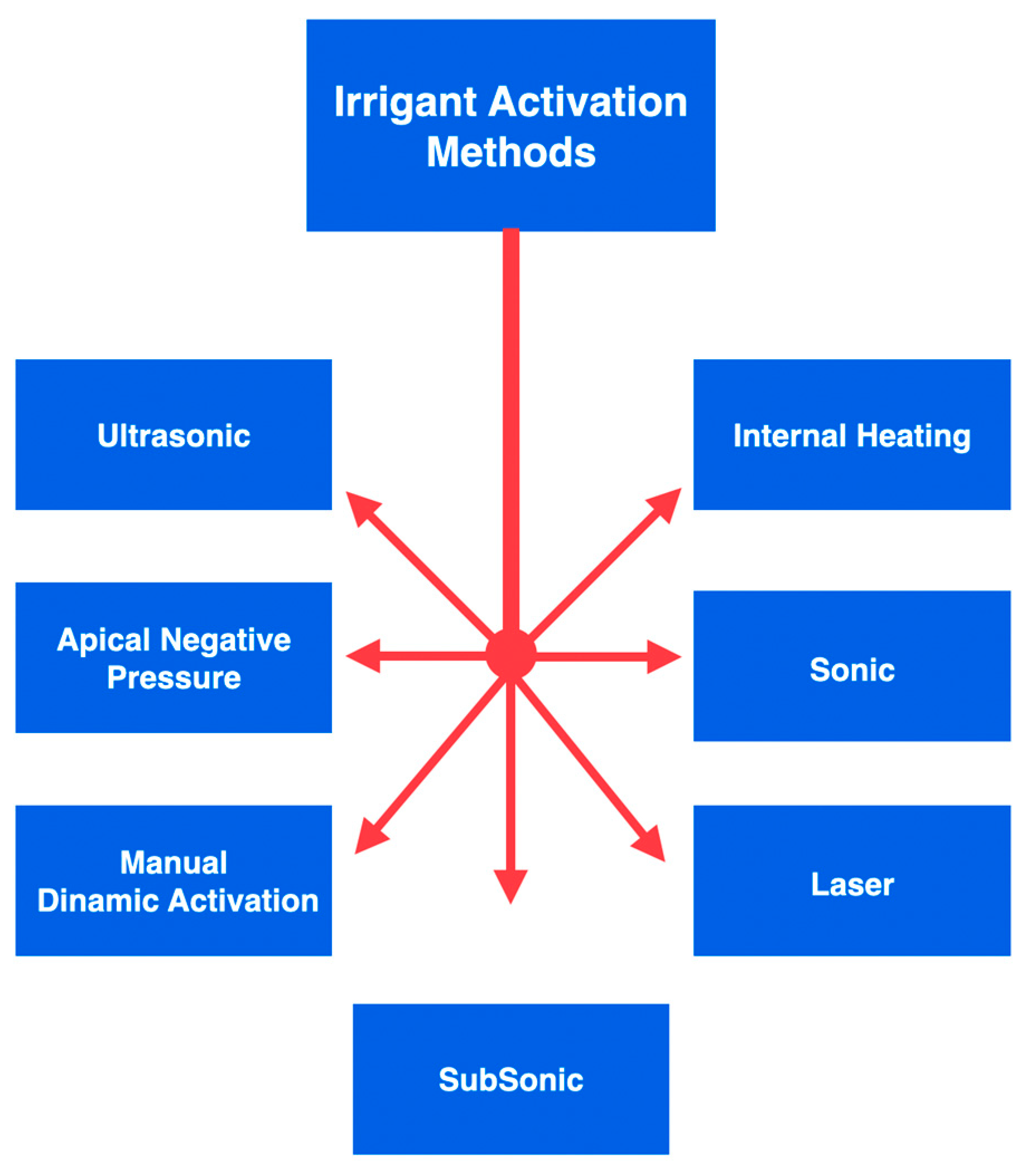
  • Manual dynamic activation;
  • Heating (internal or external);
  • Negative apical pressure (EndoVac, Kerr Endodontics, Gilbert, AZ, USA; Rinsendo, Dürr Dental, Bietigheim, Germany);
  • Subsonic technique (EndoAcivator, Dentsply Maillefer, Ballaigues, Switzerland);
  • Sonic technique (EDDY, VDW, München, Germany);
  • Passive ultrasonic irrigation (PUI);
  • Laser techniques [10,11,12,13,14].
Bacteria and their byproducts are the main etiological agents of periapical pulp infection. The cleaning part is the most delicate phase, where the bacterial load is lowered [15,16,17,18]. The main factors that define the composition of the microbial flora are the anaerobic environment, the interactions among various bacteria, and the availability of nutrients [19]. The number of bacterial species is usually limited in the initial phase of endodontic infection. According to the literature, the number of bacterial species in an infected root canal can vary between 1 and 12. It also appears that there is a correlation between the size of the periapical lesion and the number of bacterial species present in the root canal [20,21]. It is generally believed that the persistence of endodontic infection is related to difficulties encountered during the initial treatment and thus to incomplete removal of damaged tissues or bacteria. Normally, only one or a few bacterial species are found in the root canals of dental elements with persistent pathology [19,22,23]. They are mostly Gram-positive microorganisms with equal distribution of facultative and obligate anaerobes [24]. The microbial flora is distinctly different from that typical of untreated tooth infections, which is polymicrobial with similar proportions of Gram-positive and Gram-negative species, and a predominance of obligate anaerobes [25,26,27,28]. Despite the differences between species isolated from different channels with the same clinical presentation (persistent periapical disease), many studies agree that enterococci and streptococci are highly prevalent [29]. Enterococcus faecalis is considered a specific opportunistic pathogen of persistent periapical pathology. While in the untreated infected canal, the microbial flora can find an abundance of nourishment, it can suffer famine in the endodontically well-treated canal. Ideally, all the necrotic pulp originally present will have been eliminated, leaving dry and poor conditions for the survival of the remaining microbial species. These species must endure hunger and a static environment but can sometimes find some nourishment in the exudate coming from the periapical tissue [30]. Persistent species survived antimicrobial treatments, entered the canal during treatment, and settled where other species could not. If the coronal seal has defects, there may be bacterial infiltration, and therefore, there is the possibility of new infection of the canal. Inside the endodontic space, there are free bacteria in planktonic form and microorganisms united in a very organized structure, the biofilm, which can oppose the removal action with different mechanisms [31]. The biofilm formation mechanism is known: on a protein layer that forms a conditioning film, the bacteria in planktonic form adhere, which subsequently stratify. The bacteria in the biofilm are immersed in a matrix called the glycocalyx, which acts as a mechanical barrier against antibacterial agents. Biofilm is 1000 times more resistant than bacteria in planktonic form. The biofilm can be broken up and removed thanks to the activation of the irrigating agents [32,33]. The endodontic space is not only composed of the main root canals, different and complex three-dimensional anatomies also form it (Figure 2).
Lateral canals: canals of small, medium, and large dimensions that start from the main canal at any height and communicate with the periodontium. They can be located in the root canal’s coronal third, middle third, and apical third. They can also be present in the furcation area in the case of multi-rooted teeth. By communicating with the periodontium, they can give rise to lateral lesions.
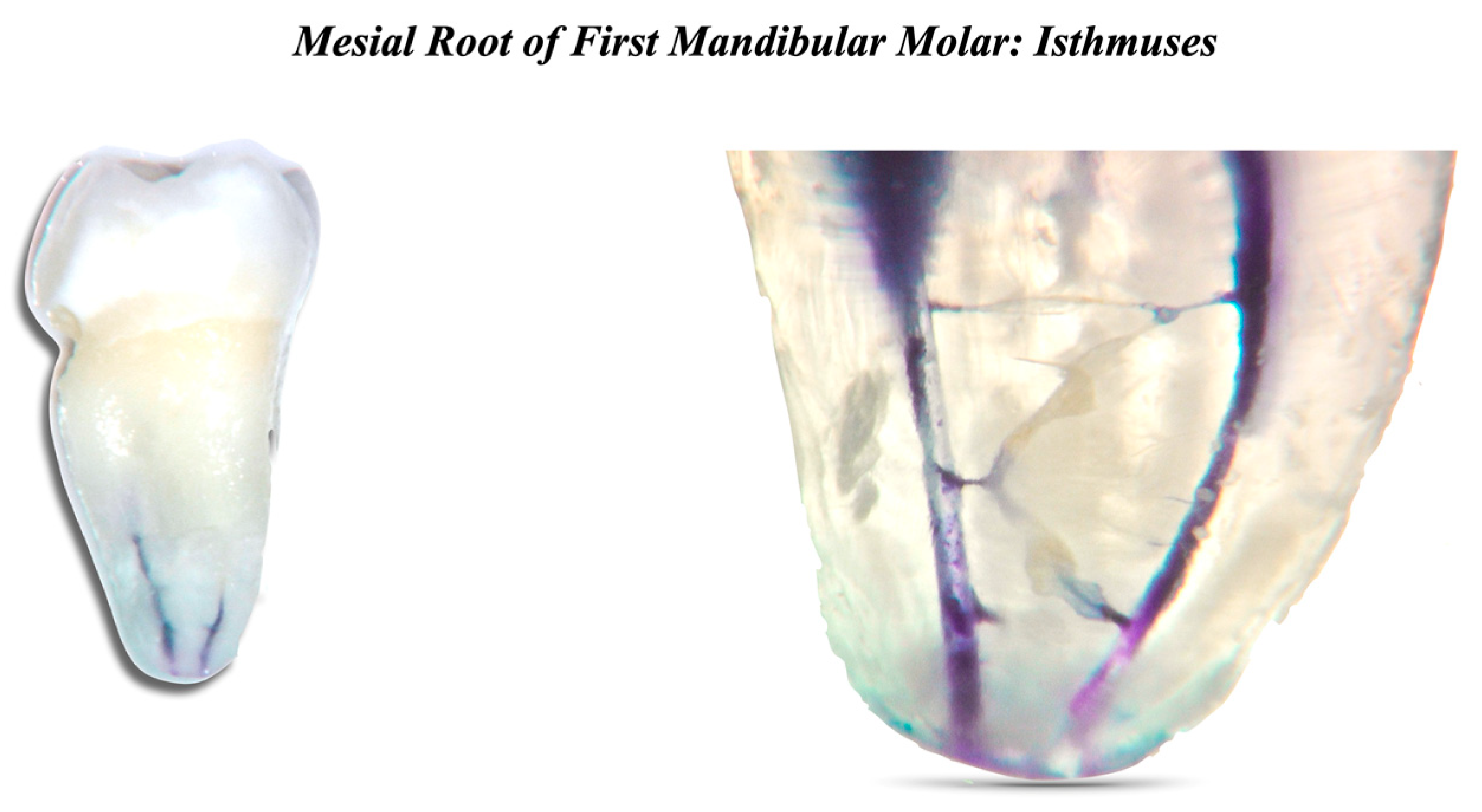
Isthmuses: connections of small, medium, and large dimensions between two main canals. Isthmuses contain pulp tissue and may also retain bacteria in the case of necrotic teeth. Additionally, the incomplete removal of the infected tissues within these areas can jeopardize the treatment outcome after performing endodontic therapy [34].
Loop: a small area that leaves the main canal and then reconnects to it.
Ramifications: microscopic extensions from the main canal at any height.
Apical delta: bifurcation of the main canal at the apical level.
POE: Portal of exits. The main canal can have more than one apical exit of different sizes.
Different anatomical complex variations are present together within the endodontic space.
Dentinal tubules: located in the dentin, of fundamental importance, as their internal, bacteria, and their products can enter at different depths [35,36,37,38].
Bacteria can penetrate hundreds of microns inside the dentinal tubules, so the mechanical and chemical removal of infected dentin can play a fundamental role in endodontic treatment’s short- and long-term success [39]. The literature shows that the instrumentation alone is insufficient to eradicate the bacterial load within those microscopic hidden areas [40,41].
This review aims to discuss the traditional and recent root canal irrigation methods and their effectiveness when used after conventional or conservative mechanical shaping.

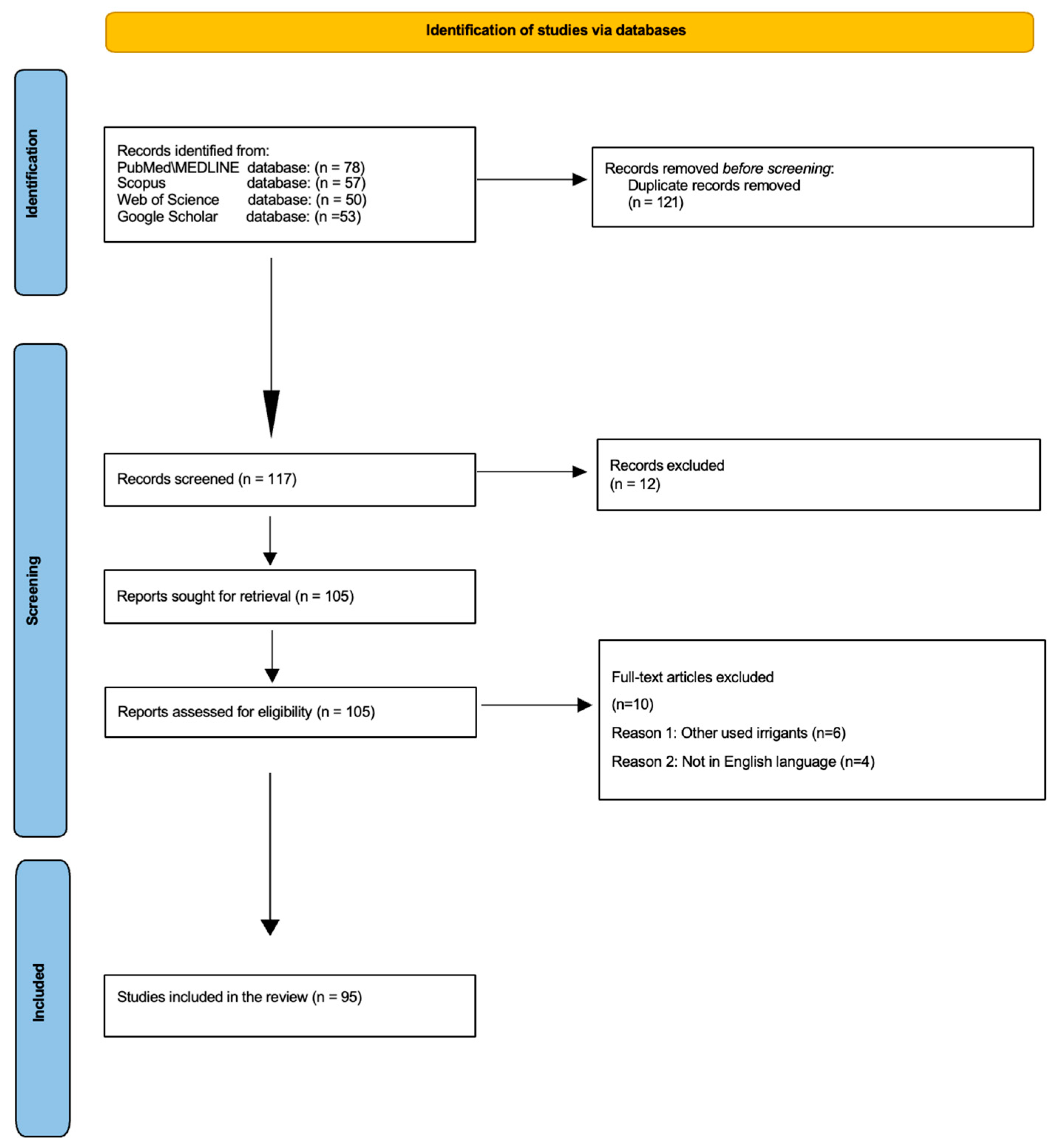
2. Materials and Methods

An electronic investigation of the literature was conducted to search the following websites: PubMed, Web of Science, Scopus, and Google Scholar databases. Specifically, a mix of these free-text words was employed: NaOCl, Sodium Hypochlorite, (ethylene diamine tetra acetic acid), EDTA, cleaning, activation, and irrigation methods. Several articles were chosen from the search results based on their importance to the subject. In the context of the activation of irrigants in endodontic treatment, a focus on clinical application and or research with a scientific foundation was made.
Study Selection and Eligibility Criteria
The inclusion and exclusion criteria that were established before the start of the search and adhered to when selecting studies are shown below.
Inclusion criteria:
Studies published in the English language.
In vivo and in vitro studies.
Reviews and systematic reviews.
Studies that examine the effects of NaOCl, EDTA, and CHX.
Studies highlighting suitable clinical techniques to be adopted to activate the irrigants.
Exclusion criteria:
Studies that were not published in the English language.
Studies that used other irrigants.
The initial electronic search identified 238 potential articles. In particular, 78 records were found using MEDLINE/PubMed, 57 were found using Scopus, 50 were found using Web of Science, and 53 were found using Google Scholar databases. Once duplicates were removed, of the 238 title abstracts identified, only 117 title abstracts were screened. Of these 117 title abstracts, 12 were removed, and 105 full-text articles were assessed for eligibility. After excluding ten full-text articles, only ninety-five were useful for the present review following the inclusion and exclusion criteria (Figure 3).

3. Types of Irrigants and Irrigant Activation Methods

During the cleansing phase, 5.25% NaOCl, 17% EDTA, and 2% chlorhexidine (CHX) are available among the most commonly used irrigants [42].

3.1. Sodium Hypochlorite

NaOCl is the most widely used irrigant in endodontics thanks to its low cost and proven organoleptic and antiseptic power.
It was first used as a root canal irrigant in 1920 [43]. When in contact with water, sodium hypochlorite dissociates into sodium hydroxide and hypochlorous acid or active chlorine. Hypochlorous acid represents the active part of the irrigating solutions, and owing to its very small molecular structure, it is able to pass the bacterial cell membrane, causing its lysis easily. The concentration of active chlorine in commercial solutions varies a lot, and this is the reason that should push the dentist to buy hypochlorite solutions prepared by pharmaceutical companies exclusively for endodontic use. The lower the active chlorine concentration, the longer the solution will be required to dissolve the damaged tissues [44]. Sodium hypochlorite solutions must be stored at a temperature of around 4 °C and protected from light. The dissolving action of sodium hypochlorite is greater on necrotic tissues than on vital tissues. An ideal concentration of NaOCl is 5.25% because it represents the best compromise between toxicity and proteolytic power. Increasing the concentration would not only increase the dissolutive capacity but also increase the toxicity. However, thanks to the heating and agitation of this irrigant, it is possible to enhance its actions without increasing concentrations [45].
On the other hand, it must be said that it is not always active against Enterococcus faecalis, a protagonist in 70% of cases of post-treatment pathology. Its antibacterial characteristics are very valid for Gram-negative bacteria and microorganisms of primary infection. Another disadvantage is the inability to eliminate the smear layer [46]. The presence of exudate, pulp, and bacterial residues quickly weakens and consumes the active chlorine present in the irrigating solutions. Therefore, continuous turnover is essential to allow adequate effectiveness of the solution. If, on the other hand, its action on biofilm is considered, it is important that NaOCl is used at high concentrations (5.25–6%) and is activated to increase its effectiveness [47].

3.2. Chlorhexidine (CHX)

The CHX solutions used in endodontics are generally between 0.2% and 2.0%. High concentrations, such as those used for endodontic purposes, have a bactericidal action due to the destruction of bacterial cytoplasm by forming cross-links between proteins [48]. Initially, its effectiveness against various bacterial species, including E. Faecalis, the reduced cytotoxicity for periapical tissues, and the minimal consequences related to the possible extrusion of the irrigant beyond the apex has prompted the use of chlorhexidine as an alternative to the sodium hypochlorite. It also appears that heated chlorhexidine used as a root canal irrigant, but at a concentration of less than 2%, has greater antibacterial efficacy. Despite its effectiveness as an endodontic irrigant, chlorhexidine cannot be indicated as a reference irrigant during routine endodontic treatments because it cannot dissolve damaged tissues and is not active against the inorganic components of the smear layer, and is more active against Gram+ versus Gram−. CHX has a high adhesion capacity to dentinal surfaces, thus continuing its antibacterial action, which appears to be directly proportional to the time of application of the drug and its concentration. This feature is called “substantivity” [49]. Some commercially available chlorhexidine solutions dedicated to endodontics are added to a surfactant agent (cetrimide), capable of lowering the surface tension of the solution, thus allowing easier entry into the tubular system of the active ingredient, where it can perform, in addition due to the direct antibacterial action, also categorised as substantivity activity [50].

3.3. EDTA (Ethylene-Diamino-Tetra-Acetic Acid)

3.3.1. Smear Layer

The formation of the smear layer or smear layers is due to the mechanical cutting action of the dentin by manual and rotating endodontic instruments. The debris produced stratifies on the inner walls of the root canals [51]. The smear layer can also penetrate several microns into the dentinal tubules. It is made up of two components: organic particles and inorganic particles. The organic component is represented by necrotic or vital pulp tissue, odontoblastic processes, bacteria, and blood cells, while the inorganic component consists of calcified tissue [52]. It is evident that its removal is of fundamental importance to allow the irrigants to enter and act in the lateral macro and micro anatomies and obtain a good adaptation of the filling material to the canal walls [53].

3.3.2. EDTA

The 17% EDTA is very important for removing the inorganic component of the smear layer. EDTA can be used both during shaping and at its completion. EDTA was introduced in endodontics by Nygaard-Ostby in 1957. Today, 17% of EDTA solutions are associated with a detergent for greater flow [54,55].

3.3.3. Chemical Properties of EDTA

EDTA is a chelating substance; it can capture calcium ions through a coordinative or dative bond, forming so-called “chelate complexes”, giving rise to the ethylenediaminetetraacetate salt of calcium [56]. Therefore, this substance acts on the tooth’s hard tissues and the inorganic component of the smear layer. The most effective concentration is between 10% and 17% [57]. Given its strong demineralising action, directly proportional to the degree of acidity, EDTA is normally supplied in the form of buffered solutions with pH values of 6.5–7. Buffered EDTA is active not only on calcium ions but also on the non-collagen protein component of dentin [58].

3.4. Interactions

It is essential to avoid the combination of the various irrigants. For example, CHX mixed with NaOCl forms parachloroaniline, a potentially carcinogenic substance. EDTA together with CHX forms a non-carcinogenic substance but is capable of occluding the dentinal tubules. Finally, the union between NaOCl and EDTA should be avoided, as they deactivate each other [59]. Mixing chelators and NaOCl reduces the pH, affecting the amount of free chlorine in the solution and causing an increase in hypochlorous acid and chlorine gas with a consequent decrease in hypochlorite ion. This union leads to a loss of reactive free chlorine in the NaOCl solution. It is advisable to use a physiological solution or sterile water between one irrigating agent and another to solve these problems [60]. Finally, during endodontic treatments, gaseous blocks called vapour locks can form inside the root canals, compromising the cleansing phase if not removed because they prevent contact with the irrigants with the tissues and bacteria [61].

3.5. Activation of Irrigants

Among the most used techniques for activating irrigants are the following:
Activation of irrigants utilising ultrasonic inserts (25–40 kHz): Through a phenomenon called acoustic streaming, this technique allows an intense agitation of the irrigant, ensuring better antibacterial activity and greater tissue dissolution action. The limits of this technique are two: passivity, and the extrusion of the irrigant beyond the apex. By passivity, it means that the insert should work freely inside the channel without having many contacts. If, in addition to contact with the walls, non-smooth and pointed inserts are added, the conditions worsen [62].
Activation of irrigants using sonic inserts (<20 kHz): This technique allows a remarkable degree of cleansing of the endodontic space but is less powerful than ultrasonic activation except that the use times are not prolonged. One of the advantages of this technique is the lesser extrusion of irrigant beyond the apex [63].
The activation of irrigants can employ subsonic inserts (frequencies lower than sonic activation). With this technique, good results are obtained but inferior to the irrigant’s sonic and ultrasonic activation [64].
Controlled intracanal heating: The heated NaOCl increases its antibacterial, dissolving, and sliding properties. It has always been recommended to preheat it to 50 °C and then introduce it inside the root canal. Preheating it is useless because it quickly stabilises at body temperature in a few seconds. Precisely, it must be heated directly inside the channel through controlled heat sources to make the most of the above features [65]. Between 96 and 120 °C is where NaOCl boils [66]. A heated NaOCl solution has a stronger ability than the solution at room temperature to dissolve the pulp tissue and clean the root canal [45].
Further heating of sodium hypochlorite improves its effectiveness against E. faecalis and its capacity to disintegrate necrotic pulp tissue [67]. Heat carriers (System-B—Endodontic Heat Source, Kerr Endodontics, Gilbert, AZ, USA, or comparable) can be used for intracanal heating procedures. Mainly, the technique is performed by adjusting the device’s maximum temperature between 150 and 180 degrees Celsius, and the tip’s dimension is 30/04. An endodontic needle is used to inject sodium hypochlorite directly into the root canal. Afterwards, the heat carrier is inserted up to 3 mm from the working length before being turned on. This technique is repeated five times; in each cycle, the heat carrier’s activation lasts 5 s and is followed by a 5 s interval. During the procedure, the NaOCl solution carriers make brief upward and downward movements with a few mm of amplitude to empower the activation process. The irrigation solution is changed with a brand-new one following each of the abovementioned cycles [68,69]. Clinicians advise heated NaOCl intracanal ultrasonic activation because it achieves superior dentin tubule penetration and canal purity compared to syringe activation or ultrasonic activation alone [61]. In the side dentin tubules that remained contaminated after using either ultrasonic activation or thermal activation alone, better pulp dissolution was obtained when thermal and ultrasonic activation were combined [70].
Laser: An adequate strategy in cleansing the root canal space; however, it comes with particular limitations: the elevated equipment cost and the hazards of irrigant extrusion beyond the apex. Chiefly, the lasers employed for irrigant activation in endodontics are diode laser, neodymium-doped yttrium aluminium garnet (ND: YAG) laser, erbium-doped yttrium aluminium garnet (Er: YAG) laser, and erbium, chromium-doped yttrium, scandium, gallium, and garnet (Er, Cr: YSGG) laser.
The activation strategies of lasers are assorted depending on the tip position: Laser-Activated Irrigation (LAI) where the laser tip is placed within the canal, while in Photon-Induced Photoacoustic Streaming (PIPS), the laser tip is limited to the pulp chamber. Regarding the latter method, Er: YAG laser is especially utilised [71,72].
Negative pressure: This technique allows for better cleansing in the apical third than positive pressure systems. A further advantage is almost no extrusion beyond the tip of the irrigant. The only limitation of this technique is a preparation with an apical diameter of a minimum equal to 0.35 ISO and a taper of at least 0.4, which is only sometimes obtainable, especially in long, narrow, and curved canals [73,74].
MDA (Manual Dynamic Activation): This technique uses a gutta-percha cone inserted 1 mm prior to the working length, followed by a moderate pumping motion into the irrigated root canal. This motion creates a series of short vertical strokes with a 2 mm amplitude at a rate of 100 strokes/minute. Dynamic movements increase intracanal pressures, which cancels out the vapour lock and significantly improves the replacement rate of the irrigant. Consequently, it allows fresh parts of the solution to be used and their beneficial effect to be extended to a larger part of the system. After root canal preparation, root canal activation (NaOCl or EDTA solution) is recommended. A very simple technique to perform but not comparable to previous techniques in terms of effectiveness [75].
Moreover, the activation of the irrigants can be performed mechanically, utilising different types of files mounted on the micromotor. Very encouraging results are being obtained using these techniques.
These techniques allow us to have satisfactory results (removal of vapour locks, biofilm disintegration). Above all, they allow the irrigants to travel the main canals and the different anatomies that compose them—shaping the main canal and irrigating it simply without the aid of these techniques results in not removing tissues and bacteria present in the three-dimensional anatomies [76].

4. Effectiveness of Activation Methods in Conservative Shaping

Conservative canal shaping combined with a powerful irrigation technique, 3D cleaning, aids in obtaining excellent results in terms of cleaning, as proven by numerous research studies. The heated NaOCl is activated using ultrasonics as part of the protocol. Regarding the risk of excessive heat generation or irrigant extrusion, this 3D cleaning method is considered safe [77]. For illustration, during the internal heating step, the increase in temperature at the external root surface was under 42 °C, while the temperature at which periodontal tissue damage could happen was 47 °C [71]. Despite the decrease in the irrigant’s viscosity when heated, extrusion of NaOCl was not detected throughout the 3D cleaning procedure [78]. The authors claim that using 17% EDTA followed by 3D cleaning enabled adequate cleaning and disinfecting of the root canal. This protocol has also been demonstrated to achieve noticeably better cleaning and disinfection than passive ultrasonic irrigation [79,80,81,82,83]. The smear layer produced during instrumentation minimises the penetration of disinfectants into the dentinal tubules and their antibacterial activity in dentin. Irrigation with 17% EDTA removes the smear layer in the dentinal tubules, resulting in cleaner canals and better penetration of warm NaOCl into the dentinal tubules and lateral anatomies after conservative root canal preparation [84]. In a study by Amato et al., it was reported that in canals with a conservative shape (size 30 at the tip and 4% taper), 3D cleaning was the only irrigation protocol allowing pulpal tissue dissolution in artificial lateral canals [85]. In addition, EDTA at 17% allows better elimination of the biofilm by breaking its attachment to the root canal walls [86]. In addition, the lower viscosity of NaOCl caused by heating the irrigant to 150 °C could cause turbulent flow, resulting in the microfilm detaching from the canal walls. A recent study found that a considerably large canal preparation (40/04) in oval-shaped canals was comparable in cleanness to conservative NiTi (20/04) rotary canal instrumentation combined with passive ultrasonic irrigation [87]. Considering the additional advantages of 17% EDTA irrigation, intracanal heating, and ultrasonic NaOCl agitation, 3D cleaning would increase the cleaning capacity of root canal preparations, including conservative preparations.
Furthermore, recent research has demonstrated that smaller conical instruments will contact lesser root canal walls, which may decrease the ability to remove biofilm thoroughly [88]. However, a study by Ghassan Yared revealed that touching the root canal walls was not required to kill bacteria [89]. Therefore, the action of 17% EDTA and 3D cleaning might have compensated for the lower proportion of intact, uninstrumented root canal walls, resulting in an adequate level of disinfection [89].

5. Discussion

When considering the different complexities of the endodontic space, it is natural to move towards cleansing techniques that allow a three-dimensional activation of the irrigant. The fundamental objectives of endodontic treatment are removing damaged tissues, eliminating bacteria present throughout the endodontic space, and preventing post-treatment recontamination [90,91,92,93]. The parameters to be respected for correct endodontic therapy are the following (Table 1):
Once an appropriate diagnosis has been made, it is of primary importance to find all root canals and clean them three-dimensionally to ensure short and long-term success. The modern literature agrees that the protocols available today for the shaping and cleansing phase cannot completely remove the microorganisms and their products contained in the complex system of the endodontic space. A significant percentage of failures are likely attributable to undetected root canals and insufficient tissue or bacteria eliminated. In addition to an adequate knowledge of the anatomy of the teeth being treated, it is essential to take advantage of modern technologies and techniques to obtain safe and reproducible results [94]. In several studies, the ultrasonic activation of NaOCl after intracanal rewarming with a system-B type heat carrier has proven more effective than intracanal rewarming alone. The results demonstrated that the ultrasonic activation of intracanal-heated NaOCl improved the penetration of the irrigant into the dentinal tubules much more than ultrasonic activation alone. Histological analysis also revealed that this technique produced much less debris than syringe-only irrigation [77]. The heated NaOCl increases the available chlorine, resulting in a superior collagen dissolution. Ultrasonic activation alone also increases the temperature of the irrigant, but this occurs only near the tip of the instrument. When activating the NaOCl using ultrasound or heating, it is also important to consider its catalytic decomposition. This technique implies that the solution must always be renewed to have active chlorine available [68]. Considering that this technique does not increase the external root surface temperature beyond 42.5 °C, it can be considered safe for the periodontal ligament [68]. Conservative root canal shaping combined with a powerful irrigation technique, 3D cleaning, aids in obtaining excellent results in terms of cleaning, as proven by numerous research studies. This approach is significantly important in difficult cases such as severe curvatures where minimally invasive shaping reduces the percentage risk of breaking a file. At the same time, an effective cleaning will be obtained.
Always bearing in mind that to ensure both short and long-term success, it is essential to reduce the bacterial load within the complex endodontic space as much as possible, it is natural to orient ourselves towards the use of performing irrigant activation protocols [62,95].
The limitations of this review are that detailed information on the real influence of irrigation time, irrigant volume, and irrigant activation time is not available. Future studies should also examine these parameters in detail, focus on clinically relevant comparisons, and have enough sample sizes to reach reliable conclusions. Moreover, the conclusions may only be properly supported with a systematic review method for the selected articles.

6. Conclusions

The current state of modern endodontics needs to indicate that an excellent endodontic irrigation procedure contributes directly to the endodontic therapy’s clinical success. Mainly, there are numerous irrigants, for example, NaOCl, EDTA, CHX, and distilled water, to be combined with multiple activation strategies. Furthermore, some techniques include manual dynamic activation, heating, negative apical pressure, subsonic, sonic and ultrasonic, and laser procedures. The most efficient of the mentioned techniques in smear layer removal are heating, sonic, or ultrasonic. The literature indicates a greater benefit of irrigant activation than its absence. However, it is not possible to indicate an effective way of activation.

Author Contributions

Conceptualization, D.A. and A.I.; methodology, A.I. and A.B.; software, M.P.; validation, A.I., D.A. and A.A.; formal analysis, M.P.; investigation, F.G.; resources, A.B.; data curation, D.A. and M.P.; writing—original draft preparation, A.I.; writing—review and editing, D.A.; visualization, A.A.; supervision, A.I.; project administration, A.I. All authors have read and agreed to the published version of the manuscript.

Funding

This research received no external funding.

Institutional Review Board Statement

Not applicable.

Informed Consent Statement

Not applicable.

Conflicts of Interest

The authors declare no conflict of interest.

References

  1. Wong, J.; Cheung, G.S.P.; Lee, A.H.C.; McGrath, C.; Neelakantan, P. PROMs following Root Canal Treatment and Surgical Endodontic Treatment. Int. Dent. J. 2023, 73, 28–41. [Google Scholar] [CrossRef]
  2. Baumgartner, J.C.; Cuenin, P.R. Efficacy of Several Concentrations of Sodium Hypochlorite for Root Canal Irrigation. J. Endod. 1992, 18, 605–612. [Google Scholar] [CrossRef] [PubMed]
  3. Iandolo, A.; Pisano, M.; Scelza, G.; Abdellatif, D.; Martina, S. Three-Dimensional Evaluation of the Root Apex of Permanent Maxillary Premolars: A Multicentric Study. Appl. Sci. 2022, 12, 6159. [Google Scholar] [CrossRef]
  4. Kumar, N.; Maher, N.; Amin, F.; Ghabbani, H.; Zafar, M.S.; Rodríguez-Lozano, F.J.; Oñate-Sánchez, R.E. Biomimetic Approaches in Clinical Endodontics. Biomimetics 2022, 7, 229. [Google Scholar] [CrossRef]
  5. Ali, A.; Bhosale, A.; Pawar, S.; Kakti, A.; Bichpuriya, A.; Agwan, M.A. Current Trends in Root Canal Irrigation. Cureus 2022, 14, e24833. [Google Scholar] [CrossRef]
  6. Vivekananda Pai, A.R. Factors Influencing the Occurrence and Progress of Sodium Hypochlorite Accident: A Narrative and Update Review. J. Conserv. Dent. 2023, 26, 3–11. [Google Scholar] [CrossRef]
  7. Peters, O.A.; Boessler, C.; Zehnder, M. Effect of Liquid and Paste-Type Lubricants on Torque Values during Simulated Rotary Root Canal Instrumentation. Int. Endod. J. 2005, 38, 223–229. [Google Scholar] [CrossRef]
  8. Wong, S.; Mundy, L.; Chandler, N.; Upritchard, J.; Purton, D.; Tompkins, G. Antibacterial Properties of Root Canal Lubricants: A Comparison with Commonly Used Irrigants. Aust. Endod. J. 2014, 40, 111–115. [Google Scholar] [CrossRef] [PubMed]
  9. Liu, R.; Hou, B.X.; Wesselink, P.R.; Wu, M.-K.; Shemesh, H. The incidence of root microcracks caused by 3 different single-file systems versus the ProTaper system. J. Endod. 2013, 39, 1054–1056. [Google Scholar] [CrossRef]
  10. Park, E.; Shen, Y.A.; Haapasalo, M. Irrigation of the apical root canal. Endod. Top. 2012, 27, 54–73. [Google Scholar] [CrossRef]
  11. Boessler, C.; Peters, O.A.; Zehnder, M. Impact of Lubricant Parameters on Rotary Instrument Torque and Force. J. Endod. 2007, 33, 280–283. [Google Scholar] [CrossRef] [PubMed]
  12. Jahromi, M.Z.; Fathi, M.H.; Zamiran, S. Experimental Study of Smear Layer and Debris Remaining following the Use of Four Root Canal Preparation Systems Using Scanning Electron Microscopy. J. Islam. Dent. Assoc. Iran 2013, 25, 235–241. [Google Scholar]
  13. Jena, A.; Sahoo, S.K.; Govind, S. Root Canal Irrigants: A Review of Their Interactions, Benefits, and Limitations. Compend. Contin. Educ. Dent. 2015, 36, 256–261. [Google Scholar]
  14. Pisano, M.; Di Spirito, F.; Martina, S.; Sangiovanni, G.; D’Ambrosio, F.; Iandolo, A. Intentional Replantation of Single-Rooted and Multi-Rooted Teeth: A Systematic Review. Healthcare 2022, 11, 11. [Google Scholar] [CrossRef]
  15. Narayanan, L.L.; Vaishnavi, C. Endodontic microbiology. J. Conserv. Dent. 2010, 13, 233–239. [Google Scholar] [CrossRef] [PubMed]
  16. Siqueira, J.F., Jr. Microbiology of apical periodontitis. In Essential Endodontology; Pitt Ford, T., Ed.; Blackwell: Oxford, UK, 2008; pp. 135–139. [Google Scholar]
  17. Olivieri, J.G.; García Font, M.; Stöber, E.; de Ribot, J.; Mercadé, M.; Duran-Sindreu, F. Effect of Manual Dynamic Activation with Citric Acid Solutions in Smear Layer Removal: A Scanning Electron Microscopic Evaluation. J. Dent. Sci. 2016, 11, 360–364. [Google Scholar] [CrossRef] [PubMed]
  18. Williamson, A.E.; Cardon, J.W.; Drake, D.R. Antimicrobial Susceptibility of Monoculture Biofilms of a Clinical Isolate of Enterococcus Faecalis. J. Endod. 2009, 35, 95–97. [Google Scholar] [CrossRef]
  19. Siqueira, J.F., Jr.; Rôças, I.N. Present status and future directions: Microbiology of endodontic infections. Int. Endod. J. 2022, 55, 512–530. [Google Scholar] [CrossRef]
  20. Haupt, F.; Meinel, M.; Gunawardana, A.; Hülsmann, M. Effectiveness of Different Activated Irrigation Techniques on Debris and Smear Layer Removal from Curved Root Canals: A SEM Evaluation. Aust. Endod. J. 2020, 46, 40–46. [Google Scholar] [CrossRef]
  21. Ruksakiet, K.; Hanák, L.; Farkas, N.; Hegyi, P.; Sadaeng, W.; Czumbel, L.M.; Sang-ngoen, T.; Garami, A.; Mikó, A.; Varga, G.; et al. Antimicrobial Efficacy of Chlorhexidine and Sodium Hypochlorite in Root Canal Disinfection: A Systematic Review and Meta-Analysis of Randomized Controlled Trials. J. Endod. 2020, 46, 1032–1041.e7. [Google Scholar] [CrossRef]
  22. Zamany, A.; Safavi, K.; Spångberg, L.S.W. The Effect of Chlorhexidine as an Endodontic Disinfectant. Oral Surg. Oral Med. Oral Pathol. Oral Radiol. Endod. 2003, 96, 578–581. [Google Scholar] [CrossRef] [PubMed]
  23. Iandolo, A.; Amato, A.; Martina, S.; Abdellatif, D.A.; Pantaleo, G. Management of severe curvatures in root canal treatment with the new generation of rotating files using a safe and predictable protocol. Open Dent J. 2020, 14, 421–425. [Google Scholar] [CrossRef]
  24. Iandolo, A.; Simeone, M.; Riccitiello, F. The preparation of coronal isthmus is a fundamental step for long term success. G. Ital. Endod. 2012, 26, 150–154. [Google Scholar] [CrossRef]
  25. Boutsioukis, C.; Arias-Moliz, M.T. Present Status and Future Directions—Irrigants and Irrigation Methods. Int. Endod. J. 2022, 55 (Suppl. S3), 588–612. [Google Scholar] [CrossRef]
  26. Rito Pereira, M.; Silva, G.; Semiao, V.; Silverio, V.; Martins, J.N.R.; Pascoal-Faria, P.; Alves, N.; Dias, J.R.; Ginjeira, A. Experimental Validation of a Computational Fluid Dynamics Model Using Micro-Particle Image Velocimetry of the Irrigation Flow in Confluent Canals. Int. Endod. J. 2022, 55, 1394–1403. [Google Scholar] [CrossRef]
  27. Pladisai, P.; Ampornaramveth, R.S.; Chivatxaranukul, P. Effectiveness of Different Disinfection Protocols on the Reduction of Bacteria in Enterococcus Faecalis Biofilm in Teeth with Large Root Canals. J. Endod. 2016, 42, 460–464. [Google Scholar] [CrossRef] [PubMed]
  28. Stojicic, S.; Zivkovic, S.; Qian, W.; Zhang, H.; Haapasalo, M. Tissue Dissolution by Sodium Hypochlorite: Effect of Concentration, Temperature, Agitation, and Surfactant. J. Endod. 2010, 36, 1558–1562. [Google Scholar] [CrossRef]
  29. Boutsioukis, C.; Lambrianidis, T.; Kastrinakis, E. Irrigant Flow within a Prepared Root Canal Using Various Flow Rates: A Computational Fluid Dynamics Study. Int. Endod. J. 2009, 42, 144–155. [Google Scholar] [CrossRef]
  30. Abou-Rass, M.; Piccinino, M.V. The Effectiveness of Four Clinical Irrigation Methods on the Removal of Root Canal Debris. Oral Surg. Oral Med. Oral Pathol. 1982, 54, 323–328. [Google Scholar] [CrossRef]
  31. Boutsioukis, C.; Lambrianidis, T.; Kastrinakis, E.; Bekiaroglou, P. Measurement of Pressure and Flow Rates during Irrigation of a Root Canal Ex Vivo with Three Endodontic Needles. Int. Endod. J. 2007, 40, 504–513. [Google Scholar] [CrossRef] [PubMed]
  32. Silva, P.V.; Guedes, D.F.C.; Pécora, J.D.; da Cruz-Filho, A.M. Time-Dependent Effects of Chitosan on Dentin Structures. Braz. Dent. J. 2012, 23, 357–361. [Google Scholar] [CrossRef]
  33. Nair, P.N. On the causes of persistent apical periodontitis, a review. Int. Endod. J. 2006, 39, 249–281. [Google Scholar] [CrossRef]
  34. Yamada, R.S.; Armas, A.; Goldman, M.; Lin, P.S. A Scanning Electron Microscopic Comparison of a High Volume Final Flush with Several Irrigating Solutions: Part 3. J. Endod. 1983, 9, 137–142. [Google Scholar] [CrossRef]
  35. Kakehashi, S.; Stanley, H.R.; Fitzgerald, R.J. The effects of surgical exposures of dental pulps in germ-free and conventional laboratory rats. Oral Surg. Oral Med. Oral Pathol. 1965, 20, 340–349. [Google Scholar] [CrossRef]
  36. Berber, V.B.; Gomes, B.P.F.A.; Sena, N.T.; Vianna, M.E.; Ferraz, C.C.R.; Zaia, A.A.; Souza-Filho, F.J. Efficacy of Various Concentrations of NaOCl and Instrumentation Techniques in Reducing Enterococcus Faecalis within Root Canals and Dentinal Tubules. Int. Endod. J. 2006, 39, 10–17. [Google Scholar] [CrossRef] [PubMed]
  37. Siqueira, J.F.; Rôças, I.N.; Favieri, A.; Lima, K.C. Chemomechanical Reduction of the Bacterial Population in the Root Canal after Instrumentation and Irrigation with 1%, 2.5%, and 5.25% Sodium Hypochlorite. J. Endod. 2000, 26, 331–334. [Google Scholar] [CrossRef] [PubMed]
  38. Yesilsoy, C.; Whitaker, E.; Cleveland, D.; Phillips, E.; Trope, M. Antimicrobial and Toxic Effects of Established and Potential Root Canal Irrigants. J. Endod. 1995, 21, 513–515. [Google Scholar] [CrossRef] [PubMed]
  39. Önçaǧ, Ö.; Hoşgör, M.; Hilmioǧlu, S.; Zekioǧlu, O.; Eronat, C.; Burhanoǧlu, D. Comparison of Antibacterial and Toxic Effects of Various Root Canal Irrigants. Int. Endod. J. 2003, 36, 423–432. [Google Scholar] [CrossRef]
  40. Parente, J.M.; Loushine, R.J.; Susin, L.; Gu, L.; Looney, S.W.; Weller, R.N.; Pashley, D.H.; Tay, F.R. Root Canal Debridement Using Manual Dynamic Agitation or the EndoVac for Final Irrigation in a Closed System and an Open System. Int. Endod. J. 2010, 43, 1001–1012. [Google Scholar] [CrossRef] [PubMed]
  41. Pitt Ford, T.R.; Riccucci, D.; Saunders, E.M.; Stabholz, A.; Suter, B. Quality Guidelines for Endodontic Treatment: Consensus Report of the European Society of Endodontology. Int. Endod. J. 2006, 39, 921–930. [Google Scholar]
  42. Haapasalo, M.; Shen, Y.; Wang, Z.; Gao, Y. Irrigation in endodontics. Br. Dent. J. 2014, 216, 299–303. [Google Scholar] [CrossRef] [PubMed]
  43. Clarkson, R.M.; Moule, A.J. Sodium hypochlorite and its use as an endodontic irrigant. Aust. Dent. J. 1998, 43, 250–256. [Google Scholar] [CrossRef] [PubMed]
  44. Orlowski, N.B.; Schimdt, T.F.; Teixeira, C.D.S.; Garcia, L.D.F.R.; Savaris, J.M.; Tay, F.R.; Bortoluzzi, E.A. Smear Layer Removal Using Passive Ultrasonic Irrigation and Different Concentrations of Sodium Hypochlorite. J. Endod. 2020, 46, 1738–1744. [Google Scholar] [CrossRef]
  45. Zehnder, M. Root canal irrigants. J. Endod. 2006, 32, 389–398. [Google Scholar] [CrossRef]
  46. Cunningham, W.T.; Balekjian, A.Y. Effect of temperature on collagen-dissolving ability of sodium hypochlorite endodontic irrigant. Oral Surg. Oral Med. Oral Pathol. 1980, 49, 175–177. [Google Scholar] [CrossRef]
  47. Plotino, G.; Cortese, T.; Grande, N.M.; Leonardi, D.P.; Di Giorgio, G.; Testarelli, L.; Gambarini, G. New Technologies to Improve Root Canal Disinfection. Braz. Dent. J. 2016, 27, 3–8. [Google Scholar] [CrossRef]
  48. Gomes, B.P.; Vianna, M.E.; Zaia, A.A.; Almeida, J.F.; Souza-Filho, F.J.; Ferraz, C.C. Chlorhexidine in endodontics. Braz. Dent. J. 2013, 24, 89–102. [Google Scholar] [CrossRef] [PubMed]
  49. Basrani, B.; Santos, J.M.; Tjäderhane, L.; Grad, H.; Gorduysus, O.; Huang, J.; Lawrence, H.P.; Friedman, S. Substantive antimicrobial activity in chlorhexidine-treated human root dentin. Oral Surg. Oral Med. Oral Pathol. Oral Radiol. Endod. 2002, 94, 240–245. [Google Scholar] [CrossRef] [PubMed]
  50. Bernardi, A.; Teixeira, C.S. The properties of chlorhexidine and undesired effects of its use in endodontics. Quintessence Int. 2015, 46, 575–582. [Google Scholar]
  51. Caron, G.; Nham, K.; Bronnec, F.; MacHtou, P. Effectiveness of Different Final Irrigant Activation Protocols on Smear Layer Removal in Curved Canals. J. Endod. 2010, 36, 1361–1366. [Google Scholar] [CrossRef]
  52. Iandolo, A.; Pisano, M.; Abdellatif, D.; Amato, A.; Giordano, F.; Buonavoglia, A.; Sangiovanni, G.; Caggiano, M. Effectiveness of Different Irrigation Techniques on Post Space Smear Layer Removal: SEM Evaluation. Prosthesis 2023, 5, 539–549. [Google Scholar] [CrossRef]
  53. Torabinejad, M.; Handysides, R.; Khademi, A.A.; Bakland, L.K. Clinical implications of the smear layer in endodontics: A review. Oral Surg. Oral Med. Oral Pathol. Oral Radiol. Endod. 2002, 94, 658–666. [Google Scholar] [CrossRef]
  54. Jaiswal, S.; Gupta, S.; Nikhil, V.; Bhadoria, A.; Raj, S. Effect of Intracanal and Extracanal Heating on Pulp Dissolution Property of Continuous Chelation Irrigant. J. Conserv. Dent. 2021, 24, 544–548. [Google Scholar]
  55. Teixeira, C.S.; Felippe, M.C.S.; Felippe, W.T. The Effect of Application Time of EDTA and NaOCI on Intracanal Smear Layer Removal: An SEM Analysis. Int. Endod. J. 2005, 38, 285–290. [Google Scholar] [CrossRef]
  56. Taweewattanapaisan, P.; Jantarat, J.; Ounjai, P.; Janebodin, K. The Effects of EDTA on Blood Clot in Regenerative Endodontic Procedures. J. Endod. 2019, 45, 281–286. [Google Scholar] [CrossRef]
  57. Wright, P.P.; Kahler, B.; Walsh, L.J. The Effect of Heating to Intracanal Temperature on the Stability of Sodium Hypochlorite Admixed with Etidronate or EDTA for Continuous Chelation. J. Endod. 2019, 45, 57–61. [Google Scholar] [CrossRef]
  58. Violich, D.R.; Chandler, N.P. The smear layer in endodontics—A review. Int. Endod. J. 2010, 43, 2–15. [Google Scholar] [CrossRef] [PubMed]
  59. Krishnan, U.; Saji, S.; Clarkson, R.; Lalloo, R.; Moule, A.J. Free active chlorine in sodium hypochlorite solutions admixed with octenidine, smearoff, chlorhexidine, and EDTA. J. Endod. 2017, 43, 1354–1359. [Google Scholar] [CrossRef] [PubMed]
  60. Haapasalo, M.; Shen, Y.; Qian, W.; Gao, Y. Irrigation in endodontics. Dent. Clin. N. Am. 2010, 54, 291–312. [Google Scholar] [CrossRef] [PubMed]
  61. Wright, P.P.; Kahler, B.; Walsh, L.J. Alkaline Sodium Hypochlorite Irrigant and Its Chemical Interactions. Materials 2017, 10, 1147. [Google Scholar] [CrossRef]
  62. Paixão, S.; Rodrigues, C.; Grenho, L.; Fernandes, M.H. Efficacy of sonic and ultrasonic activation during endodontic treatment: A Meta-analysis of in vitro studies. Acta Odontol. Scand. 2022, 80, 588–595. [Google Scholar] [CrossRef]
  63. Macedo, R.G.; Verhaagen, B.; Rivas, D.F.; Versluis, M.; Wesselink, P.; Van Der Sluis, L. Cavitation Measurement during Sonic and Ultrasonic Activated Irrigation. J. Endod. 2014, 40, 580–583. [Google Scholar] [CrossRef]
  64. Mortman, R.E. Technologic advances in endodontics. Dent. Clin. N. Am. 2011, 55, 461–480. [Google Scholar] [CrossRef]
  65. Sirtes, G.; Waltimo, T.; Schaetzle, M.; Zehnder, M. The Effects of Temperature on Sodium Hypochlorite Short-Term Stability, Pulp Dissolution Capacity, and Antimicrobial Efficacy. J. Endod. 2005, 31, 669–671. [Google Scholar] [CrossRef] [PubMed]
  66. Bukiet, F.; Soler, T.; Guivarch, M.; Camps, J.; Tassery, H.; Cuisinier, F.; Candoni, N. Factors affecting the viscosity of sodium hypochlorite and their effect on irrigant flow. Int. Endod. J. 2013, 46, 954–961. [Google Scholar] [CrossRef] [PubMed]
  67. Yared, G.; Al Asmar Ramli, G. Antibacterial Ability of Sodium Hypochlorite Heated in the Canals of Infected Teeth: An Ex Vivo Study. Cureus 2020, 12, e6975. [Google Scholar] [CrossRef]
  68. Iandolo, A.; Simeone, M.; Orefice, S.; Rengo, S. 3D cleaning, a perfected technique: Thermal profile assessment of heated NaOCl. G. Ital. Endod. 2017, 31, 58–61. [Google Scholar] [CrossRef]
  69. Jain, S.; Patni, P.M.; Jain, P.; Raghuwanshi, S.; Pandey, S.H.; Tripathi, S.; Soni, A. Comparison of Dentinal Tubular Penetration of Intracanal Heated and Preheated Sodium Hypochlorite Through Different Agitation Techniques. J. Endod. 2023, 49, 686–691. [Google Scholar] [CrossRef] [PubMed]
  70. Damade, Y.; Kabir, R.; Gaddalay, S.; Deshpande, S.; Gite, S.; Bambale, S.; Dubey, N. Root canal debridement efficacy of heated sodium hypochlorite in conjunction with passive ultrasonic agitation: An ex vivo study. J. Dent. Res. Dent. Clin. Dent. Prospect. 2020, 14, 235–238. [Google Scholar] [CrossRef]
  71. Korkut, E.; Torlak, E.; Gezgin, O.; Özer, H.; Sener, Y. Antibacterial and Smear Layer Removal Efficacy of Er:YAG Laser Irradiation by Photon-Induced Photoacoustic Streaming in Primary Molar Root Canals: A Preliminary Study. Photomed. Laser Surg. 2018, 36, 480–486. [Google Scholar] [CrossRef]
  72. Koch, J.D.; Jaramillo, D.E.; DiVito, E.; Peters, O.A. Irrigant Flow during Photon-Induced Photoacoustic Streaming (PIPS) Using Particle Image Velocimetry (PIV). Clin. Oral Investig. 2016, 20, 381–386. [Google Scholar] [CrossRef]
  73. Loroño, G.; Zaldivar, J.R.; Arias, A.; Cisneros, R.; Dorado, S.; Jimenez-Octavio, J.R. Positive and negative pressure irrigation in oval root canals with apical ramifications: A computational fluid dynamics evaluation in micro-CT scanned real teeth. Int. Endod. J. 2020, 53, 671–679. [Google Scholar] [CrossRef]
  74. Konstantinidi, E.; Psimma, Z.; Chávez de Paz, L.E.; Boutsioukis, C. Apical negative pressure irrigation versus syringe irrigation: A systematic review of cleaning and disinfection of the root canal system. Int. Endod. J. 2017, 50, 1034–1054. [Google Scholar] [CrossRef]
  75. Hemalatha, S.; Srinivasan, A.; Srirekha, A.; Santhosh, L.; Champa, C.; Shetty, A. An in vitro radiological evaluation of irrigant penetration in the root canals using three different irrigation systems: Waterpik WP-100 device, passive irrigation, and manual dynamic irrigation systems. J. Conserv. Dent. 2022, 25, 403–408. [Google Scholar] [CrossRef] [PubMed]
  76. Susila, A.; Minu, J. Activated Irrigation vs. Conventional non-activated Irrigation in Endodontics—A Systematic Review. Eur. Endod. J. 2019, 4, 96–110. [Google Scholar] [CrossRef] [PubMed]
  77. Di Spirito, F.; Pisano, M.; Caggiano, M.; Bhasin, P.; Lo Giudice, R.; Abdellatif, D. Root Canal Cleaning after Different Irrigation Techniques: An Ex Vivo Analysis. Medicina 2022, 58, 193. [Google Scholar] [CrossRef] [PubMed]
  78. Martina, S.; Pisano, M.; Amato, A.; Abdellatif, D.; Iandolo, A. Modern rotary files in minimally invasive endodontics: A case report. Front. Biosci. 2021, 13, 299–304. [Google Scholar] [CrossRef]
  79. Iandolo, A.; Abdellatif, D.; Pantaleo, G.; Sammartino, P.; Amato, A. Conservative shaping combined with three-dimensional cleaning can be a powerful tool: Case series. J. Conserv. Dent. 2020, 23, 648–652. [Google Scholar] [CrossRef]
  80. Mozo, S.; Llena, C.; Forner, L. Review of Ultrasonic Irrigation in Endodontics: Increasing Action of Irrigating Solutions. Med. Oral Patol. Oral Cir. Bucal. 2012, 17, e512–e516. [Google Scholar] [CrossRef]
  81. Nagendrababu, V.; Jayaraman, J.; Suresh, A.; Kalyanasundaram, S.; Neelakantan, P. Effectiveness of Ultrasonically Activated Irrigation on Root Canal Disinfection: A Systematic Review of in Vitro Studies. Clin. Oral Investig. 2018, 22, 655–670. [Google Scholar] [CrossRef]
  82. Iandolo, A.; Pisano, M.; Abdellatif, D.; Sangiovanni, G.; Pantaleo, G.; Martina, S.; Amato, A. Smear Layer and Debris Removal from Root Canals Comparing Traditional Syringe Irrigation and 3D Cleaning: An Ex Vivo Study. J. Clin. Med. 2023, 12, 492. [Google Scholar] [CrossRef] [PubMed]
  83. Zamparini, F.; Spinelli, A.; Chersoni, S.; Buonavoglia, A.; Gandolfi, M.G.; Prati, C. Root canal retreatment of the compromised tooth or extraction and implant rehabilitation? Dent. Cadmos 2021, 89, 2–21. [Google Scholar] [CrossRef]
  84. Iandolo, A.; Amato, M.; Abdellatif, D.; Barbosa, A.F.A.; Pantaleo, G.; Blasi, A.; Franco, V.; Silva, E.J.N.L. Effect of different final irrigation protocols on pulp tissue dissolution from an isthmus model. Aust. Endod. J. 2021, 47, 538–543. [Google Scholar] [CrossRef]
  85. Troiano, G.; Perrone, D.; Dioguardi, M.; Buonavoglia, A.; Ardito, F.; Lo Muzio, L. In vitro evaluation of the cytotoxic activity of three epoxy resin-based endodontic sealers. Dent. Mater. J. 2018, 37, 374–378. [Google Scholar] [CrossRef]
  86. Chandler, N.; Chellappa, D. Lubrication during root canal treatment. Aust. Endod. J. 2019, 45, 106–110. [Google Scholar] [CrossRef]
  87. Lee, O.Y.S.; Khan, K.; Li, K.Y.; Shetty, H.; Abiad, R.S.; Cheung, G.S.P.; Neelakantan, P. Influence of apical preparation size and irrigation technique on root canal debridement: A histological analysis of round and oval root canals. Int. Endod. J. 2019, 52, 1366–1376. [Google Scholar] [CrossRef]
  88. Mohammadzadeh Akhlaghi, N.; Rahimifard, N.; Moshari, A.; Vatanpour, M.; Darmiani, S. The Effect of Size and Taper of Apical Preparation in Reducing Intra-Canal Bacteria: A Quantitative SEM Study. Iran Endod. J. 2014, 9, 61–65. [Google Scholar]
  89. Yared, G.; Ramli, G.A. Ex vivo ability of a noninstrumentation technique to disinfect oval-shaped canals. J. Conserv. Dent. 2020, 23, 10–14. [Google Scholar] [CrossRef] [PubMed]
  90. Abdellatif, D.; Amato, A.; Calapaj, M.; Pisano, M.; Iandolo, A. A novel modified obturation technique using biosealers: An ex vivo study. J. Conserv. Dent. 2021, 24, 369–373. [Google Scholar] [CrossRef] [PubMed]
  91. Desai, P.; Himel, V. Comparative Safety of Various Intracanal Irrigation Systems. J. Endod. 2009, 35, 545–549. [Google Scholar] [CrossRef]
  92. Tay, F.R.; Gu, L.S.; Schoeffel, G.J.; Wimmer, C.; Susin, L.; Zhang, K.; Arun, S.N.; Kim, J.; Looney, S.W.; Pashley, D.H. Effect of vapor lock on root canal debridement by using a side-vented needle for positive-pressure irrigant delivery. J. Endod. 2010, 36, 745–750. [Google Scholar] [CrossRef] [PubMed]
  93. Amato, M.; Pantaleo, G.; Abdellatif, D.; Blasi, A.; Lo Giudice, R.; Iandolo, A. Evaluation of cyclic fatigue resistance of modern Nickel–Titanium rotary instruments with continuous rotation. G. Ital. Endod. 2017, 31, 78–82. [Google Scholar]
  94. Buonavoglia, A.; Zamparini, F.; Lanave, G.; Pellegrini, F.; Diakoudi, G.; Spinelli, A.; Lucente, M.S.; Camero, M.; Vasinioti, V.I.; Gandolfi, M.G.; et al. Endodontic Microbial Communities in Apical Periodontitis. J. Endod. 2023, 49, 178–189. [Google Scholar] [CrossRef] [PubMed]
  95. Urban, K.; Donnermeyer, D.; Schäfer, E.; Bürklein, S. Canal Cleanliness Using Different Irrigation Activation Systems: A SEM Evaluation. Clin. Oral Investig. 2017, 21, 2681–2687. [Google Scholar] [CrossRef]
Figure 1. An overview of the graphic division of irrigant activation methods.
Clinpract 13 00094 g001
Figure 2. Mesial root of first mandibular molar. Two root canals and many interconnections can be seen.
Clinpract 13 00094 g002
Figure 3. PRISMA flowchart depicting the article selection process.
Clinpract 13 00094 g003
Table 1. An overview of the phases of root canal treatment.
Endodontic Treatment Phases
1.
Diagnosis
2.
Isolation of the operating field
3.
The opening of the pulp chamber
4.
The shaping of the main root canals
5.
Three-dimensional cleansing of the endodontic space
6.
Three-dimensional obturation of root canals
7.
Post-endodontic restoration
Disclaimer/Publisher’s Note: The statements, opinions and data contained in all publications are solely those of the individual author(s) and contributor(s) and not of MDPI and/or the editor(s). MDPI and/or the editor(s) disclaim responsibility for any injury to people or property resulting from any ideas, methods, instructions or products referred to in the content.

