Next Article in Journal
Toxicity and Disruption of Quorum Sensing in Aliivibrio fisheri by Environmental Chemicals: Impacts of Selected Contaminants and Microplastics
Previous Article in Journal
Effect of 3β, Hydroxy-lup-20(29)-en-28-oic Acid on 7,12-Dimethylbenz(a) Anthracene Impaired Cellular Homeostasis in Extrahepatic Organs of Sprague Dawley Rats
 
 
Journal of Xenobiotics is published by MDPI from Volume 10 Issue 1 (2020). Previous articles were published by another publisher in Open Access under a CC-BY (or CC-BY-NC-ND) licence, and they are hosted by MDPI on mdpi.com as a courtesy and upon agreement with PAGEPress.
Brief Report

Dose-Response Relationships in Gene Expression Profiles in a Harbor Seal B Lymphoma Cell Line Exposed to 17α-Ethinyl Estradiol

by
Christine Kleinert
1,*,
Matthieu Blanchet
1,
François Gagné
2 and
Michel Fournier
1
1
INRS-Institut Armand-Frappier, Edifice 18, 531 Blvd. des Prairies, Laval, QC H7V 1B7, Canada
2
Saint-Laurent Centre, Environment Canada, Montreal, QC, Canada
*
Author to whom correspondence should be addressed.
J. Xenobiot. 2017, 7(1), 6702; https://doi.org/10.4081/xeno.2017.6702 (registering DOI)
Submission received: 11 March 2017 / Revised: 19 April 2017 / Accepted: 28 April 2017 / Published: 23 May 2017

Abstract

:
The determination of changes in gene expression profiles with xenobiotic dose will allow identifying biomarkers and modes of toxicant action. The harbor seal (Phoca vitulina) 11B7501 B lymphoma cell line was exposed to 1, 10, 100, 1000, 10,000, or 25,000 µg/L 17α-ethinyl estradiol (EE2, the active compound of the contraceptive pill) for 24 h. Following exposure, RNA was extracted and transformed into cDNA. Transcript expression in exposed vs. control lymphocytes was analyzed via RT-qPCR to identify genes with altered expression. Our analysis indicates that gene expression for all but the reference gene varied with dose, suggesting that different doses induce dis- tinct physiological responses. These find- ings demonstrate that RT-qPCR could be used to identify immunotoxicity and rela- tive dose in harbor seal leukocytes.

Introduction

Harbor seals are exposed to various anthropogenic stressors in the environment due to their coastal habitat, e.g. chemical pollution, litter (plastics and nanoparticles), noise, and climate change [1,2,3]. Since exposure to persistent organic pollutants and metals has been shown to suppress the seal’s immune system [4,5], it is important to under- stand the relative contributions of other stressors to immunosuppression as well.
Pharmaceuticals are a compound class that has only recently been analyzed for its immunosuppressive potential in marine mammal leukocytes [6,7]. The synthetic estro- gen 17α-ethinyl estradiol (EE2), which is the active compound of the contraceptive pill, has been the focus of this study. Firstly, it is an environmental xenobiotic and fre- quently found in municipal effluents and surface waters [8]. EE2 was found at concen- trations up to 42 ng/L in effluent samples from Ontario [9], up to 831 ng/L in surface water samples from the USA [10] and at con- centrations up to 2.32±1.45 µg/L in plasma of free-ranging neonate bull shark (Carcharhinus leucas) from Florida with the latter indicating accumulative poten- tial [11]. Secondly, EE2 has previously affected lymphocyte proliferation and the cell cycle of the harbor seal 11B7501 B lymphoma cell line and harbor seal peripheral blood mononuclear cells (Kleinert et al. T lym- phocyte-proliferative responses of harbor seal (Phoca vitulina) PBMCs exposed to pharmaceuticals in vitro; 2017; unpublished data) [6]. Lastly, B lymphocytes and lym- phomas are regulated by estrogens through estrogen receptors (ERα and ERß) in mammals [12,13]. An active ERß receptor acts as an anti-proliferative and pro-apoptotic stimu- lus in non-Hodgkin lymphoma in humans [14].
The model used in this study was the harbor seal 11B7501 B lymphoma cell line. This lymphoma cell line is an ideal proxy for immunocompetence assessments in marine mammals since confounding factors like physiological state and inflammation of an individual are of no concern.
New molecular biomarkers have recent- ly been developed to identify early biologi- cal effects using minimally invasive blood samples [15,16,17]. Since pharmaceuticals are designed to have a low acute toxicity, these potentially more sensitive methods might be more relevant in assessing immunotoxi- city for this class of compounds and help infer underlying mechanisms of toxic action. The objectives of this study were to determine the immunomodulation and cyto- toxicity of EE2 and gene expression profiles in a harbor seal B lymphoma cell line.

Materials and Methods

The harbor seal 11B7501 B lymphoma cell line (CRL-1940 purchased from ATCC, Manassas, VA) was maintained as previous- ly described [18]. 17α-ethinyl estradiol (EE2) was dissolved in dimethyl sulfoxide (DMSO) (both Sigma-Aldrich, Oakville, Canada). The final concentration of DMSO in the samples was 0.1%.
For the in vitro exposures, 1×10⁶ of cells were incubated with EE2 in 24-well plates for 24 h. EE2 concentrations were 0, 1, 10, 100, 1000, 10,000, or 25,000 µg/L.
Concentrations ranging from 12,500 to 25,000 µg/L have previously affected the lymphocyte proliferation and cell cycle of the 11B7501 cell line [6]. We chose four addi- tional lower concentrations to assess if RT- qPCR is a more sensitive method for detec- tion of immunomodulation. In a parallel set of experiments the cell line was exposed to the same concentrations of EE2 in the pres- ence of 5 µg/mL of the mitogen lipopolysaccharide (LPS). LPS activated B lymphocytes non-specifically (induced proliferation) and was meant to simulate the cell activity for proliferation as if under pathogen exposure.
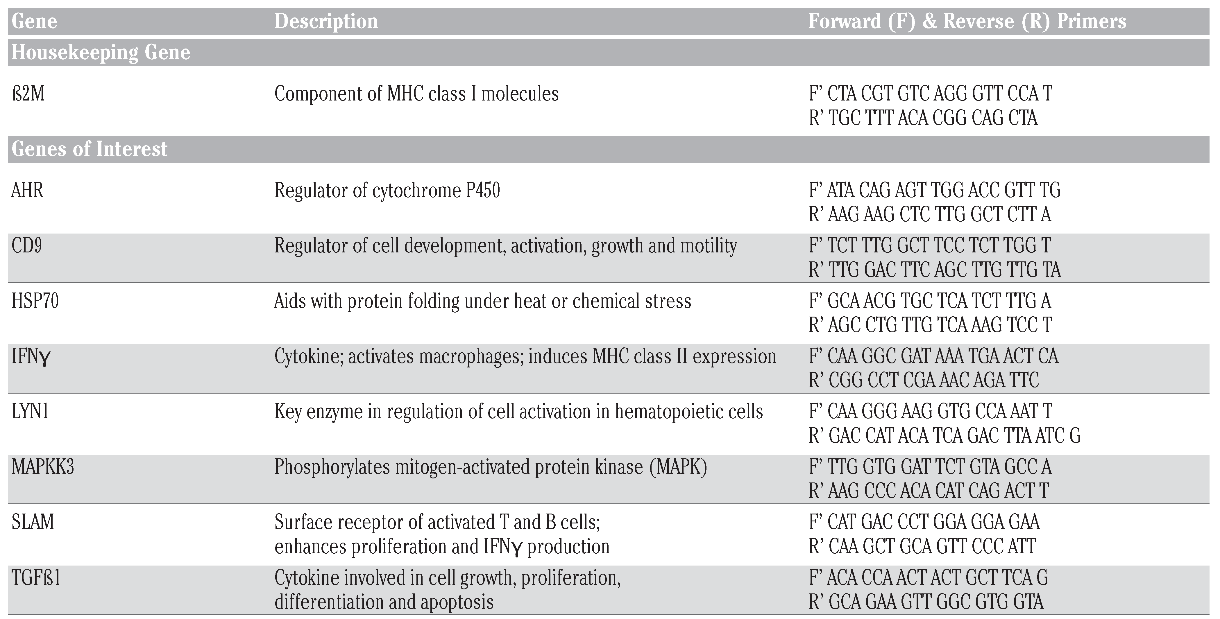
RNA was extracted using the Aurum total RNA kit (Bio-Rad, Mississauga, ON). NanoDrop-normalized (A260 nm) levels of total RNA were reverse transcribed with the iScript cDNA synthesis kit (Bio-Rad, Mississauga, ON). cDNA were used for quantitative polymerase chain reaction (qPCR) using Ssofast Evagreen supermix (Bio-Rad, Mississauga, ON). qPCR was initiated with 95°C for 30 s, followed by 40 cycles with denaturation at 95°C for 5 s, and the primer specific annealing temperature (62°C) for 15 s. Melting curves were creat- ed by denaturation at 95°C followed by 61 steps during which the temperature was increasing steadily for 0.5°C every 5 s, starting at 65°C and ending at 95°C, to exclude the measurement of non-specific PCR products and primer dimers and to determine true amplification. Results were analyzed using the comparative Ct method. Primers are shown in Table 1.
The reference gene ß2M was evaluated for its stability with bestkeeper (stability value =0.073) and was chosen to normalize mRNA transcription levels of the genes of interest. Genes of cells exposed to EE2 only were normalized with the ß2M 0 µg/L EE2 negative control, whereas genes of cells exposed to EE2 and LPS were normalized with the ß2M 0 µg/L EE2+LPS negative control.
Results are obtained from two inde- pendent experiments with two technical replicates for each sample.
Viability assays were set up in parallel to observe if EE2 concentrations were cyto- toxic to the 11B7501 B lymphoma cell line. After 24 h, viability of cells was evaluated by adding 0.8 μg/mL propidium iodide (PI) (Sigma-Aldrich, Oakville, Canada) to the cell suspension. A FACSCalibur (Becton Dickinson, San Jose, CA, USA) with an air- cooled argon laser providing an excitation at 488 nm was used. For each sample 5000 events were acquired at a fluorescence emission of 620 nm (FL3). The cell popula- tion was electronically gated in a FSC/SSC dot plot and the fluorescence frequency dis- tribution histogram was obtained. The per- centage of dead cells was determined as the proportion of cells with an elevated fluores- cence using a marker. Data collection and analysis were performed with the CellQuest Pro software (Version 4.0.1). The results were expressed in percentage of viable cells.
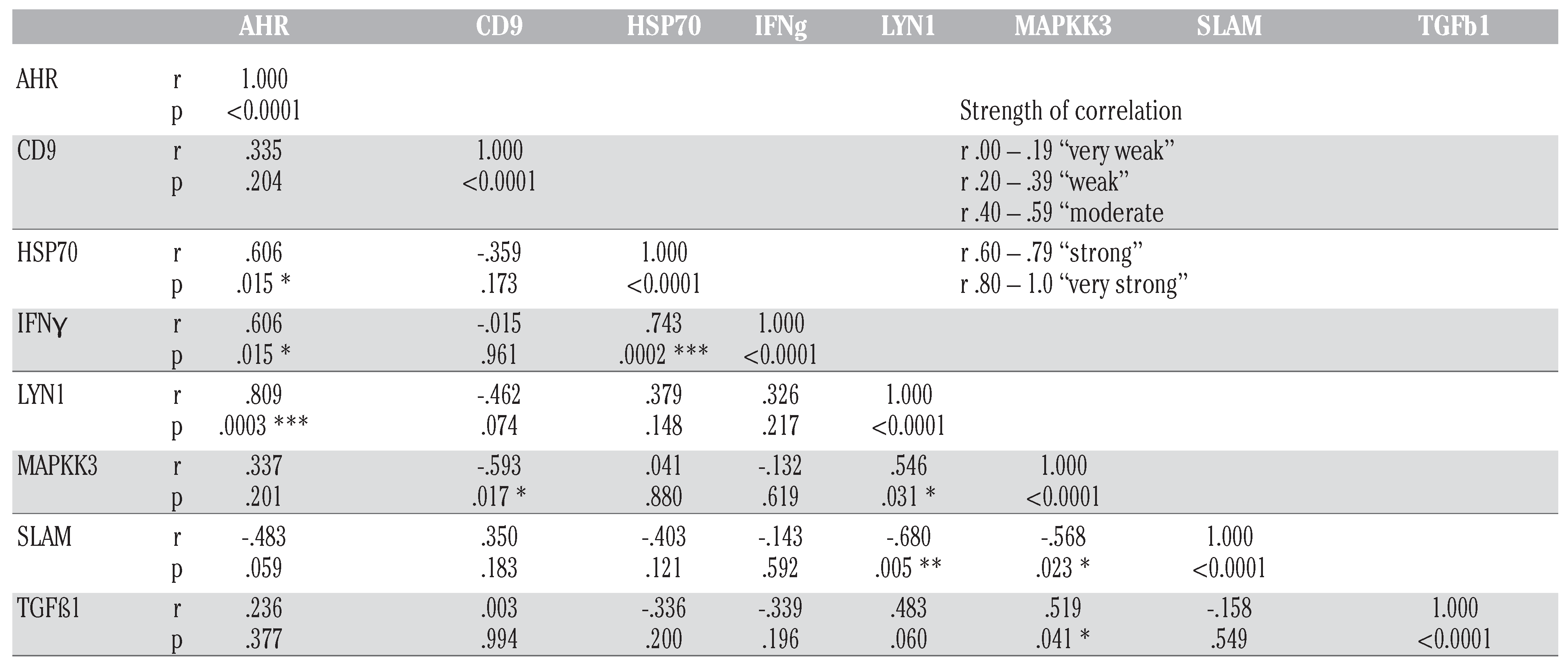
Differences between controls and treat- ed groups were evaluated by one-way and two-way ANOVA followed by Dunnett’s post hoc test. To evaluate correlations of inter-relationships of genes of interest a Spearman rank order correlation analysis was conducted and results are summarized in Table 3. The calculations were performed using GraphPad Prism 6 for Mac (GraphPad Software). The level of signifi- cance was set at P≤0.05.

Results and Discussion

A range of eight immune-relevant (CD9, IFNγ, LYN1, MAPKK3, SLAM, TGFß1), pollutant-associated (AHR) and stress (HSP70) biomarkers as well as a ref- erence gene (ß2M) were selected to analyze differential gene expression in the harbor seal 11B7501 B lymphoma cell line exposed to EE2. We limited our choice of genes to sequences that have already been validated in the harbor seal.
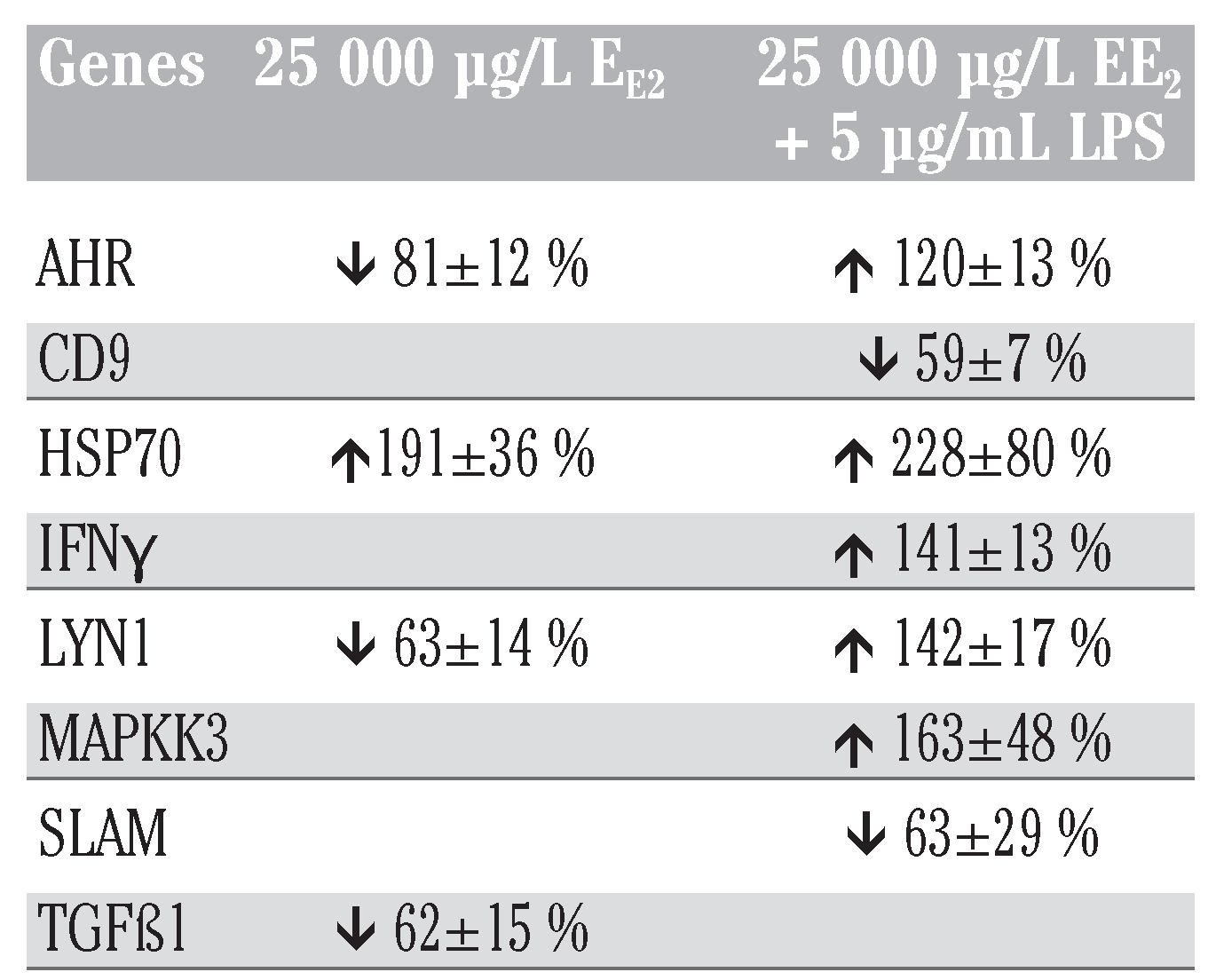
To obtain an overview of potential effects on the mRNA expression level of the chosen genes, we analyzed the control sam- ples and the highest exposure concentration of EE2 (25,000 µg/L) for the genes of inter- est (Figure 1A). The mRNA expression of all but one (ß2M) gene was impacted at 25,000 µg/L EE2. Genes at this concentration were both up- and downregulated. Lymphocytes exposed to EE2 without stim- ulation from LPS downregulated four out of five genes (Table 2). Lymphocytes exposed to EE2 and the mitogen LPS were more like- ly to have changes in gene expression, and genes were mainly upregulated (6 of 9) (Table 2).
HSP70 protects proteins from stress by aiding protein folding. HSP70 mRNA was upregulated two-fold compared to controls upon exposure to EE2 alone as well as EE2 and LPS (Table 2). Since HSP70 showed the most marked change in gene expression of all genes tested, we further evaluated the remaining exposure concentrations. The analysis revealed that HSP70 was induced only at the highest concentration (Figure 1B). Previous studies in harbor seal blood demonstrated negative (cadmium, Cd) and positive (lead, Pb) correlations between trace metal concentrations in blood and HSP70 expression [1]. These trends are in accordance with previous work that demon- strated that xenobiotic exposure could lead to immunosuppression or acute and chronic inflammatory processes that could cause hypersensitivities or autoimmune dis- eases [19]. When assessing HSP70 in blood of free-ranging animals it has to be taken into account that capture and sampling stress might alter the physiological levels of HSP70 in these animals. HSP70 and corti- sol were correlated in adult seals, and habit- uation (i.e. decreasing levels of HSP70 over time) was observed in rehabilitation processes where seals have been handled frequently [1,15,17,20]. Moreover, effects on HSP70 expression strongly correlated with AHR expression (r=0.606) and IFNγ expression (r=0.743) (Table 3).
Interferon gamma (IFNγ) and signaling lymphocytic activation molecule (SLAM) are associated with susceptibility to mam- malian infectious diseases [21]. IFNγ is a cytokine that activates macrophages and induces class II major histocompatibility complex (MHC II) expression. In our study, IFNγ expression was upregulated after exposure to 25,000 µg/L EE2 + LPS, but not when the mitogen LPS was not present (Figure 1A, Table 2). Since the +LPS nega- tive control was identical to the -LPS nega- tive control, this upregulation can be attrib- uted to a combined effect of EE2 with LPS. Furthermore, effects on IFNγ expression displayed strong correlations with pattern changes in AHR expression (r=0.606) and HSP70 expression (r=0.743) (Table 3).
SLAM is a surface receptor on activated B and T lymphocytes. In our study, its expression was not affected by 25,000 µg/L EE2 alone, but it was one of the few genes that was significantly downregulated when additionally exposed to LPS. It has been shown to enhance IFNγ production, and is the primary cellular receptor for Morbillivirus [22]. Interestingly, SLAM and IFNγ mRNA levels showed inverse trends in regulation after exposure to EE2 and LPS. While the exposure to the EE2 or LPS alone did not change the mRNA expression, the additional stimulus and stress of a potential pathogen (LPS) impacted the B lymphoma cell line. The increase in IFNγ can therefore be attributed to the presence of LPS, and not due to an increased production due to SLAM. The Spearman rank order correla- tion analysis revealed negative correlations with expression of SLAM and LYN1 (r= –0.680) as well as MAPKK3 (r= –0.568) (Table 3).
Tyrosine-protein kinase LYN1 is involved in the regulation of cell activation of hematopoietic cells. LYN1 expression was upregulated after exposure to 25,000 µg/L EE2 + LPS, but significantly downregulated when LPS was not present (Figure 1A, Table 2). These results are in accordance with previous work demonstrat- ing an upregulation of LYN in murine macrophages after LPS as well as IFNγ stimulation [23]. Effects on LYN1 expression very strongly correlated with AHR expres- sion changes (r=0.809) and moderately to strongly correlated with expression changes of MAPKK3 (r=0.546) and SLAM (r=–0.680), respectively (Table 3).
Mitogen-activated protein kinase kinase 3 (MAPKK3) activates MAPK3, a protein involved in the regulation of cellular processes such as proliferation and differen- tiation in lymphocytes [24]. MAPKK3 mRNA was upregulated in response to 25,000 µg/L EE2 + LPS exposure, but not when the mito- gen LPS was not present (Figure 1A, Table 2). A change in the MAP kinase pathway could impact both B cell receptor or T cell receptor signaling and T cell develop- ment [25,26,27]. MAPKK3 expression changes correlated with expression changes in four other genes (Table 3). Moderate positive and negative correlations were observed with CD9 (r= –0.593), LYN1 (r=0.546), SLAM (r= –0.568) and TGFß1 (r=0.519).
The observed downregulation of the transmembrane protein CD9 at 25,000 µg/L EE2 + LPS is potentially related to the inac- tivation of histone deacetylases by LPS [28]. The Spearman rank order correlation analy- sis revealed CD9 expression changes corre- lation only with one other gene (MAPKK3, moderate negative correlation, r= –0.593) (Table 3).
TGFß1, a cytokine involved in cell growth, proliferation, differentiation and apoptosis, is the only gene affected by EE2 alone, but not EE2 + LPS. A moderate cor- relation was observed with MAPKK3 (r=0.519) (Table 3).
As the differential gene expression pat- terns revealed (Figure 1A), it is crucial to test the immunotoxicity of a compound with and without stimulation from a poten- tial pathogen like LPS, since immunotoxic- ity of a compound can vary quite consider- ably in the two exposure scenarios.
Viability of the harbor seal 11B7501 B lymphoma cell line after 24 h was 91±3% in the negative controls and 83±2% in the neg- ative controls of cells exposed with LPS (Figure 1C). The cell line was significantly impacted only in one exposure at 25,000 µg/L EE2 without LPS reducing it to 76±8%. It is problematic to conclude envi- ronmentally relevant effects of EE2 on gene expression of harbor seal lymphocytes in this study, since the maximum dose tested was quite high. Furthermore, it should be investigated to what extent hormone levels in FBS might have influenced the bioavail- ability of EE2 to the cells, by comparing with charcoal stripped FBS.
Quantitative PCR has rarely been uti- lized in toxicity testing in the marine mam- mal immune system [1], while the method is more common in other species. To our knowledge this is the second study after Lehnert et al., 2016 that has used this method to assess the toxic mechanisms of action of gene expression in marine mam- mal immune cells. To date, only few mRNA sequences are known for pinnipeds in com- parison to e.g. rodent sequences. It is there- fore important to continue the effort to sequence the gene expression transcript and continue using marine mammal lymphocyte cell lines or blood samples from free-rang- ing animals to further the knowledge of immunotoxic action under contaminant load in marine mammals.

Conclusions

In conclusion, the observed effect of EE2 depends on the activation status of the lymphocyte, therefore pinpointing com- plexity of such studies. Indeed, the present results show that two genes were expressed differently with or without LPS. Two genes reacted similarly with or without LPS. One gene was downregulated with EE2 alone, while two genes were downregulated and three upregulated with EE2+LPS. Correlations of gene expression changes between genes further indicate the com- plexity of immunotoxic mechanisms and the need for in depth studies elucidating mechanisms of toxic action. We estimate that gene expression analysis is a useful tool in marine mammal immunotoxicological research that should be further developed and used.

Research Highlights

-
First study to assess effect of EE2 on gene expression profiles in harbor seal leukocytes.
-
Development and validation of mRNA primers for harbor seal.
-
Gene expression of selected sequences varied in a dose-response depended manner.

Acknowledgments

the Research Chair in Environmental Immunotoxicology (MF) supported this study. We want to also thank Professor Patrick Labonté from the INRS-IAF for lending us equipment and lab space to validate the primers.

Contributions

CK planned and conducted experiments, wrote manuscript; MB designed primers, planned experiments; FG supervised the project, corrected manuscript; MF provided funding and lab space.

Conflicts of Interest

the authors declare no conflict of interest.

References

  1. Lehnert, K.; Ronnenberg, K.; Weijs, L.; Covaci, A.; Das, K.; Hellwig, V.; et al. Xenobiotic and Immune-Relevant Molecular Biomarkers in Harbor Seals as Proxies for Pollutant Burden and Effects. Archiv Environ Contam Toxicol 2016, 70, 106–120. [Google Scholar]
  2. Harwood, J. Marine mammals and their environment in the twenty-first century. J Mammal 2001, 82, 630–640. [Google Scholar] [CrossRef]
  3. Weijs, L.; Das, K.; Siebert, U.; Neels, H.; Blust, R.; Covaci, A. PCBs, PBDEs and their hydroxylated metabolites in serum of free-ranging harbour seals (Phoca vitulina): levels and profiles. Organohalogen Compounds 2008, 70, 000837. [Google Scholar]
  4. Ross, P.; DeSwart, R.; Addison, R.; Van Loveren, H.; Vos, J.; Osterhaus, A. Contaminant-induced immunotoxicity in harbour seals: Wildlife at risk? Toxicology 1996, 112, 157–169. [Google Scholar] [CrossRef]
  5. Das, K.; Siebert, U.; Gillet, A.; Dupont, A.; Dipoi, C.; Fonfara, S.; et al. Mercury immune toxicity in harbour seals: links to in vitro toxicity. Environ Health- Glob 2008, 7. [Google Scholar]
  6. Kleinert, C.; Lacaze, E.; Mounier, M.; De Guise, S.; Fournier, M. Immunotoxic effects of single and combined pharma- ceuticals exposure on a harbor seal (Phoca vitulina) B lymphoma cell line. Marine Pollution Bulletin, 2017; [Epub ahead of print]. [Google Scholar]
  7. Kleinert, C.; Mournier, M.; Fortier, M.; Brousseau, P.; De Guise, S.; Fournier, M. Several pharmaceuticals impaired har- bor seal lymphocytes (Phoca vitulina) in vitro. J Xenobiotics 2013, 3, 5. [Google Scholar] [CrossRef]
  8. Allinson, M.; Shiraishi, F.; Salzman, S.; Allinson, G. In vitro and immunological assessment of the estrogenic activity and concentrations of 17beta-estradiol, estrone, and ethinyl estradiol in treated effluent from 45 wastewater treatment plants in Victoria, Australia. Archiv Environ Contam Toxicol 2010, 58, 576–586. [Google Scholar] [CrossRef]
  9. Ternes, T.; Stumpf, M.; Mueller, J.; Haberer, K.; Wilken, R.; Servos, M. Behavior and occurrence of estrogens in municipal sewage treatment plants - I. Investigations in Germany, Canada and Brazil. Sci Total Environ 1999, 225, 81–90. [Google Scholar] [CrossRef] [PubMed]
  10. Kolpin, D.; Furlong, E.; Meyer, M.; Thurman, E.; Zaugg, S.; Barber, L.; et al. Pharmaceuticals, hormones, and other organic wastewater contaminants in US streams, 1999-2000: a national recon- naissance. Environ Sci Technol 2002, 36, 1202–1211. [Google Scholar] [CrossRef] [PubMed]
  11. Gelsleichter, J. Evaluating the risks that pharmaceutical-related pollutants pose to Caloosahatchee River wildlife: observations on the bull shark, Carcharhinus leucas. Fort Myers: Charlotte Harbor National Estuary Program, 2009. [Google Scholar]
  12. Medina, K.; Strasser, A.; Kincade, P. Estrogen influences the differentiation, proliferation, and survival of early B- lineage precursors. Blood 2000, 95, 2059–2067. [Google Scholar] [CrossRef]
  13. Grimaldi, C.; Cleary, J.; Dagtas, A.; Moussai, D.; Diamond, B. Estrogen alters thresholds for B cell apoptosis and acti- vation. J Clin Investig 2002, 109, 1625–1633. [Google Scholar] [CrossRef]
  14. Yakimchuk, K.; Iravani, M.; Hasni, M.; Rhonnstad, P.; Nilsson, S.; Jondal, M.; et al. Effect of ligand-activated estrogen receptor beta on lymphoma growth in vitro and in vivo. Leukemia 2011, 25, 1103–1110. [Google Scholar] [CrossRef] [PubMed]
  15. Lehnert, K.; Muller, S.; Weirup, L.; Ronnenberg, K.; Pawliczka, I.; Rosenberger, T.; et al. Molecular bio- markers in grey seals (Halichoerus gry- pus) to evaluate pollutant exposure, health and immune status. Marine Pollut Bull 2014, 88, 311–318. [Google Scholar] [CrossRef]
  16. Müller, S.; Lehnert, K.; Seibel, H.; Driver, J.; Ronnenberg, K.; Teilmann, J.; et al. Evaluation of immune and stress status in harbour porpoises (Phocoena pho- coena): can hormones and mRNA expression levels serve as indicators to assess stress? BMC Vet Res 2013, 9. [Google Scholar] [CrossRef] [PubMed]
  17. Weirup, L.; Müller, S.; Ronnenberg, K.; Rosenberger, T.; Siebert, U.; Lehnert, K. Immune-relevant and new xenobiotic molecular biomarkers to assess anthro- pogenic stress in seals. Marine Environ Res 2013, 92, 43–51. [Google Scholar] [CrossRef]
  18. Frouin, H.; Fortier, M.; Fournier, M. Toxic effects of various pollutants in 11B7501 lymphoma B cell line from harbour seal (Phoca vitulina). Toxicology 2010, 270, 66–76. [Google Scholar] [CrossRef]
  19. Kakuschke, A.; Prange, A. The influence of metal pollution on the immune sys- tem a potential stressor for marine mammals in the North Sea. Int J Comparat Psychol 2007, 20, 2. [Google Scholar]
  20. Fonfara, S.; Kakuschke, A.; Rosenberger, T.; Siebert, U.; Prange, A. Cytokine and acute phase protein expression in blood samples of harbour seal pups. Marine Biol 2008, 155, 337–345. [Google Scholar] [CrossRef]
  21. Pacheco, A.; Cardoso, C.; Moraes, M. IFNG+874T/A, IL10-1082G/A and TNF-308G/A polymorphisms in associ- ation with tuberculosis susceptibility: a meta-analysis study. Hum Genet 2008, 123, 477–484. [Google Scholar] [CrossRef]
  22. McCarthy, A.; Shaw, M.; Jepson, P.; Brasseur, S.; Reijnders, P.; Goodman, S. Variation in European harbour seal immune response genes and susceptibil- ity to phocine distemper virus (PDV). Infect Genet Evol 2011, 11, 1616–1623. [Google Scholar] [CrossRef]
  23. Boulet, I.; Ralph, S.; Stanley, E.; Lock, P.; Dunn, A.; Green, S.; et al. Lipopolysaccharide-and interferon- gamma-induced expression of hck and lyn tyrosine kinases in murine bone marrow-derived macrophages. Oncogene 1992, 7, 703–710. [Google Scholar]
  24. Neale, J.; Kenny, T.; Gershwin, M. Cloning and sequencing of protein kinase cDNA from harbor seal (Phoca vitulina) lym- phocytes. Clin Devel Immunol 2004, 11, 157–163. [Google Scholar]
  25. Alberolaila, J.; Forbush, K.; Seger, R.; Krebs, E.; Perlmutter, R. Selective requirement for map kinase activation in thymocyte differentiation. Nature 1995, 373, 620–623. [Google Scholar] [CrossRef] [PubMed]
  26. Li, W.; Whaley, C.; Mondino, A.; Mueller, D. Blocked signal transduction to the ERK and JNK protein kinases in aner- gic CD4(+) T cells. Science 1996, 271, 1272–1276. [Google Scholar] [CrossRef]
  27. Abbas, A.; Lichtman, A. Cellular and molecular immunology, 5 ed.; Elsevier Science, Saunders: Philadelphia, PA, 2003. [Google Scholar]
  28. Jin, Y.; Tachibana, I.; Takeda, Y.; He, P.; Kang, S.; Suzuki, M.; et al. Statins decrease lung inflammation in mice by upregulating tetraspanin CD9 in macrophages. PLoS One 2013, 8, 9. [Google Scholar] [CrossRef]
Figure 1. (A) Fold change in gene expression after 24 h exposure to 0 (NC) and 25 mg/L EE2 as measured by qPCR. A fold change of 1 indicated no change in comparison to the reference gene ß2M. (B) Dose-response gene expression changes of HSP70 mRNA after 24 h exposure to EE2. (C) Viability of the 11B7501 B lymphoma cell line after 24 h exposure to EE2 and EE2 + LPS. Results are expressed as Mean ± SD. (* p<0.05; ** p<0.01; *** p<0.001; **** p<0.0001). Abbreviations: EE2=17α-ethinyl estradiol; LPS=lipopolysaccharide; ßACT=beta actin; YWHAZ=14-3-3 protein zeta/delta; AHR=aryl hydrocarbon receptor; CD9=Cluster of dif- ferentiation 9; HSP70=70 kDa heat shock protein; IFNγ=interferon gamma; LYN1=Lck/Yes novel tyrosine kinase; MAPKK3=mitogen- activated protein kinase kinase; SLAM=signaling lymphocytic activation molecule; TGFß1=transforming growth factor beta 1.
Jox 07 06702 g001
Table 1. Primers used for qPCR analysis. All primers are listed from 5’ to 3’.
Jox 07 06702 i001
ß2M=beta-2 microglobulin; AHR=aryl hydrocarbon receptor; CD9=Cluster of differentiation 9; HSP70=70 kDa heat shock protein; IFNγ=interferon gamma; LYN1=Lck/Yes novel tyrosine kinase; MAPKK3=mitogen- activated protein kinase kinase; SLAM=signaling lymphocytic activation molecule; TGFß1=transforming growth factor beta 1.
Table 2. Summary of significant gene expression changes in the 11B7501 B lym- phoma cell line. Up- or downregulation is described as the percentage of expression relative to the negative controls of ß2M. The ß2M negative control (-EE2/-LPS) was used to normalize genes exposed to EE2 only, whereas the ß2M negative control (-EE2/+LPS) was used to normalize genes exposed to EE2 and LPS.
Jox 07 06702 i002
EE2=17α-ethinyl estradiol; LPS=lipopolysaccharide; AHR=aryl hydrocarbon receptor; CD9=Cluster of differentiation 9; HSP70=70 kDa heat shock protein; IFNγ=interferon gamma; LYN1=Lck/Yes novel tyrosine kinase; MAPKK3=mitogen-activated protein kinase kinase; SLAM=signaling lymphocytic activation molecule; TGFß1=trans- forming growth factor beta 1.
Table 3. Spearman correlation coefficient hemi-matrix for inter-relationships of genes of interest. Significant correlations (p < 0.05 *, p < 0.01 **, p < 0.001 ***, p < 0.0001 ****) are in bold.
Jox 07 06702 i003