Next Article in Journal
Distribution of Dichlorodiphenyltrichloroethane and Hexachlorocyclohexane in Urban Soils and Risk Assessment
Previous Article in Journal
Congener Specific Distribution and Health Risk Assessment of Polychlorinated Biphenyls in Urban Soils
 
 
Journal of Xenobiotics is published by MDPI from Volume 10 Issue 1 (2020). Previous articles were published by another publisher in Open Access under a CC-BY (or CC-BY-NC-ND) licence, and they are hosted by MDPI on mdpi.com as a courtesy and upon agreement with PAGEPress.
Article

Reduction of Azo Dyes by Flavin Reductase from Citrobacter freundii A1

by
Mohd Firdaus Abdul-Wahab
1,2,
Giek Far Chan
1,2,
Abdull Rahim Mohd Yusoff
3 and
Noor Aini Abdul Rashid
1,2,*
1
Department of Biological Sciences, Faculty of Biosciences and Bioengineering, Malaysia
2
Nanoporous and Mesoporous Materials for Biological Applications Research Group (NAMBAR), Sustainability Research Alliance, Malaysia
3
Institute of Environmental and Water Resource Management, Water Research Alliance, Universiti Technologi Malaysia, Johor, Malaysia
*
Author to whom correspondence should be addressed.
J. Xenobiot. 2013, 3(1), e2; https://doi.org/10.4081/xeno.2013.e2
Submission received: 3 October 2012 / Revised: 30 November 2012 / Accepted: 30 November 2012 / Published: 18 December 2012

Abstract

:
Citrobacter freundii A1 isolated from a sewage treatment facility was demonstrated to be able to effectively decolorize azo dyes as pure and mixed culture. This study reports on the investigation on the enzymatic systems involved. An assay performed suggested the possible involvement of flavin reductase (Fre) as an azo reductase. A heterologously-expressed recombinant Fre from C. freundii A1 was used to investigate its involvement in the azo reduction process. Three model dyes were used, namely Acid Red 27 (AR27), Direct Blue 15 (DB15) and Reactive Black 5 (RB5). AR27 was found to be reduced the fastest by Fre, followed by RB5, and lastly DB15. Redox mediators nicotinamide adenine dinucleotide (NADH) and riboflavin enhance the reduction, suggesting the redox activity of the enzyme. The rate and extent of reduction of the model dyes correlate well with the reduction potentials (Ep). The data presented here strongly suggest that Fre is one of the enzymes responsible for azo reduction in C. freundii A1, acting via an oxidation-reduction reaction.

Introduction

Azo dyes are known to pose hazards to human health and the environment, as these dyes do not occur naturally. Thus, all the industrially-produced azo dyes are classified as xenobiotics.[1] Various treatment methods have been used to obviate the problems posed by these dyes and their derivatives. These include physical, chemical and biological methods.[2,3,4] Among the biological meth- ods used is enzymatic approach, and several enzymes have been reported to degrade azo dyes, including peroxidase,[5] nitric oxide syn- thase,[6] laccase,[4] and azo reductase.[7]
The enzyme NAD(P)H:flavin oxidoreduc- tase or flavin reductase (Fre) evolved as non- enzymatic reduction of free flavins by nicoti- namide adenine dinucleotide phospate and nicotinamide adenine dinucleotide (NADH) is rather slow and requires organism to pos- sess a system to catalyze the reaction.[8] Fre catalyzes the reduction of various flavins, namely riboflavin, flavin mononucleotide and flavin adenine dinucleotide (FAD) at the expense of reduced pyridine nucleotides.[9] Fre from Sphingomonas sp. has been shown to possess an azo reductase activity, with the cell extract showing higher activity compared to the whole-cells.[10] One reason being that the cell membranes limit the uptake of high- ly polar sulfonated azo compounds. Fre could act as an azo reductase under both aerobic and anaerobic conditions.[10] Due to this, it was hypothesized that Fre could act as one of the azo reductases in the biodegradation of azo compounds by C. freundii A1. There have been no previous reports on Fre as azo reduc- tase in C. freundii to our knowledge.
The potential use of bacterial culture (pure or mixed) to treat dye-containing wastewater has been widely researched, with various bacterial strains reported to be able to decol- orize azo dyes. These include several methanogenic bacteria, Pseudomonas sp., Aeromonas sp., Bacillus sp., Citrobacter sp., and Sphingomonas sp.[7,11,12,13] under anaerobic, anoxic and aerobic conditions. Recently, we reported on the capability of a novel NAR-2 bacterial consortium, consisting of Citrobacter freundii A1, Enterococcus cas- selifla us C1 and Enterobacter cloacae L17, able to decolorize Amaranth (Acid Red 27) within 30 minutes under microaerophilic condition.[14] The fate of Amaranth degrada- tion by the consortium has also been explored in detail in the report which had led to the proposal of its biodegradation path- ways.[14] These unprecedented observations suggested that a thorough research on the mechanism of azo dye degradation and min- eralization by the consortium is necessary for future applications, including all the enzymatic systems potentially involved in each strain.
We present here the attempt to character- ize an enzyme system in C. freundii A1, which relates closely to the ability of the bac- terium to decolorize azo dyes in both pure and mixed cultures. The first enzymatic sys- tem discovered which relates to its ability to decolorize in mixed culture is a precursor to the synthesis of autoinducer-2.[15] This sug- gested the role of autoinducer-2 synthase (luxS) gene product in the decolorization process.15 Strain A1 was originally isolated from a sewage oxidation pond and has been shown to be able to decolorize azo dyes even as pure culture.[14,15] Recently, the draft genome sequence of this bacterium has been published, which further assists on the investigation of the decolorization mecha- nism.[16]
This paper describes an investigation on the cellular enzymatic system involved in the azo dye decolorization by C. freundii A1, one of the bacteria used in NAR-2 consortium,[14] in order to further understand the mecha- nism. It also addresses the question: is Fre one of the azo reductases of C. freundii A1? The model dyes used in this study are Acid Red 27 (AR27, Amaranth), Reactive Black 5 (RB5, Remazol Black B) and Direct Blue 15 (DB15) (Figure 1), all of which are industri- ally important dyes. This research, which constitutes part of a bigger study on the mechanism of azo dye degradation by the NAR-2 consortium, is necessary for a more effective method to treat dye-related waste- water.

Materials and Methods

Preparation of cell extracts, azoreductase and flavin reductase assays

Fresh overnight culture of C. freundii A1 in P5 medium (K2HPO4 35.2 g/L, KH2PO4 20.9 g/L, NH4Cl 2 g/L, glucose 10 g/L, nutrient broth 2% (w/v), trace elements, pH 7.0) was harvested by centrifugation, and washed 2-3 times in cold potassium phosphate buffer (50 mM, pH 7.5). The supernatant was kept at 4°C prior to detec- tion of extracellular activity. The cells were first re-suspended in the same potassium phosphate buffer, and were maintained as resting cell. The remaining cell suspension was sonicated for a total of 2 min, with 2 min- interval after each 15-S treatment. The cells were incubated on ice during the sonication, using Vibra Cell™ ultrasonic processor (Sonics & Materials, Newtown, CT, USA) using 600 W power output at 0% amplitude. The homogenate was centrifuged at 5000 rpm at 4°C for 30 min. The supernatant was the crude cell-free extract while the pellet contained the cell debris. Protein concentration was deter- mined by Lowry method with bovine serum albumin as standard.[17] The absorbance was read at 750 nm using Cary 100 UV-Visible Spectrophotometer (Varian).
The azoreductase activity of C. freundii A1 was detected and measured using a modified procedure.[18] The cell extract (150 μL) was added to a sparged anaerobic solution contain- ing Tris-HCl buffer (50 mM, pH 7.5), Acid Red 27 (0.03 mM) and FAD (0.05 mM). The reac- tion mix was flushed with nitrogen gas to cre- ate an anaerobic condition. The reaction (1 mL) was initiated by the addition of NADH (0.2 mM), followed by 10 min incubation. Reaction progression was monitored using Cary 100 UV- Visible Spectrophotometer (Varians) at the AR27 λmax (521 nm). The azo reductase activi- ty was calculated from the decrease in absorbance, corresponding to the reduction of azo dyes by the enzyme. One unit (U) of the enzyme is defined as the amount catalyzing the reduction of 1 nmol of azo dye per min. The specific activity is defined as units per mg pro- tein. Flavin reductase assay was performed as previously reported by Chan et al.[19]

Over expression and protein extraction

Over expression and extraction of recombi- nant Fre was performed as reported previous- ly.[19] Briefly, Fre was expressed in recombinant E. coli BL21(DE3)pLysS. Upon IPTG induction, it was grown for further 4 h at 37°C. The har- vested cells were washed and lysed by sonica- tion. As the recombinant Fre was expressed as inclusion bodies, it was first solubilized and refolded using the Protein Refolding Kit (Novagen, EMD Chemicals, Gibbstown, NJ, USA), and purified using nickel affinity chro- matography. Protein concentration was deter- mined using the Lowry assay,[17] and analyzed on 15% SDS-PAGE.

Reduction of azo dyes by Fre

One hundred and fifty μL of the recombi- nant enzyme was added to the reaction mix- ture with Tris-HCl (20 mM, pH 7.5), NADH (0.2 mM), riboflavin (15 μM) and the respective azo compounds at a final concentration of 0.1 mM. Reduction rate was determined at 30°C from the decrease in UV-vis absorbance at λmax of each of the dyes (AR27 at 521 nm, DB15 at 585 nm, RB5 at 596 nm) over the period of 30 min. Experiments were performed in several repeats and the best results were plotted. Dye concentrations were determined from the standard curves plotted. Percentage (extent) and rate of dye reduction by Fre were extracted from the plots.

Voltammetric analysis

Voltammetric method involves the applica- tion of a variable potential difference between a reference electrode, and a microelectrode (working electrode) at which an electrochemi- cal reaction is induced (Ox + ne → Red). ‘Ox’ and ‘Red’ represent the oxidized and reduced species respectively, while n is the number of electron. As the potential at the working elec- trode reaches a value such that a species pres- ent in solution is either reduced or oxidized, the current in the circuit increases. This phe- nomenon is depicted in a oltammogram, which is essentially a current-voltage curve, I=f(E), that corresponds to a voltage scan over a range that induces oxidation or reduction of the analytes.[20] The voltammetric technique used in this study is the stripping oltammetry. This technique was chosen due to its high sen- sitivity and is among the most widely used voltammetric techniques when analyzing dyes and azo dyes.[21,22,23] In stripping voltammetry, a fraction of the electroactive analyte present in the sample is deposited by electrolysis on an electrode in a stirred solution held at a con- stant potential (pre-concentration stage). Then the stirring is terminated and the poten- tial difference between the working and refer- ence electrode determines the change in potential applied to the working electrode. The analyte is re-dissolved (the stripping stage) by sweeping the potential negatively (cathodic stripping) or positively (anodic stripping).[20] The reduction or oxidation of analytes can be measured as current surge and represented as a peak in a voltammogram. An increased sen- sitivity and better distinction between analytes can be obtained using a differential pulse volt- age during the stripping stage. In cathodic stripping voltammetry, the more negative the reduction potential, the more difficult it is for the analyte to be reduced.
Cyclic and differential pulse stripping voltammetry was performed using Autolab PGSTAT30 (EcoChemie, Utrecht, The Netherlands) connected to a VA 663 stand (Metrohm, Herisau, Switzerland) at the scan rate of 0.2 V sec-1. Glassy carbon electrode was used as the working electrode completed by means of Ag/Au auxiliary electrode filled with 3 M KCl and Pt electrode as the reference elec- trode. The experiments were performed in 0.04 M Britton-Robinson buffer (BRb) pH 7 at dye concentrations ranging from 0.001 to 0.1 mM. Dye stock solutions were prepared at the con- centration of 1 mM in deionized water. All the solutions were purged with nitrogen gas for 300 s to obtain anaerobic condition prior to determination and thereafter, a blanket of nitrogen gas was maintained throughout the experiment. The buffer and sample mixture was further degassed for 30 s before each measurement was made.

Results

The azo reductase and flavin reductase activities of C. freundii A1 cell extracts

The azo reductase activity of various cell extracts is shown in Figure 2. The azo reductase was found to have an intracellular localization, as can be seen from the high specific activity in the cell-free extract. Fre assay was also per- formed on all the cell fractions (Figure 2) to find out the possibility of it being one of the azo reductases in C. freundii A1. It was found that the cell-free extract possesses the highest Fre activity.

Reduction of azo dyes by the recombinant Fre

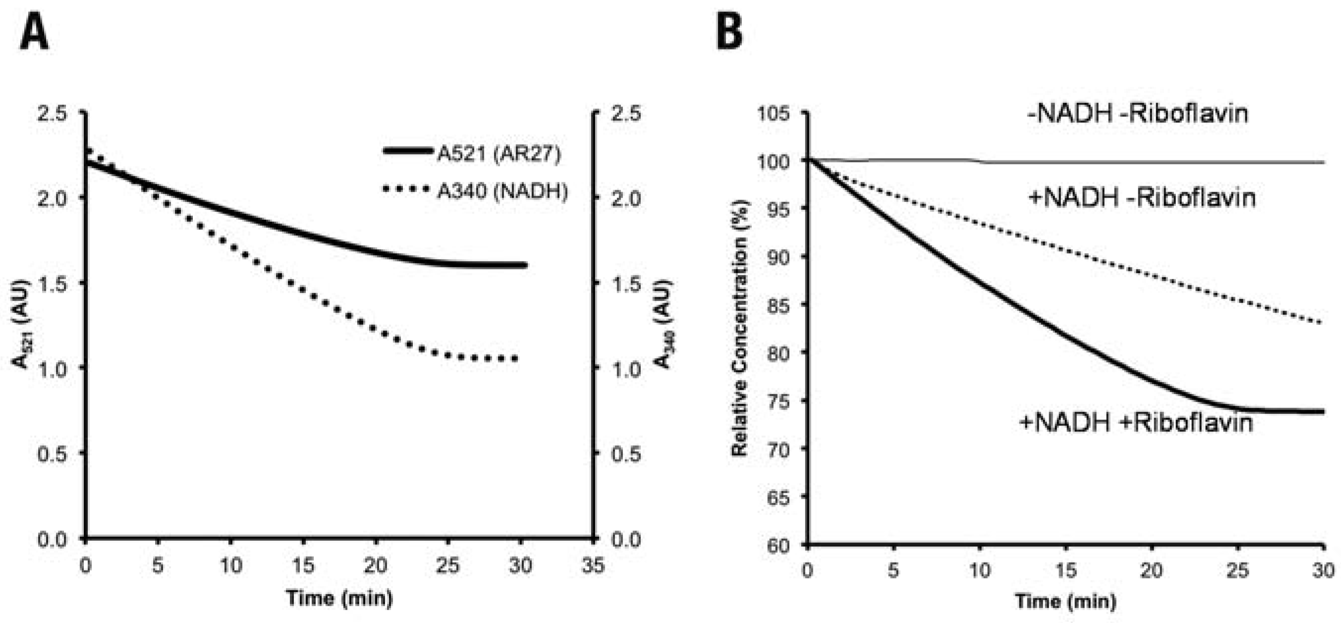
A recombinant Fre, cloned and purified as described previously,[19] was reacted with three model dyes with different structural complexi- ty. Figure 3A shows the reduction of AR27 by Fre, monitored at its maximum absorbance wavelength (λmax). The absorbance at 340 nm (A340) was also monitored simultaneously to investigate the effects of AR27 reduction on NADH concentration. NADH is an electron donor used in many enzymatic systems. Decrease in A340 signifies the oxidation of NADH to NAD+,[24] suggesting a continuous uti- lization of NADH in the process. We observed that the oxidation of NADH ended simultane- ously with AR27 reduction. This is probably due to the complete consumption of riboflavin (an electron acceptor), which acts by accepting electron from a redox enzyme and transferring it to the substrate. In order to further confirm that Fre isolated from C. freundii A1 is a redox enzyme, and is critical in azo dye reduction, we then tested the requirements for both NADH and/or riboflavin for AR27 reduction. The result is shown in Figure 3b. The presence of both NADH and riboflavin was found to signif- icantly increase the reduction rate of the dye. AR27 is reduced at approximately 1.15% min-1 in the first 20 min (estimated using the tan- gent method), compared to 0.60% min-1 in the presence of NADH alone. NADH was found to contribute to reduction, even without riboflavin, due to the direct non-enzymatic reaction. The reduction of RB5 and DB15 were also investigated, and the results are summa- rized in Table 1. Reduction behavior is similar in the cases of allother model dyes (data not shown).

Voltammetric analysis of the azo dyes

Voltammetric measurement was employed to determine the azo bond reduction potentials (Ep) of the azo dyes to further support any involvements of an electron transfer system. The non-corrected dye reduction potentials obtained are summarized in Table 1. The Ep determined was based on the Ag/AgCl refer- ence electrode, and can be used as is. Potentials at several pHs were initially deter- mined, but the data at pH 7 were deliberately chosen as they closely resemble the pH of the buffer solution in the enzymatic reaction ves- sel. From Table 1, AR27 is found to be reduced at the lowest (least negative) Ep, followed by RB5, then DB15. As described in Materials and Methods, the more negative Ep signifies the more difficult it is for the azo bond to receive electron(s) to be reduced electrochemically.

Discussion

C. freundii A1 has recently been shown to be able to decolorize azo dye Amaranth (AR27) efficiently as part of a three-bacteria consor- tium NAR-2.[14] We have also shown before, that it can decolorize even as pure culture but at a slower rate.15 This prompted us to further investigate the decolorization mechanism, including the enzymatic systems involved, for future applications in dye-containing waste- water treatment. The presence of this xenobi- otic compound in wastewater is not only aes- thetically unpleasant, but also poses hazards to aquatic microflora due to the toxic reduction products.
The ability of C. freundii A1 to decolorize azo dyes may be due to the presence of azoreduc- tase(s), which reduces the azo bond to give amine-containing products. It has also been reported that under anaerobic condition, unspecific cytoplasmic azoreductases would act via the intermediate formation of free reduced soluble flavins.10 This suggests that flavin reductase (Fre) could be one of the enzymes responsible for azo reduction in C. freundii A1. We have previously isolated the fre gene, cloned into an expression vector,[19,25] and the Fre protein was overexpressed in E. coli, before being extracted and characterized.[19]
In the present study, we investigated the ability of the recombinant enzyme to decol- orize azo dyes and also the requirements of cofactors for the enzyme to work in itro. Fre, in general, acts by transferring reducing equiv- alents or electrons from NADH to flavin (FAD or riboflavin) that serve as redox mediator, fol- lowed by the transfer of electrons to the dye molecule. Chemical reaction of the dye with reduced riboflavin is then responsible for the reduction.[26] We did not observe any direct reduction by the enzyme itself (data not shown). Reduction is observed immediately upon reaction of the enzyme with an azo dye (Figure 3). These confirm the azoreductase ability of the recombinant Fre. The redox activ- ity of Fre was then investigated by simultane- ously monitoring the NADH oxidation. The observations that NADH is concomitantly oxi- dized with azo reduction suggest that the cofactor transfers electron to Fre, before being transferred to riboflavin for direct chemical reduction of the azo bonds (Figure 4). The reduction of AR27 in the absence of riboflavin is probably a non-enzymatic reaction by NADH, as reported by others.[27,28] A general scheme to illustrate the reduction of azo dye in the C. fre- undii Fre system based on our observations is shown in Figure 4. This reaction scheme is classified as indirect (redox mediator cat- alyzed) biological reduction.[29]
Voltammetric analysis of the three different dyes used in this study shows that the reducibil-ity of the azo dyes used follows the trend: AR27>RB5>DB15, with AR27 having the high-est reducibility while DB15 the lowest. We pro- pose that the difference in reduction rate of the three model dyes is due to the neighbor group effect of the substituents on the dye ring sys- tem.[30,31] AR27 is the simplest to reduce as it is a monoazo dye and the azo bond is sterically less hindered (Figure 1) by the substituents. The different Ep values of RB5 and DB15 may proba- bly be due to i) the structure of DB15,which is more complex compared to RB5; ii) The pres- ence of different substituents at the ortho- posi-tion relative to the azo bonds (Figure 1). This position is occupied by an amino(NH2)- group in RB5 and a hydroxyl(OH)-group in DB15. The different electronic effects exerted by these sub- stituents onto the ring system might affect the transfer of electron to the respective azo bonds.
It is widely accepted that reduction potential of compounds usually affects the activity of redox enzymes acting on the compound.[32,33] In our case, we found that both the rate of reduc- tion and extent of reduction (percentage) by Fre of the three dyes tested follow the trend shown by their respective Ep values. AR27 has the high- est rate and extent of reduction, and DB15 the lowest. These results strongly support our hypothesis that Fre is the redox enzyme acting as one of the azo reductases in the decoloriza- tion mechanism of C. freundii A1.

Conclusions

We described here the investigation on the enzymatic system involved in the azo dye biodegradation of C. freundii strain A1. Fre was found to be able to act as an azo reductase by reducing aromatic mono- and diazo- dyes in the presence of riboflavin and NADH as elec- tron transfer mediators. The trend of reduction correlates well with the reduction potential, supporting the involvement of an electron transfer system and the redox activity of the enzyme. This further supports the role of Fre as an azo reductase of C. freundii. Further research is currently under way to investigate other enzymatic systems that might be involved in the decolorization mechanism, as the possibility of a multi-enzyme system could not be ruled out completely. The findings from this research will assist the understanding of the decolorization mechanisms and pathways in pure and mixed cultures of C. freundii A1 for use in wastewater treatment.

Author Contributions

MFA-W designed and performed the experiments, and wrote the manuscript; GFC designed and performed the experiments; ARMY and NAR designed the experiments, read and approved the manuscript.

Acknowledgments

the authors would like to acknowledge the Ministry of Science, Technology and Innovation Malaysia (MOSTI) for research grant under Vot. 74199 and UTM for the financial support under the UTM-PTP scholarship, awarded to MFA-W.

Conflicts of Interest

the authors declare no conflict of interests.

References

  1. Stolz, A. Basic and applied aspects in the microbial degradation of azo dyes. Appl Microbiol Biotechnol 2001, 56, 69–80. [Google Scholar] [CrossRef] [PubMed]
  2. Pedro Silva J, Sousa S, Rodrigues J; et al. Adsorption of Acid Orange 7 dye in aqueous solutions by spent brewery grains. Sep Purif Technol 2004, 40, 309–315. [Google Scholar] [CrossRef]
  3. Arslan-Alaton, I.; Ferry, J.L. Application of polyoxotungstates as environmental cata- lysts: Wet air oxidation of acid dye Orange II. Dyes Pigments 2002, 54, 25–36. [Google Scholar] [CrossRef]
  4. Liu, W.; Chao, Y.; Yang, X.; Bao, H.; Qian, S. Biodecolorization of azo, anthraquinonic and triphenylmethane dyes by white-rot fungi and a laccase-secreting engineered strain. J Ind Microbiol Biotechnol 2004, 31, 127–132. [Google Scholar] [CrossRef] [PubMed]
  5. Heinfling, A.; Ruiz-Dueñas, F.J.; Martínez, M.J.; Bergbauer, M.; Szewzyk, U.; Martínez, A.T. A study on reducing substrates of man- ganese-oxidizing peroxidases from Pleurotus eryngii and Bjerkandera adusta. FEBS Lett 1998, 428, 141–146. [Google Scholar] [CrossRef]
  6. Miyajima, M.; Sagami, I.; Daff, S.; Taiko Migita, C.; Shimizu, T. Azo reduction of methyl red by neuronal nitric oxide synthase: The impor- tant role of FMN in catalysis. Biochem Biophys Res Commun 2000, 275, 752–758. [Google Scholar] [CrossRef]
  7. Chang, J.-S.; Chou, C.; Lin, Y.-C.; Lin, P.-J.; Ho, J.-Y.; Lee Hu, T. Kinetic characteristics of bacteri- al azo-dye decolorization by Pseudomonas luteola. Water Res 2001, 35, 2841–2850. [Google Scholar] [CrossRef]
  8. Ingelman, M.; Ramaswamy, S.; Niviere, V.; Fontecave, M.; Eklund, H. Crystal structure of NAD(P)H:flavin oxidoreductase from Escherichia coli. Biochemistry 1999, 38, 7040–7049. [Google Scholar] [CrossRef]
  9. Spyrou, G.; Haggard-Ljungquist, E.; Krook, M.; Jornvall, H.; Nilsson, E.; Reichard, P. Characterization of the flavin reductase gene (fre) of Escherichia coli and construc- tion of a plasmid for overproduction of the enzyme. J. Bacteriol 1991, 173, 3673–3679. [Google Scholar] [CrossRef]
  10. Russ, R.; Rau, J.; Stolz, A. The function of cytoplasmic flavin reductases in the reduc- tion of azo dyes by bacteria. Appl Environ Microbiol 2000, 66, 1429–1434. [Google Scholar] [CrossRef]
  11. Pandey, A.; Singh, P.; Iyengar, L. Bacterial decolorization and degradation of azo dyes. Int Biodeter Biodegr 2007, 59, 73–84. [Google Scholar] [CrossRef]
  12. Elbanna, K.; Hassan, G.; Khider, M.; Mandour, R. Safe biodegradation of textile azo dyes by newly isolated lactic acid bacteria and detection of plasmids associated with degradation. J Bioremed Biodegr 2010, 1, 110. [Google Scholar]
  13. Khalid, A.; Arshad, M.; Crowley, D.E. Biodegradation potential of pure and mixed bacterial cultures for removal of 4- nitroaniline from textile dye wastewater. Water Res 2009, 43, 1110–1116. [Google Scholar] [CrossRef] [PubMed]
  14. Chan, G.F.; Rashid, N.A.A.; Chua, L.S.; Abllah, N.; Nasiri, R.; Ikubar, M.R.M. Communal microaerophilic-aerobic biodegradation of Amaranth by novel NAR-2 bacterial consor- tium. Bioresour Technol 2012, 105, 48–59. [Google Scholar] [CrossRef] [PubMed]
  15. Chan, G.F.; Rashid, N.A.A.; Yusoff, A.R.M.; Chua, L.S. Biosynthesis of autoinducer-2 as the possible mechanism to enhance decolouri- sation of azo dye by Citrobacter freundii A1. J Biol Sci 2012, 12, 91–97. [Google Scholar] [CrossRef]
  16. Chan, G.F.; Gan, H.M.; Rashid, N.A.A. Genome sequence of Citrobacter sp. strain A1, a dye-degrading bacterium. J Bacteriol 2012, 194, 5485–5486. [Google Scholar] [CrossRef]
  17. Lowry, O.H.; Rosebrough, N.J.; Farr, A.L.; Randall, R.J. Protein measurement with the Folin phenol reagent. J Biol Chem 1951, 193, 265–275. [Google Scholar] [CrossRef]
  18. Keck, A.; Klein, J.; Kudlich, M.; Stolz, A.; Knackmuss, H.J.; Mattes, R. Reduction of azo dyes by redox mediators originating in the naphthalenesulfonic acid degradation pathway of Sphingomonas sp. strain BN6. Appl Environ Microbiol 1997, 63, 3684–3690. [Google Scholar] [CrossRef]
  19. Chan, G.F.; Rashid, N.A.A.; Yusoff, A.R.M. Expression, purification and characteriza- tion of flavin reductase from Citrobacter freundii A1. Ann Microbiol 2012, 1–9. [Google Scholar]
  20. Rouessac, F.; Rouessac, A. Chemical analy- sis: Modern instrumentation and tech- niques. John Wiley and Sons Ltd: Hoboken, NJ, USA, 2000. [Google Scholar]
  21. Yusoff, A.R.H.M.; Fogg, A.G.; Ahmad, R. Cathodic stripping voltammetry of 2,3-dichloro- quinoxaline and 1,4-dichlorophthalazine reactive dyes and their hydrolysis prod- ucts: Reactive Red 41 and Reactive Red 96. Talanta 1998, 47, 797–801. [Google Scholar] [CrossRef]
  22. Guaratini, C.C.I.; Fogg, A.G.; Zanoni, M.V.B. Assessment of the application of cathodic stripping voltammetry to the analysis of diazo reactive dyes and their hydrolysis products. Dyes Pigments 2001, 50, 211–221. [Google Scholar] [CrossRef]
  23. Florian, M.; Yamanaka, H.; Carneiro, P.A.; Zanoni, M.V.B. Determination of brilliant blue FCF in the presence and absence of erythrosine and quinoline yellow food colours by cathodic stripping voltammetry. Food Addit Contam 2002, 19, 803–809. [Google Scholar] [CrossRef]
  24. Keller, R.J.; Coulombe, R.A.; Sharma, R.P.; Grover, T.A.; Piette, L.H. Oxidation of NADH by vanadium compounds in the presence of thiols. Arch Biochem Biophys 1989, 271, 40–48. [Google Scholar] [CrossRef] [PubMed]
  25. Abdul-Wahab, M.F.; Abdul Rashid, N.A.; Yusoff, A.R.M. Degradation of Acid Red 27 by the recombinant flavin reductase. In: KUSTEM 5th Annual Seminar on Sustainability Science and Management, Kuala Terengganu, Malaysia, 2006.
  26. Field, J.A.; Brady, J. Riboflavin as a redox mediator accelerating the reduction of the azo dye mordant yellow 10 by anaerobic granular sludge. Water Sci Technol 2003, 48, 187–193. [Google Scholar] [CrossRef] [PubMed]
  27. Zhu, M.; Huang, X.; Shen, H. Aromatic azo compounds as spectrophotometric kinetic assay substrate for HRP. Talanta 2001, 53, 927–935. [Google Scholar] [CrossRef] [PubMed]
  28. Mohan, S.V.; Prasad, K.K.; Rao, N.C.; Sarma, P.N. Acid azo dye degradation by free and immobilized horseradish peroxidase (HRP) catalyzed process. Chemosphere 2005, 58, 1097–1105. [Google Scholar] [CrossRef]
  29. van der Zee, F.P.; Bisschops, I.A.E.; Blanchard, V.G.; Bouwman, R.H.M.; Lettinga, G.; Field, J.A. The contribution of biotic and abiotic processes during azo dye reduction in anaerobic sludge. Water Res 2003, 37, 3098–109. [Google Scholar] [CrossRef]
  30. Zollinger, H. Color chemistry: Synthesis, properties and applications of organic dyes and pigments; VCH Verlagsgesellschaft mbH & VCH Publishers Inc.: New York, NY, USA, 1991. [Google Scholar]
  31. Santos de Paula, F.; Cioletti, A.G.; da Silva Filho, J.F.; Santana, A.E.G.; dos Santos, A.F.; et al. Electrochemical studies of biologically active arylazoxy compounds: The relation- ship between redox potentials and mollus- cicidal activities. J Electroanal Chem 2003, 544, 25–34. [Google Scholar] [CrossRef]
  32. Zille, A.; Ramalho, P.; Tzanov, T.; Millward, R.; Aires, V.; Cardoso, M.H.; et al. Predicting dye biodegradation from redox potentials. Biotechnol Prog 2004, 20, 1588–1592. [Google Scholar] [CrossRef]
  33. Guo, J.; Zhou, J.; Wang, D.; Xiang, X.; Yu, H.; Tian, C.; et al. Correlation of anaerobic biodegradability and the electrochemical characteristic of azo dyes. Biodegradation 2006, 17, 341–346. [Google Scholar] [CrossRef]
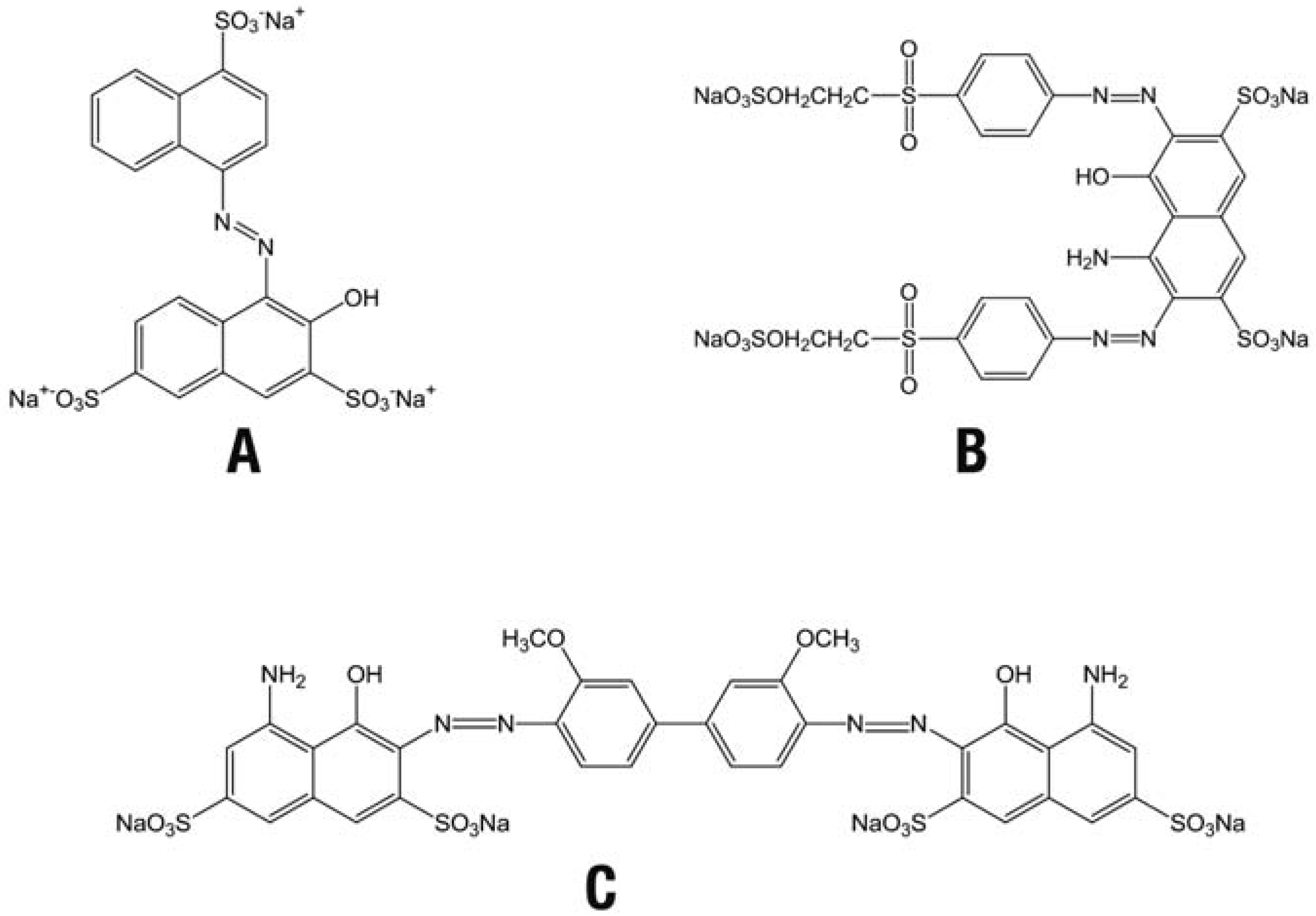
Figure 1. Structures of the azo dyes used in this study. A) Acid Red 27 (AR27), B) Reactive Black 5 (RB5), C) Direct Blue 15 (DB15).
Jox 03 e2 g001
Figure 2. The azo reductase and flavin reductase activities of different cell fractions of C. freundii A1.
Jox 03 e2 g002
Figure 3. A) Reduction of AR27 by Fre and the simultaneous oxidation of nicotinamide adenine dinucleotide (NADH); B) Reduction of AR27 by Fre in the presence of NADH and/or riboflavin.
Jox 03 e2 g003
Figure 4. Proposed scheme depicting the reduction of an azo dye in the Fre system medi- ated by nicotinamide adenine dinucleotide (NADH) and riboflavin, reproduced (with slight modifications) from Field and Brady (2003), with permission. Dotted line indicates the non-enzymatic reduction by NADH.
Jox 03 e2 g004
Table 1. Summary of the reduction data of the azo dyes by Fre, and the reduction potentials determined using voltammetry.
Jox 03 e2 g005
*In the presence of both nicotinamide adenine dinucleotide and riboflavin, estimated using the tangent method; °relative to initial concentration, after reaction at 30°C for 30 min. AR27, Acid Red 27; RB5, Reactive Black 5; DB15, Direct Blue 15; BRb, Britton-Robinson buffer.
Disclaimer/Publisher’s Note: The statements, opinions and data contained in all publications are solely those of the individual author(s) and contributor(s) and not of MDPI and/or the editor(s). MDPI and/or the editor(s) disclaim responsibility for any injury to people or property resulting from any ideas, methods, instructions or products referred to in the content.