
Article

An Integrated Gene Therapy Strategy for γ-Globin Addition and HbF Reactivation in β-Thalassemia
by
Kiriaki Paschoudi
1,2,†,
Ninos Ioannis Vasiloudis
1,2,†,
Fotios Papadopoulos
1,2,
Xenia Nikolaou
1,2,
Anastasia Papadopoulou
1,2,
Pavel Sova
3,
Evangelia Yannaki
2,4 and
Nikoletta Psatha
1,*
1
Department of Genetics, Development & Molecular Biology, School of Biology, Aristotle University of Thessaloniki, 54124 Thessaloniki, Greece
2
Gene and Cell Therapy Center and Hematology Clinic/Bone Marrow Transplant Unit, “George Papanikolaou” Hospital, 57010 Thessaloniki, Greece
3
Altius Institute for Biomedical Sciences, Seattle, WA 98121, USA
4
Division of Hematology, Department of Medicine, University of Washington, Seattle, WA 98195, USA
*
Author to whom correspondence should be addressed.
These authors contributed equally to this work.
Thalass. Rep. 2026, 16(3), 19; https://doi.org/10.3390/thalassrep16030019
Submission received: 15 July 2026 / Revised: 19 August 2026 / Accepted: 21 August 2026 / Published: 1 September 2026
(This article belongs to the Section Innovative Treatment of Thalassemia)

Abstract

Background: β-Thalassemia and other β-hemoglobinopathies arise from defective β-globin production, and reactivating fetal hemoglobin (HbF) is a well-established strategy to ameliorate disease severity. Two principal gene-therapy approaches—erythroid-specific γ-globin gene addition and shRNA/shmiRNA-mediated knockdown of the γ-globin repressor BCL11A—have each shown clinical promise, but their combination within a single vector remains largely unexplored. Methods: Here we developed and compared three compact, C1-insulated lentiviral vectors driven by the micro-locus control region (μLCR): a BCL11A-targeting shmiRNA vector, a γ-globin cDNA vector, and a dual construct combining both elements. Results: All vectors were produced at high titers, and incorporating both cassettes into a single construct did not compromise vector production or cell viability. In CD34+ hematopoietic stem and progenitor cells from healthy donors and β-thalassemia patients, transduction did not impair proliferation, erythroid differentiation, or progenitor colony formation. All three vectors significantly increased the proportion of HbF-expressing cells, with the combined shmiRNA + γ-globin cDNA vector achieving the highest induction, reaching approximately 25% HbF-positive cells in the enucleated erythroid population of thalassemic cells versus approximately 10% in untransduced controls. Notably, all constructs significantly reduced reactive oxygen species levels, indicating alleviation of oxidative stress. Conclusions: Together, these findings support the feasibility and therapeutic potential of combining complementary HbF-inducing mechanisms within a single lentiviral vector for β-thalassemia.

1. Introduction

Beta-hemoglobinopathies are the most common inherited recessive disorders caused by mutations in the β-globin gene, with about 400,000 affected births each year. More than 300 mutations have been identified, leading either to reduced or absent β-globin production (β+ or β0 thalassemia) or to abnormal β-globin variants such as sickle cell disease (SCD) [1,2].
Disease severity is strongly influenced by genetic modifiers, particularly hereditary persistence of fetal hemoglobin (HPFH), which sustains γ-globin expression and increase fetal hemoglobin (HbF), ameliorating the clinical phenotype [3,4,5]. Allogeneic hematopoietic stem cell transplantation (allo-HSCT) was until recently the only established curative treatment for β-thalassemia; however, its broader application is limited by donor availability, and transplant-related complications [6].
Over the last three decades, gene therapy has emerged as a promising curative alternative for patients with β-hemoglobinopathies. These approaches are based on the autologous transplantation of genetically modified hematopoietic stem and progenitor cells (HSPCs). The procedure involves mobilizing CD34+ HSPCs from the bone marrow into the bloodstream using agents such as G-CSF combined with Plerixafor in β-thalassemia patients [7,8,9,10,11,12]. After leukapheresis, the collected HSPCs are genetically corrected ex vivo, by either gene addition or genome editing, and reinfused into the patient following conditioning [13,14].
Several lentiviral vectors have been developed and clinically evaluated for β-thalassemia [15]. Among these, LentiGlobin BB305 [16] is the most clinically advanced gene addition platform, demonstrating durable transgene expression and sustained transfusion independence in the majority of patients [17,18,19,20,21]. The vector carries a modified β-globin gene (βA-T87Q), including coding and non-coding sequences, which produces a distinguishable and fully functional b-globin variant. Expression of the transgene is driven by the β-globin promoter and the core HS2, HS3, and HS4 elements of the β-globin locus control region (named μLCR), enabling high-level, erythroid-specific expression. This platform led to the development of betibeglogene autotemcel, marketed as Zynteglo, an autologous CD34+ cell-based gene therapy approved for transfusion-dependent β-thalassemia.
Despite its clinical success, the broader implementation of this approach has been limited by high cost and reimbursement challenges, which in turn contributed to the withdrawal of Zynteglo from the European market [22]. In parallel, the large LCR-based regulatory architecture, along with the large globin expression cassettes, has been shown to reduce viral titers, increase manufacturing demands, and require larger quantities of vectors for efficient HSPC transduction [23,24,25]. Furthermore, integration-related safety concerns also remain, particularly in vectors lacking additional regulatory elements, such as chromatin insulators.
In addition to β-globin gene addition, γ-globin gene addition was among the earliest gene-therapy strategies investigated for β-thalassemia. Initial studies demonstrated that erythroid-specific γ-globin expression could improve globin-chain balance and correct disease-related phenotypes in murine β-thalassemia models, although efficacy was influenced by vector copy number, integration-site position effects, and the composition of the regulatory elements included in the vector [26,27]. Subsequent studies showed that γ-globin gene transfer could generate therapeutically relevant HbF levels in erythroid progeny derived from β-thalassemia patient CD34+ cells and improve erythroid maturation and survival [28,29,30,31,32].
The emergence of approaches that reactivate the endogenous HBG genes has nevertheless reduced the translational momentum of γ-globin gene addition. In particular, genome editing of the erythroid-specific BCL11A (a key γ-globin repressor) enhancer [33,34,35,36,37,38,39] and erythroid-restricted BCL11A knockdown using lentiviral shRNA or shmiR constructs [40,41,42,43] can induce HbF from the endogenous β-globin locus without requiring the introduction of an additional globin transgene. The latter offers an additional advantage of a highly compact therapeutic sequence, substantially smaller than a genomic β-globin or γ-globin expression cassette thereby facilitating vector production or the incorporation of additional functional elements. The strong preclinical and clinical performance of BCL11A-targeting approaches has increasingly directed the field towards endogenous HbF reactivation. However, their efficacy and safety depend on the magnitude and lineage specificity of BCL11A suppression [37,38,44,45,46,47,48,49]. Ubiquitous or extensive depletion can adversely affect hematopoietic stem and progenitor cell function, while complete or profound loss in erythroid cells has been associated with impaired precursor expansion, increased apoptosis, and defective terminal maturation or enucleation [48,50]. These observations highlight the need for erythroid-restricted and carefully controlled BCL11A knockdown and provide a rationale for directly comparing this approach with γ-globin addition, both individually and in combination.
In this study, we performed a direct side-by-side comparison of lentiviral strategies based on γ-globin addition, BCL11A-targeting shmiRNA, or their combination within a single vector under the control of the μLCR. To limit vector size and facilitate incorporation of both therapeutic components, γ-globin was delivered as a compact cDNA rather than as the entire genomic sequence. Across healthy donor- and β-thalassemia patient-derived erythroid cultures, all three vectors increased the proportion of HbF-expressing cells without adversely affecting progenitor capacity or erythroid differentiation. The bifunctional vector produced the highest mean proportion of HbF-positive cells and γ-globin chain production in patient derived erythroid cells, whereas γ-globin cDNA addition alone achieved a comparable response and was associated with robust erythroid expansion. These findings support the feasibility of integrating direct γ-globin provision and endogenous HbF reactivation within a single compact lentiviral platform for β-thalassemia.

2. Materials and Methods

The study was conducted in accordance with the Declaration of Helsinki, and the protocol was approved by the Research Ethics and Deontology Committee of the Aristotle University of Thessaloniki (OGETHERA: 326425/2023) on 13 December 2023.

2.1. Viral Vectors

To systematically evaluate the therapeutic efficacy of HbF reactivation, we engineered a panel of three compact, self-inactivating lentiviral vectors (Figure 1A). The constructs were designed to test three distinct genetic modalities: (1) indirect HbF induction via post-transcriptional silencing of the BCL11A repressor using a specific shmiRNA (BCL11A shmiRNA vector) [43]; (2) direct γ-globin gene addition via a cDNA expression cassette (γ-globin cDNA vector) [51]; and (3) a dual-functional, combinatorial approach integrating both the BCL11A-targeting shmiRNA and the γ-globin cDNA into a single vector architecture (BCL11A shmiRNA + γ-globin cDNA vector). All transgene cassettes were cloned within a pRRLSIN.cPPT.PGK-GFP.WPRE plasmid (a gift from Didier Trono; Addgene plasmid # 12252), modified to incorporate the C1 dual-function chromatin insulator [52], a beta globin promoter and the HS2,HS3, HS4 μLCR as previously described [53].

2.2. Lentiviral Production and Titration

VSV.G-pseudotyped lentiviral vectors (LVs) were produced by transient co-transfection of 293FT cells using the calcium phosphate precipitation method. For lentiviral production we used a plasmid mixture consisting of the packaging plasmid psPAX2, the envelope plasmid pVSV-G, and the transfer vector carrying the corresponding transgene (BCL11A shmiRNA, γ-globin cDNA, BCL11A shmiRNA + γ-globin cDNA).
After viral production the viral titers were quantified by quantitative real-time PCR (qPCR) using an ABI 7500 Real-Time PCR System (Applied Biosystems, Waltham, MA, USA). The following primers and probes were used for viral genome quantification:
  • Gag Forward: 5′-GGAGCTAGAACGATTCGCAGTTA-3′
  • Gag Reverse: 5′-GGTTGTAGCTGTCCCAGTATTTGTC-3′
  • Gag Probe: 5′-FAM-ACAGCCTTCTGATGTTTCTAACAGGCCAGG TAMRA-3′
  • hAlb Forward: 5′-TGAAACATACGTTCCCAAAGAGTTT-3′
  • hAlb Reverse: 5′-CTCTCCTTCTCAGAAAGTGTGCATAT-3′
  • hAlb Probe: 5′-VIC-TGCTGAAACATTCACCTTCCATGCAGA-TAMRA-3′
PCR reactions were conducted in a total volume of 25 μL using TaqMan Universal PCR Master Mix (Applied Biosystems, Waltham, MA, USA). Amplification was carried out with an initial incubation at 50 °C for 2 min, followed by 95 °C for 10 min, and 40 amplification cycles consisting of 95 °C for 15 s and 60 °C for 1 min. Lentiviral vector copy number (VCN) per cell was determined by normalization to the endogenous albumin (ALB) gene.

2.3. Cell Culture of Primary CD34+ Culture

Human CD34+ hematopoietic stem and progenitor cells a HSPCs were obtained from normal donors (n = 3) and patients with intermedia β-thalassemia (n = 2) β++ and severe β-thalassemia (n = 1) β00 (Table 1).
Patient-derived cells were harvested during prior mobilization clinical trials at George Papanikolaou Hospital in Thessaloniki, Greece [9,10]. Cells were cultured at a concentration of (3–5) × 105 cells/mL in (IMDM + Glutamax, GibcoTM, Grand Island, NY, USA) supplemented with 1% Penicillin/Streptomycin (pen/strep, GibcoTM, Grand Island, NY, USA), 1% Glutamine (L-glutamine, GibcoTM, Grand Island, NY, USA), 5% AB plasma (obtained from “G. Papanikolaou” Hospital, Thessaloniki, Greece), 10 ug/mL Insulin (Sigma Aldrich, St. Louis, MO, USA), 2 IU/mL Heparin (Sigma Aldrich, St. Louis, MO, USA), 330 ug/mL Holo-Transferrin (Sigma-Aldrich, St. Louis, MO, USA) This formulation is hereafter referred to as the basal medium. The basal medium was supplemented with 100 ng/mL h-SCF (R&D Systems, Minneapolis, MN, USA), 100 ng/mL h-TPO (Peprotech, Cranbury, NJ, USA), and 100 ng/mL h-Flt3 (Peprotech, Cranbury, NJ, USA), together with the small molecules 35 nM UM171 (Stem Cell Technologies, Vancouver, BC, Canada), 100 nM LY2228820 (Stem Cell Technologies, Vancouver, BC, Canada), and 1 μM SR1 (Stem Cell Technologies, Vancouver, BC, Canada) [54]. Lentiviral transduction was performed at an MOI of 30 on Retronectin-coated plates (Takara Bio, Kusatsu, Shiga, Japan). The transduction medium was supplemented with 8   μ g / m L protamine sulfate (Sigma-Aldrich, St. Louis, MO, USA) and 0.5   m g / m L Synperonic (Sigma-Aldrich, St. Louis, MO, USA). Each donor-derived sample was processed and transduced independently, using separately produced batches of the corresponding lentiviral vectors. Repeated cultures or technical measurements from the same donor were not considered independent biological replicates.

2.4. In Vitro Erythroid Differentiation (ED) of CD34+ Cells

In vitro erythroid differentiation of CD34+ cells was carried out in three sequential stages as previously described [55,56]. The basal medium was supplemented with hydrocortisone, IL-3 (Peprotech, Cranbury, NJ, USA), hSCF (R&D Systems, Minneapolis, MN, USA), and EPO (R&D Systems, Minneapolis, MN, USA) during Stage I. In Stage II, only hSCF and EPO were added, while Stage III utilized EPO as the sole supplement.

2.5. Colony Forming Unit (CFU) Assay

The colony-forming potential of C D 34 + cells was evaluated using a methylcellulose assay. In brief, 2500 cells in 300   μ L of AMEM (Corning, Corning, NY, USA) were mixed with 2   m L of MethoCult H4434 (Stemcell Technologies, Vancouver, BC, Canada). After 14 days of incubation, CFU-GM and BFU-E colonies were classified and enumerated.

2.6. Flow Cytometry

To assess erythroid differentiation during ex vivo culture, 50.000 cells were stained with anti-human CD235a-PE (ExBio, Vestec, Czech Republic) and anti-human CD36-APC (ExBio, Vestec, Czech Republic) antibodies based on manufacturer instruction and analyzed by flow cytometry. To detect intracellular human γ -globin, cells were fixed in 4% paraformaldehyde and permeabilized by sequential washes of 1:1 acetone/water, 100% acetone, and 1:1 acetone/water. Cells were then labeled with an anti-human γ -globin antibody (clone 51.7, Santa Cruz Biotechnology, Dallas, TX, USA). Enucleated erythroid cells were identified by the absence of staining using Nuclear Red (Thermo Fisher Scientific, Waltham, MA, USA).

2.7. Analysis of Oxidative Stress Species (ROS)

Intracellular ROS production was determined using the CellROX Deep Red Flow Cytometry Assay Kit (Invitrogen, Thermo Fisher Scientific, Waltham, MA, USA) according to the manufacturer’s instructions. Briefly, at the final stage of ex vivo differentiation, 1 × 10 6 thalassemic cells were stained with CellROX Deep Red reagent at a final concentration of 500   n M and incubated for 45 min at 37 °C. Following probe oxidation by intracellular ROS, fluorescence intensity was quantified via flow cytometry.

2.8. VCN

Vector copy number (VCN) was quantified by real-time quantitative PCR (qPCR) using an ABI 7500 Real-Time PCR System (Applied Biosystems, Waltham, MA, USA). Genomic DNA was analyzed using TaqMan assays targeting the vector gag sequence, with the endogenous human albumin (hALB) gene serving as the reference for normalization. The following primers and probes were used:
  • Gag F: 5′-GGAGCTAGAACGATTCGCAGTTA-3′
  • Gag R: 5′-GGTTGTAGCTGTCCCAGTATTTGTC-3′
  • Gag probe: 5′-FAM-ACAGCCTTCTGATGTTTCTAACAGGCCAGG-TAMRA-3′
  • hALB F: 5′-TGAAACATACGTTCCCAAAGAGTTT-3′
  • hALB R: 5′-CTCTCCTTCTCAGAAAGTGTGCATAT-3′
  • hALB probe: 5′-VIC-TGCTGAAACATTCACCTTCCATGCAGA-TAMRA-3′
Each DNA sample was analyzed in a 25 μL reaction containing TaqMan Universal PCR Master Mix (Applied Biosystems). The thermal cycling conditions consisted of an initial incubation at 50 °C for 2 min, followed by denaturation at 95 °C for 10 min and 40 amplification cycles of 95 °C for 15 s and 60 °C for 1 min. VCN was calculated based on the ratio of vector gag copies to the endogenous hALB gene, allowing estimation of the number of vector copies per cell.

2.9. HPLC

Globin chain expression in ex vivo–differentiated thalassemic cells (day 18) was analyzed by liquid chromatography using a Shimadzu LC-2060C 3D system (Shimadzu Corporation, Kyoto, Japan). Fitted with a GmbH MultiHigh Bio 300 column (250 mm × 3 mm). Separation was performed using a 38–60% gradient of 0.1% trifluoroacetic acid in water/acetonitrile at a flow rate of 1 mL/min.

2.10. Statistical Analysis

All quantitative data are presented as mean ± standard error of the mean (SEM) of at least three independent replicates. Statistical analyses were performed using R (version 4.4.3). To compare the efficacy of the gene therapy strategies, we employed one-way ANOVA followed by Tukey’s honestly significant difference (HSD) post hoc test for multiple comparisons among treatment groups, as well as two-tailed Student’s t-tests where appropriate (such as comparing individual treatment groups directly against the UNTD control). A p-value of less than 0.05 was considered statistically significant. All data visualization and statistical evaluations were conducted within the RStudio environment.

3. Results

3.1. Development of Erythroid Specific Lentiviral Vectors for HbF Reactivation

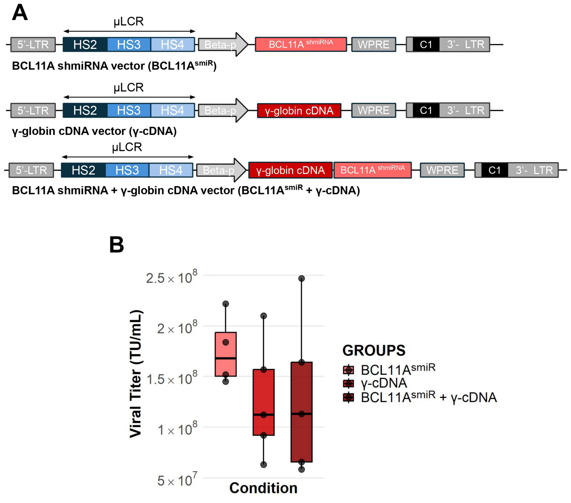
We developed three compact lentiviral vectors designed to reactivate fetal hemoglobin (HbF) expression through distinct gene therapy strategies (Figure 1A). The first vector (BCL11A shmiRNA) encodes a BCL11A-targeting shmiRNA to suppress the γ-globin repressor BCL11A [43]. The second vector (γ-globin cDNA) contains the γ-globin cDNA, enabling direct gene addition [51]. The third vector combines both the BCL11A-targeting shmiRNA and the γ-globin cDNA within a single construct (BCL11A shmiRNA + γ-globin cDNA).
In all vectors, transgene expression is driven by the μLCR, comprising the HS2, HS3, and HS4 regulatory elements, in combination with a minimal β-globin promoter. This design ensures robust erythroid-specific expression. The dual-function chromatin insulator C1 [52] was incorporated in all vectors, to provide enhancer-blocking and barrier activity and to reduce integration-site-dependent effects on transgene expression and potentially mitigate enhancer-mediated insertional genotoxicity.
Lentiviral vector production was efficient, yielding high titers across all constructs (BCL11A shmiRNA: 1.72 ± 0.14 × 10⁸ TU/mL; γ-globin cDNA: 1.30 ± 0.21 × 10⁸ TU/mL; combined BCL11A shmiRNA + γ-globin cDNA: 1.30 ± 0.28 × 10⁸ TU/mL) (Figure 1B). Notably, no significant differences in viral titers were observed among the three vectors, indicating that the incorporation of both the BCL11A-targeting shmiRNA and γ-globin cDNA within a single construct did not adversely affect vector production. One-way ANOVA showed no significant difference in titer among the three conditions (p = 0.86), and Tukey’s HSD post hoc test found no significant pairwise differences (all adjusted p > 0.05), indicating that construct design (shRNA alone, γ-cDNA alone, or the combined cassette) did not measurably affect viral packaging or production efficiency.

3.2. Evaluation of Gamma Globin Induction in Healthy Donor Derived CD34+ Primary Cells

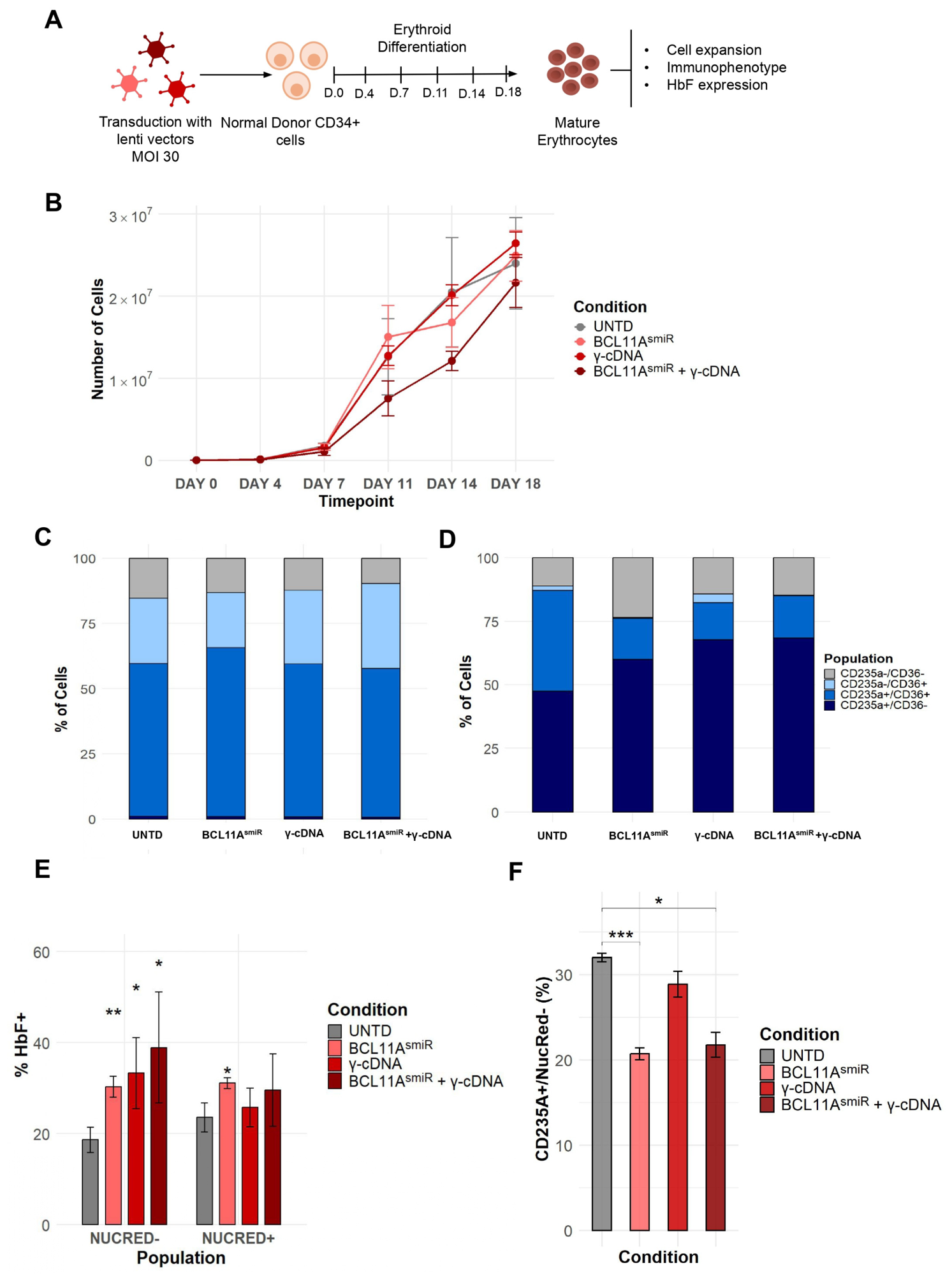
To evaluate the performance of the three lentiviral vectors in primary cells, mobilized CD34+ hematopoietic stem and progenitor cells from healthy donors were transduced at a multiplicity of infection (MOI) of 30, with untransduced cells serving as controls. Forty-eight hours post-transduction, cells were cultured under erythroid differentiation conditions (Figure 2A). Transduction with any of the three lentiviral vectors did not affect cell expansion compared with untransduced controls throughout the culture period (Figure 2B). Erythroid differentiation and maturation were assessed by flow cytometry through the analysis of the erythroid markers CD36 and CD235a on days 7 and 14 of differentiation. No significant differences were observed between transduced and untransduced cells at either time point, indicating that the overall erythroid differentiation profile was preserved following transduction (Figure 2C,D).
The therapeutic impact of the lentiviral vectors was assessed by quantifying the frequency of HbF-expressing cells within the NucRed- and NucRed+ populations. Across both populations, all three vectors (BCL11A shmiRNA, γ-globin cDNA, and the combined BCL11A shmiRNA + γ-globin cDNA) demonstrated a consistent increase in HbF+ cells compared to the untransduced (UNTD) control (Figure 2E). Within the NucRed- population, all three vectors showed a statistically significant increase in HbF+ cells relative to the UNTD control (NucRed- HbF+ cells: UNTD 18.63 ± 1.60%; BCL11A shmiRNA 30.28 ± 1.34%, p = 0.005; γ-globin cDNA 33.30 ± 4.51%, p = 0.037; BCL11A shmiRNA + γ-globin cDNA 38.93 ± 7.05%, p = 0.048). Furthermore, within the NucRed+ population, the BCL11A shmiRNA vector also exhibited a statistically significant increase in the percentage of HbF+ cells compared to the control, whereas the increases observed with the other two vectors did not reach statistical significance (NucRed + HbF+ cells: UNTD 23.57 ± 1.86%; BCL11A shmiRNA 31.09 ± 0.70%, p = 0.019; γ-globin cDNA 25.77 ± 2.46%; BCL11A shmiRNA + γ-globin cDNA 29.59 ± 4.60%) (Figure 2E). To assess whether BCL11A knockdown and/or γ-globin cDNA expression affected terminal erythroid maturation, we quantified the proportion of enucleated cells (CD235a+/NucRed−) by flow cytometry in healthy donor-derived erythroid cultures. Untransduced (UNTD) cells enucleated at a mean rate of 32.0% ± 0.7% (n = 3). Enucleation was significantly reduced in cells transduced with the BCL11A-targeting shRNA vector alone (BCL11AsmiR: 20.7% ± 1.2%, n = 3; p = 0.0014, unpaired two-tailed t-test) and in cells receiving the combined BCL11AsmiR + γ-cDNA vector (21.8% ± 2.5%, n = 3; p = 0.012) relative to UNTD controls. In contrast, cells transduced with the γ-cDNA vector alone did not differ significantly from UNTD (28.9% ± 2.6%, n = 3; p = 0.21). Together, these data suggest that BCL11A knockdown, whether alone or combined with γ-globin cDNA overexpression, is associated with a modest reduction in enucleation efficiency, whereas γ-globin cDNA expression alone does not appreciably impair terminal maturation in this system.

3.3. Assessment of Vector Efficacy in Thalassemia Patient-Derived CD34+ Hematopoietic Stem and Progenitor Cells

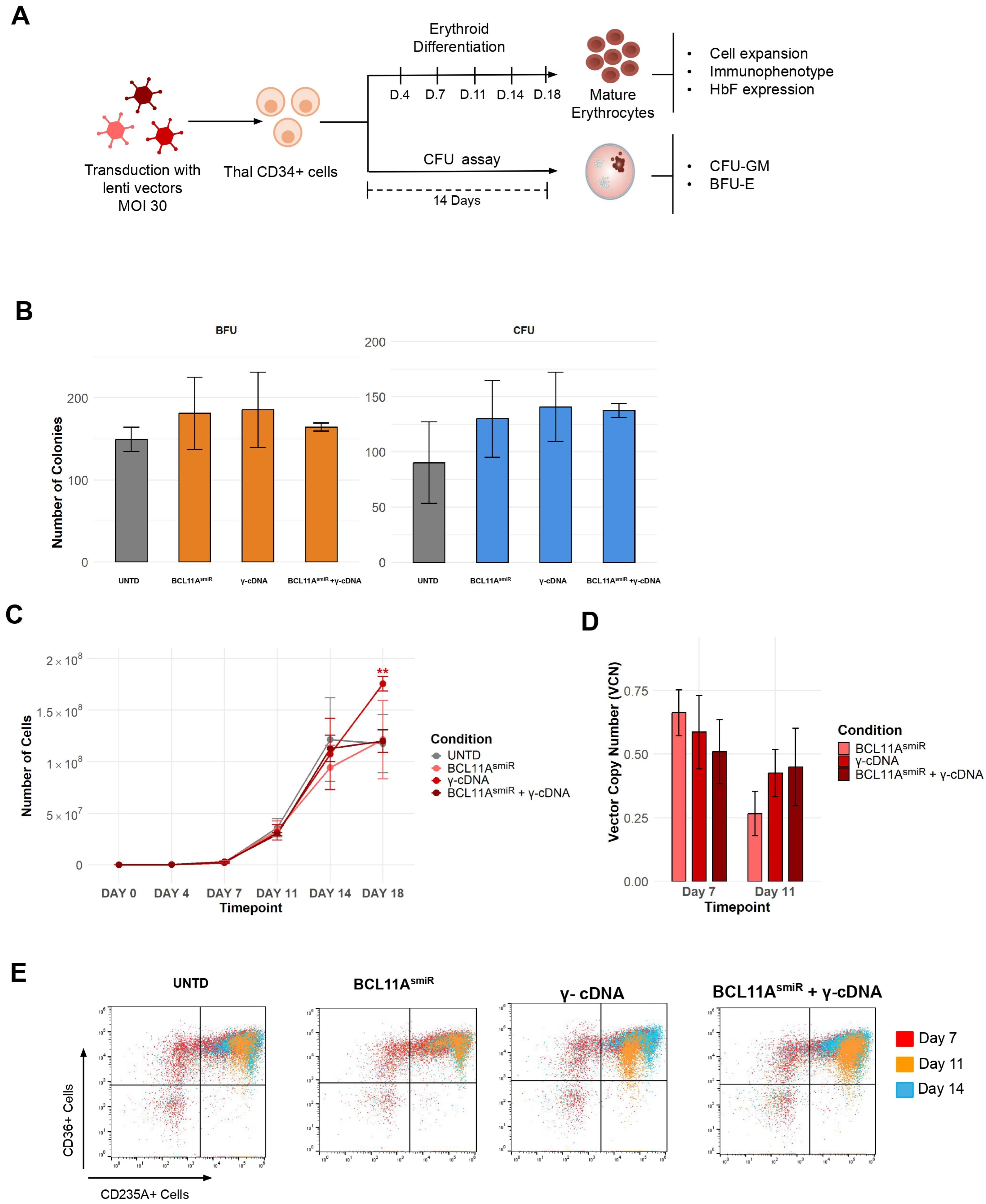
To evaluate the performance of the three lentiviral vectors in a disease model, mobilized CD34+ hematopoietic stem and progenitor cells from patients with β-thalassemia were transduced at a multiplicity of infection (MOI) of 30, with untransduced cells serving as controls. Forty-eight hours post-transduction, cells were cultured under erythroid differentiation conditions to assess maturation and phenotypic expression. Concurrently, a portion of the transduced cells was subjected to colony-forming unit (CFU) assays to evaluate myeloid and erythroid progenitor function, specifically quantifying burst-forming unit-erythroid (BFU-E) and colony-forming unit-granulocyte macrophage (CFU-GM) colonies (Figure 3A). Absolute numbers of BFU-E and CFU-GM colonies per 2500 seeded cells were quantified for each experimental condition. Colony formation was maintained following transduction with all three vectors, with no reduction in either erythroid or myeloid colony output compared with the UNTD control, indicating preservation of progenitor capacity (Figure 3B).
The proliferative capacity of the cells was monitored over an 18-day culture period across all experimental conditions, including the untransduced (UNTD) control, BCL11A shmiRNA, γ-globin cDNA, and the combined BCL11A shmiRNA + γ-globin cDNA (Figure 3C). The expansion of the cells was similar across all conditions (Day 18 cell counts: UNTD 117.5 ± 14.1 × 10⁶; BCL11A shmiRNA 121.4 ± 17.0 × 10⁶; γ-globin cDNA 175.6 ± 3.6 × 10⁶; combined 119.9 ± 7.8 × 10⁶ cells), demonstrating that transduction with the three vectors did not exert a negative effect on cell growth. Notably, the γ-globin cDNA vector resulted in the highest cell yields at Day 18, reaching approximately 1.5-fold that of the untransduced control (175.6 ± 3.6 × 10⁶ vs. 117.5 ± 14.1 × 10⁶ cells; p = 0.007) with markedly low inter-experiment variability. This increased erythroid output was specific to the γ-globin cDNA condition, whereas the BCL11A shmiRNA and combined-vector groups remained comparable to UNTD cell.
To determine whether differences in vector performance could be attributed to differences in gene transfer efficacy, vector copy number (vcn) was quantified at different time points during erythroid differentiation. VCN was comparable across all three conditions at both timepoints (BCL11AsmiR: 0.82 ± 0.30 at day 7, 0.27 ± 0.09 at day 11; γ-cDNA: 0.59 ± 0.14 at day 7, 0.43 ± 0.09 at day 11; BCL11AsmiR + γ-cDNA: 0.51 ± 0.13 at day 7, 0.45 ± 0.15 at day 11). One-way ANOVA showed no significant difference in VCN among conditions at either day 7 (p = 0.15) or day 11 (p = 0.10), and Tukey’s HSD post hoc test found no significant pairwise differences at either timepoint (all adjusted p > 0.05), indicating that transduction efficiency was similar across the three vector conditions (Figure 3D).
Flow cytometric analysis of CD235a and CD36 expression was utilized to assess the impact of vector transduction on the erythroid differentiation and maturation of thalassemic CD34+ cells (Figure 3E). The data indicates that lentiviral transduction with either vector did not fundamentally alter the lineage commitment of these cells, as the distribution of cell populations remained relatively consistent across experimental conditions. Furthermore, the differentiation trajectory remained normal across all groups, mirroring the observations in healthy primary cells, with no notable differences in the maturation profile compared to the untransduced (UNTD) control (Day 14 CD235a+/CD36- cells: UNTD 13.2 ± 1.9%; BCL11A shmiRNA 19.0 ± 5.0%; γ-globin cDNA 16.2 ± 3.0%; combined 11.7 ± 2.5%). These findings suggest that the genetic modifications do not negatively impact the erythroid differentiation or maturation potential of the patient-derived cells.

3.4. Fetal Hemoglobin Induction and Oxidative Stress in Transduced Patient-Derived Cells

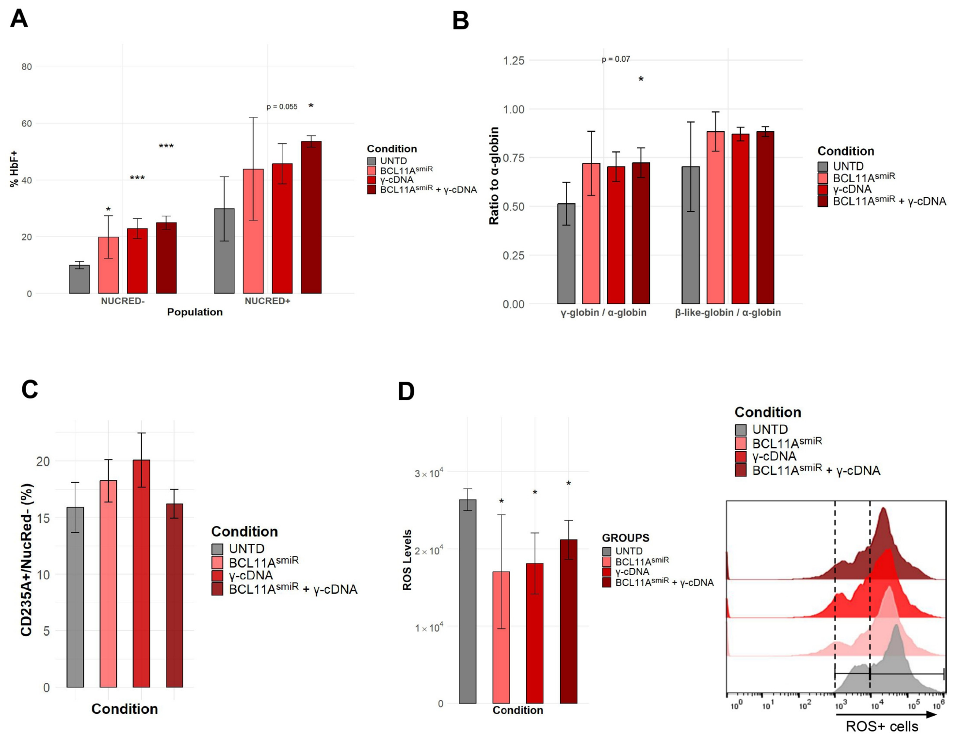
The therapeutic efficacy of the BCL11A shmiRNA and γ-globin cDNA vectors, as well as their combined effect, was evaluated by quantifying the percentage of HbF-expressing cells within the NucRed− and NucRed + populations following erythroid differentiation (Figure 4A). In the NucRed − population, all experimental conditions demonstrated a statistically significant increase in HbF expression compared to the untransduced (UNTD) control, which exhibited a baseline of 9.92 ± 0.63%. Specifically, the BCL11A shmiRNA, γ-globin cDNA, and combined BCL11A shmiRNA + γ-globin cDNA conditions yielded mean HbF levels of 19.74 ± 3.36% (p = 0.038), 22.77 ± 1.78% (p < 0.001), and 24.85 ± 1.65% (p < 0.001), respectively. Within the NucRed + population, the UNTD control baseline was 29.79 ± 5.69%, while the BCL11A shmiRNA, γ-globin cDNA, and combined vector conditions reached mean levels of 43.78 ± 8.11%, 45.72 ± 3.52%, and 53.55 ± 1.45%, respectively. While these differences did not reach conventional statistical significance, the γ-globin cDNA and combined vector conditions approached significance (p = 0.055 and p = 0.050, respectively). Thus, the combined construct produced the highest mean percentage of HbF+ cells, although its effect was not statistically superior to that of individual vectors.
To directly evaluate globin-chain composition, HPLC analysis was performed in terminally differentiated β-thalassemia patient derived-erythroid cells. HPLC analysis showed increased γ-globin/α-globin ratios following vector treatment (Figure 4C). The BCL11A shmiRNA vector alone and the γ-globin cDNA alone did not produce a statistically significant increase (p = 0.14 and p = 0.07, respectively), whereas the combined BCL11A shmiRNA + γ-globin cDNA vector reached statistical significance compared to UNTD cells (0.51 ± 0.11 vs. 0.72 ± 0.08, p = 0.053, p = 0.05). Analysis of the total β-like-globin/α-globin ratio did not reveal statistically significant differences among conditions; nevertheless, all three vector-treated groups showed mean ratios closer to 1:1 than UNTD cells, consistent with a shift toward a more balanced non-α/α-globin chain balance.
Terminal erythroid maturation was further assessed by quantifying the proportion of enucleated (CD235a+/NucRed) cells at the end of the differentiation culture (Figure 4C). All three lentiviral vectors supported enucleation at levels comparable to the untransduced control, with no statistically significant differences between conditions (CD235a+/NucRed cells: UNTD 15.90 ± 2.23%; BCL11A shmiRNA 18.24 ± 1.87%, p = 0.444; γ-globin cDNA 20.06 ± 2.40%, p = 0.251; BCL11A shmiRNA + γ-globin cDNA 16.21 ± 1.29%, p = 0.931).
To assess the impact of vector transduction on the redox status of thalassemic erythroid cells, Reactive Oxygen Species (ROS) levels were quantified across all groups (Figure 4D). The assessment of oxidative stress revealed that the untransduced (UNTD) control cells exhibited the highest mean ROS MFI levels at 26,376.8 ± 712.54. In contrast, all transduced conditions demonstrated a statistically significant reduction in mean ROS levels compared to the UNTD control. Specifically, the BCL11A shmiRNA, γ-globin cDNA, and combined dual-function-vector conditions resulted in mean ROS levels of 17,020.5 ± 3686.44 (p = 0.047), 18,107.0 ± 2276.63 (p = 0.011), and 21,176.0 ± 1778.02 (p = 0.027), respectively. Thus, increased γ-globin expression was associated with reduced intracellular ROS across all three vector-treated groups. However, the magnitude of HbF induction did not directly correlate with the extent of ROS reduction, as the combined vector produced the highest mean HbF-positive fraction but not the greatest reduction in ROS.

4. Discussion

Gene therapy has now entered clinical practice for β-hemoglobinopathies, establishing autologous HSPC modification as a viable therapeutic approach. In β-thalassemia, lentiviral β-globin gene addition [16,57] has demonstrated durable clinical benefit and led to the approval of betibeglogene autotemcel [58,59]. Erythroid-restricted BCL11A silencing using a lentiviral shmiRNA vector [16,42,43,49] has also progressed to clinical evaluation, providing proof of principle for therapeutic HbF reactivation. γ-Globin gene addition has likewise been investigated for several decades and has shown efficacy in preclinical models and patient-derived cells [16,30,60,61,62]; however, its recent translational development has focused predominantly on sickle cell disease rather than β-thalassemia [63]. Against this background, a direct comparison of these HbF-inducing strategies in a β-thalassemia context, both individually and in combination, remains particularly relevant. In the present proof-of-concept study, we directly compared three lentiviral strategies designed to increase γ-globin expression in β-thalassemia: γ-globin cDNA addition, BCL11A-targeting shmiRNA-mediated endogenous HbF reactivation, and the combination of both mechanisms within a single vector.
A central concern in the development of multifunctional lentiviral vectors is that increasing cassette size and regulatory complexity may impair vector production and transduction efficiency. This limitation is particularly relevant to globin gene therapy, where the inclusion of large LCR-derived regulatory regions and genomic globin sequences has historically contributed to reduced viral titers and increased manufacturing demands. Accordingly, several efforts have focused on reducing vector size while preserving high-level erythroid-specific expression. These have included systematic minimization of the β-globin LCR, as exemplified by the development of novel compact HS2-, HS3-, and HS4-derived regulatory elements by Kohn and colleagues, which improved vector titer and gene-transfer efficiency compared with conventional β-globin vectors [25]. Other approaches have sought to replace the canonical LCR altogether with alternative erythroid enhancers. In our recent study, high-throughput functional screening identified compact erythroid regulatory elements capable of replacing the μLCR in a therapeutic β-globin vector, increasing viral titers and HSPC transducibility while maintaining correction of the β-thalassemic phenotype [53]. A complementary strategy is to reduce the size of the therapeutic cassette itself. In the present study, this was achieved by using either a compact BCL11A-targeting shmiRNA, a γ-globin cDNA rather than the full genomic γ-globin gene, or both elements within a single construct. Importantly, incorporation of both elements did not significantly reduce viral titer, and vcn analysis in β-thalassemia patient-derived cells showed comparable gene-transfer levels among the three vectors at Days 7 and 11. Thus, the higher mean HbF induction observed with the combined construct cannot be attributed simply to increased vector integration or transduction efficiency. The compact cassette design also enabled incorporation of the C1 chromatin insulator, which provides enhancer-blocking and barrier activities and may help limit integration-site-dependent effects on transgene expression and enhancer-mediated effects on neighboring genes [52].
Multifunctional lentiviral vectors have previously been explored as a means of combining complementary therapeutic mechanisms within a single construct. For example, ATM1.1 couples βA-T87Q gene addition with an intron-embedded miR-E targeting BCL11A, thereby increasing total functional hemoglobin while simultaneously reactivating endogenous HbF [64]. Other strategies have combined two HbF-inducing RNA-interference mechanisms, such as the simultaneous targeting of BCL11A and ZNF410 [65], or paired globin gene addition with suppression of a disease-causing globin transcript [66,67]. These studies support the feasibility of incorporating multiple therapeutic activities into a single lentiviral platform. In the present study, the combined construct showed the highest mean HbF induction in patient-derived erythroid cells, although its advantage over the individual strategies was modest and did not establish statistical superiority. Interestingly, HPLC analysis was consistent with a modest additional effect of the combined strategy on γ-globin production, with the combined vector showing the strongest increase in the γ-globin/α-globin ratio. The performance of the individual constructs is also informative. Importantly, γ-globin cDNA addition alone performed comparably to BCL11A silencing, supporting direct γ-globin provision as a relevant therapeutic strategy in β-thalassemia. Although differences in the total β-like-globin/α-globin ratio were not statistically significant, all vector-treated conditions shifted toward a more balanced 1:1 ratio. Together, these findings suggest that combining direct γ-globin addition with endogenous HbF reactivation may provide a modest benefit in globin-chain output, while the present data do not support a claim of clear superiority over either individual approach. Beyond HbF induction, vector treatment was associated with changes in globin-chain composition and reduced intracellular ROS in β-thalassemia patient-derived cells. Although these findings are consistent with an improvement in the underlying erythroid phenotype, the relationship between γ-globin induction, globin-chain balance, and oxidative stress cannot be established from the present data.
Overall, our findings demonstrate that γ-globin cDNA addition, erythroid-restricted BCL11A silencing, and their combination are all capable of increasing HbF without adversely affecting HSPC expansion, progenitor capacity, or erythroid differentiation. The combined vector retained high viral titers and comparable vcn, and produced the highest mean HbF induction, supporting the concept that simultaneous direct γ-globin expression and endogenous HBG reactivation can be integrated into a single compact lentiviral platform. However, the advantage over the single-function vectors was modest and does not establish superiority across all biological readouts. Given the proof-of-concept nature and limited patient cohort of the present study, further evaluation in larger and genetically diverse cohorts, together with assessment of integration-site distribution, long-term HSC function and engraftment, and in vivo efficacy, will be required to determine the therapeutic relevance of these findings. Nevertheless, this work provides a foundation for multifunctional HbF-inducing vectors and supports their further development as a potential strategy for β-thalassemia.

Author Contributions

K.P. and N.I.V.: Investigation; methodology; formal analysis; visualization; writing—original draft. F.P. and X.N.: Investigation. A.P.: validation; supervision. P.S.: Methodology; Investigation. E.Y.: Resources; validation; supervision; writing—review and editing. N.P.: Conceptualization; methodology; project administration; supervision; funding acquisition; writing—original draft. All authors have read and agreed to the published version of the manuscript.

Funding

This project was carried out within the framework of the National Recovery and Resilience Plan Greece 2.0, funded by the European Union—NextGenerationEU (Implementation body: HFRI, Grant no: 15358 to NP).

Institutional Review Board Statement

The study was conducted in accordance with the Declaration of Helsinki, and the protocol was approved by the Research Ethics and Deontology Committee of the Aristotle University of Thessaloniki (OGETHERA: 326425/2023) on 13 December 2023.

Informed Consent Statement

Informed consent was obtained from all subjects involved in the study.

Data Availability Statement

All data generated or analyzed during this study are included in this published article. Additional information is available upon request.

Conflicts of Interest

The authors have no conflicts of interest to declare.

References

  1. Higgs, D.R.; Engel, J.D.; Stamatoyannopoulos, G. Thalassaemia. Lancet 2012, 379, 373–383. [Google Scholar] [CrossRef] [Scilit] [PubMed]
  2. Cao, A.; Galanello, R. Beta-thalassemia. Genet. Med. 2010, 12, 61–76. [Google Scholar] [CrossRef] [Scilit] [PubMed]
  3. Martyn, G.E.; Wienert, B.; Yang, L.; Shah, M.; Norton, L.J.; Burdach, J.; Kurita, R.; Nakamura, Y.; Pearson, R.C.M.; Funnell, A.P.W.; et al. Natural regulatory mutations elevate the fetal globin gene via disruption of BCL11A or ZBTB7A binding. Nat. Genet. 2018, 50, 498–503. [Google Scholar] [CrossRef] [Scilit] [PubMed]
  4. Steinberg, M.H. Fetal Hemoglobin in Sickle Hemoglobinopathies: High HbF Genotypes and Phenotypes. J. Clin. Med. 2020, 9, 3782. [Google Scholar] [CrossRef] [Scilit] [PubMed]
  5. Forget, B.G. Molecular Basis of Hereditary Persistence of Fetal Hemoglobin. Ann. N. Y. Acad. Sci. 1998, 850, 38–44. [Google Scholar] [CrossRef] [Scilit] [PubMed]
  6. Testa, U.; Castelli, G.; Pelosi, E. Curative Approach to the Treatment of Beta-Thalassemia and Sickle Cell Disease with Hematopoietic Stem Cell Transplantation. J. Clin. Med. 2026, 15, 1379. [Google Scholar] [CrossRef] [Scilit] [PubMed]
  7. Psatha, N.; Sgouramali, E.; Gkountis, A.; Siametis, A.; Baliakas, P.; Constantinou, V.; Athanasiou, E.; Arsenakis, M.; Anagnostopoulos, A.; Papayannopoulou, T.; et al. Superior Long-Term Repopulating Capacity of G-CSF+Plerixafor-Mobilized Blood: Implications for Stem Cell Gene Therapy by Studies in the Hbbth-3 Mouse Model. Hum. Gene Ther. Methods 2014, 25, 317–327. [Google Scholar] [CrossRef] [Scilit] [PubMed]
  8. Karponi, G.; Psatha, N.; Lederer, C.W.; Adair, J.E.; Zervou, F.; Zogas, N.; Kleanthous, M.; Tsatalas, C.; Anagnostopoulos, A.; Sadelain, M.; et al. Plerixafor+G-CSF–mobilized CD34+ cells represent an optimal graft source for thalassemia gene therapy. Blood 2015, 126, 616–619. [Google Scholar] [CrossRef] [Scilit] [PubMed]
  9. Yannaki, E.; Karponi, G.; Zervou, F.; Constantinou, V.; Bouinta, A.; Tachynopoulou, V.; Kotta, K.; Jonlin, E.; Papayannopoulou, T.; Anagnostopoulos, A.; et al. Hematopoietic Stem Cell Mobilization for Gene Therapy: Superior Mobilization by the Combination of Granulocyte–Colony Stimulating Factor Plus Plerixafor in Patients with β-Thalassemia Major. Hum. Gene Ther. 2013, 24, 852–860. [Google Scholar] [CrossRef] [Scilit] [PubMed]
  10. Yannaki, E.; Papayannopoulou, T.; Jonlin, E.; Zervou, F.; Karponi, G.; Xagorari, A.; Becker, P.; Psatha, N.; Batsis, I.; Kaloyannidis, P.; et al. Hematopoietic Stem Cell Mobilization for Gene Therapy of Adult Patients With Severe β-Thalassemia: Results of Clinical Trials Using G-CSF or Plerixafor in Splenectomized and Nonsplenectomized Subjects. Mol. Ther. 2012, 20, 230–238. [Google Scholar] [CrossRef] [Scilit] [PubMed]
  11. Boulad, F.; Wang, X.; Qu, J.; Taylor, C.; Ferro, L.; Karponi, G.; Bartido, S.; Giardina, P.; Heller, G.; Prockop, S.E.; et al. Safe mobilization of CD34+ cells in adults with β-thalassemia and validation of effective globin gene transfer for clinical investigation. Blood 2014, 123, 1483–1486. [Google Scholar] [CrossRef] [Scilit] [PubMed]
  12. Baiamonte, E.; Barone, R.; Stefano, R.D.; Iacono, M.L.; Spina, B.; Contino, F.; Maggio, R.D.; Sacco, M.; Vitrano, A.; Feo, S.; et al. Hematopoietic Stem Cell Mobilization for Gene Therapy: The Combination of G-CSF+Plerixafor in Patients with Beta-Thalassemia Major Provides High Yields of CD34+ Cells with Primitive Signatures. Blood 2015, 126, 4412. [Google Scholar] [CrossRef] [Scilit]
  13. Williams, D.A.; Kohn, D.B.; Thrasher, A.J. Ex vivo modification of hematopoietic stem and progenitor cells for gene therapy. Mol. Ther. 2025, 33, 2141–2153. [Google Scholar] [CrossRef] [Scilit] [PubMed]
  14. Rosanwo, T.O.; Bauer, D.E. Editing outside the body: Ex vivo gene-modification for β-hemoglobinopathy cellular therapy. Mol. Ther. 2021, 29, 3163–3178. [Google Scholar] [CrossRef] [Scilit] [PubMed]
  15. Kohn, D.B.; Chen, Y.Y.; Spencer, M.J. Successes and challenges in clinical gene therapy. Gene Ther. 2023, 30, 738–746. [Google Scholar] [CrossRef] [Scilit] [PubMed]
  16. Negre, O.; Bartholomae, C.; Beuzard, Y.; Cavazzana, M.; Christiansen, L.; Courne, C.; Deichmann, A.; Denaro, M.; de Dreuzy, E.; Finer, M.; et al. Preclinical Evaluation of Efficacy and Safety of an Improved Lentiviral Vector for the Treatment of β-Thalassemia and Sickle Cell Disease. Curr. Gene Ther. 2015, 15, 64–81. [Google Scholar] [CrossRef] [Scilit] [PubMed]
  17. Thompson, A.A.; Kwiatkowski, J.; Rasko, J.; Hongeng, S.; Schiller, G.J.; Anurathapan, U.; Cavazzana, M.; Ho, P.J.; von Kalle, C.; Kletzel, M.; et al. Lentiglobin Gene Therapy for Transfusion-Dependent β-Thalassemia: Update from the Northstar Hgb-204 Phase 1/2 Clinical Study. Blood 2016, 128, 1175. [Google Scholar] [CrossRef] [Scilit]
  18. Kwiatkowski, J.L.; Walters, M.C.; Hongeng, S.; Yannaki, E.; Kulozik, A.E.; Kunz, J.B.; Sauer, M.G.; Thrasher, A.J.; Thuret, I.; Lal, A.; et al. Betibeglogene autotemcel gene therapy in patients with transfusion-dependent, severe genotype β-thalassaemia (HGB-212): A non-randomised, multicentre, single-arm, open-label, single-dose, phase 3 trial. Lancet 2024, 404, 2175–2186. [Google Scholar] [CrossRef] [Scilit] [PubMed]
  19. Kwiatkowski, J.L.; Walters, M.C.; Hongeng, S.; Locatelli, F.; Rasko, J.E.J.; Cavazzana, M.; Chen, Y.; Colvin, R.A.; Thompson, A.A. Long-Term Efficacy and Safety of Betibeglogene Autotemcel Gene Therapy for the Treatment of Transfusion-Dependent β-Thalassemia: Results in Patients with up to 6 Years of Follow-up. Blood 2020, 136, 51–52. [Google Scholar] [CrossRef] [Scilit]
  20. Thompson, A.A.; Walters, M.C.; Kwiatkowski, J.L.; Hongeng, S.; Porter, J.B.; Sauer, M.G.; Thrasher, A.J.; Thuret, I.; Elliot, H.; Tao, G.; et al. Northstar-2: Updated Safety and Efficacy Analysis of Lentiglobin Gene Therapy in Patients with Transfusion-Dependent β-Thalassemia and Non-β0/β0 Genotypes. Blood 2019, 134, 3543. [Google Scholar] [CrossRef] [Scilit]
  21. Lal, A.; Locatelli, F.; Kwiatkowski, J.L.; Kulozik, A.E.; Yannaki, E.; Porter, J.B.; Thuret, I.; Sauer, M.G.; Elliot, H.; Chen, Y.; et al. Northstar-3: Interim Results from a Phase 3 Study Evaluating Lentiglobin Gene Therapy in Patients with Transfusion-Dependent β-Thalassemia and Either a β0 or IVS-I-110 Mutation at Both Alleles of the HBB Gene. Blood 2019, 134, 815. [Google Scholar] [CrossRef] [Scilit]
  22. Zynteglo | European Medicines Agency (EMA). 2022. Available online: https://www.ema.europa.eu/en/medicines/human/EPAR/zynteglo (accessed on 14 July 2026).
  23. Sweeney, N.P.; Vink, C.A. The impact of lentiviral vector genome size and producer cell genomic to gag-pol mRNA ratios on packaging efficiency and titre. Mol. Ther.-Methods Clin. Dev. 2021, 21, 574–584. [Google Scholar] [CrossRef] [Scilit] [PubMed]
  24. Han, J.; Tam, K.; Ma, F.; Tam, C.; Aleshe, B.; Wang, X.; Quintos, J.P.; Morselli, M.; Pellegrini, M.; Hollis, R.P.; et al. Β-Globin Lentiviral Vectors Have Reduced Titers due to Incomplete Vector RNA Genomes and Lowered Virion Production. Stem Cell Rep. 2021, 16, 198–211. [Google Scholar] [CrossRef] [Scilit] [PubMed]
  25. Morgan, R.A.; Unti, M.J.; Aleshe, B.; Brown, D.; Osborne, K.S.; Koziol, C.; Ayoub, P.G.; Smith, O.B.; O’Brien, R.; Tam, C.; et al. Improved Titer and Gene Transfer by Lentiviral Vectors Using Novel, Small β-Globin Locus Control Region Elements. Mol. Ther. 2020, 28, 328–340. [Google Scholar] [CrossRef] [Scilit] [PubMed]
  26. Persons, D.A.; Allay, E.R.; Sawai, N.; Hargrove, P.W.; Brent, T.P.; Hanawa, H.; Nienhuis, A.W.; Sorrentino, B.P. Successful treatment of murine β-thalassemia using in vivo selection of genetically modified, drug-resistant hematopoietic stem cells. Blood 2003, 102, 506–513. [Google Scholar] [CrossRef] [Scilit] [PubMed]
  27. Hanawa, H.; Hargrove, P.W.; Kepes, S.; Srivastava, D.K.; Nienhuis, A.W.; Persons, D.A. Extended β-globin locus control region elements promote consistent therapeutic expression of a γ-globin lentiviral vector in murine β-thalassemia. Blood 2004, 104, 2281–2290. [Google Scholar] [CrossRef] [Scilit] [PubMed]
  28. Wilber, A.; Hargrove, P.W.; Kim, Y.-S.; Riberdy, J.M.; Sankaran, V.G.; Papanikolaou, E.; Georgomanoli, M.; Anagnou, N.P.; Orkin, S.H.; Nienhuis, A.W.; et al. Therapeutic levels of fetal hemoglobin in erythroid progeny of β-thalassemic CD34+ cells after lentiviral vector-mediated gene transfer. Blood 2011, 117, 2817–2826. [Google Scholar] [CrossRef] [Scilit] [PubMed]
  29. Wilber, A.; Nienhuis, A.W.; Persons, D.A. Transcriptional regulation of fetal to adult hemoglobin switching: New therapeutic opportunities. Blood 2011, 117, 3945–3953. [Google Scholar] [CrossRef] [Scilit] [PubMed]
  30. Drakopoulou, E.; Georgomanoli, M.; Lederer, C.W.; Panetsos, F.; Kleanthous, M.; Voskaridou, E.; Valakos, D.; Papanikolaou, E.; Anagnou, N.P. The Optimized γ-Globin Lentiviral Vector GGHI-mB-3D Leads to Nearly Therapeutic HbF Levels In Vitro in CD34+ Cells from Sickle Cell Disease Patients. Viruses 2022, 14, 2716. [Google Scholar] [CrossRef] [Scilit] [PubMed]
  31. Papanikolaou, E.; Georgomanoli, M.; Stamateris, E.; Panetsos, F.; Karagiorga, M.; Tsaftaridis, P.; Graphakos, S.; Anagnou, N.P. The New Self-Inactivating Lentiviral Vector for Thalassemia Gene Therapy Combining Two HPFH Activating Elements Corrects Human Thalassemic Hematopoietic Stem Cells. Hum. Gene Ther. 2012, 23, 15–31. [Google Scholar] [CrossRef] [Scilit] [PubMed]
  32. Drakopoulou, E.; Georgomanoli, M.; Lederer, C.W.; Kleanthous, M.; Costa, C.; Bernadin, O.; Cosset, F.-L.; Voskaridou, E.; Verhoeyen, E.; Papanikolaou, E.; et al. A Novel BaEVRless-Pseudotyped γ-Globin Lentiviral Vector Drives High and Stable Fetal Hemoglobin Expression and Improves Thalassemic Erythropoiesis In Vitro. Hum. Gene Ther. 2019, 30, 601–617. [Google Scholar] [CrossRef] [Scilit] [PubMed]
  33. Bauer, D.E.; Kamran, S.C.; Lessard, S.; Xu, J.; Fujiwara, Y.; Lin, C.; Shao, Z.; Canver, M.C.; Smith, E.C.; Pinello, L.; et al. An Erythroid Enhancer of BCL11A Subject to Genetic Variation Determines Fetal Hemoglobin Level. Science 2013, 342, 253–257. [Google Scholar] [CrossRef] [Scilit]
  34. Vierstra, J.; Reik, A.; Chang, K.-H.; Stehling-Sun, S.; Zhou, Y.; Hinkley, S.J.; Paschon, D.E.; Zhang, L.; Psatha, N.; Bendana, Y.R.; et al. Functional footprinting of regulatory DNA. Nat. Methods 2015, 12, 927–930. [Google Scholar] [CrossRef] [Scilit] [PubMed]
  35. Canver, M.C.; Smith, E.C.; Sher, F.; Pinello, L.; Sanjana, N.E.; Shalem, O.; Chen, D.D.; Schupp, P.G.; Vinjamur, D.S.; Garcia, S.P.; et al. BCL11A enhancer dissection by Cas9-mediated in situ saturating mutagenesis. Nature 2015, 527, 192–197. [Google Scholar] [CrossRef] [Scilit] [PubMed]
  36. Fu, B.; Liao, J.; Chen, S.; Li, W.; Wang, Q.; Hu, J.; Yang, F.; Hsiao, S.; Jiang, Y.; Wang, L.; et al. CRISPR–Cas9-mediated gene editing of the BCL11A enhancer for pediatric β0/β0 transfusion-dependent β-thalassemia. Nat. Med. 2022, 28, 1573–1580. [Google Scholar] [CrossRef] [Scilit] [PubMed]
  37. Psatha, N.; Reik, A.; Phelps, S.; Zhou, Y.; Dalas, D.; Yannaki, E.; Levasseur, D.N.; Urnov, F.D.; Holmes, M.C.; Papayannopoulou, T. Disruption of the BCL11A Erythroid Enhancer Reactivates Fetal Hemoglobin in Erythroid Cells of Patients with β-Thalassemia Major. Mol. Ther.-Methods Clin. Dev. 2018, 10, 313–326. [Google Scholar] [CrossRef] [Scilit]
  38. Frangoul, H.; Altshuler, D.; Cappellini, M.D.; Chen, Y.-S.; Domm, J.; Eustace, B.K.; Foell, J.; la Fuente, J.D.; Grupp, S.; Handgretinger, R.; et al. CRISPR-Cas9 Gene Editing for Sickle Cell Disease and β-Thalassemia. N. Engl. J. Med. 2020, 384, 252–260. [Google Scholar] [CrossRef] [Scilit]
  39. Psatha, N.; Georgakopoulou, A.; Li, C.; Nandakumar, V.; Georgolopoulos, G.; Acosta, R.; Paschoudi, K.; Nelson, J.; Chee, D.; Athanasiadou, A.; et al. Enhanced HbF reactivation by multiplex mutagenesis of thalassemic CD34+ cells in vitro and in vivo. Blood 2021, 138, 1540–1553. [Google Scholar] [CrossRef] [Scilit] [PubMed]
  40. Guda, S.; Brendel, C.; Renella, R.; Du, P.; Bauer, D.E.; Canver, M.C.; Grenier, J.K.; Grimson, A.W.; Kamran, S.C.; Thornton, J.; et al. MiRNA-embedded shRNAs for Lineage-specific BCL11A Knockdown and Hemoglobin F Induction. Mol. Ther. 2015, 23, 1465–1474. [Google Scholar] [CrossRef] [Scilit] [PubMed]
  41. Uchida, N.; Ferrara, F.; Drysdale, C.M.; Yapundich, M.; Gamer, J.; Nassehi, T.; DiNicola, J.; Shibata, Y.; Wielgosz, M.; Kim, Y.-S.; et al. Sustained fetal hemoglobin induction in vivo is achieved by BCL11A interference and coexpressed truncated erythropoietin receptor. Sci. Transl. Med. 2021, 13, eabb0411. [Google Scholar] [CrossRef] [Scilit] [PubMed]
  42. Esrick, E.B.; Achebe, M.; Armant, M.; Bartolucci, P.; Ciuculescu, M.F.; Daley, H.; Dansereau, C.; Caprio, G.D.; Goncalves, B.; Hebert, N.; et al. Validation of BCL11A As Therapeutic Target in Sickle Cell Disease: Results from the Adult Cohort of a Pilot/Feasibility Gene Therapy Trial Inducing Sustained Expression of Fetal Hemoglobin Using Post-Transcriptional Gene Silencing. Blood 2019, 134, LBA–5. [Google Scholar] [CrossRef] [Scilit]
  43. Brendel, C.; Guda, S.; Renella, R.; Bauer, D.E.; Canver, M.C.; Kim, Y.-J.; Heeney, M.M.; Klatt, D.; Fogel, J.; Milsom, M.D.; et al. Lineage-specific BCL11A knockdown circumvents toxicities and reverses sickle phenotype. J. Clin. Investig. 2016, 126, 3868–3878. [Google Scholar] [CrossRef] [Scilit] [PubMed]
  44. Frangoul, H.; Locatelli, F.; Sharma, A.; Bhatia, M.; Mapara, M.; Molinari, L.; Wall, D.; Liem, R.I.; Telfer, P.; Shah, A.J.; et al. Exagamglogene Autotemcel for Severe Sickle Cell Disease. N. Engl. J. Med. 2024, 390, 1649–1662. [Google Scholar] [CrossRef] [Scilit]
  45. Locatelli, F.; Lang, P.; Wall, D.; Meisel, R.; Corbacioglu, S.; Li, A.M.; la Fuente, J.D.; Shah, A.J.; Carpenter, B.; Kwiatkowski, J.L.; et al. Exagamglogene Autotemcel for Transfusion-Dependent β-Thalassemia. N. Engl. J. Med. 2024, 390, 1663–1676. [Google Scholar] [CrossRef] [Scilit]
  46. Wang, K.; Wang, J.; Feng, R.; Dudnyk, K.; Kim, Y.J.; Lim, J.Y.S.; Lee, M.; Zhang, Y.; Gao, X.; Cheng, Y.; et al. Silencing of BCL11A by disrupting enhancer-dependent epigenetic insulation. Blood 2026, 147, 1470–1484. [Google Scholar] [CrossRef] [Scilit] [PubMed]
  47. Luc, S.; Huang, J.; McEldoon, J.L.; Somuncular, E.; Li, D.; Rhodes, C.; Mamoor, S.; Hou, S.; Xu, J.; Orkin, S.H. Bcl11a Deficiency Leads to Hematopoietic Stem Cell Defects with an Aging-like Phenotype. Cell Rep. 2016, 16, 3181–3194. [Google Scholar] [CrossRef] [Scilit] [PubMed]
  48. Jang, Y.; Feng, R.; Palmer, L.E.; Mayuranathan, T.; Yao, Y.; Mayberry, K.; Zhou, S.; Xu, J.; Gossett, J.M.; Kang, G.; et al. BCL11A-deficient human erythropoiesis is impaired in vitro and after xenotransplantation into mice. Blood Adv. 2025, 9, 2722–2732. [Google Scholar] [CrossRef] [Scilit] [PubMed]
  49. Esrick, E.B.; Lehmann, L.E.; Biffi, A.; Achebe, M.; Brendel, C.; Ciuculescu, M.F.; Daley, H.; MacKinnon, B.; Morris, E.; Federico, A.; et al. Post-Transcriptional Genetic Silencing of BCL11A to Treat Sickle Cell Disease. N. Engl. J. Med. 2020, 384, 205–215. [Google Scholar] [CrossRef] [Scilit]
  50. Chang, K.-H.; Smith, S.E.; Sullivan, T.; Chen, K.; Zhou, Q.; West, J.A.; Liu, M.; Liu, Y.; Vieira, B.F.; Sun, C.; et al. Long-Term Engraftment and Fetal Globin Induction upon BCL11A Gene Editing in Bone-Marrow-Derived CD34+ Hematopoietic Stem and Progenitor Cells. Mol. Ther.-Methods Clin. Dev. 2017, 4, 137–148. [Google Scholar] [CrossRef] [Scilit] [PubMed]
  51. Li, C.; Psatha, N.; Wang, H.; Singh, M.; Samal, H.B.; Zhang, W.; Ehrhardt, A.; Izsvák, Z.; Papayannopoulou, T.; Lieber, A. Integrating HDAd5/35++ Vectors as a New Platform for HSC Gene Therapy of Hemoglobinopathies. Mol. Ther.-Methods Clin. Dev. 2018, 9, 142–152. [Google Scholar] [CrossRef] [Scilit] [PubMed]
  52. Papayanni, P.-G.; Psatha, N.; Christofi, P.; Li, X.-G.; Melo, P.; Volpin, M.; Montini, E.; Liu, M.; Kaltsounis, G.; Yiangou, M.; et al. Investigating the Barrier Activity of Novel, Human Enhancer-Blocking Chromatin Insulators for Hematopoietic Stem Cell Gene Therapy. Hum. Gene Ther. 2021, 32, 1186–1199. [Google Scholar] [CrossRef] [Scilit] [PubMed]
  53. Psatha, N.; Sova, P.; Georgolopoulos, G.; Paschoudi, K.; Iwata, M.; Bloom, J.; Ulyanova, T.; Wang, H.; Kirtsou, A.; Vasiloudis, N.-I.; et al. Large-scale discovery of potent, compact and erythroid specific enhancers for gene therapy vectors. Nat. Commun. 2025, 16, 4325. [Google Scholar] [CrossRef] [Scilit] [PubMed]
  54. Psatha, N.; Georgolopoulos, G.; Phelps, S.; Papayannopoulou, T. Brief Report: A Differential Transcriptomic Profile of Ex Vivo Expanded Adult Human Hematopoietic Stem Cells Empowers Them for Engraftment Better than Their Surface Phenotype. Stem Cells Transl. Med. 2017, 6, 1852–1858. [Google Scholar] [CrossRef] [Scilit] [PubMed]
  55. Giarratana, M.-C.; Kobari, L.; Lapillonne, H.; Chalmers, D.; Kiger, L.; Cynober, T.; Marden, M.C.; Wajcman, H.; Douay, L. Ex vivo generation of fully mature human red blood cells from hematopoietic stem cells. Nat. Biotechnol. 2005, 23, 69–74. [Google Scholar] [CrossRef] [Scilit] [PubMed]
  56. Giarratana, M.-C.; Rouard, H.; Dumont, A.; Kiger, L.; Safeukui, I.; Pennec, P.-Y.L.; François, S.; Trugnan, G.; Peyrard, T.; Marie, T.; et al. Proof of principle for transfusion of in vitro–generated red blood cells. Blood 2011, 118, 5071–5079. [Google Scholar] [CrossRef] [Scilit] [PubMed]
  57. Negre, O.; Eggimann, A.-V.; Beuzard, Y.; Ribeil, J.-A.; Bourget, P.; Borwornpinyo, S.; Hongeng, S.; Hacein-Bey, S.; Cavazzana, M.; Leboulch, P.; et al. Gene Therapy of the β-Hemoglobinopathies by Lentiviral Transfer of the βA(T87Q)-Globin Gene. Hum. Gene Ther. 2016, 27, 148–165. [Google Scholar] [CrossRef] [Scilit] [PubMed]
  58. Locatelli, F.; Thompson, A.A.; Kwiatkowski, J.L.; Porter, J.B.; Thrasher, A.J.; Hongeng, S.; Sauer, M.G.; Thuret, I.; Lal, A.; Algeri, M.; et al. Betibeglogene Autotemcel Gene Therapy for Non–β0/β0 Genotype β-Thalassemia. N. Engl. J. Med. 2021, 386, 415–427. [Google Scholar] [CrossRef] [Scilit] [PubMed]
  59. ZYNTEGLO | FDA. 2022. Available online: https://www.fda.gov/vaccines-blood-biologics/zynteglo (accessed on 2 June 2026).
  60. Pawliuk, R.; Westerman, K.A.; Fabry, M.E.; Payen, E.; Tighe, R.; Bouhassira, E.E.; Acharya, S.A.; Ellis, J.; London, I.M.; Eaves, C.J.; et al. Correction of Sickle Cell Disease in Transgenic Mouse Models by Gene Therapy. Science 2001, 294, 2368–2371. [Google Scholar] [CrossRef] [Scilit]
  61. Shadid, M.; Shrestha, A.; Malik, P. Preclinical safety assessment of modified gamma globin lentiviral vector-mediated autologous hematopoietic stem cell gene therapy for hemoglobinopathies. PLoS ONE 2024, 19, e0306719. [Google Scholar] [CrossRef] [Scilit] [PubMed]
  62. Perumbeti, A.; Higashimoto, T.; Urbinati, F.; Franco, R.; Meiselman, H.J.; Witte, D.; Malik, P. A novel human gamma-globin gene vector for genetic correction of sickle cell anemia in a humanized sickle mouse model: Critical determinants for successful correction. Blood 2009, 114, 1174–1185. [Google Scholar] [CrossRef] [Scilit] [PubMed]
  63. Grimley, M.; Davies, S.M.; Shrestha, A.; Shova, A.; Asnani, M.; Kent, M.; Sayani, F.; Quinn, C.T.; Niss, O.; Lutzko, C.; et al. Lentiviral gene therapy with reduced-intensity conditioning for sickle cell disease: A phase 1/2 trial. Nat. Med. 2025, 31, 2204–2212. [Google Scholar] [CrossRef] [Scilit] [PubMed]
  64. Lourenco, S.P.; Jarocha, D.; Ghiaccio, V.; Guerra, A.; Abdulmalik, O.; La, P.; Zezulin, A.; Smith-Whitley, K.; Kwiatkowski, J.L.; Guzikowski, V.; et al. Inclusion of a short hairpin RNA targeting BCL11A into a β-globin expressing vector allows concurrent synthesis of curative adult and fetal hemoglobin. Haematologica 2021, 106, 2740–2745. [Google Scholar] [CrossRef] [Scilit] [PubMed]
  65. Liu, B.; Brendel, C.; Vinjamur, D.S.; Zhou, Y.; Harris, C.; McGuinness, M.; Manis, J.P.; Bauer, D.E.; Xu, H.; Williams, D.A. Development of a double shmiR lentivirus effectively targeting both BCL11A and ZNF410 for enhanced induction of fetal hemoglobin to treat β-hemoglobinopathies. Mol. Ther. 2022, 30, 2693–2708. [Google Scholar] [CrossRef] [Scilit] [PubMed]
  66. Brusson, M.; Chalumeau, A.; Martinucci, P.; Romano, O.; Felix, T.; Poletti, V.; Scaramuzza, S.; Ramadier, S.; Masson, C.; Ferrari, G.; et al. Novel lentiviral vectors for gene therapy of sickle cell disease combining gene addition and gene silencing strategies. Mol. Ther.-Nucleic Acids 2023, 32, 229–246. [Google Scholar] [CrossRef] [Scilit] [PubMed]
  67. Koniali, L.; Flouri, C.; Kostopoulou, M.I.; Papaioannou, N.Y.; Papasavva, P.L.; Naiisseh, B.; Stephanou, C.; Demetriadou, A.; Sitarou, M.; Christou, S.; et al. Evaluation of Mono- and Bi-Functional GLOBE-Based Vectors for Therapy of β-Thalassemia by HBBAS3 Gene Addition and Mutation-Specific RNA Interference. Cells 2023, 12, 2848. [Google Scholar] [CrossRef] [Scilit] [PubMed]
Figure 1. Design and production of BCL11A shmiRNA and γ-globin cDNA lentiviral vectors. (A) Schematic of the lentiviral vector constructs. Each contains 5′- and 3′-LTRs flanking a micro-locus control region (µLCR; HS2, HS3, HS4) driving expression from the β-globin promoter (Beta-p), followed by a WPRE element and chromatin insulator (C1). Three constructs were generated: a BCL11A shmiRNA vector (BCL11AsmiR), a γ-globin cDNA vector (γ-cDNA), and a combined dual-expression vector (BCL11AsmiR + γ-cDNA) co-expressing both elements from a single cassette. (B) Viral titers (TU/mL) obtained for each construct following vector production. Box plots show median and interquartile range with whiskers to min/max; dots represent individual production runs. Titers were comparable across all three constructs. Statistical comparisons across the three conditions were performed using a one-way ANOVA, followed by Tukey’s HSD test for all pairwise post hoc comparisons.
Thalassrep 16 00019 g001
Figure 2. Evaluation of safety and efficacy of lenti-viral vectors in healthy donor HSPCs. Erythroid expansion, fetal hemoglobin induction, and differentiation profile in normal donor-derived cells. (A) Experimental workflow for lentiviral transduction and erythroid differentiation. Normal donor CD34+ cells were transduced with lenti vectors at an MOI of 30 and subjected to an 18-day erythroid differentiation protocol. Samples were collected across multiple time points (Days 0, 4, 7, 11, 14, and 18) to assess cell expansion, immunophenotype, and HbF expression. (B) Cell expansion rate over an 18-day culture period. Normal donor cells were cultured and evaluated at indicated timepoints following treatment with indicated vectors (UNTD, BCL11A shmiRNA vector (BCL11AsmiR), γ-globin cDNA vector (γ-cDNA), BCL11A shmiRNA + γ-globin cDNA vector (BCL11AsmiR + γ-cDNA)). Data points represent the mean ± standard error (SE). (C,D) Flow cytometric analysis of erythroid differentiation at Day 7 (C) and Day 14 (D). Stacked bar plots depict the relative percentages of cell populations defined by CD235a and CD36 surface marker expression: double negative (CD235a-/CD36-, grey), early erythroid (CD235a-/CD36+, light blue), mid-erythroid (CD235a+/CD36+, Fmedium blue), and late erythroid (CD235a+/CD36-, dark navy blue). Bars represent the mean percentage of replicates per condition. (E) Percentage of fetal hemoglobin (HbF)-positive cells evaluated in nucleated erythroid populations (NucRec- and NucRed+). (F) Percentage of enucleated cells referred to as the %CD235A+/NucRed- cells. Statistical significance was determined compared to the UNTD control (* p < 0.05, ** p < 0.01, *** p < 0.001).
Thalassrep 16 00019 g002
Figure 3. Therapeutic evaluation of vector-treated Thalassemia patients-derived erythroid cells. (A) Beta-thalassemia patients derived CD34+ cells were transduced with the lenti viral vectors (MOI 30) and subsequently seeded in erythroid differentiation medium and in semi-solid methylcellulose culture (B) Clonogenic capacity of treated cells evaluated by CFU-GM and BFU-E colony-forming assays. (C) Erythroid expansion kinetics over an 18-day culture period. Thalassemia cells treated with indicated vectors (BCL11A shmiRNA vector (BCL11AsmiR), γ-globin cDNA vector (γ-cDNA) and BCL11A shmiRNA + γ-globin cDNA vector (BCL11AsmiR + γ-cDNA)) were compared to untransduced (UNTD) controls. Data represents the mean ± standard error (SE). (D) Vector copy number (VCN) in transduced cells at day 7 and day 11 post-transduction. Vector copy number was determined by quantitative PCR at day 7 and day 11 post-transduction in cells receiving the BCL11A-targeting micro-RNA-embedded shRNA vector alone (BCL11AsmiR), the γ-globin cDNA vector alone (γ-cDNA), or the combined vector (BCL11AsmiR + γ-cDNA); untransduced (UNTD) cells were excluded as a reference given the absence of vector integration. Bars represent the mean VCN for each condition, error bars denote the standard deviation, and individual points represent independent biological replicates (n = 4 per condition at each timepoint). Statistical comparisons across the three conditions were performed using a one-way ANOVA at each timepoint, followed by Tukey’s HSD test for all pairwise post hoc comparisons. VCN did not differ significantly among conditions at either day 7 (one-way ANOVA, p = 0.26) or day 11 (p = 0.10), and no pairwise comparison reached significance by Tukey’s HSD post hoc test (all adjusted p > 0.05). (E) Flow cytometric analysis of erythroid differentiation progression at Day 7, Day 11, and Day 14. FACS analysis denotes the relative frequencies of distinct differentiation stages based on CD235a and CD36 expression: double-negative (CD235a-/CD36-), early erythroid (CD235a-/CD36+), mid-erythroid (CD235a+/CD36+), and late erythroid (CD235a+/CD36-). For all bar graphs, bars represent the mean ± SE. Statistical significance was determined (** p < 0.01).
Thalassrep 16 00019 g003
Figure 4. Efficient HbF reactivation in beta-thalassemia patient-derived HSPCs. (A) Percentage of fetal hemoglobin (HbF)-expressing cells within the nucleated erythroid population at Day 14. (B) γ-globin and β-like-globin relative to α-globin, determined by reverse-phase HPLC. Globin chain composition was quantified by reverse-phase high-performance liquid chromatography (HPLC) in untransduced (UNTD) cells and cells transduced with the BCL11AsmiR vector alone or the γ-cDNA vector alone. Two ratios are shown for each condition: total γ-globin (the sum of the Gγ and Aγ isoforms) relative to α-globin, and total β-like-globin relative to α-globin. Bars represent the mean ratio per condition, error bars denote the standard deviation, and individual points represent independent biological replicates (n = 4 per condition). Statistical significance was assessed by two-tailed unpaired Student’s t-test against the UNTD reference group, performed separately within each ratio type; asterisks denote statistical significance and non-significant comparisons are not annotated. (C) Erythroid differentiation of normal human CD34+ cells. Flow cytometric analysis of CD235a expression versus nuclear condensation (NucRed) following transduction with the indicated vectors, demonstrating the maturation kinetics of normal donor-derived cells. (D) Intracellular accumulation of reactive oxygen species (ROS) measured by mean fluorescence intensity (MFI) at Day 18, assessing the mitigation of oxidative stress. For all bar graphs, bars represent the mean ± SE. Statistical significance was determined * p < 0.05, *** p < 0.001.
Thalassrep 16 00019 g004
Table 1. Clinical characteristics of the β-thalassemia donors included in the study. Genotype and mobilization regimen are shown for each independent patient-derived CD34+ HSPC sample.
DonorsGenotypeMobilization Scheme
b-thal 1B0/B0G-CSF + Plerixafor
b-thal 2IVSI-110/IVSI-110G-CSF + Plerixafor
b-thal 3IVSI-110/IVSI-110G-CSF + Plerixafor
Disclaimer/Publisher’s Note: The statements, opinions and data contained in all publications are solely those of the individual author(s) and contributor(s) and not of MDPI and/or the editor(s). MDPI and/or the editor(s) disclaim responsibility for any injury to people or property resulting from any ideas, methods, instructions or products referred to in the content.