Abstract
Background and aim: We conducted a review to determine the efficacy of amlodipine alongside iron chelators on serum ferritin levels and liver T2-weighted magnetic resonance imaging (MRI T2*) in β-thalassemia patients. Methods: Systematic search was conducted in multiple databases, including Web of Science, PubMed, Scopus, Embase, Cochrane Library, ClinicalTrials.gov, the Iranian Registry of Clinical Trials (IRCT), ProQuest, OpenGrey, and Web of Science Conference Proceedings Citation Index. The search was closed in January 2023. Primary outcomes were comprised of liver MRI T2* (millisecond (msec)) and serum ferritin levels (ng/mL). Results: Seven studies (n = 227) were included in the study. The pooled Cohen’s d for serum ferritin was estimated at −0.46, 95% confidence interval (CI) −1.11 to 0.19 and p = 0.16 (I2 86.23%, p < 0.0001). The pooled mean difference for serum ferritin was −366.44 ng/mL, 95% CI −844.94 to 112.05, and p = 0.13 (I2 81.63%, p < 0.0001). After a meta-regression based on the length of using amlodipine, a coefficient for the mean difference was also −23.23 ng/mL and 95% CI −155.21 to 108.75. The coefficient obtained from a meta-regression as per the amlodipine dose at 5 mg/day than 2.5 to 5 mg/day anchored at −323.49 ng/mL and 95% CI −826.14 to 1473.12. A meta-regression according to the baseline values of serum ferritin discovered a coefficient of 1.25 ng/mL and 95% CI 0.15 to 2.35. Based on two included studies (n = 96), the overall Cohen’s d for liver MRI T2* was 2.069, 95% CI −0.896 to 5.035, and p = 0.17 (I2 96.31%, p< 0.0001). The synthesized mean difference for liver MRI T2* was 8.76 msec, 95% CI −4.16 to 21.67, and p = 0.18 (I2 98.38%, p < 0.000). Conclusion: At a very low level of evidence, probably using amlodipine at a dose of 2.5 to 5 mg a day, up to a year, alongside iron chelators slightly decreases serum ferritin levels in iron-overloaded thalassemia cases by nearly 366 ng/mL (23 ng/mL per month). The liver MRI T2* might also rise to 8.76 msec upon co-therapy with amlodipine.
1. Introduction
Thalassemia syndromes are among the most common hemoglobin disorders worldwide. Severe and chronic hemolytic anemia ensuing from a decrease in hemoglobin β-chain synthesis ends in developing β-thalassemia, ranging from non-transfusion-dependent to transfusion-dependent [1]. α-thalassemia arises from mutations in the genes responsible for α-globin protein synthesis, and the severity of anemia and manifestations is contingent upon the extent of mutations and the nature of affected genes. Similarly, β-thalassemia involves two genes, and the specific mutations within the genes dictate the intensity of the disorder [1]. β-thalassemia has spread from the Middle East to the Mediterranean and Southeast Asia; approximately 80 million people may carry the defect genes across the world [2].
Transfusion-dependent β-thalassemia patients require lifelong red-cell transfusions, often given monthly or bi-monthly [3]. Chronic red-cell transfusions in these patients develop iron overload in such tissues as the cardiac, liver, and endocrine glands, ultimately damaging these organs [4]. The cardiac muscle is especially paramount in patients with β-thalassemia because it is sensitive to iron siderosis and toxicity, responsible for death in half of this population [5,6]. Myocardial hemosiderosis can be accompanied by a rise in oxidative stress and inflammation in the cardiomyocytes, driving to heart failure, most often with a poor prognosis [7,8].
Iron chelation therapies are largely used to reduce the surplus iron in β-thalassemia patients. Iron-chelating drugs, such as deferoxamine, deferasirox, and deferiprone, are routinely applied to subside the iron burden [9,10]. Despite advances in the treatment of iron overload, existing strategies have limitations, including dose escalation, concomitant side effects, and the lack of homogeneous access due to their high cost and toxicity [11]. On the other hand, using the conventional iron chelators may not always be effective as iron can ingress cells through multiple potential gateways in various tissues, encompassing the transferrin 1 receptor (TfR1), T-type calcium channel (TTCC), L-type calcium channel (LTCC), divalent metal transporter 1 (DMT1), and ZRT, IRT-like proteins (ZIP) [12].
Experimental studies have displayed that voltage-gated calcium channel blockers (CCBs) can prevent iron absorption in cardiomyocytes [13,14,15,16]. Amlodipine is a dihydropyridine-type calcium blocker that may impede calcium influx. Amlodipine is available at a lower price, with oral administration, proven safety, and a long half-life (35 to 50 h) [5,17]. Some clinical trials have shown the significant impacts of amlodipine on reducing iron overload. The lack of sample size in the prior studies on β-thalassemia cases necessitates conducting a review on this issue [4,5,17,18].
A meta-analysis [19] on three randomized controlled trials (n = 130 patients) unveiled a non-significant difference in serum ferritin between amlodipine and control groups, with a weighted mean difference (WMD) of −0.16, and 95% confidence interval (CI) −0.51 to 0.19. Moreover, a recent systematic review found that amlodipine can significantly improve cardiac T2-weighted magnetic resonance imaging (MRI T2*) and myocardial iron concentration (MIC) and even reduce the incidence of cardiomyopathy-related iron overload in thalassemia patients without significant side effects [20]. The values of Hedges’ g for cardiac MRI T2* and MIC were 0.36, 95% CI 0.10–0.62 and −0.82, 95% CI −1.40 to −0.24, respectively.
Out of the need to understand the beneficial effects of amlodipine added to the iron chelator agents, a systematic review of the primary research can help develop an appropriate program and protocol for prescribing amlodipine so as to decrease sequala induced by iron surplus, particularly myocardial hemosiderosis in β-thalassemia patients. Therefore, the current study purposed to investigate the impact of combination therapy with amlodipine on iron excess in this population.
2. Methods
The current systematic review was consistent with the Preferred Reporting Items For Systematic Review and Meta-Analysis (PRISMA) 2020 Statement [21].
2.1. Eligibility Criteria
A randomized controlled trial was the inclusion criterion, with the following PICO (a) population: patients with β-thalassemia, dependent or non-dependent on red-cell transfusions, (b) intervention: amlodipine therapy alongside standard treatment with any iron chelators, (c) comparison: only standard treatment with any iron-chelating drugs, (d) outcomes: serum ferritin level (ng/mL) and liver MRI T2* (millisecond (msec)).
2.2. Systematic Search
A comprehensive systematic search was carried out across the following databases: Web of Science, PubMed, Scopus, Embase, Cochrane Library, ClinicalTrials.gov, and the Iranian Registry of Clinical Trials (IRCT). We searched ProQuest, OpenGrey, and Web of Science Conference Proceedings Citation Index to explore grey literature as well. A manual search in Google Scholar was also undertaken to assess the reference lists provided by the included studies. The reviewer concentrated on key terms related to “amlodipine”, “iron overload”, and “β-thalassemia”, regardless of language. The search was closed in January 2023.
2.3. Search Strategy
The following terms extracted by MeSH, Emtree, and Entry Terms were combined to be systematically searched in the cited databases.
(“amlodipine” OR “Istin” OR “Norvasc” OR “Astudal” OR “Amlor” OR “amlopin” OR “amloc”) AND (“iron” OR “ferritin” OR “ferritin” OR “TIBC” OR “total iron binding capacity” OR “hepatic MRIT2*” OR “liver T2*” OR “liver MRI” OR “T2-weighted liver magnetic resonance imaging” OR “T2-weighted liver MRI”).
2.4. Selection Process
EndNote X7 software (Version 17) helped remove duplicates after sorting the records out. At first, two investigators autonomously assessed the studies’ eligibility based on the titles and abstracts. Next, they scrutinized the full texts. In case of any disagreement, the project’s leader would advise (the correspondence).
2.5. Data Collection Process
Data were separately extracted by four independent authors for each included study via a standard data extraction Excel sheet from which the following basic information was extracted: study characteristics (the authors, year, the study design, and the sample size), the dose of intervention, dependency on red-cell transfusion, the measuring tools for the outcomes, the follow-up time, the controls’ data, the values of the outcomes, the key findings, the type of iron chelators, and the adverse effects.
2.6. Risk of Bias Assessment
We utilized the Cochrane Risk of Bias Tool version 2.0. Two autonomous reviewers qualified the included clinical trials and determined the potential risk of bias for the following categories: random sequence generation, allocation concealment, blinding of participants and personnel, blinding the outcome assessors, incomplete outcome data, selective reporting bias, and other sources of bias. Low, High, or Unclear terms were used. If there was at least one high-risk rating out of seven points, the study was viewed as a high-risk study [22]. Likewise, being unclear for three items meant a high risk of bias, followed by a moderate risk of bias, possessing two unclear labels, and a low risk of bias was identified as having one unclear label at most. The first author resolved any disagreement.
2.7. Level of Evidence Assessment
The Grading of Recommendations Assessment, Development, and Evaluation (GRADE) approach was adopted to evaluate the quality of evidence. The evidence was categorized as high, moderate, low, and very-low quality based on the risk of bias, inconsistency, indirectness, imprecision, and publication bias in the included studies [23].
2.8. Strategy for Data Synthesis
All data were analyzed using Stata version 17 (StataCorp, College Station, TX, USA). A meta-analysis was utilized for serum ferritin values and liver MRI T2* for the post-measured values between the intervention and placebo groups. Using a random-effects model, the pooled effect sizes for these outcomes were Cohen’s d and mean difference with 95% CI. Multiple rounds of meta-regression were also applied to explore the association between serum ferritin levels and the dose plus duration of amlodipine, along with the extent to which the potential relationships would be robust. A meta-regression was eventually executed to determine how much the baseline values of serum ferritin were linked with the synthesized effect size. The baseline values of serum ferritin in the amlodipine arm were regarded. The statistical heterogeneity was assessed using I2. The trim-and-fill method and the Egger test were used to assess the potential publication bias for each outcome. Quality analysis was executed to find the source of heterogeneity, which rested on the results of the risk of bias assessment. Having removed El-Haggar’s study, a sensitivity analysis was also undertaken to explore the influence of the alone study carried out on non-transfusion-dependent thalassemia on the effect sizes. Another analysis was conducted when Fernandes’s (2013) study, a high-risk-of-bias study, was swiped out. For all statistical tests, the significance threshold was 0.05.
3. Results
3.1. Search Findings
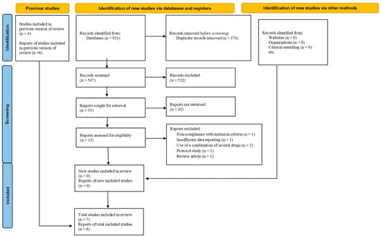
Among 921 results found in the databases, 13 studies were evaluated according to eligibility and presented in full text after the screening. Eventually, seven studies (n = 291) with eligible criteria were included in the current systematic review. In addition, seven studies were excluded for various reasons, namely non-compliance with inclusion criteria [24], insufficient data reporting [25], using a combination of several drugs [26,27], a protocol study [28], and a review article [29]. The data selection process has been represented in the PRISMA diagram (Figure 1).

Figure 1.
PRISMA diagram.
3.2. Studies Characteristics
The included studies were published between 2013 and 2023, of which seven studies were conducted in four countries: Iran [7,17], Egypt [4,5], Brazil [12,18], and India [30]. Two hundred and twenty-seven subjects participated in the intervention group (n = 113) and the control group (n = 114). The average age of participants was between 13 and 31 years old, and the time of amlodipine treatment was between 3 and 12 months. The dose of amlodipine used in the three studies [5,7,18] was 5 mg/day; in the rest [4,12,17,30], it was 2.5 to 5 mg/day. All studies used deferoxamine, deferasirox, and/or deferiprone as iron chelators. The characteristics of the included clinical trials have been illustrated in Table 1.

Table 1.
The characteristics of included clinical trials.
3.3. Outcomes
Serum Ferritin
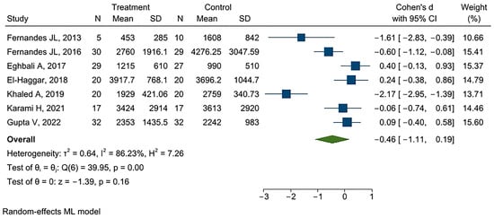
Seven studies provided data for serum ferritin (n = 291) [4,5,12,17,18,30,31]. The pooled Cohen’s d for serum ferritin was estimated at −0.46, 95% CI −1.11 to 0.19, and p = 0.16 (I2 86.23%, p < 0.0001) (Figure 2).

Figure 2.
Forest plot of Cohen’s d for serum ferritin [4,5,7,12,17,18,30].
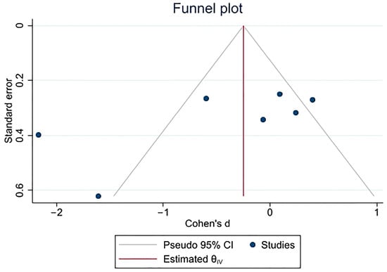
The p-value for the Egger test was gained at 0.02 (β −5.72, SE 2.38), while the trim and fill analysis of the publication bias for ferritin outcome led to no change in the estimated effect size (−0.46, 95% CI −1.17 to 0.24) (Figure 3).

Figure 3.
Funnel plot of Cohen’s d for serum ferritin.
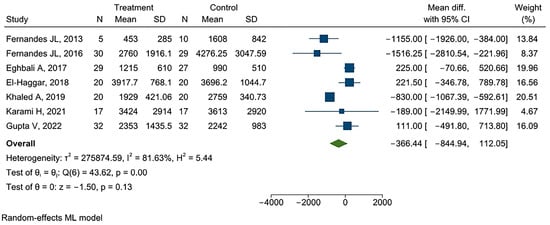
Furthermore, the pooled mean difference for serum ferritin reached −366.44 ng/mL, 95% CI −844.94 to 112.05, and p = 0.13 (I2 81.63%, p < 0.0001). The coefficient for the mean difference was also −23.23 ng/mL, 95% CI −155.21 to 108.75 when a meta-regression operated upon considering the time during which amlodipine was used, R-squared 0.0%, and p = 0.73 (I2 77.13%; p < 0.0001). The coefficient coming from a meta-regression with a dose of amlodipine 5 mg/day than 2.5–5 mg/day was found at −323.49 ng/mL, 95% CI −826.14 to 1473.12, R-squared 0.0%, and p = 0.58 (I2 87.12%; p < 0.0001). I2 remained high once a meta-regression with the baseline values of serum ferritin was implemented (68.20%; p < 0.0001). The coefficient was 1.25 ng/mL and 95% CI 0.15 to 2.35 (R-squared 44.16%, p = 0.07). The Egger test demonstrated a non-significant publication bias (β −0.87, SE 1.117, and p = 0.43). After running the trim-and-fill publication bias analysis, the mean difference turned into −377.23 ng/mL, 95% CI −901.89 to 147.43 (Figure 4 and Figure S1).

Figure 4.
Forest plot of mean difference for serum ferritin [4,5,7,12,17,18,30].
3.4. Quality and Sensitive Analyses
Upon performing the quality analysis, the statistical heterogeneity fell to 24.33% (p = 0.15) when the Cohen’s d for serum ferritin was calculated for the studies with a low risk of bias (−0.2, 95% CI −0.56 to 0.17, and p = 0.15). Three included studies with a moderate risk of bias provided a Cohen’s d of −0.48, 95% CI −1.78 to 0.83 (I2 92.08%, p < 0.0001) (ptest of group difference = 0.09). The combined mean difference for serum ferritin originating from the low-risk-of-bias studies was −380.35 ng/mL, 95% CI −1209.37 to 44.68 (I2 36.51%, p = 0.08), whereas that of for the moderated-risk-of-bias studies valued at −159.87 ng/mL, 95% CI −746.42 to 426.67 (I2 88.99%, p < 0.0001) (ptest of group difference = 0.13) (Figures S2 and S3).
Furthermore, after removing El-Haggar’s study (2018), the only study carried on non-transfusion-dependent thalassemia, the value of Cohen’s d changed to −0.58, 95% CI −1.31 to 0.14; p = 0.11 (I2 86.99%; p < 0.0001). The value of −484.18 ng/mL, 95% CI −1012.01 to 42.65, and p = 0.07 was the mean difference for serum ferritin (I2 82.12%, p < 0.0001) (Figures S4 and S5). When we took the study of Fernandes (2013) aside, the Cohen’s d for serum ferritin reached −0.32, 95% CI −0.98 to 0.34; p = 0.34 (I2 86.66%, p < 0.0001). The mean difference was −232.58 ng/mL, 95% CI −706.60 to 241.44, and p = 0.34 (I2 79.65%; p < 0.0001) (Figures S6 and S7).
3.5. Liver MRI T2*
According to data that originated from two included studies (n = 96) [5,12], the overall Cohen’s d for liver MRI T2* stood at 2.069, 95% CI −0.896 to 5.035, and p = 0.17 (I2 96.31%, p < 0.0001). The estimated effect size did not see changes after the trim-and-fill analysis of publication bias (Figures S8 and S9). The synthesized mean difference for liver MRI T2* was 8.76 msec, 95% CI −4.16 to 21.67, and p = 0.18 (I2 98.38%, p < 0.000). After conducting the trim-and-fill analysis of the publication bias, the estimated mean difference turned out to be the same (Figures S10 and S11).
3.6. Risk of Bias Assessment
Of seven studies, one study was labeled high risk of bias. The number of studies with moderate and low risk of bias was the same as three. The details of the risk of bias assessment are presented in Table 2.

Table 2.
The risk of bias assessment of the included trials.
3.7. Level of Evidence Assessment
The quality of evidence was very low, according to the GRADE methodology (Table 3).

Table 3.
GRADE evidence profile.
4. Discussion
Iron-overload-induced cardiomyopathy resulting from β-thalassemia patients is responsible for considerable cardiovascular morbidity and mortality. Protection against myocardial damage driven by hemosiderosis has been the core determinant that expands the life span in patients with β-thalassemia. Cardiomyopathy, and other complications stemming from iron excess account for nearly 70% of deaths in these individuals.
The study’s results discovered that using amlodipine at the dose of 2.5 to 5 mg/day for a year may subtly cut down serum ferritin levels around 366 ng/mL (very low level of evidence), but the decline was about 380 ng/mL based on the low-risk-of-bias studies. However, the heterogeneity between the included studies remained high even after running the meta-regression based on the dose and duration of amlodipine use. An increase in the length of amlodipine use might lead to a drop in serum ferritin levels as much as 23 ng/mL/month, although this relationship was not statistically significant. Amlodipine at a dose of 5 mg/day was associated with a 320 ng/mL decline in serum ferritin levels compared to those who received 2.5 to 5 mg/day, representing no significant difference in serum ferritin values between the doses. As such, thalassemia cases with iron overload may not take advantage of a greater dose of amlodipine in mitigating ferritin levels. The cases with higher serum ferritin levels may meaningfully benefit more from using amlodipine, according to the estimated mean difference drawn by the baseline serum ferritin values. Intriguingly, the heterogeneity between included studies for serum ferritin descended as the results were narrowed down to only studies with a low risk of bias, although the change was not statistically significant given the low number of studies. Additionally, the findings have cautiously also recommended amlodipine to improve liver MRI T2* around 8.76 msec among thalassemia cases with iron overload, albeit the results suffer from scarcity of the included studies, which points out the necessity of more primary studies in this context.
The effectiveness of amlodipine in reducing ferritin levels has also been demonstrated in past studies. Due to amlodipine’s effect on prolonging DMT1 opening in the kidneys, decreased urinary excretion of iron may explain amlodipine’s reduced ferritin levels [7]. Nevertheless, in some studies, the decline in ferritin was unexpected, suggesting that other relevant factors, such as subsided inflammation, might have contributed to the fall. It is also possible that the chelators can access iron extracellularly as iron atoms are prevented from entering across the sites blocked by amlodipine [12].
Reticuloendothelial macrophages phagocytose red blood cells and release labile cellular iron into the plasma for binding to transferrin. In the liver, heart, and endocrine glands, non-transferrin-bound iron (NTBI) is readily transported through calcium channels, DMT1, and TfR-2 [32]. In clinical studies, CCBs effectively removed iron from the heart in patients with iron overload-associated cardiomyopathy. It has recently been discovered that CCBs have pleiotropic pharmacological properties, including blocking iron and preventing fibrosis [7]. Zhang Y et al., through an animal study, depicted that iron overload led to the abnormal expression of L-type voltage-dependent calcium channels (LVDCC) 1C subunits, DMT-1, and TfR-2 in the liver, ending in iron inflow into hepatocytes. Treatment with CCB could partly rescue the abnormal LVDCC α1C subunit patterns and reduce high levels of DMT-1 expression in the iron-overloaded livers. CCB administration probably depletes liver tissue of Fe2+ as LVDCC and DMT-1 allow only Fe2+ to cross the cell membrane. The treatment of iron-overloaded livers with CCB increased hepcidin expression, indicating that CCB therapy might recover the ability of hepatocytes in order to assess extracellular iron [33].
Rat liver stellate cells can express LTCCs [34]. The iron-overloaded animal model with hepatic fibrosis was treated with CCBs via two interdependent ways, including blocking iron permeation into hepatocytes and inhibiting hepatic stellate cell (HSC) growth. These results are consistent with iron-overloaded mouse livers where CCBs inhibited iron inflow. Amlodipine could, therefore, be a plausible explanation for the reduced iron entrance into hepatocytes. Accordingly, calcium channel blocking with amlodipine may represent an effective treatment for major iron overload in thalassemia [12,34].
Through Fenton-type/Haber–Weiss reactions, free iron triggers H2O2 catabolism, producing various toxic free radicals in biological systems [35]. Reactive oxygen species (ROS), generated in the presence of iron, can incur damage to intracellular organelles and cause cellular dysfunctions, apoptosis, and necrosis in the target organs, leading to toxicity and dysfunctions. The cardiac toxicity arising from free iron is the consequence of highly focused LVDCCs in myocardial tissue [36,37], where the channels malfunction and an impaired excess calcium intake through these portals may occur [36]. As a result, the mitochondrial respiratory chain and subsequent energy metabolism are ultimately impaired, which ends in cardiomyopathy [38]. Several studies indicate that LTCC blockers may deter hemosiderosis by counteracting cellular iron uptake and oxidative stress [39].
Studies on the performance of chelating agents have shown that using these agents has not been sufficiently successful in ameliorating organ iron overload. The underlying reason is that the action mechanism of iron chelators may not be effective enough to excrete surplus iron and block the influx and accumulation of iron. As such, combination therapy with other drugs alongside iron-chelating agents has been argued [9]. CCBs inhibit iron influx through the cited calcium channels [28]. In addition, the potential antioxidant effects of these agents by scavenging free radicals [39,40] and the combination therapy for iron overload as Fe ionophores are also discussed [41]. Compared to other CCBs, amlodipine is inexpensive and safe [42], with a trivial effect on inotropy and atrioventricular function at regular doses [43,44]. Multiple mechanisms have been brought up for CCBs to exert antioxidant properties, including antithrombotic and anti-inflammatory properties. It is plausible that the chemical structure of these L-type CCBs contributes to their antioxidant properties [45,46].
Taking amlodipine reduced oxidative stress and modified cholesterol levels in cholesterol-fed rabbits in an in vivo study. Additionally, oxidative stress markers could be effectively decreased in diabetic patients receiving amlodipine. A trial showed that myeloperoxidase (MPO), an indicator of oxidative stress, was significantly reduced after monotherapy with amlodipine. Previously, we established that amlodipine therapy coupled with routine treatments with iron chelators could effectively benefit iron-overloaded transfusion-dependent thalassemia cases by lessening malondialdehyde (MDA) concentrations and the total antioxidant capacity (TAC) levels. Due to its ability to reduce NTBI uptake, amlodipine may be effective as a potential antioxidant, particularly when iron overload is present [46,47]. One of the underlying mechanisms explaining amlodipine’s antioxidant properties is the control of intracellular calcium overload. The antioxidant property of amlodipine dwindles oxidative stress, reduces the level of ferritin, and may modulate endothelial function.
The studies reported no adverse effects of amlodipine therapy at the dose of 5 mg daily [12,18,31]. In Khaled’s study [5], 20% of β-thalassemia patients treated with amlodipine reported dizziness, and 15% stated swollen ankles. Hypotension was not mentioned in that study in the amlodipine arm. Given the literature, treatment with amlodipine alongside the standard iron chelation therapy might provide an advantageous clinical and cardio-friendly measurement in patients with β-thalassemia, deprived of bringing significant adverse consequences and huge costs.
5. Conclusions
The co-administration of amlodipine and iron chelators probably mildly scales down serum ferritin levels amid iron-overloaded thalassemia patients by roughly 366 ng/mL (23 ng/mL per month). Using amlodipine at a dose of 5 mg/day alongside standard iron chelators might mitigate serum ferritin levels by nearly 320 ng/mL more than the potential decline may be seen in the cases who received 2.5 to 5 mg/day, with a non-significant likelihood. Patients with higher serum ferritin levels may benefit from amlodipine therapy more. Moreover, there is a sign of betterment in liver MRI T2* up to 8.76 msec among amlodipine users with iron-burdened thalassemia patients, which comes from using amlodipine at a dose of 2.5 to 5 mg a day for a year.
As the results are inconclusive and the level of evidence is very low, more well-designed clinical trials in this area need to be invigorated. As well as that, the results may be touched by the potential publication bias. Although the current work could identify the quality of the carried-out studies as the potential source of heterogeneity, the low number of studies needs to be taken into account. More studies must be considered in future analyses to find some potential parameters that are associated with the outcome variations following adjuvant therapy with amlodipine. Because amlodipine is not very effective, the co-administration of amlodipine with compounds that can specifically reduce iron entry into cells, for instance, ebselen—an antioxidant selectively blocking DMT-1—can bring more clinical efficacy to these patients. Furthermore, whether amlodipine therapy has a potential superiority in this performance with a specific iron chelator would be intriguing, which calls for more clinical trials.
Supplementary Materials
The following supporting information can be downloaded at: https://www.mdpi.com/article/10.3390/thalassrep13040021/s1, Figure S1: Funnel plot of mean difference for ferritin; Figure S2: Forest plot of Cohens’ d for serum ferritin based the risk of bias assessment; Figure S3: Forest plot of mean difference for serum ferritin based the risk of bias assessment; Figure S4: Forest plot of Cohen’s d for serum ferritin after removing El-Haggar’s study; Figure S5: Forest plot of mean difference for serum ferritin after removing El-Haggar’s study; Figure S6: Forest plot of Cohen’s d for serum ferritin after removing the study of Fernandes (2013); Figure S7: Forest plot of mean difference for serum ferritin after removing the study of Fernandes (2013); Figure S8: Forest plot of Cohens’ d for liver MRI T2; Figure S9: Funnel plot for liver MRI T2; Figure S10: Forest plot of mean difference for liver MRI T2; Figure S11: Funnel plot of mean difference for liver MRI T2.
Author Contributions
Searching the databases, data extraction phase, and preparing the manuscript draft were performed by A.A., A.N., R.J., H.B.-D., M.Z. and M.G. Statistical analysis was executed by H.D.-K. and S.B. Moreover, H.D.-K., M.N., E.S. and H.K. were responsible for critically appraising and reviewing the manuscript. H.D.-K. and M.K. edited the final manuscript. All authors have read and agreed to the published version of the manuscript.
Funding
This research received no external funding.
Institutional Review Board Statement
Not applicable.
Informed Consent Statement
The study protocol was prospectively discernable in 2021 on ResearchGate (https://www.researchgate.net/publication/352538926).
Data Availability Statement
No new data were created or analyzed in this study. Data sharing is not applicable to this article.
Conflicts of Interest
The authors declare that the research was conducted without any commercial or financial relationships construed as a potential conflict of interest.
References
- Rund, D.; Rachmilewitz, E. β-Thalassemia. N. Engl. J. Med. 2005, 353, 1135–1146. [Google Scholar] [CrossRef]
- De Sanctis, V.; Kattamis, C.; Canatan, D.; Soliman, A.T.; Elsedfy, H.; Karimi, M.; Daar, S.; Wali, Y.; Yassin, M.; Soliman, N.; et al. β-thalassemia distribution in the old world: An ancient disease seen from a historical standpoint. Mediterr. J. Hematol. Infect. Dis. 2017, 9, e2017018. [Google Scholar] [CrossRef]
- Sadaf, A.; Hasan, B.; Das, J.K.; Colan, S.; Alvi, N. Calcium channel blockers for preventing cardiomyopathy due to iron overload in people with transfusion-dependent beta thalassaemia. Cochrane Database Syst. Rev. 2018, 7, CD011626. [Google Scholar] [CrossRef]
- El-Haggar, S.M.; El-Shanshory, M.R.; El-shafey, R.A.; Dabour, M.S. Decreasing cardiac iron overload with Amlodipine and Spirulina in children with β-thalassemia. Pediatr. Hematol. Oncol. J. 2018, 3, 64–69. [Google Scholar] [CrossRef]
- Khaled, A.; Salem, H.A.; Ezzat, D.A.; Seif, H.M.; Rabee, H. A randomized controlled trial evaluating the effects of amlodipine on myocardial iron deposition in pediatric patients with thalassemia major. Drug Des. Dev. Ther. 2019, 13, 2427–2436. [Google Scholar] [CrossRef]
- Borgna-Pignatti, C.; Rugolotto, S.; De Stefano, P.; Zhao, H.; Cappellini, M.D.; Del Vecchio, G.C.; Romeo, M.A.; Forni, G.L.; Gamberini, M.R.; Ghilardi, R.; et al. Survival and complications in patients with thalassemia major treated with transfusion and deferoxamine. Haematologica 2004, 89, 1187–1193. [Google Scholar] [PubMed]
- Eghbali, A.; Kazemi, H.; Taherahmadi, H.; Ghandi, Y.; Rafiei, M.; Bagheri, B. A randomized, controlled study evaluating effects of amlodipine addition to chelators to reduce iron loading in patients with thalassemia major. Eur. J. Haematol. 2017, 99, 577–581. [Google Scholar] [CrossRef]
- Aydinok, Y.; Porter, J.B.; Piga, A.; Elalfy, M.; El-Beshlawy, A.; Kilinç, Y.; Viprakasit, V.; Yesilipek, A.; Habr, D.; Quebe-Fehling, E.; et al. Prevalence and distribution of iron overload in patients with transfusion-dependent anemias differs across geographic regions: Results from the CORDELIA study. Eur. J. Haematol. 2015, 95, 244–253. [Google Scholar] [CrossRef]
- Kontoghiorghes, G.J. Introduction of higher doses of deferasirox: Better efficacy but not effective iron removal from the heart and increased risks of serious toxicities. Expert Opin. Drug Saf. 2010, 9, 633–641. [Google Scholar] [CrossRef]
- Pennell, D.J.; Berdoukas, V.; Karagiorga, M.; Ladis, V.; Piga, A.; Aessopos, A.; Gotsis, E.D.; Tanner, M.A.; Smith, G.C.; Westwood, M.A.; et al. Randomized controlled trial of deferiprone or deferoxamine in beta-thalassemia major patients with asymptomatic myocardial siderosis. Blood 2006, 107, 3738–3744. [Google Scholar] [CrossRef]
- Viprakasit, V.; Gattermann, N.; Lee, J.W.; Porter, J.B.; Taher, A.T.; Habr, D.; Martin, N.; Domokos, G.; Cappellini, M.D. Geographical variations in current clinical practice on transfusions and iron chelation therapy across various transfusion-dependent anaemias. Blood Transfus. 2013, 11, 108. [Google Scholar] [PubMed]
- Fernandes, J.L.; Sampaio, E.F.; Fertrin, K.; Coelho, O.R.; Loggetto, S.; Piga, A.; Verissimo, M.; Saad, S.T. Amlodipine reduces cardiac iron overload in patients with thalassemia major: A pilot trial. Am. J. Med. 2013, 126, 834–837. [Google Scholar] [CrossRef]
- Oudit, G.Y.; Trivieri, M.G.; Khaper, N.; Liu, P.P.; Backx, P.H. Role of L-type Ca2+ channels in iron transport and iron-overload cardiomyopathy. J. Mol. Med. 2006, 84, 349–364. [Google Scholar] [CrossRef]
- Oudit, G.Y.; Sun, H.; Trivieri, M.G.; Koch, S.E.; Dawood, F.; Ackerley, C.; Yazdanpanah, M.; Wilson, G.J.; Schwartz, A.; Liu, P.P.; et al. L-type Ca2+ channels provide a major pathway for iron entry into cardiomyocytes in iron-overload cardiomyopathy. Nat. Med. 2003, 9, 1187–1194. [Google Scholar] [CrossRef]
- Kumfu, S.; Chattipakorn, S.; Chinda, K.; Fucharoen, S.; Chattipakorn, N. T-type calcium channel blockade improves survival and cardiovascular function in thalassemic mice. Eur. J. Haematol. 2012, 88, 535–548. [Google Scholar] [CrossRef]
- Ludwiczek, S.; Theurl, I.; Muckenthaler, M.U.; Jakab, M.; Mair, S.M.; Theurl, M.; Kiss, J.; Paulmichl, M.; Hentze, M.W.; Ritter, M.; et al. Ca2+ channel blockers reverse iron overload by a new mechanism via divalent metal transporter-1. Nat. Med. 2007, 13, 448–454. [Google Scholar] [CrossRef]
- Karami, H.; Khalilzadeh Arjmandi, H.; Salehifar, E.; Darvishi Khezri, H.; Dabirian, M.; Kosaryan, M.; Aliasgharian, A.; Akbarzadeh, R.; Aali, R.N.; Nasirzadeh, A.; et al. A Double-Blind, Controlled, Crossover Trial of Amlodipine on Iron Overload Status in Transfusion Dependent β-Thalassemia Patients. Int. J. Clin. Pract. 2021, 75, e14337. [Google Scholar] [CrossRef]
- Fernandes, J.L.; Loggetto, S.R.; Veríssimo, M.P.; Fertrin, K.Y.; Baldanzi, G.R.; Fioravante, L.A.; Tan, D.M.; Higa, T.; Mashima, D.A.; Piga, A.; et al. A randomized trial of amlodipine in addition to standard chelation therapy in patients with thalassemia major. Blood J. Am. Soc. Hematol. 2016, 128, 1555–1561. [Google Scholar] [CrossRef]
- Elfaituri, M.K.; Ghozy, S.; Ebied, A.; Morra, M.E.; Hassan, O.G.; Alhusseiny, A.; Abbas, A.S.; Sherif, N.A.; Fernandes, J.L.; Huy, N.T. Amlodipine as adjuvant therapy to current chelating agents for reducing iron overload in thalassaemia major: A systematic review, meta-analysis and simulation of future studies. Vox Sang. 2021, 116, 887–897. [Google Scholar] [CrossRef]
- Soliman, Y.; Abdelaziz, A.; Mouffokes, A.; Amer, B.E.; Goudy, Y.M.; Abdelwahab, O.A.; Badawy, M.M.; Diab, R.A.; Elsharkawy, A. Efficacy and safety of calcium channel blockers in preventing cardiac siderosis in thalassemia patients: An updated meta-analysis with trial sequential analysis. Eur. J. Haematol. 2023, 110, 414–425. [Google Scholar] [CrossRef]
- Page, M.J.; McKenzie, J.E.; Bossuyt, P.M.; Boutron, I.; Hoffmann, T.C.; Mulrow, C.D.; Shamseer, L.; Tetzlaff, J.M.; Akl, E.A.; Brennan, S.E.; et al. The PRISMA 2020 statement: An updated guideline for reporting systematic reviews. BMJ 2021, 372, n71. [Google Scholar] [CrossRef]
- Sterne, J.A.; Savović, J.; Page, M.J.; Elbers, R.G.; Blencowe, N.S.; Boutron, I.; Cates, C.J.; Cheng, H.Y.; Corbett, M.S.; Eldridge, S.M.; et al. RoB 2: A revised tool for assessing risk of bias in randomised trials. BMJ 2019, 366, l4898. [Google Scholar] [CrossRef]
- Guyatt, G.; Oxman, A.D.; Akl, E.A.; Kunz, R.; Vist, G.; Brozek, J.; Norris, S.; Falck-Ytter, Y.; Glasziou, P.; DeBeer, H.; et al. GRADE guidelines: 1. Introduction—GRADE evidence profiles and summary of findings tables. J. Clin. Epidemiol. 2011, 64, 383–394. [Google Scholar] [CrossRef]
- Chen, M.-P.; Cabantchik, Z.I.; Chan, S.; Chan GC-f Cheung, Y.-F. Iron overload and apoptosis of HL-1 cardiomyocytes: Effects of calcium channel blockade. PLoS ONE 2014, 9, e112915. [Google Scholar] [CrossRef]
- Chowdhury, D.; Alvi, N.; Tomredle, R.; Bijnens, B.; Hasan, B. Improvements in Regional Myocardial Function on 2D STE With Optimized Chelation in Patients With Thalassemia Major (TM). Circulation 2017, 136 (Suppl. S1), A21067-A. [Google Scholar] [CrossRef]
- Basavaiah, K.; Chandrashekar, U.; Prameela, H. Sensitive spectrophotometric determination of amlodipine and felodipine using iron (III) and ferricyanide. Il Farmaco 2003, 58, 141–148. [Google Scholar] [CrossRef]
- Motta, I.; Scaramellini, N.; Cappellini, M.D. Investigational drugs in phase I and phase II clinical trials for thalassemia. Expert Opin. Investig. Drugs 2017, 26, 793–802. [Google Scholar] [CrossRef]
- Shakoor, A.; Zahoor, M.; Sadaf, A.; Alvi, N.; Fadoo, Z.; Rizvi, A.; Quadri, F.; Tipoo, F.A.; Khurshid, M.; Sajjad, Z.; et al. Effect of L-type calcium channel blocker (amlodipine) on myocardial iron deposition in patients with thalassaemia with moderate-to-severe myocardial iron deposition: Protocol for a randomised, controlled trial. BMJ Open 2014, 4, e005360. [Google Scholar] [CrossRef]
- Manglani, M.V.; Kini, P.S. Management of ß-thalassemia–Consensus and controversies! Pediatr. Hematol. Oncol. J. 2017, 2, 94–97. [Google Scholar] [CrossRef]
- Gupta, V.; Kumar, I.; Raj, V.; Aggarwal, P.; Agrawal, V. Comparison of the effects of calcium channel blockers plus iron chelation therapy versus chelation therapy only on iron overload in children and young adults with transfusion-dependent thalassemia: A randomized double-blind placebo-controlled trial. Pediatr. Blood Cancer 2022, 69, e29564. [Google Scholar] [CrossRef]
- Alali, M.A.; Alanazi, K.M.; Alsayil, S.N.; Omari, Z.; Shaaban, A. Calcium Channel Blockers in Conjunction with Standard Iron-Chelating Agents for β-Thalassemia Major: Systematic Literature Search. Hemoglobin 2020, 44, 446–450. [Google Scholar] [CrossRef]
- Taher, A.T.; Saliba, A.N. Iron overload in thalassemia: Different organs at different rates. Hematology 2017, 2017, 265–271. [Google Scholar] [CrossRef]
- Zhang, Y.; Zhao, X.; Chang, Y.; Zhang, Y.; Chu, X.; Zhang, X.; Liu, Z.; Guo, H.; Wang, N.; Gao, Y.; et al. Calcium channel blockers ameliorate iron overload-associated hepatic fibrosis by altering iron transport and stellate cell apoptosis. Toxicol. Appl. Pharmacol. 2016, 301, 50–60. [Google Scholar] [CrossRef]
- Bataller, R.; Gasull, X.; Ginès, P.; Hellemans, K.; Görbig, M.N.; Nicolás, J.M.; Sancho-Bru, P.; Heras, D.D.L.; Gual, A.; Geerts, A.; et al. In vitro and in vivo activation of rat hepatic stellate cells results in de novo expression of L-type voltage-operated calcium channels. Hepatology 2001, 33, 956–962. [Google Scholar] [CrossRef]
- Byler, R.M.; Sherman, N.A.; Wallner, J.S.; Horwitz, L.D. Hydrogen peroxide cytotoxicity in cultured cardiac myocytes is iron dependent. Am. J. Physiol. Heart Circ. Physiol. 1994, 266, H121–H127. [Google Scholar] [CrossRef]
- Winegar, B.D.; Kelly, R.; Lansman, J.B. Block of current through single calcium channels by Fe, Co, and Ni. Location of the transition metal binding site in the pore. J. Gen. Physiol. 1991, 97, 351–367. [Google Scholar] [CrossRef]
- Tsushima, R.G.; Wickenden, A.D.; Bouchard, R.A.; Oudit, G.Y.; Liu, P.P.; Backx, P.H. Modulation of iron uptake in heart by L-type Ca2+ channel modifiers: Possible implications in iron overload. Circ. Res. 1999, 84, 1302–1309. [Google Scholar] [CrossRef]
- Hershko, C.; Link, G.; Cabantchik, I. Pathophysiology of Iron Overload a. Ann. N. Y. Acad. Sci. 1998, 850, 191–201. [Google Scholar] [CrossRef]
- Sugawara, H.; Tobise, K.; Kikuchi, K. Antioxidant effects of calcium antagonists on rat myocardial membrane lipid peroxidation. Hypertens. Res. 1996, 19, 223–228. [Google Scholar] [CrossRef]
- Sevanian, A.; Shen, L.; Ursini, F. Inhibition of LDL oxidation and oxidized LDL-induced cytotoxicity by dihydropyridine calcium antagonists. Pharm. Res. 2000, 17, 999–1006. [Google Scholar] [CrossRef]
- Savigni, D.L.; Morgan, E.H. Mediation of iron uptake and release in erythroid cells by photodegradation products of nifedipine. Biochem. Pharmacol. 1996, 51, 1701–1709. [Google Scholar] [CrossRef]
- Fares, H.; DiNicolantonio, J.J.; O’Keefe, J.H.; Lavie, C.J. Amlodipine in hypertension: A first-line agent with efficacy for improving blood pressure and patient outcomes. Open Heart 2016, 3, e000473. [Google Scholar] [CrossRef]
- Abernethy, D.R.; Schwartz, J.B. Calcium-antagonist drugs. N. Engl. J. Med. 1999, 341, 1447–1457. [Google Scholar] [CrossRef]
- Dougall, H.T.; McLay, J. A comparative review of the adverse effects of calcium antagonists. Drug Safety 1996, 15, 91–106. [Google Scholar] [CrossRef]
- Masumoto, K.; Takeyasu, A.; Oizumi, K.; Kobayashi, T. Studies of novel 1, 4-dihydropyridine Ca antagonist CS-905. I. Measurement of partition coefficient (log P) by high performance liquid chromatography (HPLC). Yakugaku Zasshi J. Pharm. Soc. Jpn. 1995, 115, 213–220. [Google Scholar] [CrossRef]
- Darvishi-Khezri, H.; Khalilzadeh Arjmandi, H.; Aliasgharian, A.; Shaki, F.; Zahedi, M.; Kosaryan, M.; Karami, H.; Aali, R.N.; Salehifar, E. Amlodipine: Can act as an antioxidant in patients with transfusion-dependent β-thalassemia? A double-blind, controlled, crossover trial. J. Clin. Lab. Anal. 2022, 36, e24752. [Google Scholar] [CrossRef]
- Ackerman, Z.; Oron-Herman, M.; Rosenthal, T.; Pappo, O.; Link, G.; Sela, B.-A.; Grozovski, M. Effects of amlodipine, captopril, and bezafibrate on oxidative milieu in rats with fatty liver. Dig. Dis. Sci. 2008, 53, 777–784. [Google Scholar] [CrossRef]
Disclaimer/Publisher’s Note: The statements, opinions and data contained in all publications are solely those of the individual author(s) and contributor(s) and not of MDPI and/or the editor(s). MDPI and/or the editor(s) disclaim responsibility for any injury to people or property resulting from any ideas, methods, instructions or products referred to in the content. |
© 2023 by the authors. Licensee MDPI, Basel, Switzerland. This article is an open access article distributed under the terms and conditions of the Creative Commons Attribution (CC BY) license (https://creativecommons.org/licenses/by/4.0/).