Case Report

Use of the FLEX 28 Dexamethasone-Eluting Cochlear Implant Electrode in Electric–Acoustic Stimulation: A Case Report

1
Department of Hearing Implant Sciences, Shinshu University School of Medicine, Matsumoto 390-8621, Japan
2
Department of Otorhinolaryngology-Head and Neck Surgery, Shinshu University School of Medicine, Matsumoto 390-8621, Japan
*
Author to whom correspondence should be addressed.
Audiol. Res. 2025, 15(5), 112; https://doi.org/10.3390/audiolres15050112
Submission received: 21 May 2025 / Revised: 4 August 2025 / Accepted: 3 September 2025 / Published: 8 September 2025
(This article belongs to the Section Hearing)

Abstract

Background/Objectives: During and after electric–acoustic stimulation (EAS) surgery (as well as regular cochlear implant surgery), the oral and/or intravenous administration of steroids is recommended to prevent acute inflammatory reactions and subsequent fibrosis. However, the effect does not last long. Therefore, with the hope of providing a sustained effect, a new dexamethasone (DEX)-eluting electrode (FLEX28 DEX) has recently been developed. Methods: A case study was performed at Shinshu University in February 2024 in which a DEX-eluting electrode array was utilized for a patient presenting with high-frequency hearing loss with a defined etiology (hearing loss due to a mitochondrial m.1555A > G variant). Results: Residual hearing was well preserved after EAS surgery, and post-operative impedance field telemetry was maintained at a very low level in contrast with a historical/retrospective control group (FLEX28 electrodes without DEX); therefore, it is expected that post-operative fibrosis will be minimized. Further, it was shown that the DEX-eluting electrode can also be applied to EAS. Conclusions: The DEX-eluting electrode was useful in maintaining post-operative impedance at a very low level, indicating that post-operative fibrosis could be minimized even after EAS surgery.

1. Introduction

Cochlear implantation (CI) is the most important and effective treatment for patients with severe-to-profound sensorineural hearing loss (HL). In addition, the indications for CI have been expanded to patients with ski slope-type HL with residual hearing in lower frequencies and severe-to-profound HL in higher frequencies. Von Ilberg et al. developed the concept of electric–acoustic stimulation (EAS) combining low-frequency stimulation using hearing amplification together with electrical stimulation in the high frequencies [1]. EAS has afforded great benefits to patients with residual hearing [2,3,4,5,6,7] and is now used as a standard treatment option for patients with ski slope-type HL.
Various factors have been reported to affect the post-operative hearing preservation (HP), such as surgical approach, electrode flexibility, electrode insertion speed, insertion depth angle, round window opening size, round window accessibility, steroid administration, implant age, degree of residual hearing, etiology of HL and cochlear duct length [8,9,10,11,12,13,14,15,16,17,18,19,20]. A round window approach is preferable as (1) less drilling reduces acoustic trauma, (2) it ensures electrode insertion into the Scala tympani, and (3) it preserves vestibular function [8]. Indeed, we first demonstrated that HP can be achieved even when the electrode is located under the basilar membrane [8].
In addition, steroid administration is an important factor for HP, with systemic administration used pre- and post-operatively and topical administration also used intra-operatively [21,22,23]. During and after EAS surgery (as well as regular CI surgery), the use of steroids is recommended to prevent acute inflammatory reactions and subsequent fibrosis [24]. Previous studies also described the clinical utility of intra-cochlear steroid administration using an inner ear catheter [25,26,27]. However, the effect is not long-lasting. Therefore, with the hope of providing a sustained effect, a new dexamethasone (DEX)-eluting electrode has recently been developed. The pharmacokinetics of the DEX-eluting electrode have been investigated in preclinical studies, which showed sustained release over many weeks [28,29,30,31]. In addition, several preclinical studies showed favorable outcomes for DEX in reducing electrode insertion trauma as assessed by HP, decreasing impedance change after CI insertion, changing sensory compound action potential, and decreasing fibrosis after electrode insertion, outer hair cell loss and proinflammatory cytokine expression [29,32,33,34,35,36,37]. A feasibility study (Phase I study) indicated that the post-operative impedance of the DEX-eluting electrodes (FLEX28 DEX) is lower and more stable compared to that of the conventional FLEX28 electrodes [38]. Additionally, the feasibility study showed the possibility of preserving residual hearing. To date, however, there have been no reports of the use of DEX-eluting electrodes in patients who meet the criteria for EAS. Here, we first report our experience using DEX-eluting electrodes in a patient with a defined etiology that met the indications for EAS.

2. Materials and Methods

2.1. Case

The patient was a 53-year-old female with progressive HL. She noticed her HL at around age 20 and visited the Department of Otorhinolaryngology at Shinshu University Hospital at the age of 52 due to insufficient hearing with hearing aids. Social health insurance-based genetic testing [39] for this patient was conducted in 2023. Genetic testing was performed as a part of research approved by the Shinshu University Ethical Committee (approval number: No. 717—8 March 2022). The study protocol for EAS surgery and assessment using FLEX28 DEX (MED-EL, Innsbruck, Austria) was approved by the Shinshu University Certified Review Board of Clinical Research (approval number: No. 23008—5 December 2023) and was conducted in accordance with the Declaration of Helsinki. Informed consent was obtained prior to enrollment in this study.

2.2. Control Subjects

A control group was used to understand the positioning of the data obtained from this case. We used two control subjects to compare the post-operative HP and post-operative impedance field telemetry (IFT) change. (1) For HP, we used the average pre-operative and 12-month post-operative audiograms of 22 EAS patients. All of the 22 cases received EAS surgery using conventional FLEX28 (without DEX-eluting function). The average age of control group 1 at cochlear implantation was 35.5 ± 19.1 y.o. (range from 8 to 65). Seven subjects were male and fifteen subjects were female. Five cases had CDH23-associated HL, two cases had SLC26A4-associated HL and one case each had KCNQ4-associated HL and LOXHD1-associated HL. We could not identify any genetic cause for 13 cases. All subjects in this control group fulfilled the criteria for conventional EAS (≤65 dB HL in 125, 250 and 500 Hz, ≥80 dB HL in 2000 Hz and ≥85 dB HL in 4000 and 8000 Hz) in terms of preoperative hearing level. (2) For post-operative IFT, IFT data from 15 patients using conventional FLEX28 electrodes (without DEX-eluting function) were used. The average age of control group 2 at cochlear implantation was 42.0 ± 27.1 y.o. (range from 1 to 82). Five subjects were male and ten subjects were female.

2.3. Clinical Evaluation

Evaluation of hearing thresholds was performed using pure-tone audiometry pre-operatively and at 1, 3, 6, and 12 months after EAS surgery. Audiometric thresholds at four frequencies (0.5, 1, 2, and 4 kHz) were used to calculate the pure-tone average (PTA). Hearing thresholds with EAS were evaluated using free-field audiometry at 1, 3, 6, and 12 months after EAS surgery. Speech audiometry, including the Japanese monosyllable perception test (iCI2004 monosyllable) in quiet conditions, representing 65dBSPL, and the Japanese word recognition test (iCI2004 word), representing 65dBSPL in S/N +10 dB conditions, was performed at 1, 3, 6, and 12 months after EAS surgery [40].
IFT was performed to measure electrode impedance (kΩ) for all 12 channels intra-operatively and at 1, 3, 6, and 12 months after EAS surgery. Impedance values were obtained using the MAESTRO software package (Version 9, MED-EL, Innsbruck, Austria). The severity of HL was classified into four categories: mild (PTA > 25 dB and ≤40 dB HL), moderate (>40 dB and ≤70 dB HL), severe (>70 dB and ≤90 dB HL), and profound (>90 dB).

3. Results

3.1. Clinical Details of the Patient

The patient was a 53 y.o. female. She represented a case of sporadic HL without any other affected family members. Her audiogram showed precipitous high-frequency HL with residual HL in lower frequencies. Comprehensive genetic testing using next-generation sequencing identified the genetic cause of her HL as a mitochondrial m.1555A > G variant (Figure 1).
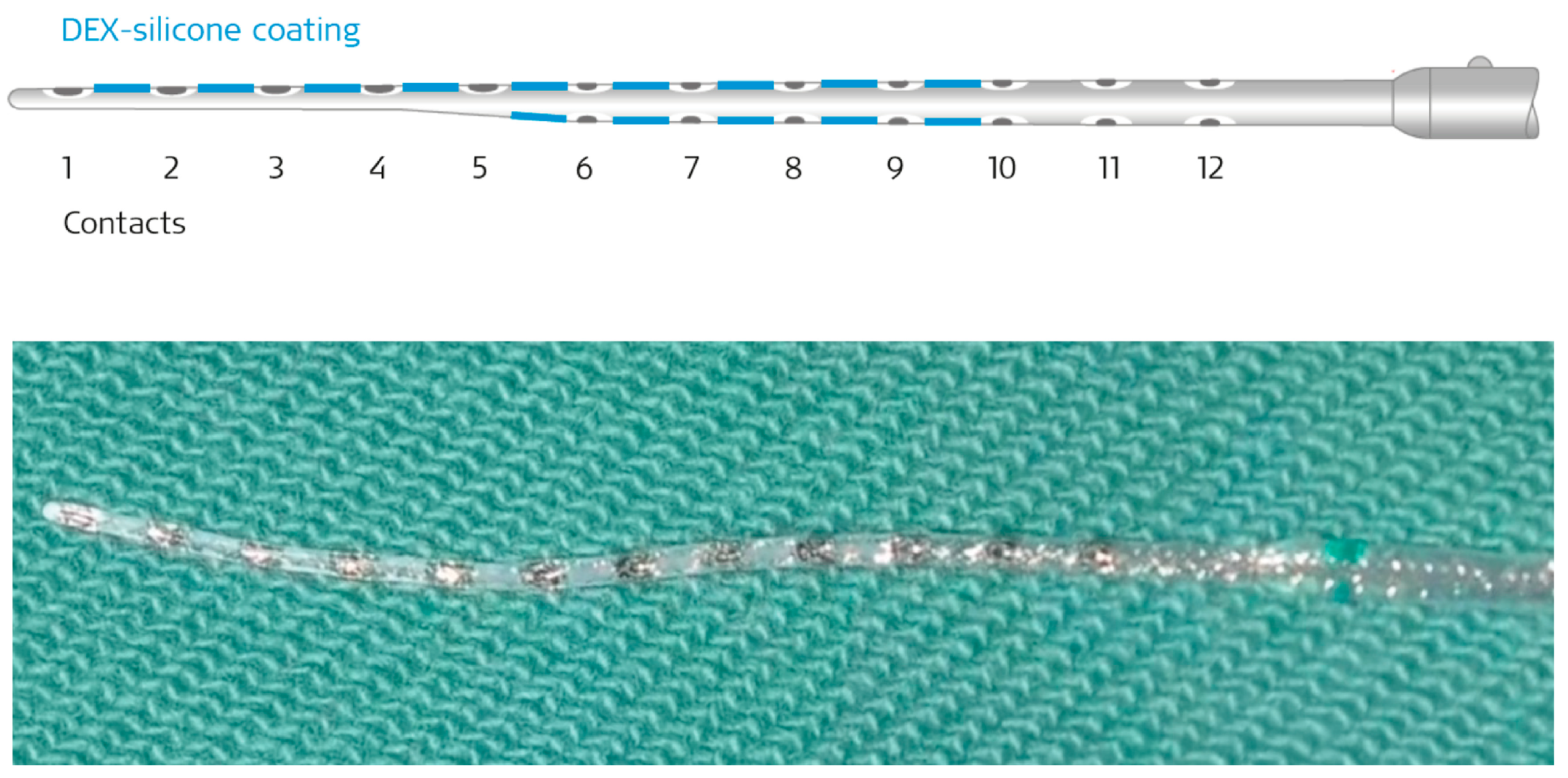
The patient wanted to receive EAS. With the hope of providing a sustained effect to reduce post-operative fibrosis surrounding the inserted electrode, and to preserve residual hearing at the lower frequencies, a new DEX-eluting electrode (FLEX28 DEX electrode array, MED-EL, Innsbruck, Austria) was used as part of a clinical trial in February 2024 (Figure 2).

3.2. Surgery and Implantation

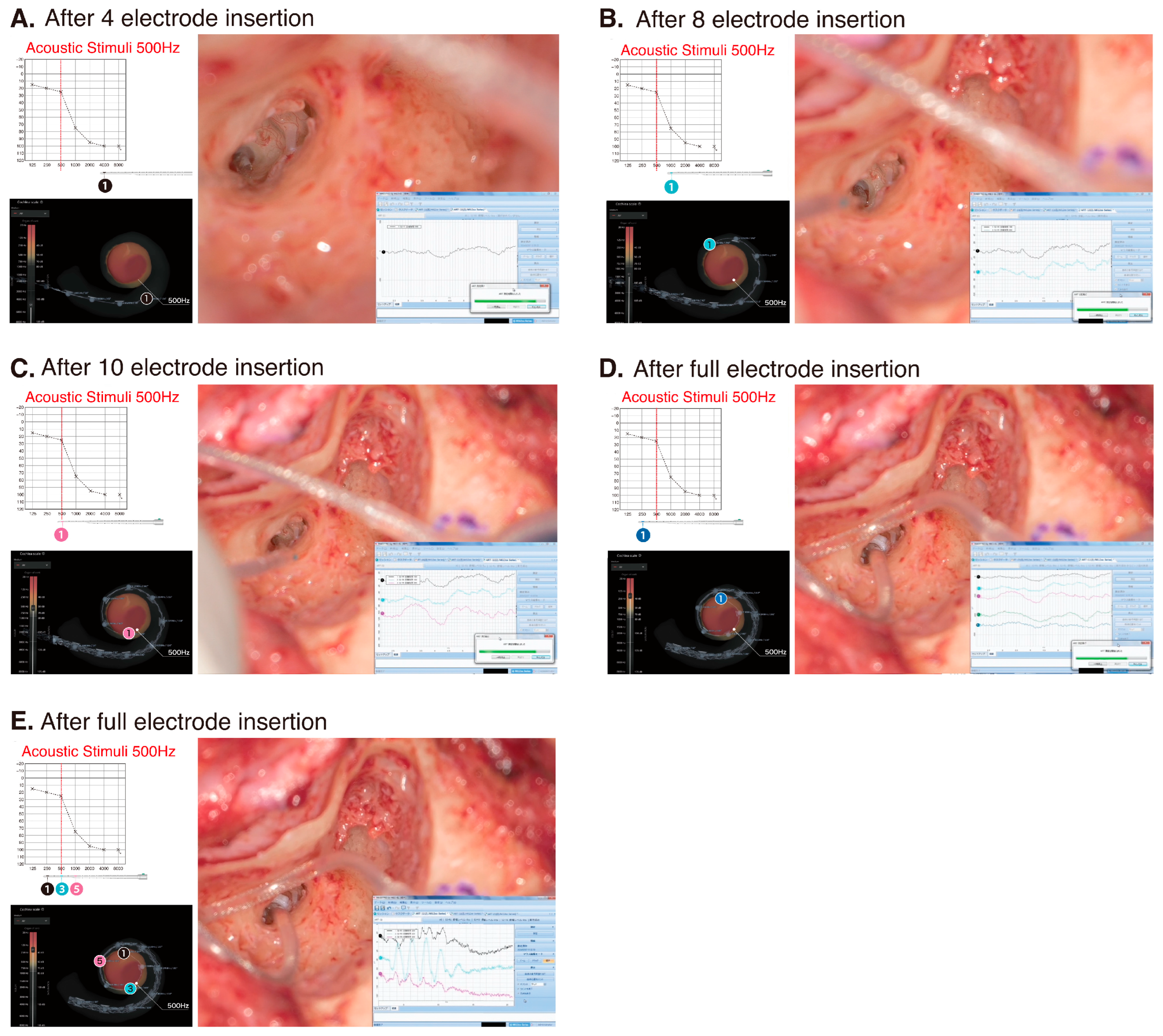
The patient underwent a less-invasive surgical procedure for HP as described previously [7]. The round window membrane was widely exposed by removing the bony over-hang of the round window niche with a low-speed drill. Subsequently, the electrode was slowly inserted via the round window membrane. During electrode insertion, intraoperative cochlear microphonics (CM) monitoring was performed to confirm low-frequency functional preservation and to determine the most appropriate electrode position (Figure 3). During surgery, a 500 Hz acoustic stimulus was applied to the external auditory canal, and the CM response was recorded from the first electrode (Ch 1). The maximum response was obtained when the first electrode reached the area responsible for 500 Hz (Figure 3C). A video of the surgical procedure is available as Supplemental Materials.

3.3. Post-Operative Hearing Outcome and Impedance Change

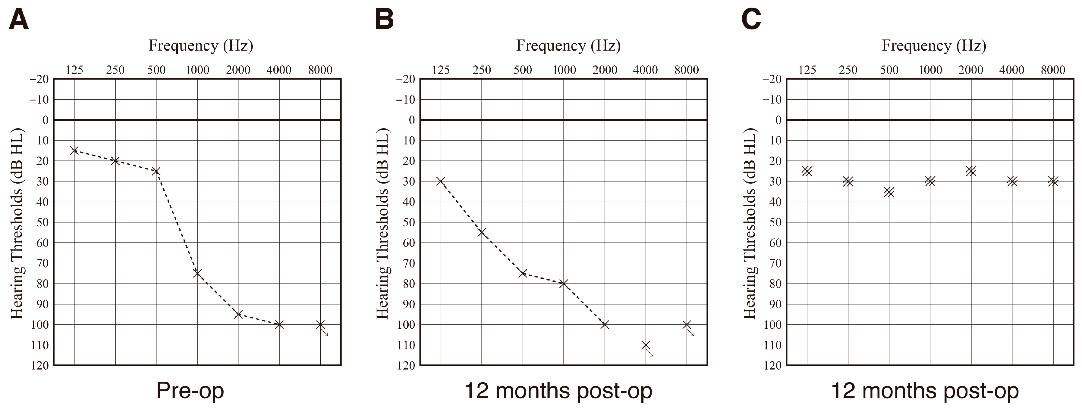
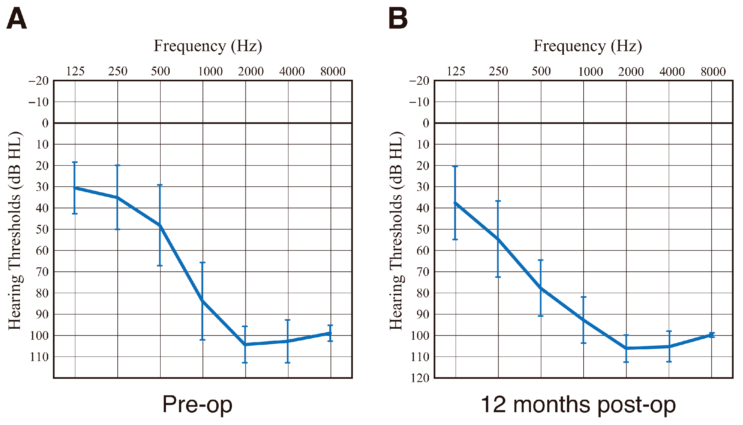
Based on the HP criteria [41], HP in this case was regarded as “partial (58.6%).” After electrode insertion, residual hearing available for acoustic stimulation was well preserved. Audiograms taken pre-operatively and at 12 months post-operatively are shown in Figure 4A,B. HP for this patient was comparable with that for 22 EAS patients using conventional FLEX28 electrodes (Figure 5). Hearing level with EAS is around 25–35 dB (Figure 4C). In addition, Japanese speech audiometry (iCI2004) also improved to 72% (monosyllable perception in quiet) and 92% (word recognition in SN + 10 dB).
The impedance measured by IFT of the patient with the DEX-eluting electrode (FLEX28 DEX) was lower and quite stable compared to that of patients with the conventional FLEX28 electrode (Figure 6). Therefore, it is expected that post-operative fibrosis will be minimized.

4. Discussion

Pre- and post-operative systemic and/or topical steroid administration is an important factor in facilitating HP [21,22,23,24]. In addition, intra-cochlear steroid administration is also performed via an inner ear catheter [25,26,27]. A double-blinded placebo-controlled trial for intra-operative systemic methylprednisolone has been performed, but no statistical differences were observed between the treatment and placebo control groups [42]; however, several clinical papers report positive outcomes for pre- and post-operative systemic and/or topical steroid administration and such steroid administration is recommended. To provide sustained steroid administration, the DEX-eluting electrode was developed. Several preclinical studies showed the DEX-eluting electrode to be useful in reducing electrode insertion trauma as assessed by HP, decreasing impedance change after CI insertion, changing compound action potential, and decreasing fibrosis after electrode insertion, outer hair cell loss and proinflammatory cytokine expression [29,32,33,34,35,36,37]. Intracochlear sustained dexamethasone release in combination with CI in two patients has been reported as a proof of concept with regard to the reduction of impedance [43]. The sustained dexamethasone release from the electrode is also useful in maintaining lower impedance after CI [44]. Recently the results of a phase I study have been reported, with the post-operative impedance of the DEX-eluting electrodes (FLEX28 DEX) shown to be lower and more stable compared to that of conventional FLEX28 electrodes [38].
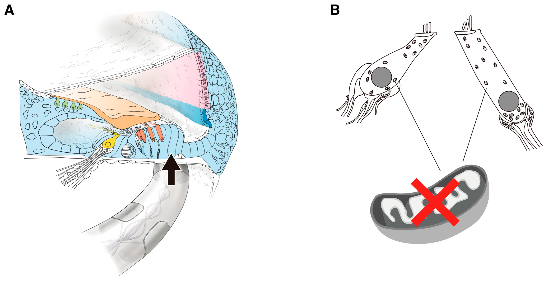
The mitochondrial m.1555A > G variant is one of the most prevalent genetic causes of maternally inherited HL among the pathogenic variants in mitochondrial DNA that is identified in different populations [39,45,46,47,48,49,50,51,52]. Additionally, it is recognized to increase the susceptibility to aminoglycoside antibiotics and the risk of aminoglycoside-induced HL [45,52,53]. In general, aminoglycoside-induced HL is bilateral and severe-to-profound in all frequencies, occurring within several days after the administration of aminoglycoside antibiotics [45]. However, HL is sometimes seen in patients with no known history of aminoglycoside administration, although in these cases the HL is usually milder and involves higher frequencies [45,52]. Mitochondria are known as organelles that produce energy (adenosine triphosphate; ATP) necessary for hearing, but mutated mitochondria show a decrease in energy production. As a result, there is a lack of energy to convert sound vibrations to electrical signals, leading to HL. As the latter group of patients usually have residual hearing at the lower frequencies, they are good candidates for EAS [54,55].
Understanding the etiology allows us to imagine what is happening inside the cochlea. With regard to possible phenomena occurring in the cochlea during electrode insertion in patients with the mitochondrial m.1555A > G variant, the following factors may result in HL due to electrode insertion: (1) electrodes inhibit the movement of the basilar membrane, and (2) mutated mitochondria cause a decrease in energy production. If inflammation induced by electrode insertion is added to this condition, the lack of energy can lead to even more severe hearing deterioration. For the second factor, DEX-eluting electrodes can be used to prevent reactions mediated by inflammation (Figure 7).
With regard to the residual hearing at lower frequencies, many factors, including age at which EAS was performed, individual cochlear volume, electrodes used, and surgical method, have been reported to be involved [8,9,10,11,12,13,14,15,16,17,18,19,20]. Of these, damage occurs within the cochlea and hearing deteriorates with the use of rigid electrodes or when cochleostomy or the extended RW approach is used. However, when flexible electrodes are used and inserted via the RW approach, there may be individual differences in HP due to differences in the position of the electrode for each case (first factor) and differences in the pathological inflammatory response (second factor) mentioned above. Of course, there is potential for inner ear trauma and deteriorated residual hearing as a result of a pathological inflammatory response.
In this clinical case, the residual hearing available for acoustic stimulation was well preserved (Figure 4). HP was comparable to that of the 22 EAS patients using conventional FLEX28 electrodes (Figure 5). Intraoperative CM monitoring was found to be useful for HP (i.e., monitoring whether outer hair cells are viable) and for confirming optimal electrode positioning. The maximum response was obtained when the first electrode reached the area responsible for 500 Hz, proving that these monitoring techniques were performed accurately [56,57]. The concept underlying HP includes minimizing operation-associated trauma and enhancing the preservation of cochlear structures, which is important in expanding future treatment strategies such as gene therapy or regeneration therapy.
The most remarkable result was the change in post-operative impedance over time. As shown in Figure 6, the impedance of the DEX-eluting electrode (FLEX28 DEX) is very low and more stable compared to that of the conventional FLEX28 electrode. As the impedance (resistance value when a current is passed through the electrode) is believed to be a marker of post-operative fibrosis surrounding the inserted electrode [35], it is expected that post-operative fibrosis will be minimized and that there will be a positive long-term effect on hearing performance with CI/EAS. The fact that the DEX-eluting electrode also had a favorable effect on EAS in this study suggests that it can also be applied (instead of conventional electrodes) for all patients in the near future.

5. Conclusions

In this study, we clearly showed the clinical utility of DEX-eluting electrodes, particularly in terms of achieving lower and more stable post-operative impedance. Our results indicate an expected reduction in post-operative fibrosis surrounding the inserted electrode. Thus, the DEX-eluting electrodes can be applied (instead of conventional electrodes) for all CI/EAS patients in the near future.

Supplementary Materials

The following supporting information can be downloaded at https://www.mdpi.com/article/10.3390/audiolres15050112/s1, Video S1: Surgical procedure of FLEX28 DEX electrode for EAS.

Author Contributions

Conceptualization, S.-i.U.; methodology, S.-i.U.; investigation, S.-i.U.; writing—original draft preparation, S.-i.U.; writing—review and editing, S.-i.U., Y.T., H.Y. and S.-y.N.; visualization, S.-y.N.; supervision, S.-i.U.; project administration, S.-i.U.; funding acquisition, S.-i.U. All authors have read and agreed to the published version of the manuscript.

Funding

This study was funded by a Health and Labor Sciences Research Grant for Research on Rare and Intractable Diseases and Comprehensive Research on Disability Health and Welfare from the Ministry of Health, Labor and Welfare of Japan (S.U. 23FC10149), and Grants-in-Aid from the Japan Agency for Medical Research and Development (AMED) (S.U. JP24ek0109741).

Institutional Review Board Statement

The study was conducted according to the guidelines of the Declaration of Helsinki and was approved by the Institutional Ethics Committee of Shinshu University School of Medicine (No. 387—4 September 2012 and No. 576—2 May 2017) and Shinshu University Certified Review Board of Clinical Research (approval number: No. 23008 and approval date: 5 December 2023).

Informed Consent Statement

Written informed consent was obtained from the patient for the publication of this paper.

Data Availability Statement

The original contributions presented in this study are included in the article/supplementary material. Further inquiries can be directed to the corresponding author.

Acknowledgments

We thank the patient who participated in this study. We also thank Soren Schilp and Stefano Morettini for providing us with valuable technical information.

Conflicts of Interest

MED-EL provided the cochlear implant system (Mi1250 SYNCHRONY 2 cochlear implant with a FLEX28 DEX electrode array and audio-processor) along with technical support.

References

  1. Von Ilberg, C.A.; Baumann, U.; Kiefer, J.; Tillein, J.; Adunka, O.F. Electric-acoustic stimulation of the auditory system: A review of the first decade. Audiol. Neurotol. 2011, 16 (Suppl. S2), 1–30. [Google Scholar] [CrossRef] [PubMed]
  2. Gstoettner, W.; Helbig, S.; Settevendemie, C.; Baumann, U.; Wagenblast, J.; Arnoldner, C. A new electrode for residual hearing preservation in cochlear implantation: First clinical results. Acta Oto-Laryngol. 2009, 129, 372–379. [Google Scholar]
  3. Prentiss, S.; Sykes, K.; Staecker, H. Partial deafness cochlear implantation at the University of Kansas: Techniques and outcomes. J. Am. Acad. Audiol. 2010, 21, 197–203. [Google Scholar] [CrossRef]
  4. Helbig, S.; Van de Heyning, P.; Kiefer, J.; Baumann, U.; Kleine-Punte, A.; Brockmeier, H.; Anderson, I.; Gstoettner, W. Combined electric acoustic stimulation with the PULSARCI(100) implant system using the FLEX(EAS) electrode array. Acta Oto-Laryngol. 2011, 131, 585–595. [Google Scholar] [CrossRef]
  5. Tamir, S.; Ferrary, E.; Borel, S.; Sterkers, O.; Bozorg Grayeli, A. Hearing preservation after cochlear implantation using deeply inserted flex atraumatic electrode arrays. Audiol. Neurotol. 2012, 17, 331–337. [Google Scholar] [CrossRef]
  6. Adunka, O.F.; Dillon, M.T.; Adunka, M.C.; King, E.R.; Pillsbury, H.C.; Buchman, C.A. Hearing preservation and speech perception outcomes with electric-acoustic stimulation after 12 months of listening experience. Laryngoscope 2013, 123, 2509–2515. [Google Scholar] [CrossRef]
  7. Usami, S.; Moteki, H.; Tsukada, K.; Miyagawa, M.; Nishio, S.Y.; Takumi, Y.; Iwasaki, S.; Kumakawa, K.; Naito, Y.; Takahashi, H.; et al. Hearing preservation and clinical outcome of 32 consecutive electric acoustic stimulation (EAS) surgeries. Acta OtoLaryngol. 2014, 134, 717–727. [Google Scholar]
  8. Usami, S.; Moteki, H.; Suzuki, N.; Fukuoka, H.; Miyagawa, M.; Nishio, S.Y.; Takumi, Y.; Iwasaki, S.; Jolly, C. Achievement of hearing preservation in the presence of an electrode covering the residual hearing region. Acta Oto-Laryngol. 2011, 131, 405–412. [Google Scholar] [CrossRef]
  9. Carlson, M.L.; Driscoll, C.L.; Gifford, R.H.; Service, G.J.; Tombers, N.M.; Hughes-Borst, B.J.; Neff, B.A.; Beatty, C.W. Implications of minimizing trauma during conventional cochlear implantation. Otol. Neurotol. 2011, 32, 962–968. [Google Scholar] [CrossRef] [PubMed]
  10. Cosetti, M.K.; Friedmann, D.R.; Zhu, B.Z.; Heman-Ackah, S.E.; Fang, Y.; Keller, R.G.; Shapiro, W.H.; Roland, J.T., Jr.; Waltzman, S.B. The effects of residual hearing in traditional cochlear implant candidates after implantation with a conventional electrode. Otol. Neurotol. 2013, 34, 516–521. [Google Scholar] [CrossRef] [PubMed]
  11. Rajan, G.P.; Kontorinis, G.; Kuthubutheen, J. The effects of insertion speed on inner ear function during cochlear implantation: A comparison study. Audiol. Neurotol. 2013, 18, 17–22. [Google Scholar] [CrossRef]
  12. Hochmair, I.; Hochmair, E.; Nopp, P.; Waller, M.; Jolly, C. Deep electrode insertion and sound coding in cochlear implants. Hear. Res. 2015, 322, 14–23. [Google Scholar] [CrossRef]
  13. Mirsalehi, M.; Mohebbi, S.; Ghajarzadeh, M.; Lenarz, T.; Majdani, O. Impact of the round window membrane accessibility on hearing preservation in adult cochlear implantation. Eur. Arch. Otorhinolaryngol. 2017, 274, 3049–3056. [Google Scholar] [CrossRef] [PubMed]
  14. Wanna, G.B.; O’Connell, B.P.; Francis, D.O.; Gifford, R.H.; Hunter, J.B.; Holder, J.T.; Bennett, M.L.; Rivas, A.; Labadie, R.F.; Haynes, D.S. Predictive factors for short- and long-term hearing preservation in cochlear implantation with conventional-length electrodes. Laryngoscope 2018, 128, 482–489. [Google Scholar] [CrossRef]
  15. Helbig, S.; Adel, Y.; Leinung, M.; Stöver, T.; Baumann, U.; Weissgerber, T. Hearing Preservation Outcomes After Cochlear Implantation Depending on the Angle of Insertion: Indication for Electric or Electric-Acoustic Stimulation. Otol. Neurotol. 2018, 39, 834–841. [Google Scholar] [CrossRef] [PubMed]
  16. Gotamco, G.L.; Sun, C.H.; Chou, Y.F.; Hsu, C.J.; Wu, H.P. Effect of Round Window Opening Size on Residual Hearing Preservation in Cochlear Implantation. Otolaryngol. Head Neck Surg. 2020, 163, 375–381. [Google Scholar] [CrossRef] [PubMed]
  17. Yoshimura, H.; Moteki, H.; Nishio, S.Y.; Miyajima, H.; Miyagawa, M.; Usami, S.I. Genetic testing has the potential to impact hearing preservation following cochlear implantation. Acta Otolaryngol. 2020, 140, 438–444. [Google Scholar] [CrossRef]
  18. Lee, G.; Lee, S.; Chung, J.H.; Choi, J.W. Preimplant Hearing Threshold: An Important Predictor of Hearing Preservation in Cochlear Implantation With Lateral Wall Electrodes. Otol. Neurotol. 2021, 42, e145–e152. [Google Scholar] [CrossRef]
  19. Hollis, E.S.; Canfarotta, M.W.; Dillon, M.T.; Rooth, M.A.; Bucker, A.L.; Dillon, S.A.; Young, A.; Quinones, K.; Pillsbury, H.C.; Dedmon, M.M.; et al. Initial Hearing Preservation Is Correlated With Cochlear Duct Length in Fully-inserted Long Flexible Lateral Wall Arrays. Otol. Neurotol. 2021, 42, 1149–1155. [Google Scholar] [CrossRef]
  20. Sladen, M.; Nichani, J.; Kluk-de Kort, K.; Saeed, H.; Bruce, I.A. Outcomes of attempted hearing preservation after cochlear implantation (HPCI): A prognostic factor (PF) systematic review of the literature. Cochlear Implants Int. 2025, 26, 12–29. [Google Scholar] [CrossRef]
  21. Rajan, G.P.; Kuthubutheen, J.; Hedne, N.; Krishnaswamy, J. The role of preoperative, intratympanic glucocorticoids for hearing preservation in cochlear implantation: A prospective clinical study. Laryngoscope 2012, 122, 190–195. [Google Scholar] [CrossRef]
  22. Cho, H.S.; Lee, K.Y.; Choi, H.; Jang, J.H.; Lee, S.H. Dexamethasone Is One of the Factors Minimizing the Inner Ear Damage from Electrode Insertion in Cochlear Implantation. Audiol. Neurotol. 2016, 21, 178–186. [Google Scholar] [CrossRef] [PubMed]
  23. Kuthubutheen, J.; Joglekar, S.; Smith, L.; Friesen, L.; Smilsky, K.; Millman, T.; Ng, A.; Shipp, D.; Coates, H.; Arnoldner, C.; et al. The Role of Preoperative Steroids for Hearing Preservation Cochlear Implantation: Results of a Randomized Controlled Trial. Audiol. Neurotol. 2017, 22, 292–302. [Google Scholar] [CrossRef]
  24. Braun, S.; Ye, Q.; Radeloff, A.; Kiefer, J.; Gstoettner, W.; Tillein, J. Protection of inner ear function after cochlear implantation: Compound action potential measurements after local application of glucocorticoids in the guinea pig cochlea. ORL J. Otorhinolaryngol. Relat. Spec. 2011, 73, 219–228. [Google Scholar] [CrossRef] [PubMed]
  25. Prenzler, N.K.; Salcher, R.; Timm, M.; Gaertner, L.; Lenarz, T.; Warnecke, A. Intracochlear administration of steroids with a catheter during human cochlear implantation: A safety and feasibility study. Drug Deliv. Transl. Res. 2018, 8, 1191–1199. [Google Scholar] [CrossRef] [PubMed]
  26. Prenzler, N.K.; Salcher, R.; Lenarz, T.; Gaertner, L.; Warnecke, A. Dose-Dependent Transient Decrease of Impedances by Deep Intracochlear Injection of Triamcinolone with a Cochlear Catheter Prior to Cochlear Implantation-1 Year Data. Front. Neurol. 2020, 11, 258. [Google Scholar] [CrossRef]
  27. Prenzler, N.K.; Salcher, R.; Lenarz, T.; Gaertner, L.; Lesinski-Schiedat, A.; Warnecke, A. Deep intracochlear injection of triamcinolone-acetonide with an inner ear catheter in patients with residual hearing. Front. Neurosci. 2023, 17, 1202429. [Google Scholar] [CrossRef] [PubMed]
  28. Farahmand Ghavi, F.; Mirzadeh, H.; Imani, M.; Jolly, C.; Farhadi, M. Corticosteroid-releasing cochlear implant: A novel hybrid of biomaterial and drug delivery system. J. Biomed. Mater. Res. Part B Appl. Biomater. 2010, 94, 388–398. [Google Scholar] [CrossRef]
  29. Liu, Y.; Jolly, C.; Braun, S.; Stark, T.; Scherer, E.; Plontke, S.K.; Kiefer, J. In vitro and in vivo pharmacokinetic study of a dexamethasone-releasing silicone for cochlear implants. Eur. Arch. Otorhinolaryngol. 2016, 273, 1745–1753. [Google Scholar] [CrossRef]
  30. Bas, E.; Bohorquez, J.; Goncalves, S.; Perez, E.; Dinh, C.T.; Garnham, C.; Hessler, R.; Eshraghi, A.A.; Van De Water, T.R. Electrode array-eluted dexamethasone protects against electrode insertion trauma induced hearing and hair cell losses, damage to neural elements, increases in impedance and fibrosis: A dose response study. Hear Res. 2016, 337, 12–24. [Google Scholar] [CrossRef]
  31. Liebau, A.; Schilp, S.; Mugridge, K.; Schön, I.; Kather, M.; Kammerer, B.; Tillein, J.; Braun, S.; Plontke, S.K. Long-term in vivo release profile of dexamethasone-loaded silicone rods implanted into the cochlea of guinea pigs. Front. Neurol. 2019, 10, 1377. [Google Scholar] [CrossRef]
  32. Vivero, R.J.; Joseph, D.E.; Angeli, S.; He, J.; Chen, S.; Eshraghi, A.A.; Balkany, T.J.; Van de Water, T.R. Dexamethasone base conserves hearing from electrode trauma-induced hearing loss. Laryngoscope 2008, 118, 2028–2035. [Google Scholar] [CrossRef] [PubMed]
  33. Niedermeier, K.; Braun, S.; Fauser, C.; Straubinger, R.K.; Stark, T. Pneumococcal meningitis post cochlear implantation: Development of an animal model in the guinea pig. Hear Res. 2012, 289, 108–115. [Google Scholar] [CrossRef]
  34. Takumi, Y.; Nishio, S.Y.; Mugridge, K.; Oguchi, T.; Hashimoto, S.; Suzuki, N.; Iwasaki, S.; Jolly, C.; Usami, S. Gene expression pattern after insertion of dexamethasone-eluting electrode into the guinea pig cochlea. PLoS ONE. 2014, 9, e110238. [Google Scholar] [CrossRef]
  35. Wilk, M.; Hessler, R.; Mugridge, K.; Jolly, C.; Fehr, M.; Lenarz, T.; Scheper, V. Impedance Changes and Fibrous Tissue Growth after Cochlear Implantation Are Correlated and Can Be Reduced Using a Dexamethasone Eluting Electrode. PLoS ONE. 2016, 11, e0147552. [Google Scholar] [CrossRef] [PubMed]
  36. Honeder, C.; Zhu, C.; Gausterer, J.C.; Schöpper, H.; Ahmadi, N.; Saidov, N.; Nieratschker, M.; Gabor, F.; Arnoldner, C. Sustained-Release Triamcinolone Acetonide Hydrogels Reduce Hearing Threshold Shifts in a Model for Cochlear Implantation with Hearing Preservation. Audiol. Neurotol. 2019, 24, 237–244. [Google Scholar] [CrossRef]
  37. Manrique-Huarte, R.; Zulueta-Santos, C.; Calavia, D.; Linera-Alperi, M.Á.; Gallego, M.A.; Jolly, C.; Manrique, M. Cochlear Implantation With a Dexamethasone Eluting Electrode Array: Functional and Anatomical Changes in Non-Human Primates. Otol. Neurotol. 2020, 41, e812–e822. [Google Scholar] [CrossRef] [PubMed]
  38. Prenzler, N.; Salcher, R.; Büchner, A.; Warnecke, A.; Kley, D.; Batsoulis, C.; Vormelcher, S.; Mitterberger-Vogt, M.; Morettini, S.; Schilp, S.; et al. Cochlear implantation with a dexamethasone-eluting electrode array: First-in-human safety and performance results. Hear Res. 2025, 461, 109255. [Google Scholar] [CrossRef]
  39. Usami, S.I.; Nishio, S.Y. The genetic etiology of hearing loss in Japan revealed by the social health insurance-based genetic testing of 10K patients. Hum. Genet. 2022, 141, 665–681. [Google Scholar] [CrossRef]
  40. Nishio, S.Y.; Tono, T.; Iwaki, T.; Moteki, H.; Suzuki, K.; Tsushima, Y.; Kashio, A.; Akamatsu, Y.; Sato, H.; Yaegashi, K.; et al. Development and validation of an iPad-based Japanese language monosyllable speech perception test (iCI2004 monosyllable). Acta Otolaryngol. 2021, 141, 267–272. [Google Scholar] [CrossRef]
  41. Skarzynski, H.; van de Heyning, P.; Agrawal, S.; Arauz, S.L.; Atlas, M.; Baumgartner, W.; Caversaccio, M.; de Bodt, M.; Gavilan, J.; Godey, B.; et al. Towards a consensus on a hearing preservation classification system. Acta Otolaryngol. Suppl. 2013, 133, 3–13. [Google Scholar] [CrossRef]
  42. O’Leary, S.J.; Choi, J.; Brady, K.; Matthews, S.; Ozdowska, K.B.; Payne, M.; McLean, T.; Rousset, A.; Lo, J.; Creber, N.; et al. Systemic methylprednisolone for hearing preservation during cochlear implant surgery: A double blinded placebo-controlled trial. Hear Res. 2021, 404, 108224. [Google Scholar] [CrossRef]
  43. Plontke, S.K.; Götze, G.; Rahne, T.; Liebau, A. Intracochlear drug delivery in combination with cochlear implants: Current aspects. HNO 2017, 65 (Suppl. S1), 19–28. [Google Scholar] [CrossRef] [PubMed]
  44. Briggs, R.; O’Leary, S.; Birman, C.; Plant, K.; English, R.; Dawson, P.; Risi, F.; Gavrilis, J.; Needham, K.; Cowan, R. Comparison of electrode impedance measures between a dexamethasone-eluting and standard Cochlear™ Contour Advance® electrode in adult cochlear implant recipients. Hear Res. 2020, 390, 107924. [Google Scholar] [CrossRef]
  45. Usami, S.; Abe, S.; Kasai, M.; Shinkawa, H.; Moeller, B.; Kenyon, J.B.; Kimberling, W.J. Genetic and clinical features of sensorineural hearing loss associated with the 1555 mitochondrial mutation. Laryngoscope 1997, 107, 483–490. [Google Scholar] [CrossRef]
  46. Lehtonen, M.S.; Uimonen, S.; Hassinen, I.E.; Majamaa, K. Frequency of mitochondrial DNA point mutations among patients with familial sensorineural hearing impairment. Eur. J. Hum. Genet. 2000, 8, 315–318. [Google Scholar] [CrossRef] [PubMed]
  47. Wu, C.C.; Chiu, Y.H.; Chen, P.J.; Hsu, C.J. Prevalence and clinical features of the mitochondrial m.1555A>G mutation in Taiwanese patients with idiopathic sensorineural hearing loss and association of haplogroup F with low penetrance in three families. Ear Hear. 2007, 28, 332–342. [Google Scholar] [CrossRef]
  48. Guo, Y.F.; Liu, X.W.; Xu, B.C.; Zhu, Y.M.; Wang, Y.L.; Zhao, F.F.; Wang, D.Y.; Zhao, Y.L.; Ji, Y.B.; Wang, Q.J. Analysis of a large-scale screening of mitochondrial DNA m.1555A>G mutation in 2417 deaf-mute students in northwest of China. Genet. Test. Mol. Biomark. 2010, 14, 527–531. [Google Scholar] [CrossRef]
  49. Subathra, M.; Ramesh, A.; Selvakumari, M.; Karthikeyen, N.P.; Srisailapathy, C.R. Genetic Epidemiology of Mitochondrial Pathogenic Variants Causing Nonsyndromic Hearing Loss in a Large Cohort of South Indian Hearing Impaired Individuals. Ann. Hum. Genet. 2016, 80, 257–273. [Google Scholar] [CrossRef]
  50. Kaheel, H.; Breß, A.; Hassan, M.A.; Shah, A.A.; Amin, M.; Bakhit, Y.H.Y.; Kniper, M. Frequency of mitochondrial m.1555A > G mutation in Syrian patients with non-syndromic hearing impairment. BMC Ear Nose Throat Disord. 2018, 18, 7. [Google Scholar] [CrossRef] [PubMed]
  51. Borisova, T.V.; Cherdonova, A.M.; Pshennikova, V.G.; Teryutin, F.M.; Morozov, I.V.; Bondar, A.A.; Baturina, O.A.; Kabilov, M.R.; Romanov, G.P.; Solovyev, A.V.; et al. High prevalence of m.1555A > G in patients with hearing loss in the Baikal Lake region of Russia as a result of founder effect. Sci. Rep. 2024, 14, 15342. [Google Scholar] [CrossRef]
  52. Gallo-Terán, J.; Salomón-Felechosa, C.; González-Aguado, R.; Onecha, E.; Fontalba, A.; Del Castillo, I.; Morales-Angulo, C. Sensorineural Hearing Loss in Patients With the m.1555A > G Mutation in the MTRNR1 Gene. Laryngoscope 2025, 135, 901–907. [Google Scholar] [CrossRef] [PubMed]
  53. Guan, M.X. Mitochondrial 12S rRNA mutations associated with aminoglycoside ototoxicity. Mitochondrion 2011, 11, 237–245. [Google Scholar] [CrossRef]
  54. Usami, S.; Miyagawa, M.; Nishio, S.Y.; Moteki, H.; Takumi, Y.; Suzuki, M.; Kitano, Y.; Iwasaki, S. Patients with CDH23 mutations and the 1555A > G mitochondrial mutation are good candidates for electric acoustic stimulation (EAS). Acta Otolaryngol. 2012, 132, 377–384. [Google Scholar] [CrossRef]
  55. Usami, S.; Miyagawa, M.; Suzuki, N.; Moteki, H.; Nishio, S.; Takumi, Y.; Iwasaki, S. Genetic background of candidates for EAS (Electric-Acoustic Stimulation). Audiol. Med. 2010, 8, 28–32. [Google Scholar] [CrossRef]
  56. Oghalai, J.S.; Tonini, R.; Rasmus, J.; Emery, C.; Manolidis, S.; Vrabec, J.T.; Haymond, J. Intra-operative monitoring of cochlear function during cochlear implantation. Cochlear Implant. Int. 2009, 10, 1–18. [Google Scholar] [CrossRef] [PubMed]
  57. Koka, K.; Riggs, W.J.; Dwyer, R.; Holder, J.T.; Noble, J.H.; Dawant, B.M.; Ortmann, A.; Valenzuela, C.V.; Mattingly, J.K.; Harris, M.M.; et al. Intra-Cochlear Electrocochleography During Cochlear Implant Electrode Insertion Is Predictive of Final Scalar Location. Otol. Neurotol. 2018, 39, e654–e659. [Google Scholar] [CrossRef] [PubMed]
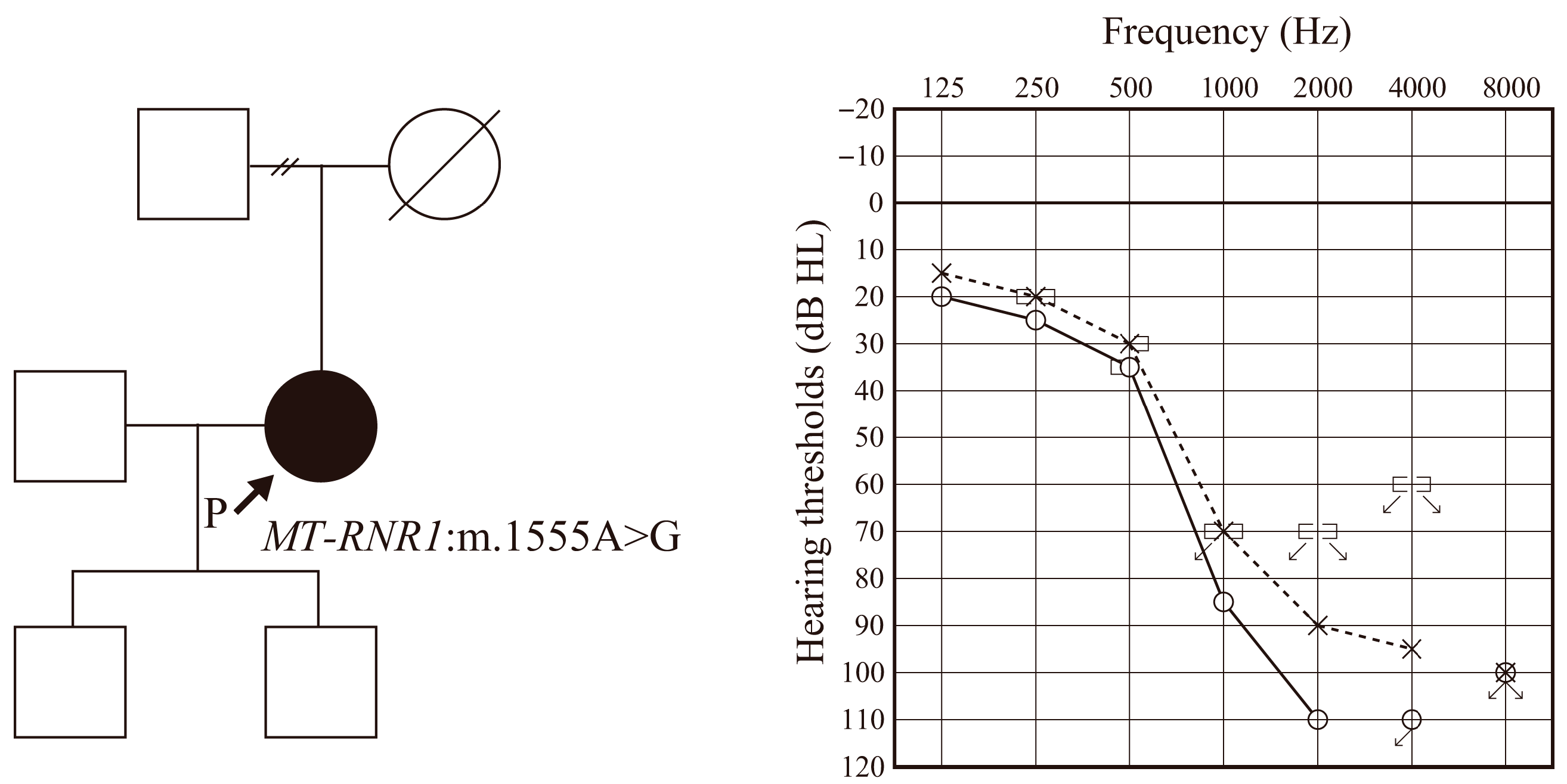
Figure 1. Pedigree, identified variant and audiogram of the patient. Squares indicate the male and circles indicate the female family members. Black indicates the affected individual and white indicates unaffected individuals. Solid line and dashed line indicates the hearing thresholds for right and left ear respectively.
Audiolres 15 00112 g001
Figure 2. Top: Illustration of a FLEX28 DEX electrode array (MED-EL, Innsbruck, Austria); the blue areas represent the coating. Bottom: The white areas between the electrodes are coated with dexamethasone-containing silicone.
Audiolres 15 00112 g002
Figure 3. The exoscopic view of the surgical field, estimated electrode position in the audiogram and cochlear microphonics during electrode insertion. (A) The tip of the electrode has been inserted into the basal turn of the cochlear. As it is still far from the area responsible for the 500 Hz, no CM response is observed. (B) The first electrode has just reached the area responsible for 1000 Hz. A weak CM reaction is observed. (C) The first electrode has just reached the area responsible for 500 Hz. The maximum CM reaction has been obtained. (D) After full insertion, the first electrode is farther away from the area responsible for 500 Hz, so the response is weaker. (E) After full insertion, CM responses were taken from the first, third, and fifth electrodes, and the largest response was from the third electrode, which was located in the area responsible for 500 Hz. See Supplemental Materials (Video S1) for more high-resolution images.
Audiolres 15 00112 g003
Figure 4. The audiogram and hearing level with EAS. (A) Audiograms taken pre-operatively and (B) at 12 months post-operatively, and (C) hearing level with EAS. Cross mark indicates the hearing threshold for each frequency.
Audiolres 15 00112 g004
Figure 5. Averaged audiogram of 22 EAS patients using conventional FLEX28 (without DEX-eluting function). (A) Audiogram taken pre-operatively and (B) at 12 months post-operatively. Error bar indicates the standard deviation.
Audiolres 15 00112 g005
Figure 6. Post-operative impedance field telemetry (IFT) taken from (A) electrode Ch 1, (B) electrode Ch 5 and (C) electrode Ch 11. Blue indicates the average of 15 patients using conventional FLEX28 electrodes (without DEX). Red indicates the IFT for the patient with FLEX28 DEX. Error bar indicates the standard deviation of 15 controls. IFT was maintained at a very low level compared with the controls.
Audiolres 15 00112 g006
Figure 7. Possible phenomena occurring within the cochlea during electrode insertion in patients with the mitochondrial m.1555A > G variant. (A) Inserted electrodes touch basilar membrane and inhibit its movement. (B) Inflammation induced by electrode insertion in addition to the lack of energy caused by mitochondrial mutation can lead to even more severe hearing deterioration.
Audiolres 15 00112 g007
Disclaimer/Publisher’s Note: The statements, opinions and data contained in all publications are solely those of the individual author(s) and contributor(s) and not of MDPI and/or the editor(s). MDPI and/or the editor(s) disclaim responsibility for any injury to people or property resulting from any ideas, methods, instructions or products referred to in the content.