1. Introduction
Numerous techniques for straightening the nasal septum have been described. The simple submucosal resection of bony or cartilaginous deviations is effective and probably the most commonly used technique for this purpose. However, severe, caudal, and dorsal deviations or cases that are combined with a closed rhinoplasty often require other or additional approaches. The search continues for a method that is universally accepted, technically convenient, conservative in terms of cartilage preservation, and long-lasting, as shown by the various publications proposing new or modified approaches.
More conservative methods, in terms of cartilage preservation and the avoidance of permanent synthetic materials, such as the crosshatching technique or the use of mattress sutures, are technically convenient. Nevertheless, they have been criticized for failures in complete correction of the deviated septum or unpredictable long-term effects. On the other hand, more invasive techniques, such as extracorporeal resection or the use of bony or septal batten grafts and adhesive materials, have been criticized for possible increased rates of complications [
1].
Herein, we describe in detail a technique that combines modifications of previously reported methods. It is simple and has visible direct and positive long-term effects. It can be applied to multiple areas such as the middle septum, the keystone area, and the dorsal and caudal part of the septum, where reduction techniques may endanger the basic nasal framework. Finally, this method also appears to offer protection against septal hematoma formation.
2. Materials and Methods
A retrospective review of prospectively collected data was performed on all patients who underwent surgery for septal deviation in the areas of interest. More specifically, patients with severe septal deviation involving the keystone area or the caudal (anterior) septum were included. Severity was defined as deviation requiring more extensive resection, with a potential risk of compromising septal support.
Patients undergoing concomitant rhinoplasty were excluded from the study, as were those with conditions significantly increasing the risk of complications, such as bleeding disorders. Both primary and revision procedures were included. Concurrent turbinopexy, which is commonly performed in the majority of septoplasty cases, was not considered an exclusion criterion.
All patients were evaluated during the immediate postoperative period up to one month after surgery, as well as during longer-term follow-up between six and twelve months postoperatively Evaluation included septal straightening and the detection of potential complications, such as septal perforation, hematoma, infection, synechiae formation, and the need for revision surgery. Because of the suturing, patients were meticulously assessed for indications of ischemia of the mucoperichondrial flap, mucosal necrosis, or delayed mucosal healing.
2.1. Surgical Technique
Ethical clearance was obtained by the Scientific Council and Ethical Committee of our hospital (approval no. 435/5 November 2025). Hemitransfixion incision, bilateral elevation of the mucoperichondrial flaps, mobilization of the septum via the separation of its inferior part from the nasal spine, and excision of the excessive portion of the inferior septum are performed as usual. Remaining deviation affecting the caudal or dorsal part of the septum is addressed with our technique, as described below:
- (a)
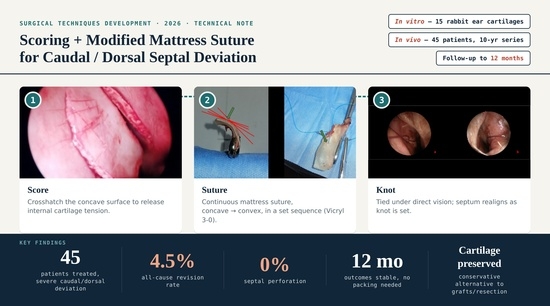
Scoring (crosshatching) of the concave side (
Figure 1).
- (b)
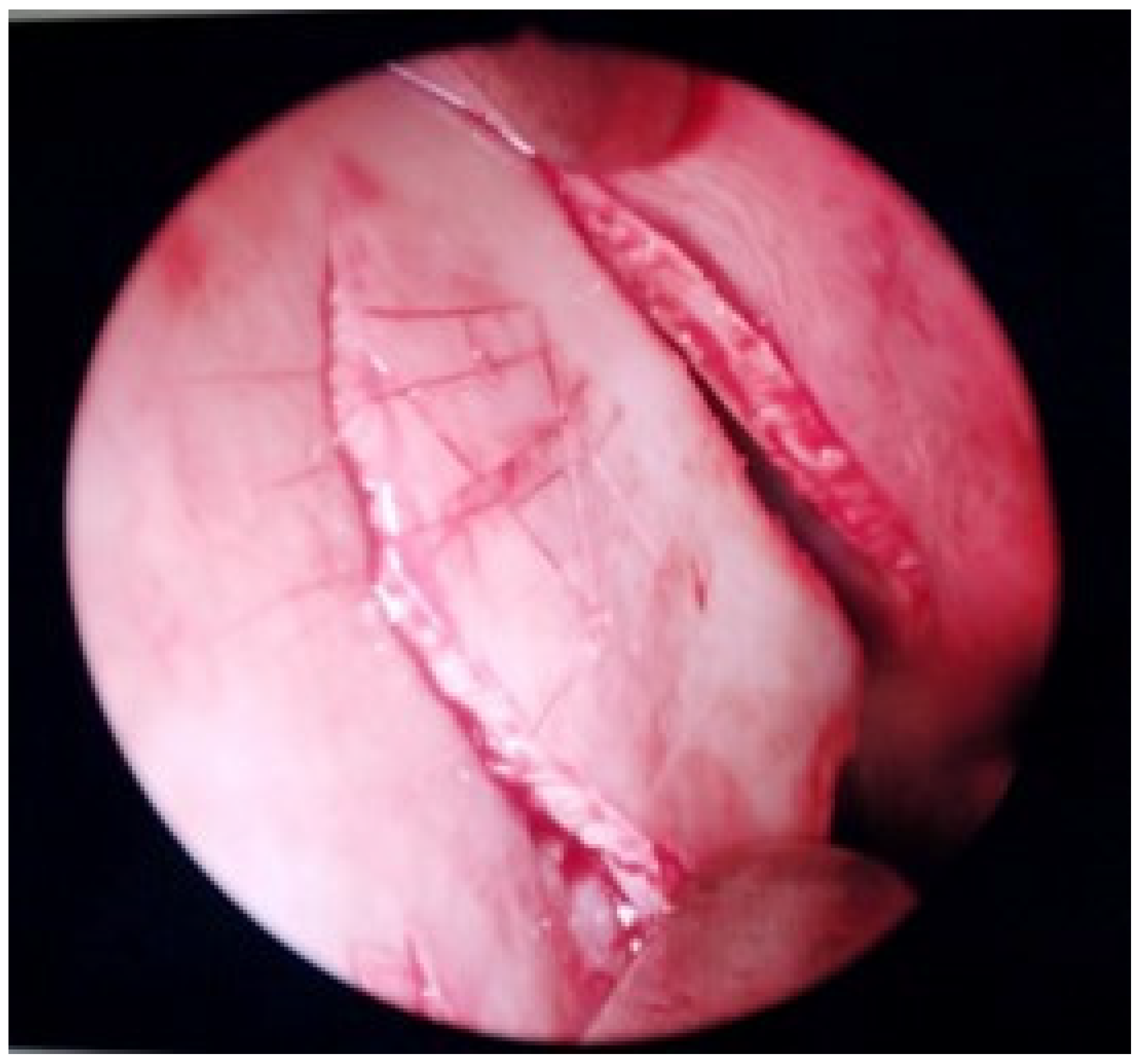
A modified continuing mattress suture that begins from the posterior (cephalic) side of the concave area (
Figure 2a). The suture is continued and inserted in the anterior (caudal) part of the convex side (
Figure 2b), and then in the superior (anatomically superoanterior) part of the concave side between the initial suturing sites, and is ended in the inferior part (anatomically inferoposterior) (
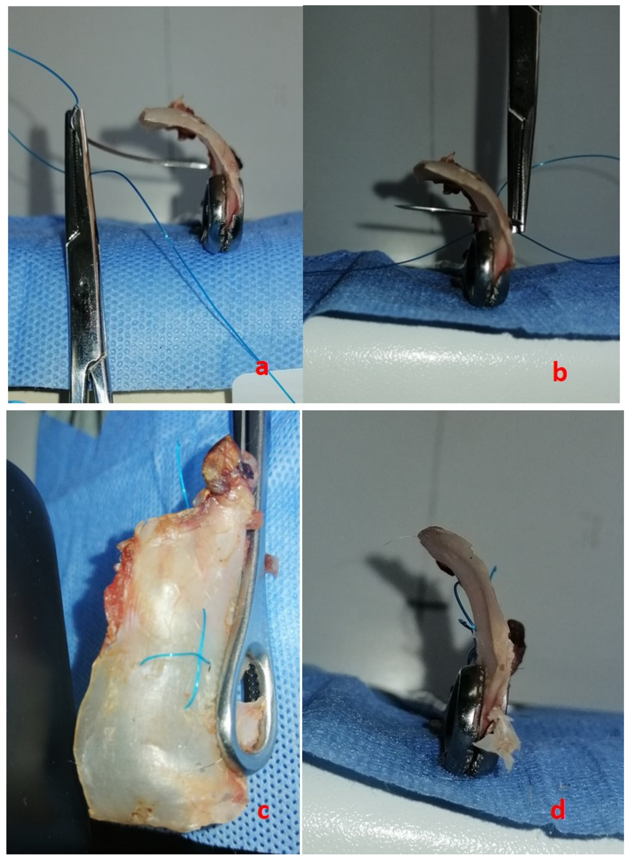
Figure 2c). Alternatively, the mattress suture can be initiated in the superior part of the concave area (
Figure 3a), reinserted in the inferior part of the convex side (
Figure 3b,c), continued in the posterior part of the concave (
Figure 3d,e), and reinserted in the anterior part of the convex (
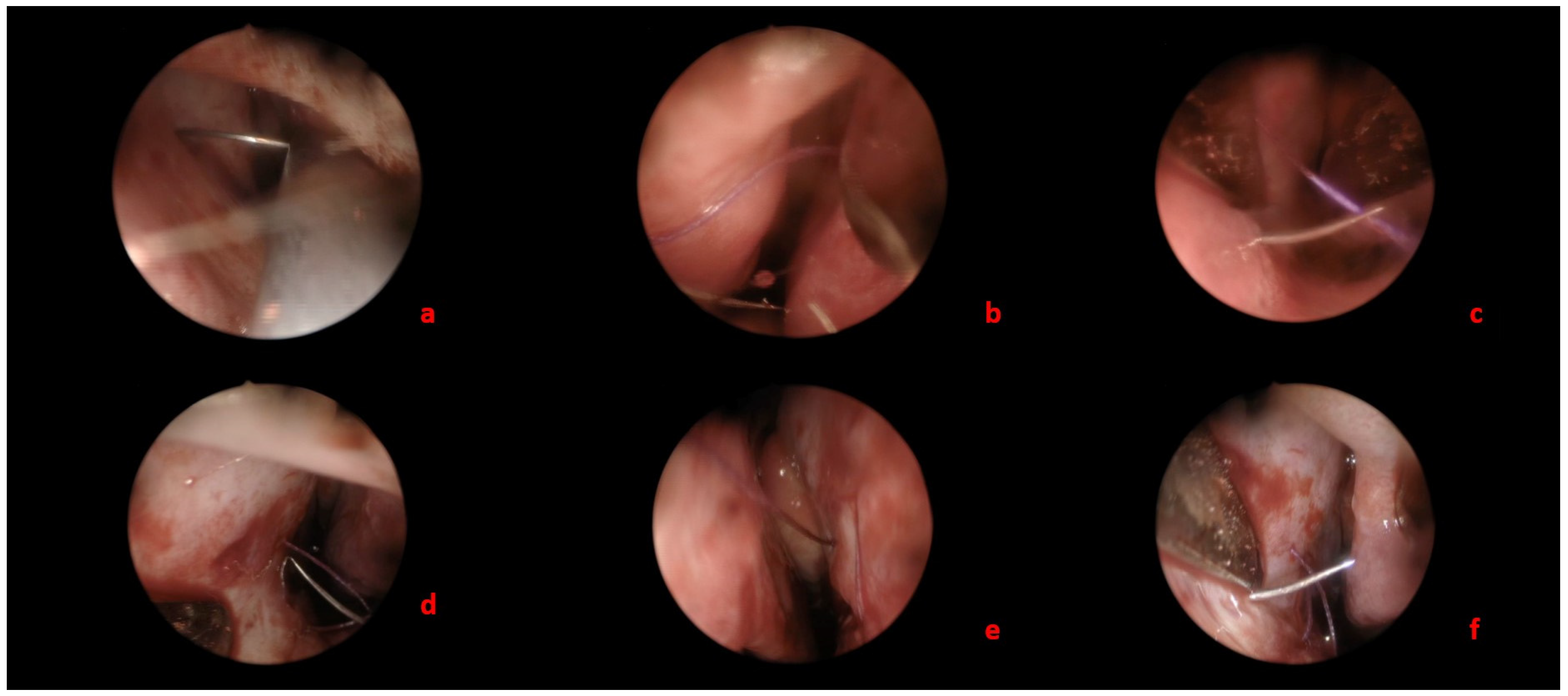
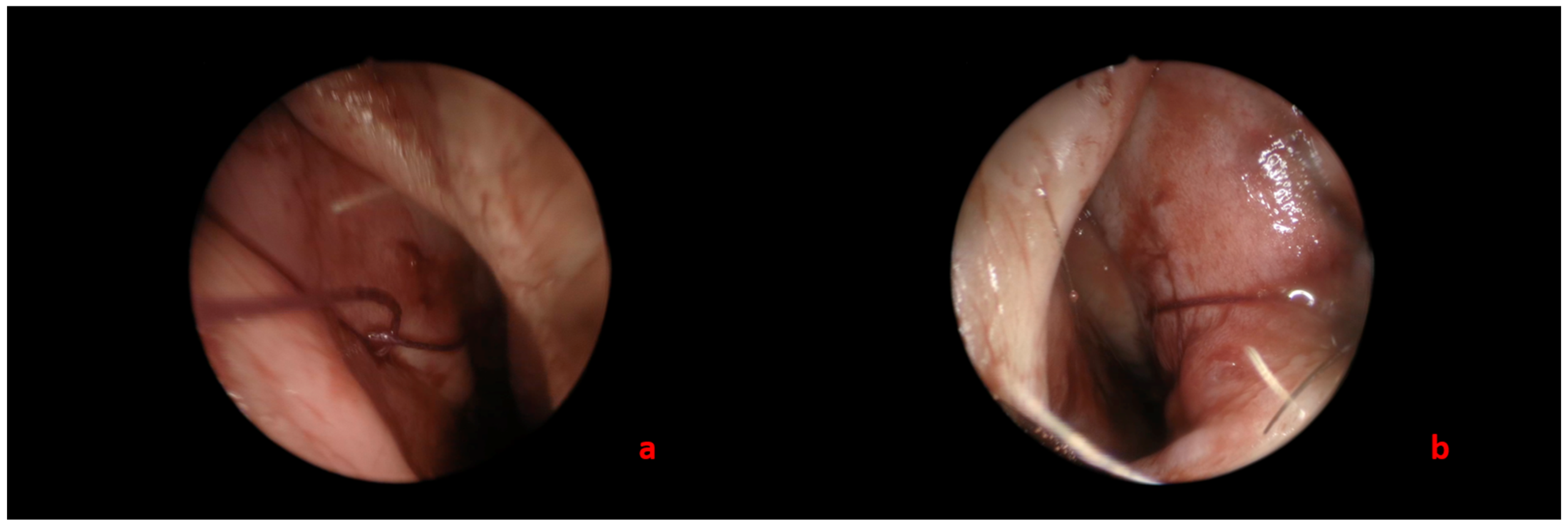
Figure 3f) so that the knotting can be performed as described previously on the concave side of the septum (
Figure 4 and
Supplementary Video S1).
- (c)
The alignment of the deviated part of the septum can be visualized directly during knotting (
Figure 2d and
Figure 4). A Vicryl 3.0 suture is utilized, and mucoperichondrial flaps of both sides are included in suturing.
Figure 1.
Scoring of the concave site of the septum.
Figure 1.
Scoring of the concave site of the septum.
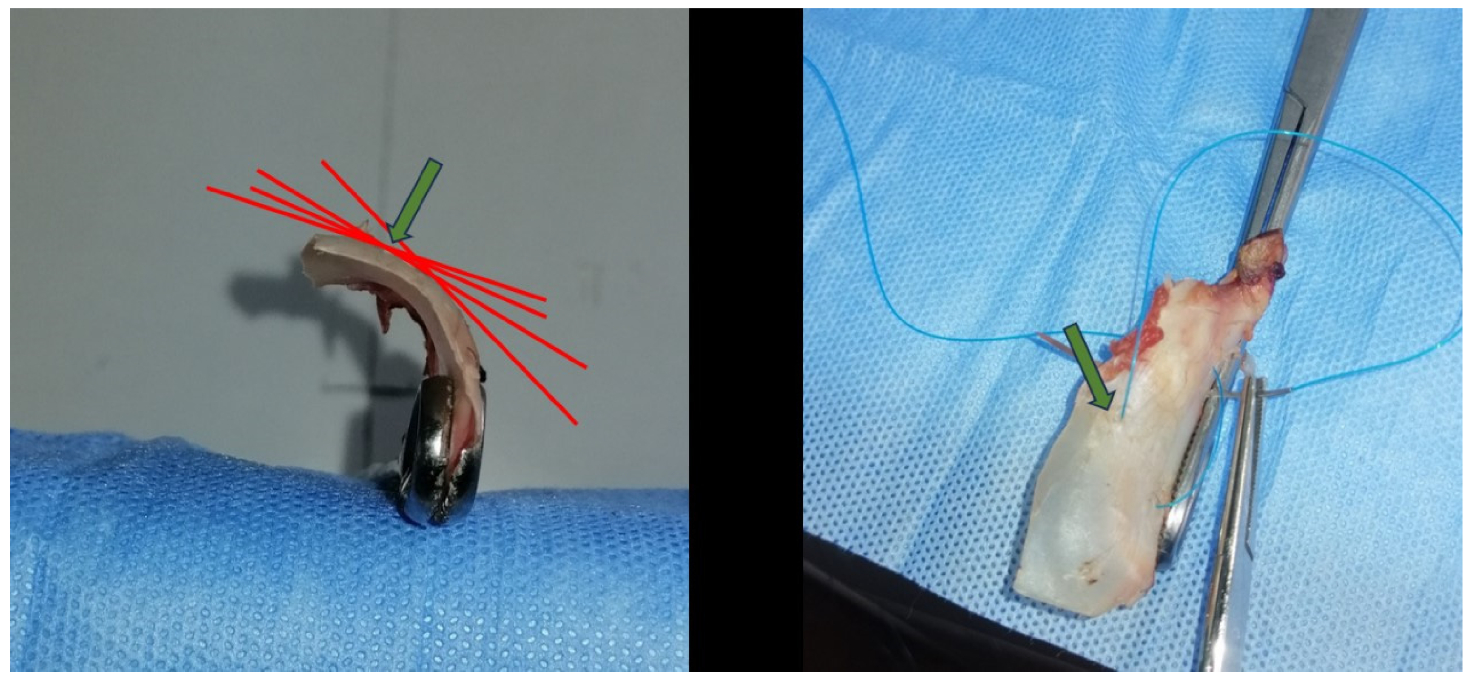
The site of suture insertion is approximated visually each time. As shown in
Figure 5, theoretically, it is close to the point where the tangents of the cartilage curve begin to merge.
A “tongue-in-groove” technique may also be added in caudal septal deviations to facilitate further straightening. In such cases, a simple repositioning of the caudal septum within the groove of the medial crura is easily performed without suture fixation. The hemitransfixion incision is then closed, and thin silicone splints are sutured on the septum and remain in the nostril for 7 days. No nasal packing is utilized in the majority of cases.
Figure 2.
Mattress suture for straightening of the cartilage in a rabbit ear. Beginnign of the mattress suture from the posterior side of the concave area (a). The suture is continued and inserted in the anterior part of the convex side (b). Final appearance of the suture (c) and of the straightening of the cartilage (d).
Figure 2.
Mattress suture for straightening of the cartilage in a rabbit ear. Beginnign of the mattress suture from the posterior side of the concave area (a). The suture is continued and inserted in the anterior part of the convex side (b). Final appearance of the suture (c) and of the straightening of the cartilage (d).
Figure 3.
Mattress suture for straightening of the cartilage (endoscopic view) that begins from the superior part of the concave area (a), reinserted in the inferior part of the convex side (b,c), continued in the posterior part of the concave (d,e), and reinserted in the anterior part of the convex (f).
Figure 3.
Mattress suture for straightening of the cartilage (endoscopic view) that begins from the superior part of the concave area (a), reinserted in the inferior part of the convex side (b,c), continued in the posterior part of the concave (d,e), and reinserted in the anterior part of the convex (f).
Figure 4.
Knotting of the suture is performed on the concave side of the septum (a). Sub-figure (b) shows the knotting as it appears in the convex side of the septum.
Figure 4.
Knotting of the suture is performed on the concave side of the septum (a). Sub-figure (b) shows the knotting as it appears in the convex side of the septum.
Figure 5.
The green arrow indicates the site of insertion. The preferred site for suture insertion each time appeared to be close to the point where the tangents (red lines) of the cartilage curve began to merge. This site was approximated visually each time, as is the case in the operation theater.
Figure 5.
The green arrow indicates the site of insertion. The preferred site for suture insertion each time appeared to be close to the point where the tangents (red lines) of the cartilage curve began to merge. This site was approximated visually each time, as is the case in the operation theater.
2.2. In Vitro Animal Model Testing
Fifteen ear cartilages extracted from eight rabbit ears were sutured with the above modified mattress suture technique with the aim of adjusting the optimal site of needle insertion in concave and convex parts of the cartilage. The sites of the sutures’ insertion were experimented with several times in almost all the ear cartilages in order to find the best straightening result. Inserting the suture close to the site where the tangents of the cartilage curve began to merge (
Figure 5) seemed to offer the best results in terms of cartilage straightening. An approximate visual estimation of the insertion point was performed, reflecting real-life conditions in the operating theater. The rabbit ear specimens were obtained from commercially sourced rabbits intended for human consumption, and no animals were sacrificed specifically for the purposes of this study. Therefore, no additional ethical approval was required for their use in this experimental setting, in accordance with institutional guidelines. For the experiment, the skin overlying the cartilage was carefully removed in order to expose the latter, which was then used for the suturing technique. The cartilage straightening was primarily assessed visually. To enhance consistency, a straight reference line was drawn on a white background, against which the degree of straightening was evaluated, as illustrated in the corresponding figures.
Supplementary Video S1 legend: The authors prefer to perform the suture technique under direct vision with the aid of a nose speculum. However, in this video, an endoscope was utilized for demonstration purposes.
4. Discussion
There are several studies on the application of mattress suturing for straightening septal deviations. In the majority of these studies, modifications of the mattress suture technique originally described by Ellis [
2] have been utilized for caudal septal deviations. This is rational, because an effective yet conservative method is needed mostly in this area of the septum. To our knowledge, apart from Ellis’ paper, two more publications exist in the English literature regarding the use of mattress sutures for caudal septal deviations [
3,
4], one for the correction of the septum around the keystone area [
5] and one for the correction of high dorsal deviations with a direct impact on the internal nasal valve [
6].
The initial concept was the application of a technique that resembles the reverse of the Mustarde procedure for protruding ears. As the author states, “instead of bending the cartilage into a harmonious shape, the bent cartilage is straightened with submucosal permanent sutures” [
2]. Several years later, Calderón-Cuéllar et al. [
3] reported a modified mattress suture technique in which they made a partial thickness incision on the concave side of the deviated septal cartilage in a dorsoventral direction. The differences from our technique are that they used a partial thickness incision and not scoring (crosshatching), with the latter being technically easier in our opinion, and that they began the suturing in the anterior part of the convex site, whereas we began it in the posterior part of the concave site. In addition, they did not use the superior and inferior part of the suturing. Still, their results are better than those obtained using the classical Hinderer technique.
More recently, a wedge resection of the caudal septal cartilage on the convex side in the cephalocaudal direction was described in 40 patients with initial good results [
4]. Similarly good results have been demonstrated in a modified mattress suture technique described for high dorsal deviations, in which the author utilizes the bony septum to stabilize the suture [
6]. This technique involves drilling two suture holes in the bony septum, which may be technically more challenging, contrary to our technique. Other modifications of the technique involve the overlapping of cartilage edges or use of batten grafts [
7] that we are trying to avoid.
Other non-mattress suturing techniques that have been published recently include the “traction-suture” technique and the “DOUS—double opposing U suture” technique [
8,
9]. The former addresses the correction of a caudal deviation by using traction in order to fixate the caudal end into the columellar pocket [
8], while the latter is performed in rhinoplasty cases in order to strengthen the caudal end and fixate it to the nasal spine [
9]. Both present very promising results and could be used alone or in combination with our method.
Last but not least, mattress suturing has been described in many publications for the avoidance of nasal packing, with equal results in relation to postoperative bleeding, mucosal adhesions, septal perforation, septal hematoma, and local infection [
10]. At the same time, it avoids the pain and discomfort that patients experience in the first postoperative period after nasal packing [
11]. In our practice, although mattress suturing has not been used primarily for the avoidance of nasal packing, it seems to confer the previously reported satisfactory results. The complication rates in relation to bleeding, hematoma formation, or septal perforation are very low in our retrospective series.
A number of limitations of the present study should be acknowledged. First, the outcome assessment was not based on validated patient-reported outcome measures or objective functional tests such as rhinomanometry. Although such tools may provide additional insight, patient-reported outcomes are inherently subjective and typically require larger sample sizes for robust interpretation, while rhinomanometry is not routinely applied in our setting and is associated with certain methodological limitations. In this context, we elected to use the revision rate as a more objective and clinically meaningful endpoint. However, we recognize that this approach, together with surgeon-reported outcomes, does not enable a comprehensive evaluation of functional improvement. In addition, the retrospective design, the lack of a structured pre-versus-postoperative comparison, and the limited reporting of follow-up completeness further restrict the strength of our conclusions. Therefore, our findings should be interpreted with caution, and the present study is best viewed as a technical report demonstrating feasibility and reproducibility rather than definitive evidence of superiority. Future prospective studies incorporating validated outcome measures and objective functional assessment are warranted to better define the clinical impact of this technique.
In conclusion, the absence of objective functional measures and validated patient-reported outcomes limits direct comparison with other techniques and should be acknowledged. Nevertheless, this study includes patients with severe dorsal or caudal septal deviations treated over a ten-year period, with favorable outcomes in terms of recurrence and complications. The described technique is not an innovative concept but, rather, a practical modification based on established principles of cartilage scoring and mattress suturing. Its simplicity and reproducibility make it a useful option in routine septoplasty practice, although further studies with more comprehensive outcome assessment are needed to better define its clinical value.