1. Introduction
Inherited epidermolysis bullosa (EB) comprises a heterogeneous group of rare genetic skin-fragility disorders characterized by mucocutaneous fragility. Even minimal friction or trauma can induce blistering and erosions that evolve into chronic wounds, scarring, and progressive functional impairment [
1,
2]. EB encompasses a broad spectrum of clinical phenotypes driven by pathogenic variants in genes encoding proteins critical for dermo-epidermal adhesion; classification is based on the level of tissue cleavage and currently recognizes EB simplex, junctional EB, dystrophic EB, and Kindler syndrome, each with multiple subtypes [
3,
4].
Hand involvement is a major determinant of disability in EB. Continuous shear forces during activities of daily living lead to recurrent blistering and erosions that heal with scarring and fibrosis, progressively causing web-space obliteration (pseudosyndactyly), flexion or extension contractures at the interphalangeal and metacarpophalangeal joints, and thumb adduction. In severe cases, the hand becomes enclosed in a mitten-like “cocoon” with loss of pinch and grasp function [
5,
6,
7]. The burden is greatest in recessive dystrophic EB, in which deficiency of collagen VII predisposes to deep blistering and scarring [
5,
6]. International clinical practice guidelines support surgery for moderate-to-severe deformity to restore prehension and improve independence; however, the evidence base is limited, recurrence is expected, and outcomes critically depend on atraumatic postoperative wound management and intensive hand therapy [
8].
Following the release of pseudosyndactyly and contractures, surgeons are left with large denuded areas that require protection while epithelialization occurs. Autologous split-thickness skin grafting can provide rapid coverage [
7], but donor-site creation is intrinsically problematic in EB because harvesting produces an additional wound in tissue that is already fragile and may heal slowly. For this reason, some teams, especially in younger children, prefer release followed by secondary intention healing to avoid donor-site morbidity, accepting a longer healing phase, meticulous dressing care, and the risk of infection or scar contracture [
6].
Skin substitutes and biologically active matrices have therefore attracted interest as strategies to reduce operative trauma and simplify postoperative care in a condition where repeated procedures may be required over a lifetime. Dermal substitutes and dermal regeneration templates have shown encouraging results in EB hand surgery, but these strategies may still require staged reconstruction, careful postoperative protection, or donor-site creation depending on the protocol used [
9,
10,
11].
Intact fish skin grafts (iFSGs) are acellular dermal matrices derived from Atlantic cod (Gadus morhua) and processed to preserve native extracellular-matrix architecture and omega-3 lipids. Experimental and clinical literature suggests that this combination may support cellular ingrowth and remodeling while contributing to anti-inflammatory and antimicrobial effects and may also reduce pain during dressing changes [
12,
13,
14,
15]. These characteristics make iFSG an appealing adjunct in EB hand surgery, where donor-site avoidance, atraumatic dressing care, and early rehabilitation are critical. In this context, we report a preliminary single-center pediatric case series using iFSG (Kerecis
® Omega3 Wound), within its general wound-management indication, as an adjunct after EB hand surgery, focusing on feasibility, early wound-healing behavior, complications, and the potential to support secondary closure without autologous donor-site creation.
2. Materials and Methods
2.1. Study Design and Patient Selection
We performed a preliminary retrospective observational case series of consecutive pediatric patients with inherited EB undergoing operative correction of hand deformities (contractures and/or pseudosyndactyly) with adjunctive application of iFSG. Consecutive cases treated at Bambino Gesù Children’s Hospital (Rome, Italy) between December 2022 and December 2025 were included. The hospital is a tertiary pediatric referral center with multidisciplinary expertise in EB care. The limited number of eligible patients reflects the rarity of dystrophic EB and the even smaller subgroup of children requiring this specific type of hand surgery during the study period. The study followed institutional standards for observational research.
Inclusion criteria were age < 18 years, clinically and/or genetically confirmed EB, and indication for surgery due to function-limiting hand deformity. Exclusion criteria were uncontrolled local infection, inability to comply with postoperative immobilization and hand therapy, and known fish allergy, which was considered a contraindication to iFSG. In the present series, all included patients were affected by dystrophic EB.
Before surgery, parents/legal guardians received detailed information regarding the rationale for iFSG use, including its use within the product’s general wound-management indication as a decellularized intact fish-skin matrix for surgical wounds and complex wound beds. They were also informed about available reconstructive alternatives, the limited evidence in pediatric EB hand surgery, potential risks including fish allergy, and the expected postoperative dressing and rehabilitation pathway. Written informed consent for treatment and for publication of anonymized clinical data and photographs was obtained.
EB subtype and baseline severity of hand deformity were documented using clinical examination and standardized photography. Preoperative optimization was multidisciplinary and included nutritional and hematologic assessment, pain management planning, and microbiologic screening and treatment when indicated.
2.2. Surgical Technique and iFSG Application Method
All procedures were performed under general anesthesia by the same senior pediatric plastic surgery team, following a standardized EB-specific protocol to minimize iatrogenic shear and pressure injury. Precautions included atraumatic positioning, generous padding, avoidance of adhesive devices, and gentle tissue handling.
The surgical strategy was tailored to the deformity pattern and typically included a combination of first web-space release to restore thumb abduction, release of pseudosyndactyly to recreate web spaces, and selective release of interphalangeal and/or metacarpophalangeal contractures with gradual extension. In one severe case, a 27-gauge needle was temporarily used for 2 weeks only as a guide to support wound healing and maintenance of the surgical correction. After meticulous hemostasis, iFSG (Kerecis
® Omega3 Wound, Solid, 7 × 10 cm; Atlantic cod-Suðurlandsbraut 18, 108 Reykjavik) was hydrated in 0.9% saline for 5 min, trimmed to match the defect with slight overlap when feasible, and applied as an acellular dermal scaffold. Fixation was achieved with fine absorbable sutures according to wound size and location. The dressing protocol was designed to maintain a moist wound environment and minimize shear during dressing changes, using a non-adherent interface layer, an absorbent secondary layer, and a soft protective wrap. An extension splint was incorporated into the postoperative dressing and custom-made in thermoplastic material to maintain finger extension and preserve the recreated web spaces. In patients 4 and 5, who had severe bilateral mitten-hand deformities and palmar wound involvement, the same general splinting principles were used, but the splints were individually adapted to avoid pressure or shear over the iFSG-treated palmar surfaces and adjacent fragile skin. Dressings were planned twice weekly for the first 3 postoperative weeks and then weekly thereafter, with schedule adjustments based on wound conditions. During the first 2 postoperative weeks, dressing changes were performed in the operating room under general anesthesia to ensure atraumatic care and adequate pain control; subsequent dressings were performed in the outpatient setting. At each change, the matrix was left in situ if adherent, and additional iFSG was applied only if uncovered areas or fragmentation were present [
14,
15]. When wounds became sufficiently stable, a structured hand-therapy program started with progressive range-of-motion exercises, followed by prolonged night splinting to reduce re-adhesion and delay recurrence. The decision to start hand therapy was made jointly by the senior plastic surgeon and the hand therapist experienced in EB care, in coordination with the wound-care team. Wounds were considered clinically stable when the matrix was adherent or sufficiently integrated, exudate was controlled, there was no active bleeding or clinical infection, and the newly forming epithelium could tolerate gentle mobilization without disruption. Long-term maintenance relied on caregiver adherence to splinting and skin-protection measures.
2.3. Outcomes and Follow-Up
Primary outcomes were time to complete re-epithelialization in days, need for iFSG re-application, need for autologous skin grafting, and early complications such as infection/colonization, bleeding, blistering, or graft non-adherence. Secondary outcomes included pain associated with dressing care, the number of dressing changes, and early recurrence signals at follow-up, including web-space narrowing, re-adhesion, or recurrent contracture. Pain during dressing care was assessed using an age-appropriate visual analog scale during awake dressing changes. The reported VAS values refer specifically to dressing-related pain and do not include dressing changes performed under general anesthesia. All children received postoperative analgesia according to institutional pediatric pain-management protocols, with rescue medication available when clinically required. Follow-up visits were scheduled according to clinical need and rehabilitation progression. ABILHAND-Kids was used as a simple parent-/caregiver-reported measure of manual ability in daily activities. Caregivers were asked to rate the child’s perceived difficulty in performing each activity without human or technical assistance, regardless of the strategy used. Responses were coded according to the official instructions as impossible, difficult, or easy; activities rated as not applicable or missing were not scored. Raw ordinal scores were then calculated [
16].
2.4. Statistical Analysis
Given the small sample size, the rarity of the condition, and the exploratory case-series design, only descriptive statistics were used. Continuous variables are reported as mean and range, or median where appropriate, and categorical variables as counts. For bilateral cases, hand-level variables were analyzed per operated hand when applicable.
3. Results
Five consecutive pediatric patients with dystrophic EB were treated during the study period; three had bilateral involvement and two had unilateral involvement, for a total of eight operated hands. These patients represented all eligible children treated with this approach at our institution during the defined period. Deformities ranged from first web-space obliteration and flexion contractures to pseudosyndactyly and bilateral mitten-hand deformities (
Table 1).
Two patients (patients 4 and 5) were twin sisters treated during the same period and represented the most severe bilateral cases. iFSG (Kerecis® Omega3 Wound, Solid 7 × 10 cm) was tailored to the defects, with an applied area ranging from 8 to 12 cm2 per hand (mean 9.6 cm2). A second iFSG application was required only once, localized to the palmar region of the left hand in patient 1 at 2 weeks; no patient required autologous split-thickness skin grafting.
We did not use K-wires routinely; in one case, a temporary 27-gauge needle was left in place for 2 weeks to guide healing. Pain during awake dressing care was low overall, with a mean visual analog scale score of 1.6 when bilateral hands were analyzed separately. Mean time to complete re-epithelialization was 47.6 days, and mean follow-up was 15.4 months.
In both twin patients (patients 4 and 5), the period of twice-weekly dressings extended beyond 5 weeks, probably because of the more severe initial deformity, with a consequent delay in healing. No different splinting concept was used in these cases; however, the thermoplastic splints were individually adapted to the severe bilateral mitten-hand deformity and palmar iFSG placement. In patient 4, worsening of the dorsal hand skin with dorsal disepithelialization was observed and attributed to prolonged dressing contact on extremely fragile skin. No allergic reactions were observed.
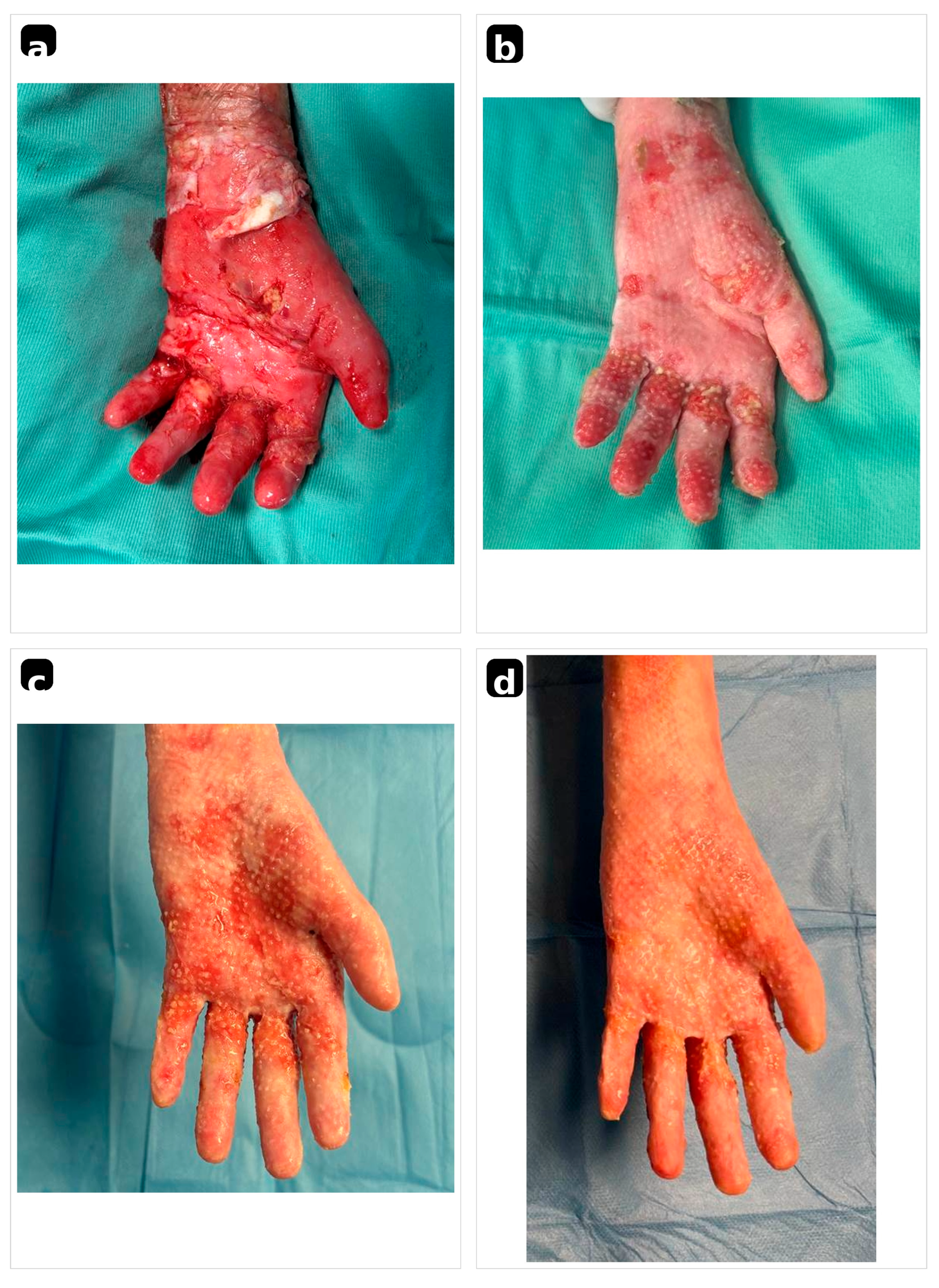
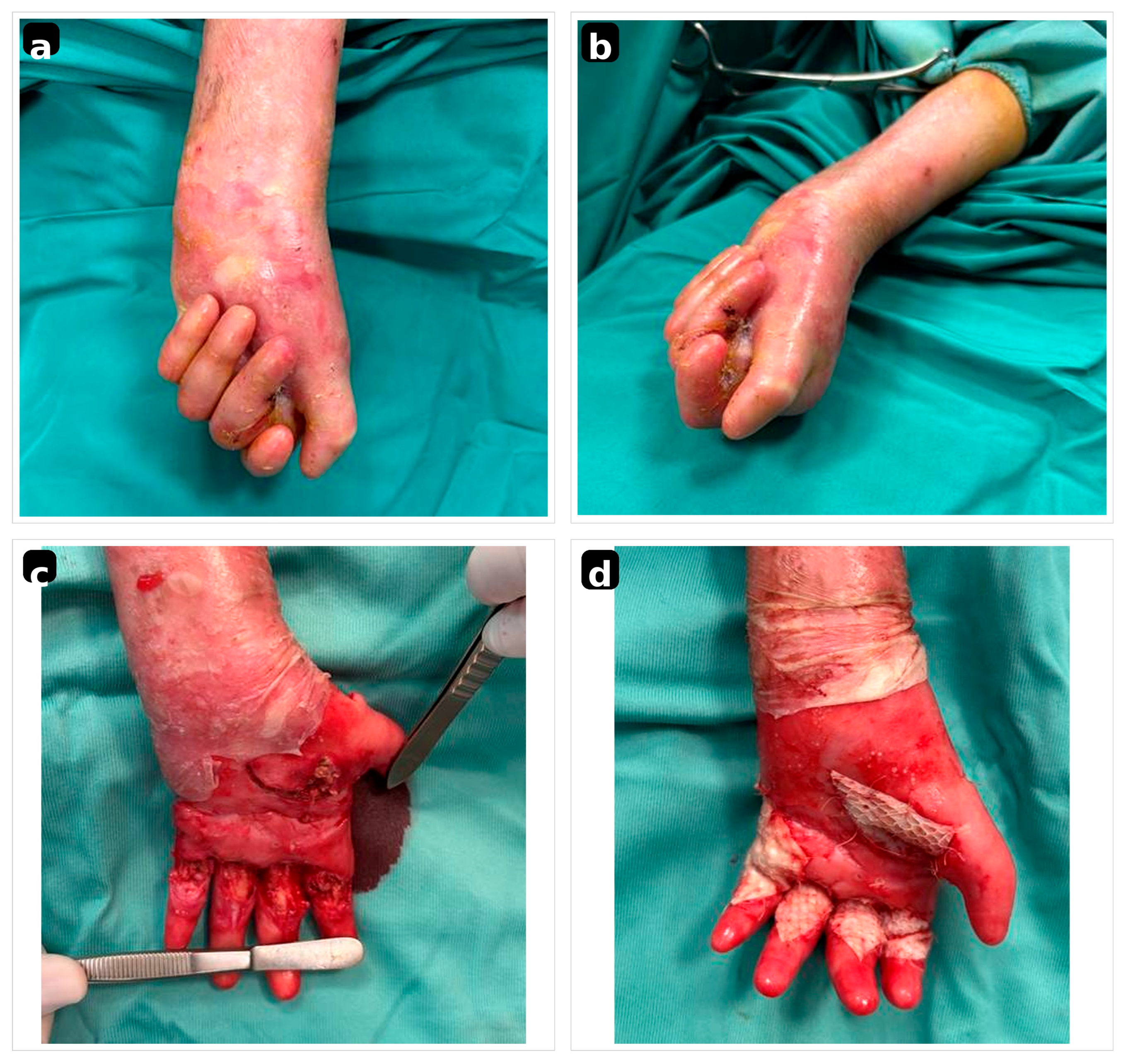
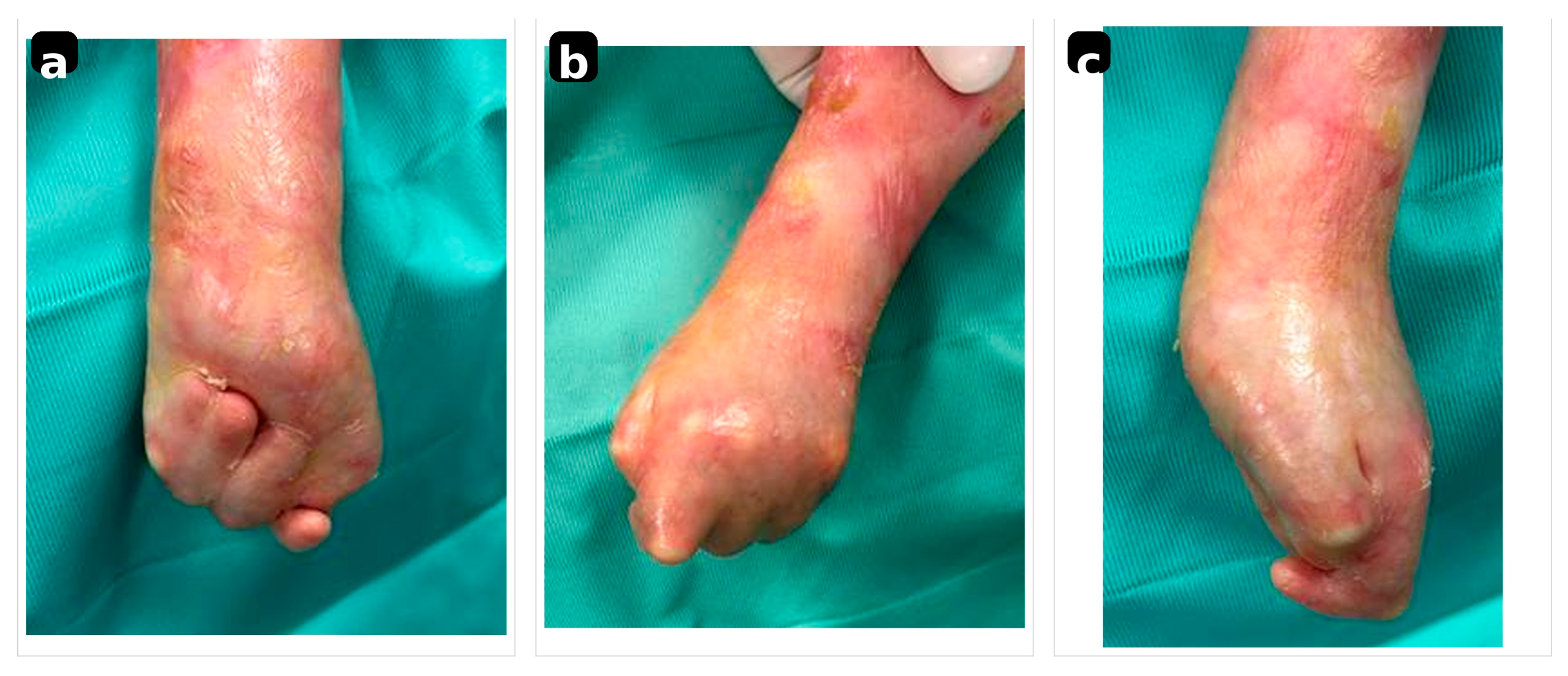
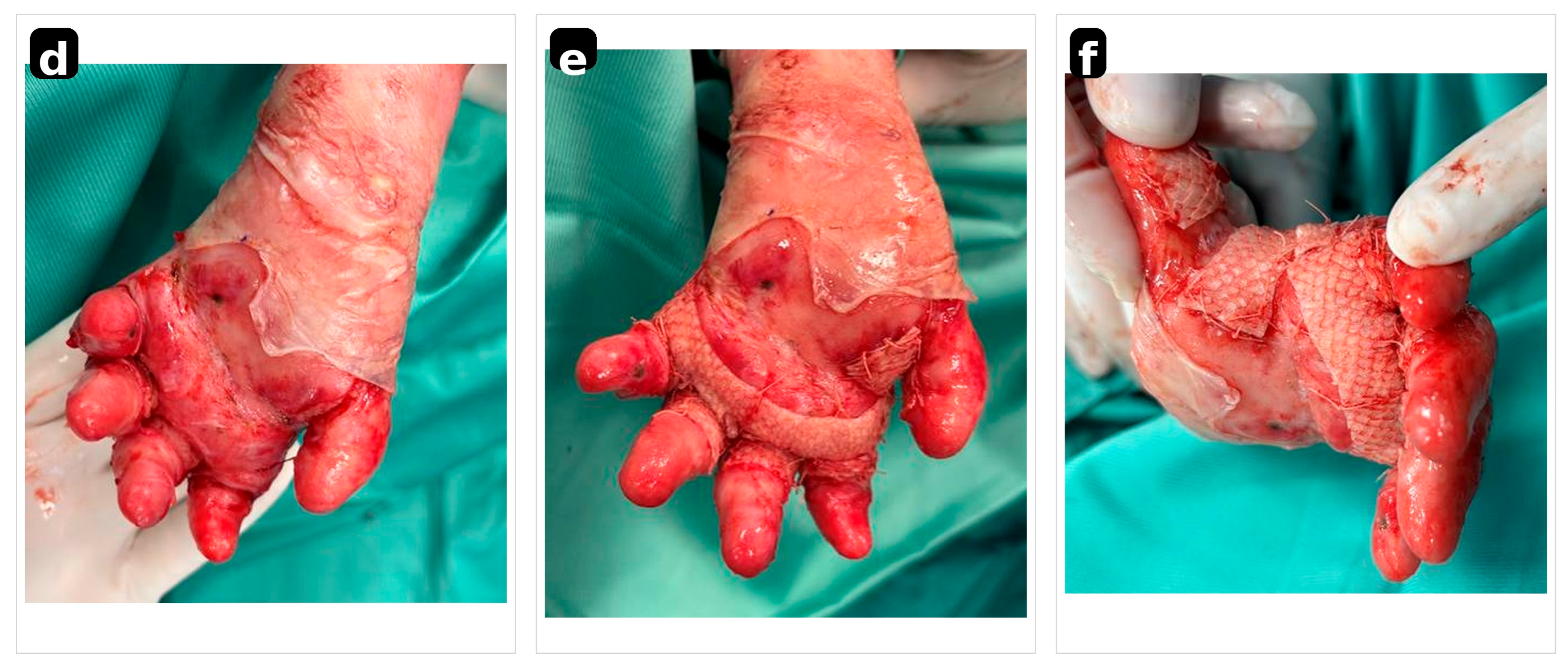
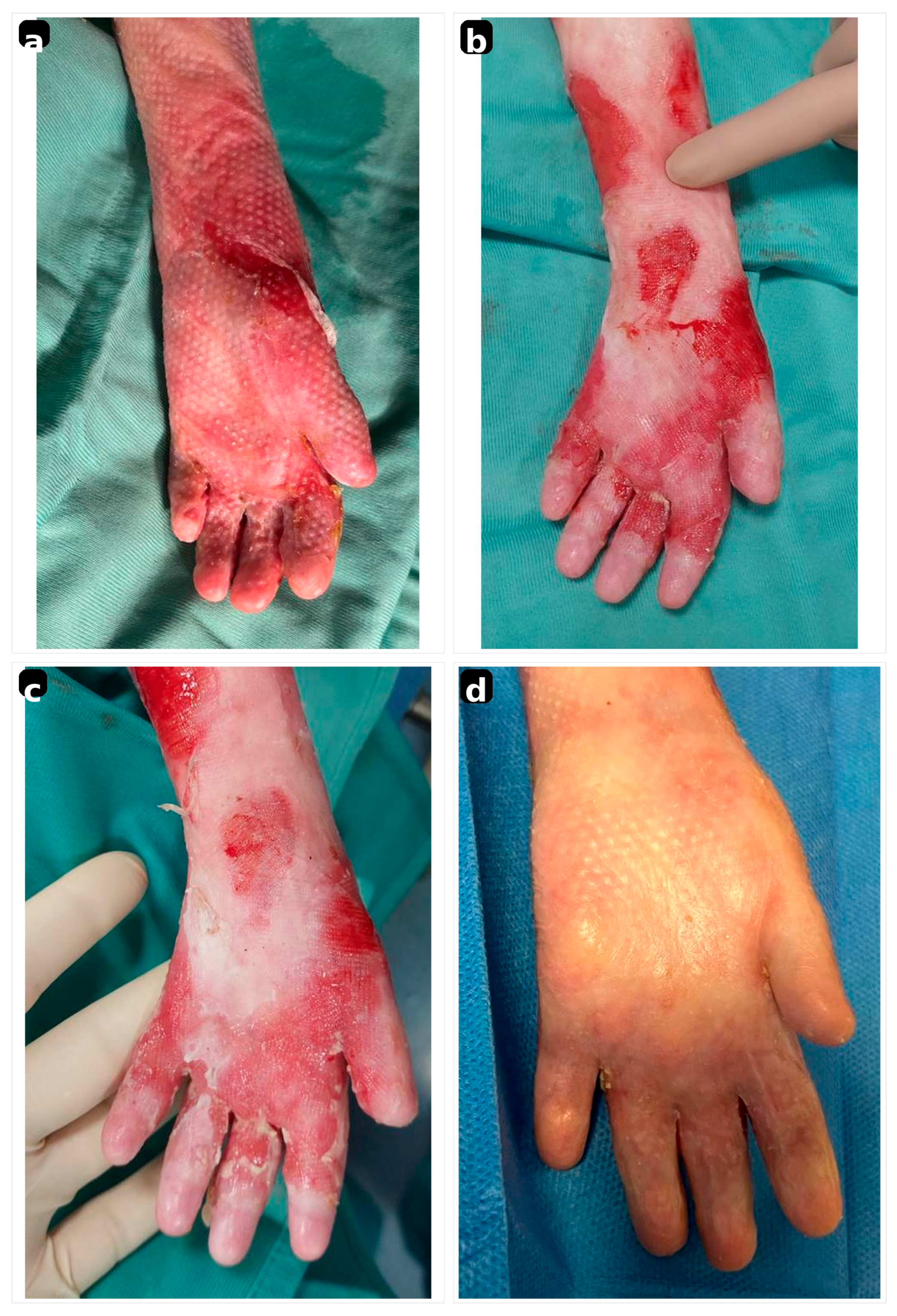
During follow-up, the first operated patient developed a partial recurrence of pseudosyndactyly in the II–III and III–IV web spaces, which did not require surgical revision. Representative sequential photographs of patient 2 (
Figure 1 and
Figure 2) and patient 4 (
Figure 3 and
Figure 4) are provided.
ABILHAND-Kids was easy to administer and well accepted by caregivers. Although the limited sample size and retrospective design preclude formal statistical analysis, scores suggested encouraging perceived postoperative manual ability in daily activities involving grasp, pinch, dressing, hygiene, and object manipulation. Lower scores were observed in the most severe bilateral mitten-hand cases.
4. Discussion
This preliminary case series describes our early experience using iFSG as an adjunct in pediatric dystrophic EB hand surgery. The small cohort must be interpreted in the context of the rarity of the underlying disease and the highly selected surgical indication. Children with dystrophic EB who require release of pseudosyndactyly or contractures represent a limited subset of an already rare condition, and available EB hand surgery literature is therefore largely composed of case series, expert recommendations, and center-specific protocols [
5,
6,
7,
8,
9,
10,
11].
A central challenge after the release of EB hand deformities is achieving stable wound coverage while minimizing additional trauma. Autologous split-thickness skin grafting may provide rapid coverage, but in EB it creates a second wound in fragile tissue and may add pain, healing burden, and potential morbidity [
7]. Secondary intention healing avoids donor-site injury but requires prolonged dressing care and may extend the period during which infection, re-adhesion, or recurrent contracture can occur [
6]. Dermal substitutes and dermal regeneration templates represent intermediate strategies, aiming to protect exposed tissues and support re-epithelialization while preserving the possibility of rehabilitation [
9,
10,
11].
In our series, cod-derived iFSG was selected to provide a biologically active scaffold over denuded surfaces after release and to avoid autologous donor-site creation. Across other wound types, iFSG has been associated with favorable healing outcomes, low rates of adverse events, and potential reduction in dressing-related pain or analgesic requirements; fish allergy remains a key contraindication [
12,
13,
14,
15]. In EB hands, these properties are conceptually attractive because postoperative care must minimize shear, pain, and repeated trauma to new epithelium. The pediatric literature is limited in two closely related domains: surgical treatment of EB hand deformities in children, and the use of cod-derived intact fish skin matrices in pediatric reconstructive wound care. Beyond the present EB hand series, Ciprandi et al. previously reported pediatric experience with acellular intact fish skin grafts in a broader cohort of acute pediatric wounds, supporting feasibility, good tolerability, and rapid healing in children; this experience provided part of the clinical basis for extending the approach to EB hand surgery [
17]. Additional pediatric evidence supports the feasibility of acellular fish skin matrices: in a 20-child series of deep dermal burns with 2-year follow-up, fish skin grafts avoided split-thickness grafting and no infections were observed, with favorable scar assessment outcomes [
18].
The present findings should not be interpreted as evidence that iFSG is superior to established techniques. Rather, they indicate that the iFSG application was technically feasible in all treated hands, that no autologous split-thickness graft was required, and that dressing-related pain during awake care remained low. Healing was slower in the two most severe bilateral mitten-hand cases, highlighting that the matrix does not overcome the intrinsic biological fragility of EB skin. Outcomes remained strongly influenced by deformity severity, infection control, dressing timing, protection of adjacent skin, splinting, rehabilitation, and caregiver adherence.
The role of parents and caregivers is particularly important in this population. Families were counseled about the rationale for iFSG use, its use within the general wound-management indication, the limited pediatric EB hand-specific evidence, the possibility of delayed healing, the need for repeated dressing changes and initial dressing care under general anesthesia, and the importance of splinting and hand therapy. This is relevant because caregiver burden, treatment acceptability, dressing burden, and perceived pain may strongly influence adherence and long-term maintenance of surgical gains [
19].
Finally, our experience reinforces the importance of early referral and multidisciplinary EB care. The most severe cases in this series were referred with advanced mitten-hand deformities, underscoring the value of preventive dressings, early hand surveillance, custom splints, rehabilitation, and coordinated follow-up in specialized centers. For children with EB, surgery should be considered part of a staged pathway rather than an isolated procedure, and recurrence should be anticipated regardless of the resurfacing strategy used.
Limitations and Future Directions
This study has several limitations. First, the sample size is small, reflecting the rarity of dystrophic EB and the even smaller subgroup of pediatric patients requiring operative correction of severe hand deformities during the study period. Second, the study was conducted at a single center and did not include a control group; therefore, no conclusion can be drawn regarding the superiority of iFSG over split-thickness skin grafting, secondary intention healing, dermal substitutes, or dermal regeneration templates. Third, follow-up remains insufficient to fully assess long-term recurrence, which is expected in EB hand surgery and is strongly influenced by rehabilitation, splinting, caregiver adherence, and disease severity. Fourth, ABILHAND-Kids was administered in a very small cohort. Future prospective multicenter studies should systematically include ABILHAND-Kids or similar validated patient-/caregiver-reported tools, together with objective measures of pinch, grasp, recurrence, pain, dressing burden, caregiver experience, recurrence-free interval, and cost-effectiveness [
10,
16].
5. Conclusions
Intact fish skin grafting was feasible in this small, preliminary pediatric dystrophic EB hand surgery series and allowed wound closure without autologous donor-site creation. These findings should be interpreted cautiously because of the rarity of the disease, the small sample size, the absence of a comparator group, and the limited follow-up. Larger multicenter studies with standardized functional, pain, recurrence, and caregiver-reported outcomes are needed to define the role of iFSG in EB hand reconstruction.
Author Contributions
Conceptualization, F.G.; methodology, F.G., E.C., G.P.D.; validation, F.G., M.Z.; formal analysis, F.G., E.C., M.C., J.M.F.; resources, F.G., E.C.; data curation, F.G., E.C.; writing—original draft preparation, E.C.; writing—review and editing, F.G., M.C., J.M.F., G.P.D.; supervision, F.G., M.Z. All authors have read and agreed to the published version of the manuscript.
Funding
This research received no external funding.
Institutional Review Board Statement
The present study represents a small retrospective observational case series based exclusively on routine clinical practice and conducted according to institutional standards of care, without any experimental intervention, prospective protocol, or modification of patient management. The iFSG (Kerecis® Omega3 Wound) was used in accordance with its intended purpose as a decellularized intact fish-skin matrix for wound management, including surgical wounds and complex wound beds, and was discussed with parents/legal guardians as an individualized reconstructive option in the context of limited disease-specific evidence.
Informed Consent Statement
Written informed consent for treatment, anonymized photography, and publication was obtained from parents/legal guardians and from patients when appropriate. Parents/legal guardians were informed that iFSG was used within its wound-management indication and were counseled about available alternatives, the limited pediatric EB hand-specific evidence, potential risks including fish allergy, and the expected postoperative dressing and rehabilitation pathway.
Data Availability Statement
The data presented in this study are available from the corresponding authors upon reasonable request.
Acknowledgments
The authors thank the patients, their families, and the multidisciplinary epidermolysis bullosa care team involved in perioperative management and rehabilitation.
Conflicts of Interest
The authors declare no conflicts of interest.
References
- Has, C.; Fischer, J. Inherited epidermolysis bullosa: New diagnostics and new clinical phenotypes. Exp. Dermatol. 2019, 28, 1146–1152. [Google Scholar] [CrossRef] [Scilit] [PubMed]
- Bachir, Y.; Daruich, A.; Marie, C.; Robert, M.P.; Bremond-Gignac, D. Eye involvement and management in inherited epidermolysis bullosa. Drugs 2022, 82, 1277–1285. [Google Scholar] [CrossRef] [Scilit] [PubMed]
- Has, C.; Bauer, J.W.; Bodemer, C.; Bolling, M.C.; Bruckner-Tuderman, L.; Diem, A.; Fine, J.-D.; Heagerty, A.; Hovnanian, A.; Marinkovich, M.P.; et al. Consensus reclassification of inherited epidermolysis bullosa and other disorders with skin fragility. Br. J. Dermatol. 2020, 183, 614–627. [Google Scholar] [CrossRef] [Scilit] [PubMed]
- Intong, L.R.A.; Murrell, D.F. Inherited epidermolysis bullosa: New diagnostic criteria and classification. Clin. Dermatol. 2012, 30, 70–77. [Google Scholar] [CrossRef] [Scilit] [PubMed]
- Bernardis, C.; Box, R. Surgery of the hand in recessive dystrophic epidermolysis bullosa. Dermatol. Clin. 2010, 28, 335–341. [Google Scholar] [CrossRef] [Scilit] [PubMed]
- Vozdvizhensky, S.I.; Albanova, V.I. Surgical treatment of contracture and syndactyly of children with epidermolysis bullosa. Br. J. Plast. Surg. 1993, 46, 314–316. [Google Scholar] [CrossRef] [Scilit] [PubMed]
- Terrill, P.J.; Mayou, B.J.; Pemberton, J. Experience in the surgical management of the hand in dystrophic epidermolysis bullosa. Br. J. Plast. Surg. 1992, 45, 435–442. [Google Scholar] [CrossRef] [Scilit] [PubMed]
- Box, R.; Bernardis, C.; Pleshkov, A.; Jessop, N.; Miller, C.; Skye, J.; O’Brien, V.; Veerkamp, M.; da Rocha, A.C.F.; Cornwall, R. Hand surgery and hand therapy clinical practice guideline for epidermolysis bullosa. Orphanet J. Rare Dis. 2022, 17, 406. [Google Scholar] [CrossRef] [Scilit] [PubMed]
- Zhou, X.; Zhang, Y.; Zhao, M.; Jian, Y.; Huang, J.; Luo, X.; Yang, J.; Sun, D. Surgical management of hand deformities in patients with recessive dystrophic epidermolysis bullosa. J. Plast. Surg. Hand Surg. 2020, 54, 33–39. [Google Scholar] [CrossRef] [Scilit] [PubMed]
- Abboud, L.; Leclerc-Mercier, S.; Bodemer, C.; Guéro, S. Hand surgery in recessive dystrophic epidermolysis bullosa: Our experience with dermal substitutes. J. Plast. Reconstr. Aesthet. Surg. 2022, 75, 314–324. [Google Scholar] [CrossRef] [Scilit] [PubMed]
- Lembo, F.; Parisi, D.; Cecchino, L.R.; Ciancio, F.; Innocenti, A.; Portincasa, A. Release of pseudosyndactyly in recessive dystrophic epidermolysis bullosa using a dermal regeneration template glove: The Foggia experience. Orphanet J. Rare Dis. 2021, 16, 52. [Google Scholar] [CrossRef] [Scilit] [PubMed]
- Seth, N.; Chopra, D.; Lev-Tov, H. Fish skin grafts with omega-3 for treatment of chronic wounds: Exploring the role of omega-3 fatty acids in wound healing and a review of clinical healing outcomes. Surg. Technol. Int. 2022, 40, 38–46. [Google Scholar] [PubMed]
- Ibrahim, M.; Ayyoubi, H.S.; Alkhairi, L.A.; Tabbaa, H.; Elkins, I.; Narvel, R. Fish skin grafts versus alternative wound dressings in wound care: A systematic review of the literature. Cureus 2023, 15, e36348. [Google Scholar] [CrossRef] [Scilit] [PubMed]
- Cherry, I.; Tarhini, L.; Doan, M.; De Buys Roessingh, A. Exploring the place of fish skin grafts with omega-3 in pediatric wound management. J. Clin. Med. 2024, 13, 112. [Google Scholar] [CrossRef] [Scilit] [PubMed]
- Karhana, S.; Khan, M.A. Omega-3 acellular fish skin grafts for chronic and complicated wounds: A systematic review of efficacy and safety. Dermatol. Pract. Concept. 2025, 15, 4945. [Google Scholar] [CrossRef] [Scilit] [PubMed]
- Eismann, E.A.; Lucky, A.W.; Cornwall, R. Hand function and quality of life in children with epidermolysis bullosa. Pediatr. Dermatol. 2014, 31, 176–182. [Google Scholar] [CrossRef] [Scilit] [PubMed]
- Ciprandi, G.; Kjartansson, H.; Grussu, F.; Baldursson, B.T.; Frattaroli, J.; Urbani, U.; Zama, M. Use of acellular intact fish skin grafts in treating acute paediatric wounds during the COVID-19 pandemic: A case series. J. Wound Care 2022, 31, 824–831. [Google Scholar] [CrossRef] [Scilit] [PubMed]
- Staubach, R.; Glosse, H.; Loff, S. The use of fish skin grafts in children as a new treatment of deep dermal burns—Case series with follow-up after 2 years and measurement of elasticity as an objective scar evaluation. J. Clin. Med. 2024, 13, 2389. [Google Scholar] [CrossRef] [Scilit] [PubMed]
- Esmaeili, M.; Mohammadi, F.; Cheragi, F.; Khazaei, S.; Sobhan, M.; Parvizi, M.M. The relationship between parental stress, resilience, and quality of life in parents of children with epidermolysis bullosa. Sci. Rep. 2025, 15, 39683. [Google Scholar] [CrossRef] [Scilit] [PubMed]
| Disclaimer/Publisher’s Note: The statements, opinions and data contained in all publications are solely those of the individual author(s) and contributor(s) and not of MDPI and/or the editor(s). MDPI and/or the editor(s) disclaim responsibility for any injury to people or property resulting from any ideas, methods, instructions or products referred to in the content. |




 ,
,