Article

“Pantaloon” Ureteroneocystostomy for Double Ureter Kidney Grafts: A Matched Single-Center Study of Perioperative and Long-Term Outcomes over 14 Years

1
Department of Transplantation, Rabin Medical Centre, Gray Faculty of Medical and Health Sciences Petah Tikva, Petah Tikva 4941492, Israel
2
Department of Nephrology, Rabin Medical Center, Petah Tikva 49100, Israel
*
Author to whom correspondence should be addressed.
These authors contributed equally to this work.
Surg. Tech. Dev. 2025, 14(3), 31; https://doi.org/10.3390/std14030031
Submission received: 25 June 2025 / Revised: 15 August 2025 / Accepted: 28 August 2025 / Published: 5 September 2025

Abstract

Background/Objectives: Double ureter kidney grafts raise concerns about increased urologic complications. Limited data exist on optimal surgical management due to small sample sizes in previous reports. This study evaluated outcomes using pantaloon ureteroneocystostomy in the largest reported cohort worldwide. Research Questions: Does pantaloon ureteroneocystostomy achieve comparable outcomes to single ureter transplants? Are long-term graft survival and function equivalent? Should this technique be adopted as standard practice? Methods: This retrospective matched cohort study involves 2210 kidney transplantations (2010–2024). Twenty-six double ureter grafts underwent pantaloon ureteroneocystostomy with dual stenting. Controls matched 1:1 for donor type, era, and recipient characteristics. The primary outcome was urologic complications. Statistical analysis included Kaplan–Meier survival curves and Mann–Whitney U tests. Results: Groups were well matched (median age: 51 vs. 52 years, 50% living donors each). Urologic complications occurred in 3.8% double ureter versus 7.7% control grafts (p = 1.000), markedly lower than 15.4% reported in recent literature. The single complication was early urinary leak, surgically repaired. No late strictures developed. The 5-year graft survival was 96.0% vs. 92.3% (p = 1.000). The final creatinine was comparable (1.25 vs. 1.28 mg/dL, p = 0.891). Conclusions: The pantaloon technique achieves superior outcomes in the largest reported double ureter cohort, with complication rates lower than previously published series. These findings support adopting this standardized approach globally to expand donor criteria while maintaining excellent outcomes.

1. Introduction

Urologic complications following renal transplantation remain a significant source of morbidity, with an incidence ranging from 3% to 20% [1,2,3,4]. The most common complications—ureteral stenosis/stricture and urinary leak—are associated with increased post-transplant morbidity and impaired graft function [5]. These complications typically occur at the distal transplant ureter due to ischemic damage from the disruption of delicate ureteral vascularization during procurement and transplantation [6].
Duplication of the ureters (duplex kidney) is a relatively common congenital anomaly with an estimated incidence of 0.8% [7,8,9,10]. Complete duplication involves two separate non-communicating renal pelvises, each with an associated ureter inserting independently into the bladder. While ureteral duplication in renal grafts requires careful surgical planning and technique, it is not a contraindication to transplantation. Although some centers have historically expressed concerns about potentially increased urologic complication rates, growing evidence demonstrates that, with appropriate surgical management, these anatomical variations can achieve excellent outcomes [11].
However, the persistent shortage of kidney grafts necessitates expanding donation criteria. The ureteroneocystostomy technique for complete duplex kidney grafts varies considerably, with no consensus on the optimal method. We describe our 14-year experience using single pantaloon anastomosis, seeking to minimize ureteral complications while maximizing graft utilization.

2. Methods

2.1. Study Design and Patient Selection

We conducted a retrospective cohort study of all kidney transplantations performed at Belenson Medical Centre Rabin Complex, between September 2008 and October 2022. The study was approved by our institutional review board (IRB #0804-23-RMC, 21 January 2025). Inclusion criteria included all adult recipients (≥18 years) undergoing kidney transplantation. Exclusion criteria included pediatric recipients, multi-organ transplants, and recipients with incomplete medical records. Double ureters were identified either on preoperative donor CT angiography (for living donors) or intraoperatively during back-table preparation (for deceased donors).
Control Selection and Matching: For each double ureter recipient, we selected a matched control using the following hierarchical criteria: (1) donor type (living/deceased)—exact match required; (2) transplant era (±1 year) to control for temporal changes in practice; (3) recipient age (±5 years); (4) donor age (±5 years); and (5) diabetic status. Matching was performed blinded to outcomes using a computerized algorithm. When multiple suitable matches existed, the closest temporal match was selected.

2.2. Data Collection

Two independent reviewers systematically extracted data from electronic medical records using a standardized form. Discrepancies were resolved by a third senior reviewer. We collected the following:
  • Recipient variables: Age, sex, BMI, primary kidney disease (diabetic nephropathy, polycystic kidney disease, glomerulonephritis, IgA nephropathy, focal segmental glomerulosclerosis, congenital anomalies, unknown), dialysis vintage, previous transplants;
  • Donor variables: Age, sex, donor type (living/deceased), cause of death (for deceased), serum creatinine, kidney anatomy (single/double ureter).
  • Transplant variables: Transplant date, HLA mismatches, cold ischemia time (CIT), warm ischemia time, number of arteries/veins, operative time.
  • Outcome variables: Delayed graft function (DGF); discharge creatinine; creatinine at 1, 3, 6, 12 months, and annually; ultrasound findings; last creatinine; date of last follow-up; graft loss; patient death.

2.3. Surgical Technique (Figure 1)

Donor Procurement: For deceased donors with double ureters, both ureters were harvested en bloc with periureteral tissue to preserve vascularity. The common vascular pedicle was carefully maintained. For living donors, preoperative CT angiography guided the surgical approach, with meticulous dissection preserving the Y-shaped ureteral confluence when present.
Recipient Operation: All procedures were performed by four experienced transplant surgeons (>50 transplants/year). The standard technique included the following:
  • Vascular anastomosis: Gibson incision, retroperitoneal approach. Venous anastomosis: end-to-side to external iliac vein using continuous 5-0 Prolene. Arterial anastomosis: end-to-side to external iliac artery using continuous 6-0 Prolene. Multiple arteries were managed by ex–vivo reconstruction when feasible or separate anastomoses.
  • Surgical technique:
    2.1.
    Single ureter technique: Lich–Gregoir ureteroneocystostomy using running 6-0 PDS (PDS II® monofilament polydioxanone suture (Ethicon)), 2–3 cm submucosal tunnel, routine 6Fr double-J stent placement [12].
    2.2.
    “Pantaloon” technique for double ureters:
    • Ureters trimmed to equal length maintaining vascularity;
    • Medial walls spatulated at 15 mm;
    • Posterior walls approximated with running 6-0 PDS creating a common channel;
    • Two 6Fr stents inserted separately;
    • Single ureteroneocystostomy as above;
    • Bladder closed in two layers (mucosa: 4-0 Vicryl continuous, detrusor: 3-0 Vicryl interrupted);
    • Jackson–Pratt drain placed routinely, removed when output < 50 mL/day.
    2.2.1
    Surgical technique—special consideration: The length of the ureter-to-ureter anastomosis was standardized at a minimum of 1.5 cm to ensure adequate luminal diameter. This length was adjusted based on patient body habitus and ureteral caliber, with larger anastomoses (up to 2.0 cm) created in patients with dilated ureters or larger body habitus to ensure optimal drainage.
    2.3
    Postoperative stent management: Ureteral stents were routinely removed at 4–6 weeks postoperatively via cystoscopy under local anesthesia. No patients required early removal due to intractable symptoms, and all stents were successfully removed without complications.
    2.4
    Postoperative stent management: For the management of stent-related symptoms, patients received (1) tamsulosin 0.4 mg daily for alpha blockade starting on postoperative day 1, (2) oxybutynin 5 mg twice daily as needed for bladder irritative symptoms, and (3) prophylactic trimethoprim–sulfamethoxazole 80/400 mg daily while stents remained in place. These medications were discontinued following stent removal unless clinically indicated.

2.4. Definitions and Outcome Assessment

Delayed graft function: Need for dialysis within 7 days after transplant (excluding single session for hyperkalemia/volume overload).
  • Urologic complications:
    • Urinary leak: Drain creatinine/serum creatinine ratio > 2, confirmed by nuclear renography or CT urography;
    • Ureteral stricture: Hydronephrosis with >50% ureteral narrowing requiring intervention;
    • Complications graded using Clavien–Dindo classification: Grades I–II (conservative management), Grade III (procedural intervention), Grade IV (organ dysfunction), Grade V (death).
    Imaging protocol: Ultrasound with Doppler on postoperative days 1 and 7, then at 1, 3, 6, 12 months, and annually. Additional imaging for rising creatinine (>20% from baseline) or hydronephrosis.

2.5. Immunosuppression

Induction with basiliximab (standard risk) or Thymoglobulin (high risk: PRA > 20%, retransplant, or donor-specific antibodies). Maintenance: tacrolimus (target trough 8–10 ng/mL months 0–3, then 5–8 ng/mL), mycophenolate mofetil (1 g twice daily), and prednisone (20 mg daily tapered to 5 mg by month 3).

2.6. Statistical Analysis

Based on literature reporting a 3% complication rate in single ureters and anticipating 15% in double ureters, 26 double ureter cases provided 80% power (alpha = 0.05, two-tailed). Missing data (<5% for primary outcomes) were handled by complete case analysis.
Continuous variables were assessed for normality (Shapiro–Wilk test) and presented as mean ± SD or median (IQR). Categorical variables were presented as n(%). Between-group comparisons used Student’s t-test or the Mann–Whitney U test for continuous variables and Fisher’s exact test for categorical variables. For comparisons with expected cell counts < 5, Fisher’s exact test was used. With rare events and small sample sizes, this test may yield p = 1.000 even when proportions appear different (e.g., 3.8% vs. 7.7%), reflecting limited statistical power rather than calculation error. This is a recognized limitation when analyzing infrequent outcomes in matched cohort studies. Graft and patient survival rates were calculated as simple proportions with 95% confidence intervals using the Wilson score method, given the small number of events. Multivariable Cox regression identified independent predictors of graft loss. Inter-rater reliability was assessed using Cohen’s kappa (κ = 0.92 for complication identification). All analyses were performed using SPSS v28.0 (IBM Corp., Armonk, NY, USA) with p < 0.05 considered significant.

3. Results

3.1. Baseline Characteristics

Among 2210 kidney transplantations performed during the study period, 26 (1.1%) involved double ureter grafts. Table 1 presents the recipient demographics of both groups following our matching protocol. The median recipient age was similar (51 vs. 52 years, p = 0.824), with predominantly male recipients in both groups (18/26 (69.2%) vs. 17/26 (65.4%), p = 0.768). Diabetes mellitus was the most common etiology in the double ureter group (9/26 (34.6%) vs. 8/26 (30.8%)), while distribution of kidney diseases showed no significant difference (p = 0.892). Median dialysis vintage was comparable (3.5 vs. 3.2 years, p = 0.713).

3.2. Transplant Characteristics

As per our matching criteria, both groups had identical donor-type distribution with 13/26 (50%) living and 13/26 (50%) deceased donors (Table 2). Cold ischemia times for deceased donors were similar between groups (median 10 vs. 11 h, p = 0.521). Multiple renal arteries were present in 8/26 (30.8%) double ureter grafts versus 7/26 (26.9%) controls (p = 0.761), demonstrating comparable anatomical complexity.
Induction therapy consisted of either basiliximab (Simulect®, Novartis; 20 mg on days 0 and 4) for standard-risk patients or rabbit anti-thymocyte globulin (Thymoglobulin®, Sanofi; 1.5 mg/kg daily for 3–5 days, total dose of 4.5–7.5 mg/kg) for high-risk patients defined as PRA > 20%, retransplant, or presence of donor-specific antibodies (Table 2).

3.3. Early Outcomes

Delayed graft function occurred exclusively in deceased donor recipients, affecting 3/13 (23.1%) in the double ureter group versus 4/13 (30.8%) in controls (p = 1.000) (Table 3). Overall urologic complication rates were 1/26 (3.8%) versus 2/26 (7.7%) (p = 1.000). The single complication in the double ureter group was an early urinary leak from pelvis perforation during stent insertion, requiring surgical repair (Clavien–Dindo Grade III). No ureteral strictures occurred in the double ureter group compared with one early stricture in controls.

3.4. Long-Term Outcomes

With median follow-up at 63 months (range 3–168), the graft function remained excellent in both groups (Table 4). The median final serum creatinine was 1.25 mg/dL versus 1.28 mg/dL (p = 0.891), with a corresponding eGFR of 58 versus 56 mL/min/1.73 m2 (p = 0.765). Notably, no late ureteral strictures developed in the double ureter group versus one (3.8%) in controls.
At 5 years, graft survival was 25/26 (96.2%) in the double ureter group versus 24/26 (92.3%) in controls, with 1 graft loss versus 2 graft losses, respectively, with 100% graft survival in living donor subgroups. Among deceased donor recipients, the 5-year graft survival was 92.3% versus 84.6% (p = 1.000). One graft loss occurred in the double ureter group (1/26 (3.8%)) versus two (2/26 (7.7%)) in controls (p = 1.000). Patient survival at 5 years was 80.8% versus 84.6% (p = 0.723), with 5/26 (19.2%) deaths in the double ureter group, all with functioning grafts.

3.5. Urologic Complications

All 26 double ureter kidney transplants underwent pantaloon-type ureteroneocystostomy with dual ureteral stent insertion. One of 26 (3.8%) patients experienced a urologic complication, occurring as an early post-operative event with no late complications observed throughout the study period. Stent-related symptoms were assessed during follow-up visits. While specific symptom scores were not prospectively collected, no patients required early stent removal or hospitalization for intractable stent-related symptoms. All patients tolerated the dual stent configuration with standard symptomatic management.
The complication occurred in a 60-year-old male diabetic recipient (BMI 24 kg/m2, 4 years’ dialysis) who underwent deceased donor transplantation with prolonged cold ischemia (12 h) and developed delayed graft function. Urinary leak diagnosed on post-operative day 1 necessitated surgical re-exploration, revealing renal pelvis perforation from stent insertion trauma, which was successfully repaired. Despite initial technical success, this patient ultimately experienced graft loss at 21 months and died 7 years after transplant, representing the only graft loss in the double ureter cohort.

4. Discussion

This matched cohort study represents the largest reported series of kidney transplants with double ureters, demonstrating that the single pantaloon anastomosis technique provides excellent outcomes without increased urologic complications compared with matched single ureter transplants. Our urologic complication rate of 3.8% in double ureter grafts was comparable to 7.7% in matched controls (p = 1.000), with no late strictures developing during a median follow-up of 63 months.
The pantaloon technique offers distinct anatomical and technical advantages. By preserving the shared blood supply between the two ureters, we minimize the risk of distal ureteral ischemia—the primary cause of urologic complications following transplantation [6,13]. The ureter’s segmental vascularization, with proximal supply from the renal artery and distal supply from internal iliac branches [14,15,16,17], remains largely intact with this approach. Furthermore, the single cystotomy simplifies the procedure, particularly beneficial in recipients with contracted bladders following prolonged dialysis.
Our results compare favorably with existing literature. While previous smaller series reported complication rates ranging from 0% to 19% [11,18,19,20], the heterogeneity in surgical techniques and limited sample sizes prevented definitive conclusions. Alberts et al. reported no complications in 11 patients using either separate anastomoses or common ostium techniques [19], while Cylke et al. documented a 19% complication rate using various approaches [11,21,22,23,24,25]. Our standardized approach with 26 cases provides more robust evidence supporting the pantaloon technique’s safety.
To our knowledge, only one prior report has described the single-anastomosis technique in double ureter kidney transplantation, involving 12 cases [26]. However, this report was published as a conference abstract only and should be interpreted as preliminary data, lacking detailed methodology or statistical validation. Our study expands upon this early observation with a matched cohort of 26 patients and comparative outcomes.
Notably, we observed no late ureteral strictures, suggesting the technique’s long-term durability.

Technical Considerations and Learning Curve

Our single urologic complication—renal pelvis perforation during stent insertion—warrants detailed analysis. This occurred early in our series (2017) in a high-risk recipient with multiple adverse factors: prolonged cold ischemia (12 h), delayed graft function, and deceased donor kidney. The perforation likely resulted from excessive force or angulation of the semi-rigid guidewire while navigating the reconstructed pantaloon anatomy.
This case highlights unique technical challenges in double ureter transplants. The pantaloon anastomosis creates an altered angle of entry into each renal pelvis, potentially increasing the risk of guidewire trauma during stent placement. Following this complication, we modified our technique to include (1) the use of hydrophilic, flexible-tip guidewires; (2) gentle rotation rather than forward pressure when meeting resistance; and (3) consideration of retrograde stent placement when antegrade insertion proves difficult.
The association of this single complication with our only graft loss underscores the potential severity of early technical complications in anatomically complex transplants. However, the absence of complications in subsequent cases (including nine performed after 2017) suggests the importance of technical refinement and experience in optimizing outcomes for double ureter kidney transplantation.
An important consideration raised by our experience is whether ureteral duplication represents a form of congenital anomaly of the kidney and urinary tract (CAKUT). Three patients (11.5%) in our double ureter cohort had CAKUT as their primary disease, which may raise questions about living donor evaluation. Current literature provides limited guidance regarding long-term risks to donors with ureteral duplication. Our practice accepts these donors, though this warrants further investigation through dedicated donor follow-up studies.
The comparable outcomes between living and deceased donor subgroups strengthen our findings. Despite higher expected complication rates in deceased donor transplants due to longer ischemia times [23], we observed no significant differences in urologic complications between donor types within the double ureter group. This suggests the pantaloon technique’s reliability across different clinical scenarios.
Alternative stenting strategies merit consideration. While we utilized dual stents in all cases, several centers have reported success with a single long stent traversing both ureters. Specifically, investigators at the University of Heidelberg described a technique using a single stent to drain both ureters, emphasizing simplified endoscopic removal and reduced bladder irritation, as described by Riedmiller et al. in 2000 [27]. More recently, teams have advocated for longer ureteric stents (22 cm versus standard 12–16 cm) in transplant recipients to optimize drainage while minimizing foreign body load in the bladder, as reported by Ribeiro et al. recently and Memarsadeghi et al. in 2005 [28,29].
The rationale for single-stent use includes (1) decreased cumulative foreign body load in the bladder, potentially reducing irritative symptoms; (2) simplified endoscopic removal requiring only one cystoscopic procedure; and (3) reduced overall procedural complexity [30,31,32].
However, our preference for dual stents was based on several considerations: (1) ability to manage each collecting system independently if complications arise, (2) reduced risk of complete obstruction if one stent migrates or occludes, (3) potential for selective stent removal if one ureter develops complications, and (4) our team’s extensive experience with this approach yielding excellent outcomes.
Notably, recent meta-analyses have not definitively established the superiority of either approach regarding complication rates or patient-reported outcomes [33,34]. Future prospective randomized trials directly comparing single versus dual stenting strategies in double ureter transplants could help optimize both graft outcomes and patient comfort. Such studies should include validated questionnaires for stent-related symptoms and quality-of-life assessments to guide evidence-based practice [35,36].
Our study has several limitations. The retrospective design introduces potential selection bias, though our consecutive inclusion of all double ureter cases and matched control selection minimize this concern. The single-center nature may limit generalizability, as surgical expertise influences outcomes. Additionally, while representing the largest series to date, the sample size remains modest for detecting small differences in complication rates. Moreover, the time span of over 14 years may be a limitation due to technique and device advances.
The clinical implications are significant. With persistent organ shortage necessitating expanded donor criteria, our results support utilizing kidneys with complete ureteral duplication without compromising outcomes. The pantaloon technique’s technical simplicity, combined with good long-term results, may provide a standardized approach for these anatomically variant grafts.

5. Conclusions

Double ureter kidney transplantation using single pantaloon anastomosis achieves equivalent outcomes for single ureter transplants when performed by experienced transplant surgeons, with no increase in urologic complications. Our results reflect outcomes from a high-volume center with surgeons performing >50 transplants annually. This technique preserves ureteral vascularity while simplifying the anastomosis through a single cystotomy. Our 14-year experience may support expanding donor acceptance criteria to include kidneys with complete ureteral duplication, potentially increasing the donor pool without compromising recipient outcomes. Future multicenter prospective studies should validate these findings and establish guidelines for living donors with ureteral duplication.

Author Contributions

Study concept and design: F.K., E.N. and A.G.; data acquisition: A.G., V.T., V.M. and M.G.; data analysis and interpretation: A.G., F.K., E.N. and D.B.; statistical analysis: A.G. and F.K.; manuscript drafting: A.G., F.K. and E.N.; critical revision: all authors; surgical procedures: F.K., E.N., V.T. and V.M.; final approval: all authors. All authors agree to be accountable for all aspects of the work and ensure that questions related to accuracy or integrity are appropriately investigated and resolved. All authors have read and agreed to the published version of the manuscript.

Funding

This research received no specific grant from any funding agency in the public, commercial, or not-for-profit sectors.

Institutional Review Board Statement

This study was approved by the Institutional Review Board of Rabin Medical Center (IRB #0804-23-RMC, 21 January 2025).

Informed Consent Statement

Patient consent was waived due to retrospective nature of this study.

Data Availability Statement

Restrictions apply to the availability of these data due to patient privacy and ethical considerations. Data are available from the corresponding author upon reasonable request and with appropriate institutional approvals.

Acknowledgments

The authors thank the operating room nursing staff of the Transplantation Department, the Nephrology Department team, and all healthcare workers in the transplant unit at Rabin Medical Center for their dedicated patient care and support of this research.

Conflicts of Interest

The authors declare no conflicts of interest.

Abbreviations

BMIbody mass index
CAKUTcongenital anomalies of kidney and urinary tract
CITcold ischemia time
CTcomputed tomography
DGFdelayed graft function
eGFRestimated glomerular filtration rate
HLAhuman leukocyte antigen
IQRinterquartile range
IRBinstitutional review board
PCKDpolycystic kidney disease
PDSpolydioxanone suture
PRApanel reactive antibody
SDstandard deviation

References

  1. Pisani, F.; Iaria, G.; D’Angelo, M.; Rascente, M.; Barletta, A.; Rizza, V.; Laurenzi, C.; Famulari, A. Urologic complications in kidney transplantation. Transpl. Proc. 2005, 37, 2521–2522. [Google Scholar] [CrossRef] [PubMed]
  2. Patel, S.; Denis, T.; Gaba, F.; Inouye, B.; White, M.; Yamamoto, T.; Shahbazov, R. Risk Factors and Rates of Occurrence of Ureteral Stricture Formation Following Renal Transplantation: A Literature Review. Exp. Clin. Transplant. 2024, 22, 655–663. [Google Scholar] [CrossRef] [PubMed]
  3. Neri, F.; Tsivian, M.; Coccolini, F.; Bertelli, R.; Cavallari, G.; Nardo, B.; Fuga, G.; Faenza, A. Urological complications after kidney transplantation: Experience of more than 1,000 transplantations. Transplant. Proc. 2009, 41, 1224–1226. [Google Scholar] [CrossRef] [PubMed]
  4. Choate, H.R.; Mihalko, L.A.; Choate, B.T. Urologic complications in renal transplants. Transl. Androl. Urol. 2019, 8, 141–147. [Google Scholar] [CrossRef]
  5. Di Carlo, H.N.; Darras, F.S. Urologic considerations and complications in kidney transplant recipients. Adv. Chronic Kidney Dis. 2015, 22, 306–311. [Google Scholar] [CrossRef]
  6. Minkovich, M.; Famure, O.; Li, Y.; Ghanekar, A.; Selzner, M.; Kim, S.J.; Lee, J.Y. Ureteral strictures post-kidney transplantation: Trends, impact on patient outcomes, and clinical management. Can. Urol. Assoc. J. 2021, 15, E524–E530. [Google Scholar] [CrossRef]
  7. Privett, J.T.; Jeans, W.D.; Roylance, J. The incidence and importance of renal duplication. Clin. Radiol. 1976, 27, 521–530. [Google Scholar] [CrossRef]
  8. Hopkins, L.M. Duplicated collecting system. Am. J. Obstet. Gynecol. 2021, 225, B12–B13. [Google Scholar] [CrossRef]
  9. Didier, R.A.; Chow, J.S.; Kwatra, N.S.; Retik, A.B.; Lebowitz, R.L. The duplicated collecting system of the urinary tract: Embryology, imaging appearances and clinical considerations. Pediatr Radiol. 2017, 47, 1526–1538. [Google Scholar] [CrossRef]
  10. Arumugam, S.; Subbiah, N.K.; Mariappan Senthiappan, A. Double Ureter: Incidence, Types, and Its Applied Significance—A Cadaveric Study. Cureus 2020, 12, e7760. [Google Scholar] [CrossRef]
  11. Cylke, R.; Karpeta, E.; Bieniasz, M.; Kosieradzki, M. Urologic Complications After Transplantation of Kidneys with Duplicated Ureter: A Retrospective Study. Transpl. Proc. 2019, 51, 779–782. [Google Scholar] [CrossRef]
  12. Barry, J.M. Surgical Techniques of Renal Transplantation. In Kidney Transplantation: Principles and Practice, 6th ed.; Morris, P.J., Knechtle, S.J., Eds.; Elsevier: Amsterdam, The Netherland, 2008; pp. 158–171. [Google Scholar]
  13. Thrasher, J.B.; Temple, D.R.; Spees, E.K. Extravesical versus Leadbetter-Politano ureteroneocystostomy: A comparison of urological complications in 320 renal transplants. J. Urol. 1990, 144, 1105–1109. [Google Scholar] [CrossRef]
  14. Apel, H.; Rother, U.; Wach, S.; Schiffer, M.; Kunath, F.; Wullich, B.; Heller, K. Transplant Ureteral Stenosis after Renal Transplantation: Risk Factor Analysis. Urol. Int. 2022, 106, 518–526. [Google Scholar] [CrossRef]
  15. Lescay, H.A.; Jiang, J.; Leslie, S.W.; Tuma, F. Anatomy, Abdomen and Pelvis Ureter. In StatPearls [Internet]; StatPearls Publishing: Treasure Island, FL, USA, 2025. [Google Scholar] [PubMed]
  16. Chait, A.; Matasar, K.W.; Fabian, C.E.; Mellins, H.Z. Vascular impressions on the ureters. Am. J. Roentgenol. 1971, 111, 729–749. [Google Scholar] [CrossRef] [PubMed]
  17. Fernbach, S.K.; Feinstein, K.A.; Spencer, K.; Lindstrom, C.A. Ureteral duplication and its complications. Radiographics 1997, 17, 109–127. [Google Scholar] [CrossRef] [PubMed]
  18. Haferkamp, A.; Dörsam, J.; Möhring, K.; Wiesel, M.; Staehler, G. Ureteral complications in renal transplantation with more than one donor ureter. Nephrol. Dial. Transplant. 1999, 14, 1521–1524. [Google Scholar] [CrossRef] [PubMed]
  19. Alberts, V.P.; Minnee, R.C.; van Donselaar-van der Pant, K.A.; Bemelman, F.J.; Zondervan, P.J.; Laguna Pes, M.P.; Idu, M.M. Duplicated ureters and renal transplantation: A case-control study and review of the literature. Transpl. Proc. 2013, 45, 3239–3244. [Google Scholar] [CrossRef]
  20. Sulikowski, T.; Zietek, Z.; Ostrowski, M.; Kamiński, M.; Sieńko, J.; Romanowski, M.; Majewski, W.; Ostrowska-Clarck, K.; Domański, L.; Róźański, J.; et al. Experiences in kidney transplantation with duplicated ureters. Transpl. Proc. 2005, 37, 2096–2099. [Google Scholar] [CrossRef]
  21. Heidari, M.; Gharaati, M.R.; Iran-Pour, E.; Simforoosh, N.; Zare, S.; Basiri, A. Transplantation of kidneys with duplicated ureters. Scand. J. Urol. Nephrol. 2010, 44, 337–340. [Google Scholar] [CrossRef]
  22. Uchida, J.; Naganuma, T.; Machida, Y.; Kitamoto, K.; Yamazaki, T.; Iwai, T.; Nakatani, T. Modified extravesical ureteroneocystostomy for completely duplicated ureters in renal transplantation. Urol. Int. 2006, 77, 104–106. [Google Scholar] [CrossRef]
  23. Perico, N.; Cattaneo, D.; Sayegh, M.H.; Remuzzi, G. Delayed graft function in kidney transplantation. Lancet 2004, 364, 1814–1827. [Google Scholar] [CrossRef]
  24. ANZDATA Registry. 46th Report, Chapter 7: Kidney Transplantation; Australia and New Zealand Dialysis and Transplant Registry: Adelaide, Australia, 2023; Available online: https://www.anzdata.org.au (accessed on 30 January 2023).
  25. Loupy, A.; Haas, M.; Roufosse, C.; Naesens, M.; Adam, B.; Afrouzian, M.; Akalin, E.; Alachkar, N.; Bagnasco, S.; Becker, J.U.; et al. The Banff 2019 Kidney Meeting Report (I): Updates on and clarification of criteria for T cell-and antibody-mediated rejection. Am. J. Transpl. 2020, 20, 2318–2331. [Google Scholar] [CrossRef]
  26. Tennak, V.; Nesher, E.; Eizner, S.; Gurevich, M.; Mehjibovsky, V.; Mor, E. Single Anastomosis of Double Ureter Kidneys—Experience in 12 Kidney Transplant Recipients. Transplantation 2018, 102, S542. [Google Scholar] [CrossRef]
  27. Riedmiller, H.; Gerharz, E.W.; Köhl, U.; Weingärtner, K. Continent urinary diversion in preparation for renal transplantation. Transplantation 2000, 70, 1713–1717. [Google Scholar] [CrossRef]
  28. Ribeiro, L.; Olsburgh, J.; Sabharwal, T. Long Transplant Ureteric Stents: Better in IR! Cardiovasc. Interv. Radiol. 2025, 48, 1218–1219. [Google Scholar] [CrossRef] [PubMed]
  29. Memarsadeghi, M.; Heinz-Peer, G.; Helbich, T.H.; Schaefer-Prokop, C.; Kramer, G.; Scharitzer, M.; Prokop, M. Unenhanced multi-detector row CT in patients suspected of having urinary stone disease: Effect of section width on diagnosis. Radiology 2005, 235, 530–536. [Google Scholar] [CrossRef] [PubMed]
  30. El-Mekresh, M.; Osman, Y.; Ali-El-Dein, B.; El-Diasty, T.; Ghoneim, M.A. Urological complications after living-donor renal transplantation. BJU Int. 2001, 87, 295–306. [Google Scholar] [CrossRef]
  31. Patel, P.; Rebollo-Mesa, I.; Ryan, E.; Sinha, M.D.; Marks, S.D.; Banga, N.; Macdougall, I.C.; Webb, M.C.; Koffman, G.; Olsburgh, J. Prophylactic ureteric stents in renal transplant recipients: A multicenter randomized controlled trial of early versus late removal. Am. J. Transpl. 2017, 17, 2129–2138. [Google Scholar] [CrossRef]
  32. Tavakoli, A.; Surange, R.S.; Pearson, R.C.; Parrott, N.R.; Augustine, T.; Riad, H.N. Impact of stents on urological complications and health care expenditure in renal transplant recipients: Results of a prospective, randomized clinical trial. J. Urol. 2007, 177, 2260–2264. [Google Scholar] [CrossRef]
  33. Visser, I.J.; van der Staaij, J.P.T.; Muthusamy, A.; Willicombe, M.; Lafranca, J.A.; Dor, F.J.M.F. Timing of ureteric stent removal and occurrence of urological complications after kidney transplantation: A systematic review and meta-analysis. J. Clin. Med. 2019, 8, 689. [Google Scholar] [CrossRef]
  34. Thompson, E.R.; Hosgood, S.A.; Nicholson, M.L.; Wilson, C.H. Early versus late ureteric stent removal after kidney transplantation. Cochrane Database Syst. Rev. 2023, 6, CD011455. [Google Scholar] [CrossRef]
  35. Vogel, T.; Utech, M.; Schmidt, F.; Keplin, W.H.; Diller, R.; Brockmann, J.; Wolters, H. Double-J versus external ureteral stents in kidney transplantation: A retrospective analysis. Nephro. Urol. Mon. 2015, 7, e27820. [Google Scholar] [CrossRef]
  36. Oudmaijer, C.A.; Muller, K.; van Straalen, E.; Minnee, R.C.; Kimenai, D.J.; Reinders, M.E.; van de Wetering, J.; IJzermans, J.N.; Terkivatan, T. Long-term Double-J stenting is superior to short-term Single-J stenting in renal transplantation: A randomized controlled trial. PLoS ONE 2025, 20, e0317991. [Google Scholar] [CrossRef]
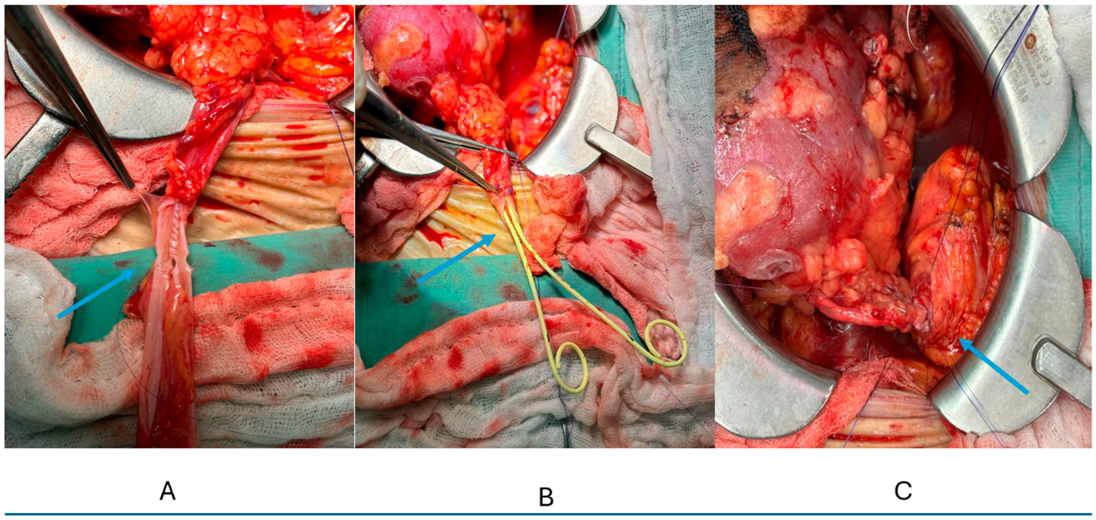
Figure 1. Intraoperative steps of pantaloon uretero-ureterostomy and bladder implantation. (A) Fashioning a common ureteral channel (“pantaloon”) by side-to-side uretero-ureteral anastomosis using a running 6-0 PDS II® monofilament polydioxanone suture (Ethicon). The blue arrow indicates the anastomosis preparation. (B) Placement of two ureteral stents, each advanced separately into one limb of the unified ureters. Follow the blue arrow. (C) Implantation of the unified ureteral channel into the recipient urinary bladder (ureteroneocystostomy; see the blue arrow).
Std 14 00031 g001
Table 1. Recipient demographics (matched cohort).
CharacteristicDouble Ureter (n = 26)Single Ureter (n = 26)p-Value
Age, years, median (IQR)51 (38–60)52 (40–61)0.824
Male sex, n (%)18 (69.2)17 (65.4)0.768
BMI, kg/m, median (IQR)25.5 (23.8–28.3)26.1 (24.2–28.8)0.642
Dialysis time, years, median (IQR)3.5 (2.0–5.0)3.2 (1.8–4.8)0.713
Preemptive transplant, n (%)2 (7.7)3 (11.5)0.640
Primary kidney disease, n (%) 0.892
- Diabetes mellitus9 (34.6)8 (30.8)
- PCKD3 (11.5)4 (15.4)
- Unknown6 (23.1)7 (26.9)
- Glomerular disease3 (11.5)4 (15.4)
- CAKUT3 (11.5)2 (7.7)
- Other2 (7.7)1 (3.8)
Data presented as median (IQR) or n (%). p-Values from Mann–Whitney U test (continuous) or Fisher’s exact test (categorical). BMI: body mass index; CAKUT: congenital anomalies of kidney and urinary tract; PCKD: polycystic kidney disease.
Table 2. Donor and transplant characteristics.
CharacteristicDouble Ureter (n = 26)Single Ureter (n = 26)p-Value
Donor type, n (%) 1.000
- Living donor13 (50.0)13 (50.0)
- Deceased donor13 (50.0)13 (50.0)
Donor age, years, median (IQR)
- Living donors42 (35–50)43 (36–51)0.812
- Deceased donors54 (45–62)55 (46–63)0.756
HLA mismatches, median (IQR)
- Living donors3 (2–4)3 (2–4)1.000
- Deceased donors5 (4–6)5 (4–6)1.000
Cold ischemia time, hours
- Living donors (by definition)1 (1–1)1 (1–1)1.000
- Deceased donors10 (8–12)11 (9–13)0.521
Multiple arteries, n (%)8 (30.8)7 (26.9)0.761
Induction therapy, n (%)
- Basiliximab (Simulect®) 20 (76.9)19 (73.1)0.751
- Thymoglobulin® (Sanofi)6 (23.1)7 (26.9)0.751
Table 3. Early post-transplant outcomes.
OutcomeDouble Ureter (n = 26)Single Ureter (n = 26)p-Value
Delayed graft function, n (%)3 (11.5)4 (15.4)1.000
- Living donor recipients0/13 (0)0/13 (0)1.000
- Deceased donor recipients3/13 (23.1)4/13 (30.8)1.000
Acute rejection < 90 days, n (%)2 (7.7)3 (11.5)1.000
Discharge creatinine, mg/dL
- All patients, median (IQR)1.26 (0.91–1.82)1.31 (0.95–1.89)0.724
- Living donors1.12 (0.85–1.45)1.15 (0.88–1.48)0.812
- Deceased donors1.68 (1.17–2.80)1.72 (1.20–2.85)0.867
Urologic complications, n (%)1 (3.8)2 (7.7)1.000
- Urinary leak1 (3.8)1 (3.8)
- Ureteral stricture < 90 days0 (0)1 (3.8)
- Vesicoureteral reflux0 (0)0 (0)
Clavien–Dindo Grade ≥ III, n (%)1 (3.8)2 (7.7)1.000
Table 4. Long-term outcomes.
OutcomeDouble Ureter (n = 26)Single Ureter (n = 26)p-Value
Follow-up time, months
- Median (IQR)63 (36–96)60 (36–92)0.812
- Range3–1686–164
Graft function at last F/U
Last creatinine, mg/dL6
- All patients, median (IQR)1.25 (1.02–1.72)1.28 (1.05–1.68)0.891
- Living donor recipients1.10 (0.94–1.34)1.12 (0.96–1.36)0.834
- Deceased donor recipients1.45 (1.15–1.94)1.48 (1.18–1.92)0.912
eGFR, mL/min/1.73 m
- All patients, median (IQR)58 (42–72)56 (44–70)0.765
Late ureteral stricture, n (%)0 (0)1 (3.8)1.000
Survival outcomes
1-year graft survival, %10096.21.000
5-year graft survival, %96.092.31.000
- Living donors1001001.000
- Deceased donors92.384.61.000
1-year patient survival, %96.296.21.000
5-year patient survival, %80.884.60.723
Graft loss, n (%)1 (3.8)2 (7.7)1.000
- Living donor recipients0/13 (0)0/13 (0)1.000
- Deceased donor recipients1/13 (7.7)2/13 (15.4)1.000
Patient death, n (%)5 (19.2)4 (15.4)1.000
- With functioning graft5 (19.2)3 (11.5)0.704
Disclaimer/Publisher’s Note: The statements, opinions and data contained in all publications are solely those of the individual author(s) and contributor(s) and not of MDPI and/or the editor(s). MDPI and/or the editor(s) disclaim responsibility for any injury to people or property resulting from any ideas, methods, instructions or products referred to in the content.