Article

Retrospective Validation Study of a Treatment Strategy for Benign Bone Lesions in the Proximal Femur

1
Department of Orthopaedic Surgery, Graduate School of Medical and Dental Sciences, Kagoshima University, 8-35-1 Sakuragaoka, Kagoshima 890-8520, Japan
2
Department of Clinical Physical Therapy, School of Health Sciences, Faculty of Medicine, Kagoshima University, 8-35-1 Sakuragaoka, Kagoshima 890-8544, Japan
*
Author to whom correspondence should be addressed.
Surg. Tech. Dev. 2025, 14(3), 29; https://doi.org/10.3390/std14030029
Submission received: 4 February 2025 / Revised: 7 April 2025 / Accepted: 15 August 2025 / Published: 22 August 2025

Abstract

Background: Benign bone tumors and tumor-like lesions in the proximal femur increase the risk of pathological fractures, often requiring surgical intervention. However, no consensus exists on the optimal treatment strategy. We developed a structured approach to guide the selection of implant types (compression hip screw [CHS] or intramedullary nail [IMN]) with or without bone grafting. This study aims to validate our treatment strategy through a retrospective analysis and a review of previous surgical outcomes. Methods: We sought to validate this strategy through a retrospective analysis of 16 patients (6 males and 10 females, mean age at surgery 37.4 years [range, 16–64 years]) with primary benign bone tumors or tumor-like conditions of the proximal femur, including the femoral head and neck. Curettage and synthetic or autologous bone graft was performed according to our treatment flowchart, utilizing either CHS or IMN for internal fixation. We compared the blood loss, operative time, time to full weight bearing, and perioperative complications between the CHS and IMN groups. Results: Blood loss did not significantly differ between the CHS and IMN groups (p = 0.11), but the operative time was significantly longer in the CHS group (p < 0.01). Two CHS cases experienced local recurrence, while no postoperative fractures were observed in either group. The median time to full weight bearing was 5 weeks, consistent with previous reports. No perioperative complications were noted. Conclusions: Our strategy achieved favorable clinical outcomes. IMN was selectively used in patients with non-aggressive benign tumors not involving the femoral head and neck, yielding good results with reduced surgical invasiveness, while in those patients with aggressive disease involving the head and neck, CHS was more appropriate. This approach may serve as a practical guide for surgical decision-making in benign proximal femoral bone tumors.

1. Introduction

Benign bone tumors and tumor-like diseases located in the proximal femur often increase the risk of pathologic fractures and require surgery. Although the proximal femur is mechanically important, there is still no consensus on the treatment strategy for benign bone tumors located in this region [1]. In particular, the methods to reconstruct the mechanical strength of the femoral cavity after curettage of the lesion are mainly divided into two groups: the grafting of bone or substitutes without any fixation and the addition of internal fixation after bone grafting. Some authors have reported good clinical outcomes of fibula strut grafting without internal fixation [2,3]. However, Maes et al. reported that the time to full weight bearing after this procedure was 12 weeks [3], resulting in the restriction of patients’ postsurgical activity for a long period. Although several groups reported the results of internal fixation using a compression hip screw (CHS) [4,5] or intramedullary nail (IMN) [6,7], there is no consensus on the optimal choice of internal fixation devices. Benign bone tumors are classified into three categories, such as benign, intermediate (locally aggressive), and intermediate (rarely metastasizing) according to the classification of the WHO [8]. Intermediate (locally aggressive) tumors often recur locally and may be associated with an infiltrative and locally destructive growth pattern [8]. Intermediate (rarely metastasizing) tumors often locally aggressive, which includes giant cell tumor (GCT). In this study, we define ‘aggressive’ bone tumors to include both intermediate categories, whereas ‘non-aggressive’ tumors include benign tumors based on the WHO classification. Therefore, ‘aggressive’ tumors include GCT, aneurysmal bone cyst (ABC), chondroblastoma, osteoblastoma, and chondromyxoid fibroma [9]. Among these tumors, GCT, ABC, and chondroblastoma occur in the proximal femur. Those ‘aggressive’ tumors require thorough curettage, often involving extensive cortical fenestration and meticulous surgical techniques to ensure complete removal [9,10,11,12]. In contrast, ‘non-aggressive’ tumors, including simple bone cysts and fibrous dysplasia, can often be managed with less invasive treatment approaches [13,14].Erol et al. proposed a treatment strategy for benign bone lesions in the proximal femur, particularly in pediatric patients [15]. However, their study did not include several aggressive tumors such as GCT and chondroblastoma, and its applicability to adult patients remains uncertain. Thus, there is a need for a systematic treatment strategy for benign proximal femoral bone lesions that takes the anatomical location and aggressiveness into consideration and helps guide the choice of the internal fixation device.
We have developed a strategy that helps surgeons decide which implant types to use with or without bone grafting, depending on the case. In this retrospective study, we analyzed treatment outcomes to validate our strategy and reviewed previous studies reporting the surgical treatment of proximal femoral bone lesions.

2. Materials and Methods

2.1. Inclusion and Exclusion Criteria

Inclusion criteria in this study were as follows: (1) bone tumor or tumor-like lesions involving that proximal femur that underwent surgical treatment; (2) preoperative image study including X-ray and CT or MRI was performed; (3) pathological diagnosis was obtained from a surgical specimen; (4) written consent was obtained from the patient or guardians. Exclusion criteria were as follows: (1) patients with active malignant disease; (2) hematological disease that may influence the amount of intraoperative bleeding.

2.2. Patients

This research was approved by the IRB of the authors’ affiliated institutions. Sixteen patients with primary benign bone tumors or tumor-like conditions of the proximal femur, including the femoral head and neck, were surgically treated at our hospital from 2005 to 2021 (Table 1). The patients comprised six male and ten female patients. Their average age at the time of surgical treatment was 37.4 years (range, 16–64 years). The pathological diagnosis was fibrous dysplasia in five patients, simple bone cyst in six, GCT in three, and chondroblastoma in two (Table 1). All participants provided their written consent.

2.3. Surgical Strategy and Fixation Device Selection
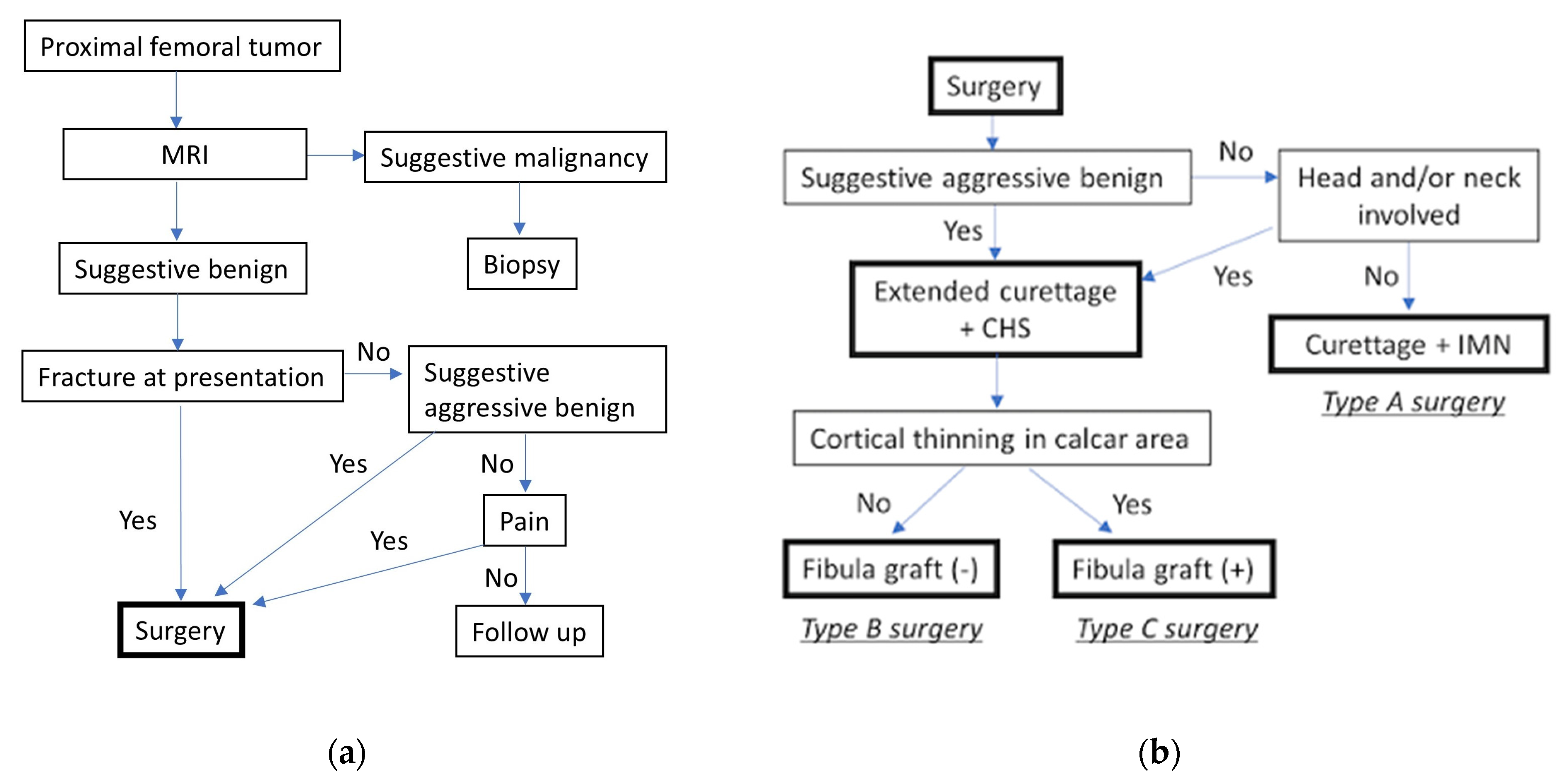
The indications for surgical treatment were decided according to our flowchart, which was based on different factors including the pathological fracture, pain on weight bearing, or suspected aggressiveness of the lesion (Figure 1a). The type of surgical procedure, including the choice of implant and addition of fibula grafting, was decided based on another flowchart (Figure 1b). The choice of surgical approach and fixation device was based on both the biological characteristics of the tumor and anatomical considerations. In cases where the lesion was in the femoral head or neck, or in the presence of an aggressive benign tumor, lateral cortical fenestration was considered necessary to facilitate thorough curettage and bone grafting. Compression hip screws (CHSs) were used to maintain adequate fixation after fenestration. Conversely, for non-aggressive benign tumors not involving the femoral head or neck, intramedullary nails (IMNs) were chosen to minimize surgical invasiveness, reduce blood loss, and maintain bone integrity. In all cases, synthetic bone substitutes such as β-tricalcium phosphate or calcium hydroxyapatite ceramic were used to reconstruct the cavity after curettage of the lesion. In cases of cortical thinning in the calcar area, non-vascularized fibula strut grafting was performed in addition to synthetic bone grafting.
Figure 1. Flowcharts for (a) surgical indication and (b) selection of surgical method. Based on the condition and symptoms, the indication and type of surgery (A~C) should be determined. MRI, magnetic resonance imaging; IMN, intramedullary nail; CHS, compression hip screw. The details of Type A~C surgeries are provided in Figure 2.
Std 14 00029 g001
Figure 2. Schematic presentation of surgical methods. (A) Curettage and intramedullary nail fixation (Type A). (B) Extended curettage and CHS fixation (Type B). (C) Strut fibula grafting in the calcar area with CHS fixation (Type C). CHS, compression hip screw.
Std 14 00029 g002

2.4. Surgical Procedure

Surgery was performed under general anesthesia with the patient in the supine position on a radiolucent table. In patients with lesions suggestive of non-aggressive benign tumors not involving the femoral head and neck, curettage was performed through the insertion hole of the IMN at the apex of the greater trochanter and the lag screw entry hole in the lateral femoral cortex. Synthetic bone was grafted into the defect after curettage, and an IMN implant was placed (Figure 2A). Extended curettage and internal fixation with a CHS were performed through a lateral skin incision as distal as necessary to the center of the greater trochanter. A cortical window was created from the lateral femoral cortex under fluoroscopy. Following curettage of the bone tumor and speed burring of the cavity wall, synthetic bone was grafted into the bone defect, and the CHS was inserted (Figure 2B). In cases of cortical thinning in the calcar area, autologous non-vascularized fibula strut grafting was performed in addition to synthetic bone grafting (Figure 2C).

2.5. Postoperative Rehabilitation and Weight-Bearing Protocol

Passive and active range-of-motion exercises were started immediately after surgery. In patients without cortical thinning or fracture, partial weight bearing was started between 1 and 7 days postoperatively. In patients with cortical thinning or fracture, partial weight loading was started between 28 and 42 days after surgery based on a radiographic assessment of the cortical bone status. The amount of loading was increased according to the severity of pain, and full loading was performed 90 days postoperatively at the latest.

2.6. Monitoring for Tumor Recurrence

Tumor recurrence was defined as radiographic evidence of lesion regrowth or cortical bone destruction detected on X-ray imaging. Routine follow-up imaging was performed, and when recurrence was suspected on X-ray, additional evaluation with CT or MRI was conducted to confirm the presence of recurrent disease.

2.7. Statistical Analysis

Statistical analysis was performed using SAS JMP Pro 16 (SAS Institute, Cary, NC, USA). The blood loss, operative time, time to partial weight bearing, and time to full weight bearing were analyzed. The Wilcoxon rank-sum test was used for nonparametric analysis, with statistical significance set at p < 0.05. To minimize potential biases inherent to the retrospective study design, standardized criteria for surgical indications were established to ensure consistency in patient selection and outcome assessment.

3. Results

3.1. Patient Characteristics and Treatment Distribution

A total of 16 patients were treated, with 11 patients undergoing CHS fixation and 6 patients receiving IMN fixation (Table 2). Among the CHS group, 5 patients underwent fibula grafting combined with synthetic bone grafting, while 6 patients received synthetic bone grafting only. In the IMN group, all patients underwent synthetic bone grafting only.

3.2. Operative Outcomes

The median intraoperative blood loss was 300 g (range, 200–1205 g) in the CHS group and 168 g (range, 90–327.5 g) in the IMN group. The median operative time was 224 min (range, 174–302 min) in the CHS group and 110 min (range, 95–140.5 min) in the IMN group. There was no significant difference in blood loss between the CHS and IMN groups (p = 0.11), but the operative time was significantly longer in the CHS group (p < 0.01).

3.3. Impact of Fibula Grafting on Surgical Outcomes

To assess the influence of fibula grafting, a subset analysis of CHS patients without fibula grafting was performed. The median blood loss was 230 g (range, 146.75–620 g) and the median operative time was 178.5 min (range, 155.5–299 min). When comparing CHS patients without fibula grafting to the IMN group, there was no significant difference in blood loss (p = 0.30), but the operative time remained significantly longer in the CHS group (p < 0.05) (Table 3).

3.4. Functional Recovery and Complications

The median time to full weight bearing was 5 weeks, with no significant difference between the CHS and IMN groups. Local tumor recurrence occurred in two CHS-treated patients with GCT, while no recurrence was observed in the IMN group. For a case with recurrent GCT, denosumab was administered, which resulted in stabilization of the tumor. In another case of GCT, recurrence was found based on MRI and CT; however, a follow-up study revealed no progression of the lesion after 6 years of recurrence. No postoperative pathological fractures occurred in either group, and no surgical site infections were observed.

3.5. Summary of Key Findings

IMN fixation resulted in a shorter operative time and less surgical invasiveness compared to CHS. CHS was required for aggressive benign tumors involving the femoral head and neck. Fibula grafting increased the operative time but had no significant effect on blood loss. No postoperative fractures or infections were observed, and the median time to full weight bearing was comparable to previous reports.

4. Discussion

The need for surgical augmentation after the curettage of benign proximal femoral lesions is controversial [16]. If the tumor involves >50% of the diameter of the femoral neck or >50% of the cortical bone of the femoral neck, some authors have recommended surgical augmentation of the proximal femur to prevent postoperative fracture [4,17].
In traumatic proximal femoral fractures, the use of IMNs reportedly results in firmer fixation, a shorter operative time, a lower reoperation rate, and less blood loss compared with the use of CHSs [18,19,20]. We propose the use of IMNs for select benign tumors of the proximal femur because of the expected lower blood loss and shorter operative time compared with CHSs, as confirmed in this study. The disadvantage of IMN fixation is that the lateral cortex cannot be widely opened for curettage or bone grafting. Because the intact lateral cortex is included in the “three-point fixation,” which is considered important to avoid mechanical failure of the IMN [21]; postoperative implant failure can be expected if the lateral cortex is opened too widely. In the present study, we demonstrated that curettage and IMN fixation for the patients selected according to our flowchart resulted in no mechanical failure or tumor recurrence. An advantage of CHS fixation is that curettage of proximal femoral tumors can be approached from the lateral side via a wide fenestration of the lateral cortex without an additional skin incision [5]. In femoral head and neck lesions, firm fixation with a CHS is necessary because the lateral cortex should be opened for curettage and adequate bone grafting. Because aggressive benign tumors such as GCT or ABC in this region have a high risk of postoperative recurrence if extended curettage through the cortical window is not performed, internal fixation with a CHS should be chosen (Figure 1). Although some groups have reported that the anterior approach is advantageous for controlling tumors in this region [6,22], Nakamura et al. [5] reported that the anterior approach carries a high risk of damage to the femoral rotator artery and postoperative necrosis of the femoral head when performing curettage of tumors in the proximal femur. Thus, IMNs can effectively achieve firm fixation with less blood loss and a shorter operative time for the treatment of non-aggressive benign tumors that do not require a fenestration of the lateral femoral cortex.
Concerning surgical invasiveness, we did not find a significant difference in intraoperative blood loss between the IMN and CHS groups. Based on the previous studies, it is still controversial whether there is a significant difference in intraoperative blood loss between the two implant types [5,23]. A study by Gao et al. on surgeries for proximal femoral metastatic bone tumors reported that the operation time was longer in the CHS group than in the IMN group, while blood loss was not significantly different [23]. The actual numerical data on blood loss associated with implant fixation for benign proximal femoral tumors was reported only by Nakamura et al., who documented a mean blood loss of 1088 g (range, 44–3200 g) in cases treated with CHS fixation [5]. In contrast, the mean blood loss in CHS-treated cases in our study was 300 g (Table 2), suggesting that our study may have included a higher proportion of cases with lower blood loss due to the small sample size. In addition, differences in surgical techniques, tumor characteristics, and patient selection criteria may have contributed to the observed differences.
An extended surgical duration can contribute to increased intraoperative bleeding and prolonged anesthesia exposure, which may lead to delayed postoperative recovery. Gao et al. reported that the length of hospital stay was longer in the CHS group than in the IMN group, possibly related to a longer operation time in the CHS group [23]. Although no significant differences in perioperative complications were observed in this study, optimizing perioperative management for patients undergoing curettage and internal fixation is essential to minimize these potential risks regardless of the choice of IMN or CHS.
In the treatment of benign lesions of the proximal femur, some authors have reported good outcomes with autologous nonvascularized fibula grafts [2,3,4]. Because good results with the use of artificial bone substitutes alone to fill the bone defect have also been recently reported [24,25], there is no consensus on the choice of materials to fill the bone defect. Our strategy is to use artificial bone; a fibular graft is recommended only in cases of cortical thinning in the calcar area. An autologous fibula strut graft is an effective bone substitute material to support mechanical weakness of the bone lesion, but it has the disadvantages of an extended duration of surgery and donor-site complications such as peroneal nerve palsy and contracture of toe extensor muscles. In our experience, patients who underwent autologous fibula grafting had a longer operative time than those who did not undergo fibula grafting. The necessity of autologous fibula transplantation should be further investigated. Furthermore, a strut allograft may be considered to minimize complications of the donor site of the autologous bone graft, although the supply of structural allografts from a cadaveric donor is very limited in Japan.
We reviewed previous studies on the surgical treatment of proximal benign bone lesions and summarized them for a comparison with our present results (Table 4). To the best of our knowledge, only one report to date [15] has analyzed the differences in outcomes and complications of benign proximal femoral bone tumors treated with or without internal fixation. Regarding the oncological outcome, the recurrence rate (12.5%) was comparable with that in other reports, although our study included cases treated using curettage without opening a cortical window for non-aggressive tumors. In terms of complications, the advantage of using internal fixation is achieving a shorter time to full weight bearing compared with surgery without internal fixation. Previous studies without internal fixation reported an average of 12–13 weeks to full weight bearing [2,3], whereas those with internal fixation reported an average of 3–6 weeks [4,5] (Table 4). The median time to full weight bearing in our study was 5 weeks, which is comparable with previous reports. No complications such as postoperative fracture or surgical site infection were observed in our study.
This study has several limitations. To enhance the clinical applicability of this treatment strategy, patient-specific factors must be carefully considered. The study population primarily comprised relatively young patients with presumably preserved bone strength. However, in elderly or osteoporotic patients, where a less invasive yet mechanically stable fixation is preferable, an alternative treatment algorithm expanding the indications for IMN may be necessary. Furthermore, this study did not compare the bone healing potential of fibular grafts with that of artificial bone. If artificial bone demonstrates non-inferior outcomes, it may be a preferable option given the risks of donor site complications. Additionally, for aggressive bone tumors such as GCT, where recurrence was observed in this study, adjuvant treatments like phenol application or cryotherapy are commonly used in addition to curettage. However, these adjuvant therapies were not incorporated into the treatment strategy proposed in this study. Moreover, as a retrospective study with a limited sample size, selection bias cannot be entirely ruled out. For instance, the duration of the surgery may not sorely be influenced by the type of implant; rather, the location or type of tumor may have affected the difficulty of the surgery. Late complications, including implant failure, were also not evaluated. Future research should focus on large-scale prospective studies with long-term follow-up to validate these findings and further optimize the treatment approach.

5. Conclusions

In conclusion, this retrospective validation study confirms the clinical feasibility of our treatment strategy for benign proximal femoral lesions, demonstrating favorable outcomes with a low complication rate. In particular, IMNs were selectively used in patients with non-aggressive benign tumors not involving the femoral head and neck, and good results were achieved with reduced surgical invasiveness. Further research is needed to evaluate its long-term efficacy in clinical practice, expand its applicability to a broader patient population, and assess the utility of fibular grafting.

Author Contributions

N.S. performed the collection and analysis of the data. H.S. analyzed the data and worked on the manuscript. S.N. contributed to the design and implementation of the research and to the writing of the manuscript. N.T. contributed to critical review and editing of the manuscript. All authors have read and agreed to the published version of the manuscript.

Funding

This research received no external funding.

Institutional Review Board Statement

The study was conducted in accordance with the Declaration of Helsinki and approved by the Institutional Review Board of Kagoshima University (protocol code 230055, approved on 4 August 2023).

Informed Consent Statement

Informed consent was obtained from all subjects involved in the study.

Data Availability Statement

The data supporting the reported results are available from the corresponding author upon reasonable request.

Acknowledgments

We thank Angela Morben for editing a draft of this manuscript.

Conflicts of Interest

The authors declare no conflicts of interest.

Abbreviations

The following abbreviations are used in this manuscript:
CHSCompression hip screw
IMNIntramedullary nail
GCTGiant cell tumor
ABCAneurysmal bone cyst
FDFibrous dysplasia
SBCSimple bone cyst
PWBPartial weight bearing
FWBFull weight bearing

References

  1. Shi, J.; Zhao, Z.; Yan, T.; Guo, W.; Yang, R.; Tang, X.; Qu, H.; Dong, S. Surgical treatment of benign osteolytic lesions in the femoral head and neck: A systematic review. BMC Musculoskelet. Disord. 2021, 22, 549. [Google Scholar] [CrossRef]
  2. George, B.; Abudu, A.; Grimer, R.J.; Carter, S.R.; Tillman, R.M. The treatment of benign lesions of the proximal femur with non-vascularised autologous fibular strut grafts. J. Bone Jt. Surg. Br. 2008, 90, 648–651. [Google Scholar] [CrossRef]
  3. Maes, D.J.A.; Kaneuchi, Y.; Abudu, A.; Stevenson, J.D. Biological reconstruction of the proximal femur after treatment of benign lesions: Comparison of functional and oncological outcomes for children and adults. Eur. J. Orthop. Surg. Traumatol. 2022, 32, 559–566. [Google Scholar] [CrossRef]
  4. Jaffe, K.A.; Launer, E.P.; Scholl, B.M. Use of a fibular allograft strut in the treatment of benign lesions of the proximal femur. Am. J. Orthop. 2002, 31, 575–578. [Google Scholar]
  5. Nakamura, T.; Matsumine, A.; Asanuma, K.; Matsubara, T.; Sudo, A. Treatment of the benign bone tumors including femoral neck lesion using compression hip screw and synthetic bone graft. SICOT J. 2015, 1, 15. [Google Scholar] [CrossRef] [PubMed]
  6. Zhang, Y.; Li, J.Z.; Lu, X.C.; Zhang, Y.; Zhang, H.S.; Shi, H.L.; Lei, Z.; Feng, G.; Fu, W.P. Intramedullary Nailing Combined with Bone Grafting for Benign Lesions of the Proximal Femur. Orthop. Surg. 2017, 9, 97–102. [Google Scholar] [CrossRef] [PubMed]
  7. Kushare, I.V.; Colo, D.; Bakhshi, H.; Dormans, J.P. Fibrous dysplasia of the proximal femur: Surgical management options and outcomes. J. Child. Orthop. 2014, 8, 505–511. [Google Scholar] [CrossRef] [PubMed]
  8. International Agency for Research on Cancer. Soft Tissue and Bone Tumours; World Health Organization: Geneva, Switzerland, 2020; Volume 3.
  9. Yenigul, A.E.; Sofulu, O.; Erol, B. Treatment of locally aggressive benign bone tumors by means of extended intralesional curettage without chemical adjuvants. SAGE Open Med. 2022, 10, 20503121221094199. [Google Scholar] [CrossRef]
  10. Rahman, M.A.; El Masry, A.M.; Azmy, S.I. Review of 16 cases of aneurysmal bone cyst in the proximal femur treated by extended curettage and cryosurgery with reconstruction using autogenous nonvascularized fibula graft. J. Orthop. Surg. 2018, 26, 2309499018783905. [Google Scholar] [CrossRef]
  11. Errani, C.; Ruggieri, P.; Asenzio, M.A.; Toscano, A.; Colangeli, S.; Rimondi, E.; Rossi, G.; Longhi, A.; Mercuri, M. Giant cell tumor of the extremity: A review of 349 cases from a single institution. Cancer Treat. Rev. 2010, 36, 1–7. [Google Scholar] [CrossRef]
  12. Wijsbek, A.E.; Vazquez-Garcia, B.L.; Grimer, R.J.; Carter, S.R.; Abudu, A.A.; Tillman, R.M.; Jeys, L. Giant cell tumour of the proximal femur: Is joint-sparing management ever successful? Bone Jt. J. 2014, 96, 127–131. [Google Scholar] [CrossRef] [PubMed]
  13. Stanton, R.P.; Ippolito, E.; Springfield, D.; Lindaman, L.; Wientroub, S.; Leet, A. The surgical management of fibrous dysplasia of bone. Orphanet J. Rare Dis. 2012, 7 (Suppl. S1), S1. [Google Scholar] [CrossRef] [PubMed]
  14. Roposch, A.; Saraph, V.; Linhart, W.E. Treatment of femoral neck and trochanteric simple bone cysts. Arch. Orthop. Trauma Surg. 2004, 124, 437–442. [Google Scholar] [CrossRef] [PubMed]
  15. Erol, B.; Topkar, M.O.; Aydemir, A.N.; Okay, E.; Caliskan, E.; Sofulu, O. A treatment strategy for proximal femoral benign bone lesions in children and recommended surgical procedures: Retrospective analysis of 62 patients. Arch. Orthop. Trauma Surg. 2016, 136, 1051–1061. [Google Scholar] [CrossRef] [PubMed]
  16. Panchwagh, Y.; Joshi, S.K.; Sancheti, P.K. Benign Aggressive Lesions of Femoral Head and Neck: Is Salvage Possible? Indian J. Orthop. 2018, 52, 51–57. [Google Scholar] [CrossRef]
  17. Günther, K.P.; Hartmann, A.; Aikele, P.; Aust, D.; Ziegler, J. Large femoral-neck cysts in association with femoroacetabular impingement: A report of three cases. J. Bone Jt. Surg. 2007, 89, 863–870. [Google Scholar] [CrossRef]
  18. Friedl, W.; Clausen, J. Experimental examination for optimized stabilisation of trochanteric femur fractures, intra- or extramedullary implant localisation and influence of femur neck component profile on cut-out risk. Chirurg 2001, 72, 1344–1352. [Google Scholar] [CrossRef]
  19. Grønhaug, K.M.L.; Dybvik, E.; Matre, K.; Östman, B.; Gjertsen, J.E. Intramedullary nail versus sliding hip screw for stable and unstable trochanteric and subtrochanteric fractures: 17,341 patients from the Norwegian Hip Fracture Register. Bone Jt. J. 2022, 104, 274–282. [Google Scholar] [CrossRef]
  20. Zang, W.; Liu, P.F.; Han, X.F. A comparative study of proximal femoral locking compress plate, proximal femoral nail antirotation and dynamic hip screw in intertrochanteric fractures. Eur. Rev. Med. Pharmacol. Sci. 2018, 22, 119–123. [Google Scholar] [CrossRef]
  21. Abram, S.G.; Pollard, T.C.; Andrade, A.J. Inadequate ‘three-point’ proximal fixation predicts failure of the Gamma nail. Bone Jt. J. 2013, 95, 825–830. [Google Scholar] [CrossRef]
  22. Hu, Y.C.; Lun, D.X.; Zhao, S.K. Combined anterior and lateral approaches for bone tumors of the femoral neck and head. Orthopedics 2012, 35, e628–e634. [Google Scholar] [CrossRef] [PubMed]
  23. Gao, H.; Bai, X.; Chen, W.; Li, Y.; Zhao, L.; Liu, C.; Liu, Z.; Wang, B. Clinical and functional comparison of dynamic hip screws and intramedullary nails for treating proximal femur metastases in older individuals. Chin. J. Cancer Res. 2020, 32, 395–402. [Google Scholar] [CrossRef] [PubMed]
  24. Zekry, K.M.; Yamamoto, N.; Hayashi, K.; Takeuchi, A.; Alkhooly, A.; Abd-Elfattah, A.S.; Fouly, E.H.; Ahmed, A.R.; Tsuchiya, H. Treatment of the benign lytic lesions of the proximal femur with synthetic bone graft. J. Orthop. Surg. Res. 2018, 13, 270. [Google Scholar] [CrossRef] [PubMed]
  25. Matsumine, A.; Myoui, A.; Kusuzaki, K.; Araki, N.; Seto, M.; Yoshikawa, H.; Uchida, A. Calcium hydroxyapatite ceramic implants in bone tumour surgery: A long-term follow-up study. J. Bone Jt. Surg. Br. 2004, 86, 719–725. [Google Scholar] [CrossRef]
Table 1. Summary of the background characteristics of the cases.
CaseAgeGenderDiagnosisLocationSurgery Type
HeadNeckTrochanterSub-
Trochanter
119MFD ++A
224FFD +A
356FSBC +A
431FSBC +A
520FSBC ++A
616MChondroblastoma+ B
736MChondroblastoma ++B
862FGCT ++ B
957FSBC ++ B
1064MSBC++ B
1137FSBC ++ B
1244MGCT ++ C
1322MGCT +++C
1423FFD++++C
1542FFD + C
1631FFD +++C
FD, fibrous dysplasia; GCT, giant cell tumor; SBC, simple bone cyst.; +, presence of the lesion at the specified anatomical site.
Table 2. Comparison between two groups with different internal fixation devices.
Internal Fixation Devicep-Value
IMN (N = 5)CHS (N = 11)
Time to PWB (day)31 (2–42.5)14 (7–30)0.78
Time to FWB (day)37 (16–68.5)30 (28–56)1.00
Bleeding (g)168 (90–327.5)300 (200–1205)0.11
Operative time (min)110 (95–140.5)224 (174–302)<0.01
IMN, intramedullary nail; CHS, compression hip screw; FWB, full weight bearing; PWB, partial weight bearing.
Table 3. Comparison between two groups of surgical methods without a fibula graft.
Internal Fixation Methodp-Value
IMN
(Type A, N = 5)
CHS Without
Fibula Graft
(Type B, N = 6)
Bleeding (g)168 (90–327.5)230 (146.75–620)0.30
Operative time (min)110 (95–140.5)178.5 (155.5–299)<0.05
IMN, intramedullary nail; CHS, compression hip screw.
Table 4. Summary of the studies based on surgical methods and clinical outcomes in comparison with our study.
Authors (Year)NInternal
Fixation
Type of GraftTime to Full Wight Bearing (Weeks)Postoperative FractureTumor
Recurrence
Other Complications
George et al. 2008 [2]17NoAutologous fibula13.5None2 (11.7%)
Maes et al.
2021 [3]
54NoAutologous fibula125 (10%)5 (9%)Peroneal nerve palsy (15%)
Jaffe et al. 2002 [4]15CHSAutologous fibula6NoneNone
Nakamura et al. 2015 [5]13CHSSynthetic bone3None1 (7.7%)
Zhang et al. 2017 [6]32NoAllogeneic boneN. S.1 (3.1%)2 (6.3%)
36IMNAllogeneic boneN. S.None1 (2.8%)
Erol et al.
2016 [15]
8NoAllogeneic or autologous bone6
(hip spica cast for 6 weeks)
NoneNoneSuperficial wound infection (n = 1), skin necrosis (n = 1)
54CHS or
elastic nail
Allogeneic or autologous bone
Our study16CHS
or
IMN
Synthetic bone with or without autologous fibula5None2 (12.5%)
CHS, compression hip screw; IMN, intramedullary nail.
Disclaimer/Publisher’s Note: The statements, opinions and data contained in all publications are solely those of the individual author(s) and contributor(s) and not of MDPI and/or the editor(s). MDPI and/or the editor(s) disclaim responsibility for any injury to people or property resulting from any ideas, methods, instructions or products referred to in the content.