Article

The Treatment of Three-Part Fractures of Humeral Head: A Retrospective Study to Compare Nail vs. Plate

by
Francesco Roberto Evola
1,2,*,
Michele Vecchio
2,
Marco Vacante
3 and
Giuseppe Evola
2,4
1
Department of Orthopaedic and Trauma Surgery, Cannizzaro Hospital, 95100 Catania, Italy
2
Department of Biomedical and Biotechnological Sciences, University of Catania, 95123 Catania, Italy
3
Unit of Internal Medicine Critical Area, Garibaldi Hospital, 95100 Catania, Italy
4
Surgery Department, Garibaldi Hospital, 95100 Catania, Italy
*
Author to whom correspondence should be addressed.
Surg. Tech. Dev. 2025, 14(3), 23; https://doi.org/10.3390/std14030023
Submission received: 1 December 2024 / Revised: 17 April 2025 / Accepted: 3 July 2025 / Published: 12 July 2025

Abstract

Background: There are no clear guidelines to support management decisions for patients with three-part fractures of the proximal humerus. The aim of the study is to identify the treatment used and to assess the functional and radiological outcomes at follow-up. Methods: A total of 126 patients were retrospectively included in the study and were divided into two groups based on the type of surgery: plate and nail group. We collected data on the patient’s sex, age, fracture type, surgery duration, fracture healing, initial and final neck–shaft angles, shoulder joint score, and complications. Results: A total of 69 patients received locking-plate internal fixation, while 77 patients underwent fixation with intramedullary nail. The two groups were comparable, with no significant differences observed in age, sex, or the number of patients. The average operation time for the locking-plate group (88.7 ± 10.5 min) was significantly longer compared to the intramedullary nail group (70.2 ± 8.3 min). The Constant–Murley score was 91.2 ± 6.7 (range 79–98) in the plate group and 90.5 ± 7.7 (range 80–98) in the nail group, with no statistically significant difference. Complications were observed in 16 patients (23.2%) of the locking-plate group and in 7 patients (9.1%) of the intramedullary nail group, with significant difference. Conclusions: Our assessment revealed no significant differences in fracture healing times, loss of reduction, or Constant–Murley scores between two groups. However, our results suggest that intramedullary nails have an advantage over locking plates in terms of reduced operation time and complications.

1. Introduction

Proximal humeral fractures constitute approximately 5–6% of all fractures in the adult population, with a marked predominance among females and individuals over the age of 60 [1,2,3,4]. With the progressive aging of the population, the incidence of these fractures is projected to increase in the coming decades, contributing to elevated healthcare costs and imposing an additional burden on healthcare systems. Although approximately 85% of proximal humeral fractures are managed non-operatively [5], displaced fractures involving the greater tuberosity are often associated with considerable functional impairment of the shoulder. In such cases, surgical management may offer superior clinical outcomes. The management of proximal humerus fractures remains complex and necessitates thorough clinical evaluation. Although multiple classification systems have been proposed in the literature, none has led to standardized or universally accepted treatment protocols [6,7]. Among these, the Neer classification is the most widely utilized, categorizing fractures according to the number of fragments and the extent of their displacement [8]. This system plays a pivotal role in prognostic assessment and in guiding therapeutic decision-making. Originally introduced in 1970, the Neer classification provides critical information regarding the risk of vascular compromise to the humeral head, which increases with both the number of fracture fragments and the degree of their displacement [8]. Within this framework, three-part fractures, comprising the greater tuberosity, humeral head, and humeral shaft, can be further subdivided into three distinct subgroups [9]. Subgroup 1 encompasses fractures characterized by valgus impaction of the humeral head; subgroup 2 includes fractures that are stable and minimally displaced; and subgroup 3 refers to those with procurvatum–varus angulation and loss of medial cortical continuity. Treatment strategies should consider not only the specific fracture type but also various factors, including the integrity of the posteromedial cortex, the status of the vascular supply, rotator-cuff function, the patient’s age, bone quality, and pre-fracture health status. However, the optimal fixation method for different fracture patterns remains a topic of ongoing debate. The locking plate is generally regarded as the gold standard for managing proximal humerus fractures, given its ability to achieve more precise reduction and stabilization of the fracture fragments. Nevertheless, its use is associated with a notable risk of complications, such as varus deformity of the humeral head, avascular necrosis, screw penetration, and loosening of the internal fixation [10,11,12]. In contrast, intramedullary nails, despite not facilitating an anatomical reduction of the humeral tuberosity, have become increasingly favored by orthopedic surgeons due to their minimally invasive insertion technique, reduced operative time, decreased blood loss, superior fracture stability, and lower incidence of complications [10,13,14,15]. However, the existing literature consists predominantly of studies with heterogeneous patient populations, including both two- or three-part fractures, as well as three- or four-part fractures of the humeral head. To date, there are no definitive guidelines for making treatment decisions in patients with three-part fractures of the proximal humerus. The objective of this study is to identify the treatment approaches employed for each subgroup of three-part proximal humerus fractures and to evaluate the functional and radiological outcomes associated with the two primary treatment modalities.

2. Materials and Methods

2.1. Patients

A retrospective analysis was conducted on patients diagnosed with proximal humerus fractures who received surgical management at our institution between June 2020 and June 2023. Owing to the retrospective design of the study and the exclusive use of anonymized, aggregated clinical data, formal approval from the institutional ethics committee was not deemed necessary. Inclusion criteria encompassed patients aged 18 years or older who provided informed consent for surgical intervention and demonstrated the physical capability to actively engage in postoperative functional rehabilitation. Eligible participants were free from debilitating comorbidities and assessed as fit to undergo anesthesia and surgical procedures. Only individuals diagnosed with three-part proximal humerus fractures and a documented minimum clinical follow-up of 12 months were included. Exclusion criteria comprised patients with pre-existing rotator-cuff pathology resulting in impaired shoulder joint function, as well as those with radiographic evidence of glenohumeral osteoarthritis prior to surgery. Additional exclusion parameters included open fractures of the proximal humerus, pathological or multi-fragmentary fractures, a prior history of fractures involving the proximal humerus, cases managed via hemiarthroplasty or total shoulder arthroplasty, patients who had undergone hardware removal, and individuals who were lost to follow-up. Written informed consent was obtained from all participants prior to their inclusion in the study. A retrospective evaluation was performed on 465 electronic medical records. Following the application of the predefined inclusion and exclusion criteria, a final cohort of 126 patients was identified. Subjects were stratified into two groups according to the surgical technique employed. Demographic characteristics and fracture classification were systematically documented for each patient.

2.2. Surgical Procedure

All 126 patients included in the study underwent surgical intervention performed by two orthopedic surgeons, each possessing equivalent expertise in both operative techniques. As a standard prophylactic measure, cefuroxime was administered intravenously 30 min prior to the procedure to minimize the risk of postoperative infection. The selection of the fixation method, locking plate versus intramedullary nail, was primarily guided by the specific fracture morphology. Intramedullary nailing was preferred in cases presenting with stable, minimally displaced fractures or unstable varus configurations with limited involvement of the tuberosities (Figure 1). Conversely, locking-plate fixation was selected as the initial approach for fractures demonstrating stable valgus alignment or in the presence of unstable varus patterns accompanied by significant tuberosity fragmentation (Figure 2). For patients undergoing locking-plate internal fixation, general anesthesia was administered, and individuals were positioned in the beach-chair configuration. The minimally invasive plate osteosynthesis (MIPO) technique was employed in all cases within this group. In instances where anatomical reduction of the humeral head was not readily achieved, one or two 2.0 mm Kirschner wires were temporarily inserted into the humeral head to facilitate fracture reduction, functioning as leverage tools. These wires also served as provisional stabilization devices during the procedure. The locking plate was positioned 5–8 mm inferior to the apex of the greater tuberosity. Fixation commenced with the placement of cortical screws to anchor the plate to the humeral diaphysis, followed by the insertion of locking screws into the proximal humerus to complete the construct. Fracture alignment was confirmed using both direct visualization and fluoroscopic assessment. In patients exhibiting metaphyseal bone loss (subgroup 1), cortico-cancellous allograft was used to fill the valgus-oriented void in the humeral head. In cases with disruption of the medial cortex (subgroup 3), calcar screws were employed to enhance structural support in the region of dense subchondral bone. Upon completion of fixation, rotator-cuff sutures were reinforced as necessary. Postoperatively, all patients were fitted with a shoulder brace, which was maintained for a duration of four weeks. For patients treated with intramedullary nail fixation, general anesthesia was administered, and the individual was placed in a supine position. A deltoid-splitting approach was employed, with the skin incision initiated at the anterolateral border of the acromion. The deltoid muscle was carefully split along the line of the incision, specifically at the junction between its anterior and middle thirds. In cases presenting with displacement of the greater or lesser tuberosity, bone fragments were anatomically repositioned and secured using 5-0 non-absorbable sutures. When fracture instability persisted following reduction, temporary stabilization was achieved using a 2.0 mm Kirschner wire. The intramedullary nail was then inserted and advanced approximately 1–2 mm into the subchondral bone. In most cases, appropriate selection of the entry point facilitated spontaneous anatomical reduction upon insertion of the nail. Proximal stabilization was attained using 2 to 4 locking screws, while distal fixation was completed with 1 to 2 screws, depending on fracture characteristics. Following the procedure, a shoulder brace was applied and maintained for a period of three weeks.

2.3. Postoperative Treatment

Perioperative antibiotic prophylaxis was routinely administered during surgery and continued for 24 h postoperatively to minimize the risk of surgical site infection. Sutures were removed approximately two weeks after surgery, following confirmation of complete wound healing. Passive shoulder mobilization exercises were initiated at the two-week mark, while an active range of motion exercises commenced around four weeks postoperatively. Rehabilitation protocols were standardized, with no significant differences in postoperative activity regimens observed between the two treatment groups.

2.4. Data Collection

Data were systematically collected regarding the patient’s sex, age, fracture classification, surgical duration, fracture healing, initial and final neck–shaft angles, shoulder joint score, and any postoperative complications. A minimum follow-up period of one year was observed, at which time shoulder function was evaluated using the Constant–Murley score [16]. This scoring system comprises a total of 100 points, allocated across four domains: 15 points for pain, 20 points for activities of daily living, 40 points for range of motion, and 25 points for strength. Based on the total score, shoulder function was classified as follows: excellent (90–100 points), good (80–89 points), moderate (70–79 points), and poor (below 70 points). Preoperative radiographic assessment was performed to evaluate the displacement of the humeral head using the anteroposterior (AP) view, with particular attention paid to the α angle (Figure 3) [13,17]. Tuberosity displacement was defined as a shift of 5 mm or more from the original position. The integrity of the calcar was assessed according to the criteria outlined by Hertel and Resch [18,19]. Postoperative radiographic evaluations were conducted at regular intervals (1, 3, 6, and 12 months) to monitor fracture healing and identify any complications, including signs of avascular necrosis, varus collapse of the humeral head, secondary displacement or loss of reduction of the tuberosity, fixation failure, or screw penetration. Fracture union was evaluated based on the complete obliteration of fracture lines and the formation of bridging trabecular bone on plain radiographs, with no evidence of loss of reduction or implant failure. Nonunion was defined as the absence of callus formation and clinical union after a minimum of six months postoperatively. Malunion was diagnosed when there was a loss of the initial anatomical reduction exceeding 15° or 5 mm. Avascular necrosis was identified by the progressive resorption and collapse of the humeral head. Radiographic assessments at both the time of presentation and during follow-up visits included a true anterior–posterior view and a scapular Y view. An axillary view was also obtained when possible. All results were independently evaluated by two orthopedic surgeons to ensure consistency and accuracy in assessment.

2.5. Statistical Analyses

Data collection and analysis were performed using Excel 2016 (Microsoft, Redmond, WA, USA), while statistical analyses were conducted with SPSS 14.0 Statistics for Windows (IBM Corp., Armonk, NY, USA). Continuous variables are presented as means, standard deviations, and ranges, while categorical data are expressed as counts and percentages. Continuous data were analyzed using the t-test, while categorical variables were evaluated using the Chi-square test and Fisher’s exact test. Pearson’s correlation coefficient was employed to assess associations between variables. Power analysis was performed using the G-Power software, with an alpha error threshold set at 0.05. A p-value of less than 0.05 was considered indicative of statistical significance.

3. Results

Between June 2020 and June 2023, a total of 465 patients with three-part proximal humerus neck fractures were admitted to our institution. After applying the predefined inclusion and exclusion criteria, 126 patients were enrolled in this study. Of these, 69 patients underwent minimally invasive open reduction and internal fixation with a locking plate, while 77 patients received limited open reduction and internal fixation using an intramedullary nail. The average age was 61.1 ± 3.2 years (range 24–78) in the locking-plate group and 62.4 ± 2.9 years (range 19–73) in the intramedullary nail group. In the locking-plate group, 22 patients (31.8%) were male, while 28 patients (36.3%) were male in the intramedullary nail group. Falls were the predominant mechanism of injury in both groups, accounting for 82.1% of cases in the locking-plate group and 81.3% in the intramedullary nail group. Patient characteristics are summarized in Table 1.
According to Neer’s classification subgroups, locking-plate fixation was performed on 49 patients in subgroup 1 and 20 patients in subgroup 3, while intramedullary nail fixation was applied to 45 patients in subgroup 2 and 32 patients in subgroup 3 (Figure 4 and Figure 5). The two groups were comparable in terms of age, sex, and number of patients, with no significant differences observed (p > 0.05). The average follow-up duration for all patients was 20.2 months (range: 14–36 months). The locking-plate group had a mean follow-up of 19.3 ± 3.3 months, while the intramedullary nail group was followed for an average of 20.8 ± 3.1 months. The average operative time for the locking-plate group was 88.7 ± 10.5 min, which was significantly longer (p = 0.03) than the 70.2 ± 8.3 min recorded for the intramedullary nail group. The Constant–Murley score was 91.2 ± 6.7 (range: 79–98) in the locking-plate group and 90.5 ± 7.7 (range: 80–98) in the intramedullary nail group. No statistically significant difference was found in the mean Constant–Murley scores between the two groups (p = 0.27).
The fracture healing time was 8.7 ± 2.2 weeks (range: 7–14) in the locking-plate group and 7.9 ± 2.4 weeks (range: 6–13) in the intramedullary nail group. Although the fracture healing time was shorter in the intramedullary nail group compared to the locking-plate group, the difference was not statistically significant (p = 0.11). In the locking-plate group, the initial neck–shaft angle was 134.6° ± 6.1°, which decreased to 132.5° ± 5.3°, reflecting an average reduction of 2.1°. In the intramedullary nail group, the initial neck–shaft angle was 135.7° ± 5.6°, which decreased to 133.4° ± 6.4°, with an average decline of 2.3°. Different studies report varying ranges for the measurement of neck–shaft angles. Boileau and Walch suggested that the normal range for the center-to-head angle (CDA) falls between 122° and 135° [20], while Vijayvargiya et al. indicated that a normal neck–shaft angle ranges from 125° to 145° [21]. Notably, the angular loss was more pronounced in subgroup 3 compared to subgroups 1 and 2. However, no statistically significant differences were observed in the initial and final neck–shaft angles between the two groups (p = 0.29 and p = 0.23, respectively).
Complications were observed in 16 patients (23.2%) in the locking-plate group. Among these, four patients developed aseptic necrosis of the humeral head, three experienced varus collapse of the proximal humerus, three exhibited screw penetration, two developed acromion impingement syndrome, two had postoperative displacement of the greater tuberosity, and two patients developed infections. In contrast, complications occurred in seven patients (9.1%) in the intramedullary nail group. Specifically, three patients experienced varus collapse of the fracture, two developed acromion impingement syndrome, one had aseptic necrosis of the humeral head, and one suffered from an infection. The difference in complication rates between the two groups was statistically significant (p = 0.02). This study’s findings are summarized in Table 2, with a detailed analysis of the results from subgroup 3 provided in Table 3.

4. Discussion

Although the majority of proximal humeral fractures, especially in elderly patients, can often be effectively managed non-surgically, there remains ongoing debate regarding the optimal treatment approach for displaced and comminuted fractures [22,23]. The management of displaced proximal humeral fractures is particularly contentious, as improper treatment can lead to significant functional disability of the affected limb [15]. The current literature on the fixation of proximal humeral head fractures lacks clear, objective guidelines to differentiate between locked plates and intramedullary nailing for both stable and unstable fractures. In the absence of evidence-based recommendations, treatment decisions are often guided by the surgeon’s clinical judgment and experience.
The open reduction and internal fixation (ORIF) approach is widely regarded as the optimal treatment for complex proximal humeral fractures, as it is associated with the best functional outcomes [10,14,24,25]. However, the use of proximal humeral locking plates can be technically challenging, particularly in elderly patients with osteoporosis. Despite the biomechanical advantages and clinical accessibility of this approach, several studies have highlighted complications such as loss of reduction and varus deformity in proximal humeral fractures treated with locking plates. The loss of medial cortical support has been identified as a major risk factor for humeral head subsidence, which can lead to loss of reduction, intra-articular screw perforation, or even plate fracture [10,12,13,26]. Reconstructing the medial column has been suggested as a potential strategy to provide additional cortical support to the humeral head, which may help mitigate complications such as loss of reduction and varus deformity. Several studies have demonstrated that the mechanical stability offered by additional medial support screws plays a pivotal role in enhancing the stability of the construct for proximal humeral fractures [27,28]. However, Bai et al. [29] reported that the supportive effect of calcar screws was not significant in varus (20°) humeral heads treated with locking plates. Furthermore, the varus traction stress exerted by the rotator cuff, combined with medial humeral calcar comminution, contributed to varus collapse and treatment failure, ultimately leading to humeral head necrosis. In addition, Tepass et al. conducted a systematic review of complications associated with various treatment modalities for proximal humeral fractures and identified plate fixation as the most frequent cause of avascular necrosis (AVN) [30]. Gregory et al. concluded that the risk of humeral head necrosis is more strongly associated with the quality of fracture reduction and the biomechanical properties of the fixation, rather than the degree of initial fracture displacement [31]. The minimally invasive plate osteosynthesis (MIPO) technique has demonstrated favorable clinical outcomes in the treatment of proximal humerus fractures, with a reduced incidence of complications, thereby representing a viable alternative to conventional open plating methods [14,32]. Although open reduction and internal fixation (ORIF) using angular locking plates remains one of the most widely adopted surgical approaches for proximal humerus fractures [3,33], previous studies have reported failure rates as high as 37% and complication rates reaching 49% [34,35]. Consequently, a considerable proportion of patients require additional surgical interventions, with up to 20% undergoing revision procedures, which may include secondary hemiarthroplasty or total shoulder arthroplasty [36,37].
Proximal humeral nails (PHNs) present several theoretical advantages over locking plates, including reduced soft tissue disruption, preservation of the already compromised vascular supply to the humeral head, and facilitation of fracture healing. Moreover, the central positioning of the intramedullary device provides biomechanical advantages by enhancing resistance to loss of reduction and varus collapse, owing to an improved lever arm effect [38]. In contrast, locking plates offer eccentric fixation relative to the humeral axis. From a biomechanical perspective, locking plates demonstrate inferior resistance to bending and torsional forces compared to intramedullary nails—an observation supported by multiple biomechanical investigations [15].
In addition to its central positioning, the proximal humeral nail (PHN) offers inherent biomechanical advantages over locking plates, including multidirectional fixation, achieved through the screw insertion trajectory; multiplanar screw configuration; and the intramedullary nature of the implant. This central fixation, combined with a lower lever arm and reduced mechanical moment, contributes to a decreased risk of postoperative varus displacement [15,39]. A critical aspect of achieving stable fixation is the insertion of a calcar screw, particularly in the dense bone region of the inferomedial humeral head. Accurate placement of the calcar screw has been identified as a key determinant of biomechanical stability [40]. Whang [14] reported a complication rate of 15.1% in patients treated with intramedullary nailing, with most complications occurring in cases of valgus proximal humeral fractures. Furthermore, a systematic review evaluating outcomes associated with PHN fixation found that 15.8% of cases required a secondary surgical procedure, predominantly due to the migration of proximal screws necessitating hardware removal [41]. The updated design of the proximal humeral interlocking intramedullary nail has led to improved early-stage clinical outcomes. This advancement features a straight nail with an entry point aligned at the apex of the humeral head, corresponding anatomically with the muscle belly of the supraspinatus. Such a configuration minimizes injury to the rotator cuff, thereby reducing postoperative complications, such as shoulder pain and restricted range of motion. Konrad et al. [42] compared the clinical outcomes of intramedullary nailing and locking-plate fixation in the treatment of three-part proximal humeral fractures. After one year of follow-up, they reported comparable clinical efficacy between the two fixation methods. However, the intramedullary nail group exhibited a lower incidence of postoperative complications compared to the locking-plate group.
The quality of fracture reduction and the biomechanical integrity of the fixation construct are regarded as more critical determinants of clinical outcomes than the degree of initial fracture displacement. According to Gadea et al. [43], when the medial hinge is preserved, locking-plate fixation is the preferred method. However, in cases where the inferomedial column is preoperatively unstable—due to the absence of a medial hinge or the presence of calcar comminution—either fixation technique may be appropriate, provided that the core principles of internal fixation are meticulously observed. These principles include proper reduction of the tuberosities, correction of varus malalignment, and secure stabilization of the calcar region.
The primary objective of this study was to underscore the importance of morphological subtypes in three-part proximal humerus fractures and to propose tailored treatment strategies for each subtype, with particular attention to addressing the challenges related to fracture stability.
Subgroup 1 consisted of valgus-impacted proximal humerus fractures. In these cases, the impaction resulting from valgization of the humeral head into the metaphysis confers a relatively stable fracture pattern. However, correction of the humeral head alignment often creates a metaphyseal void. In the absence of grafting, this space may lead to the entrapment of the greater tuberosity or recurrence of the valgus deformity. Subgroup 2 included fractures with preserved medial cortical continuity. In this group, deformities in both the sagittal and coronal planes were effectively corrected through closed reduction using intramedullary nailing. Given the lack of significant metaphyseal impaction or comminution, bone grafting was not necessary. Subgroup 3 encompassed unstable fractures characterized by metaphyseal defects and disruption of the medial cortex. While impaction of the fracture line provided some degree of mechanical stability, it was inferior to that achieved in subgroup 2. As a result, these patients exhibited angular loss, although it remained within physiological limits. Notably, fractures in subgroup 3 are associated with a higher risk of avascular necrosis, particularly when the metaphyseal bone bridge measures less than 8 mm or when there is more than 2 mm of displacement at the medial fracture interface [9]. Capriccioso et al. [44] investigated the influence of initial fracture alignment—specifically varus versus valgus displacement—on complication rates and functional outcomes. Their findings indicated that fractures presenting with initial varus displacement were associated with significantly poorer functional results. Similarly, Gadea et al. [43] emphasized that, regardless of the fixation method employed, clinical outcomes are predominantly determined by the quality of fracture reduction. In their view, the type of fixation is of secondary importance when compared to the surgeon’s ability to achieve and maintain an optimal reduction through meticulous surgical technique.
This study reports favorable clinical and radiographic outcomes following the use of intramedullary nailing in patients presenting with stable, minimally displaced fractures or unstable varus fractures with limited tuberosity involvement. Conversely, locking plates were utilized in cases of stable valgus fractures of the proximal humerus or unstable varus fractures associated with substantial tuberosity compromise. The primary aim of this investigation was to compare the clinical and radiographic results of locking-plate fixation versus intramedullary nailing in the management of three-part proximal humeral fractures. It is important to note that no control group was included for fracture subtypes 1 and 2. The two treatment groups demonstrated no statistically significant differences in fracture healing time, degree of reduction loss, initial and final neck–shaft angles, or Constant–Murley scores. However, the mean operative time was significantly greater in the locking-plate group (88.7 ± 10.5 min) compared to the intramedullary nailing group (70.2 ± 8.3 min; p = 0.03). Furthermore, the postoperative complication rate was markedly lower in the nailing group (9.1%) than in the plating group (23.2%; p = 0.02).
Given the lack of clearly established guidelines in the existing literature regarding the optimal management of these fracture types, our findings—despite being derived from heterogeneous fracture patterns in subgroups 1 and 2 and the absence of control groups—were nonetheless encouraging. As such, the choice of fixation method, whether intramedullary nailing or locking plating, appears to have been appropriately guided by the surgeon’s clinical judgment. In subgroup 3, where both fixation methods were applied to comparable fracture types, statistical analysis allowed for a more direct comparison. The results demonstrated that intramedullary nailing offered advantages over locking plates, particularly in reducing operative time and minimizing the rate of postoperative complications
This study is subject to several limitations. Firstly, its retrospective cohort design and relatively small sample size may affect the generalizability and statistical power of the findings. Secondly, although all procedures were performed by surgeons experienced in both techniques, subtle differences in surgical execution could have introduced performance bias. Moreover, since only two surgeons conducted the operations, the results may not be broadly applicable to other clinical settings. Thirdly, treatment allocation was based on the surgeon’s clinical judgment and fracture characteristics, which introduces a potential selection bias when comparing outcomes between groups. Fourthly, the absence of comparator groups for each fracture subtype limits the ability to draw definitive conclusions, with direct comparison between the two fixation methods only possible in patients with subgroup 3 fractures. Finally, the non-randomized nature of the study may have led to imbalances in treatment allocation, thereby introducing confounding variables that could have influenced the observed outcomes.

5. Conclusions

Locked plates and intramedullary nails represent the two most widely utilized fixation strategies for managing three-part proximal humeral fractures. Despite their widespread use, there remains a lack of objective, evidence-based criteria to assist surgeons in selecting the most appropriate method for a given fracture pattern. While fractures classified within subgroup 1 tend to yield favorable outcomes with plate fixation, and those in subgroup 2 respond well to intramedullary nailing, a direct comparison between these two techniques is not feasible due to the absence of specific comparator groups for each fracture subtype. In contrast, subgroup 3 included patients with comparable fracture characteristics treated with both fixation methods, enabling a more reliable comparison. Our analysis in this subgroup demonstrated no statistically significant differences between the groups in terms of fracture union time, maintenance of reduction, or Constant–Murley scores. However, intramedullary nailing was associated with significantly shorter operative times and a lower incidence of complications. To establish clearer guidelines for treatment selection, future research should focus on prospective, randomized trials with homogenous patient populations stratified by fracture type, while also accounting for pre-injury functional status, such as rotator-cuff integrity.

Author Contributions

F.R.E. and G.E. contributed to conception and design of the study; F.R.E. and M.V. (Michele Vecchio) reviewed the literature; M.V. (Michele Vecchio) and M.V. (Marco Vacante) analyzed and interpreted the data; F.R.E., M.V. (Michele Vecchio), M.V. (Marco Vacante), and G.E. contributed to the drafting the article and to making critical revisions related to important intellectual content of the manuscript; M.V. (Michele Vecchio), M.V. (Marco Vacante), and G.E. contributed to manuscript editing and review; F.R.E. revised the manuscript and approved the final version of the article. All authors have read and agreed to the published version of the manuscript.

Funding

This research received no external funding.

Institutional Review Board Statement

The Research Ethics Board has considered it not necessary to formulate the authorization for this study because the data were analyzed anonymously. Ethical review and approval were waived for this study due to it being a retrospective study.

Informed Consent Statement

Informed consent is not necessary for the study because the authors used anonymous and aggregate clinical data. Patient consent was waived due to it being a retrospective study.

Data Availability Statement

Data availability statements are available from the corresponding author upon request. The data are not publicly available due to privacy.

Conflicts of Interest

The authors declare no conflicts of interest.

References

  1. Roux, A.; Decroocq, L.; El Batti, S.; Bonnevialle, N.; Moineau, G.; Trojani, C.; Boileau, P.; de Peretti, F. Epidemiology of proximal humerus fractures managed in a trauma center. Orthop. Traumatol. Surg. Res. 2012, 98, 715–719. [Google Scholar] [CrossRef]
  2. Bosch, T.P.; Beeres, F.J.P.; Ferree, S.; Schipper, I.B.; Camenzind, R.S.; Hoepelman, R.J.; Link, B.C.; Rompen, I.F.; Babst, R.; van de Wall, B.J.M. Reverse Shoulder Arthroplasty versus Non-Operative Treatment of Three-Part and Four-Part Proximal Humerus Fractures in the Elderly Patient: A Pooled Analysis and Systematic Review. J. Clin. Med. 2024, 13, 3344. [Google Scholar] [CrossRef] [PubMed]
  3. Lanzetti, R.M.; Gaj, E.; Berlinberg, E.J.; Patel, H.H.; Spoliti, M. Reverse Total Shoulder Arthroplasty Demonstrates Better Outcomes Than Angular Stable Plate in the Treatment of Three-part and Four-part Proximal Humerus Fractures in Patients Older Than 70 Years. Clin. Orthop. Relat. Res. 2023, 481, 735–747. [Google Scholar] [CrossRef]
  4. Schumaier, A.; Grawe, B. Proximal Humerus Fractures: Evaluation and Management in the Elderly Patient. Geriatr. Orthop. Surg. Rehabil. 2018, 9, 2151458517750516. [Google Scholar] [CrossRef] [PubMed]
  5. Lazzari, B.J.; Yoo, C.J.; Kamson, A.O.; Muzio, A.E.; Lippe, R.W. Hawkins wiring for three-part fractures of the proximal humerus: A case series. Trauma Case Rep. 2022, 38, 100614. [Google Scholar] [CrossRef] [PubMed]
  6. Marsh, J.L.; Slongo, T.F.; Agel, J.; Broderick, J.S.; Creevey, W.; DeCoster, T.A.; Prokuski, L.; Sirkin, M.S.; Ziran, B.; Henley, B.; et al. Fracture and dislocation classification compendium—2007: Orthopaedic Trauma Association classification, database and outcomes committee. J. Orthop. Trauma 2007, 21 (Suppl. S10), S1–S133. [Google Scholar] [CrossRef]
  7. Greenberg, A.; Rosinsky, P.J.; Gafni, N.; Kosashvili, Y.; Kaban, A. Proximal humeral nail for treatment of 3- and 4-part proximal humerus fractures in the elderly population: Effective and safe in experienced hands. Eur. J. Orthop. Surg. Traumatol. 2021, 31, 769–777. [Google Scholar] [CrossRef]
  8. Neer, C.S. Displaced proximal humeral fractures. I. Classification and evaluation. J. Bone Jt. Surg. 1970, 52, 1077–1089. [Google Scholar] [CrossRef]
  9. Bekmezci, T.; Çepni, S.K. Subgroups and differences of fixation in 3-part proximal humerus fractures. Ulus. Travma Acil Cerrahi Derg. 2023, 29, 627–632. [Google Scholar] [CrossRef]
  10. Wang, F.; Wang, Y.; Dong, J.; He, Y.; Li, L.; Liu, F.; Dong, J. A novel surgical approach and technique and short-term clinical efficacy for the treatment of proximal humerus fractures with the combined use of medial anatomical locking plate fixation and minimally invasive lateral locking plate fixation. J. Orthop. Surg. Res. 2021, 16, 29. [Google Scholar] [CrossRef]
  11. Knežević, J.; Mihalj, M.; Čukelj, F.; Ivanišević, A. MIPO of proximal humerus fractures through an anterolateral acromial approach. Is the axillary nerve at risk? Injury 2017, 48 (Suppl. S5), S15–S20. [Google Scholar] [CrossRef] [PubMed]
  12. Ring, D. Current concepts in plate and screw fixation of osteoporotic proximal humerus fractures. Injury 2007, 38 (Suppl. S3), S59–S68. [Google Scholar] [CrossRef] [PubMed]
  13. Solberg, B.D.; Moon, C.N.; Franco, D.P.; Paiement, G.D. Surgical treatment of three and four-part proximal humeral fractures. J. Bone Jt. Surg. Am. 2009, 91, 1689–1697. [Google Scholar] [CrossRef] [PubMed]
  14. Wang, M.; Wang, X.; Cai, P.; Guo, S.; Fu, B. Locking plate fixation versus intramedullary nail fixation for the treatment of multifragmentary proximal humerus fractures (OTA/AO type 11C): A preliminary comparison of clinical efficacy. BMC Musculoskelet. Disord. 2023, 24, 461. [Google Scholar] [CrossRef]
  15. Zhu, X.; Ding, C.; Zhu, Y.; Nian, S.; Tang, H. A comparative study of locking plate combined with minimally invasive plate osteosynthesis and intramedullary nail fixation in the treatment of Neer classification of two-part and three-part fractures of the proximal humerus. Eur. J. Orthop. Surg. Traumatol. 2024, 342, 743–2749. [Google Scholar] [CrossRef]
  16. Yao, M.; Yang, L.; Cao, Z.Y.; Cheng, S.D.; Tian, S.L.; Sun, Y.L.; Wang, J.; Xu, B.P.; Hu, X.C.; Wang, Y.J.; et al. Chinese version of the Constant-Murley questionnaire for shoulder pain and disability: A reliability and validation study. Health Qual. Life Outcomes 2017, 15, 178. [Google Scholar] [CrossRef]
  17. Ponce, B.A.; Thompson, K.J.; Raghava, P.; Eberhardt, A.W.; Tate, J.P.; Volgas, D.A.; Stannard, J.P. The role of medial comminution and calcar restoration in varus collapse of proximal humeral fractures treated with locking plates. J. Bone Jt. Surg. Am. 2013, 95, e116. [Google Scholar] [CrossRef]
  18. Hertel, R.; Hempfing, A.; Stiehler, M.; Leunig, M. Predictors of humeral head ischemia after intracapsular fracture of the proximal humerus. J. Shoulder Elbow Surg. 2004, 3, 427–433. [Google Scholar] [CrossRef]
  19. Resch, H. Proximal humeral fractures: Current controversies. J. Shoulder Elbow Surg. 2011, 20, 827–832. [Google Scholar] [CrossRef]
  20. Boileau, P.; Walch, G. The three-dimensional geometry of the proximal humerus. Implications for surgical technique and prosthetic design. J. Bone Jt. Surg. Br. 1997, 79, 857–865. [Google Scholar] [CrossRef]
  21. Vijayvargiya, M.; Pathak, A.; Gaur, S. Outcome Analysis of Locking Plate Fixation in Proximal Humerus Fracture. J. Clin. Diagn. Res. 2016, 10, RC01-5. [Google Scholar] [CrossRef]
  22. Handoll, H.H.; Brorson, S. Interventions for treating proximal humeral fractures in adults. Cochrane Database Syst. Rev. 2015, 11, CD000434. [Google Scholar] [CrossRef]
  23. Rangan, A.; Handoll, H.; Brealey, S.; Jefferson, L.; Keding, A.; Martin, B.C.; Goodchild, L.; Chuang, L.H.; Hewitt, C. Surgical vs nonsurgical treatment of adults with displaced fractures of the proximal humerus: The PROFHER randomized clinical trial. JAMA 2015, 313, 1037–1047. [Google Scholar] [CrossRef]
  24. Vallier, H.A. Treatment of proximal humerus fractures. J. Orthop. Trauma 2007, 21, 469–476. [Google Scholar] [CrossRef] [PubMed]
  25. Erasmo, R.; Guerra, G.; Guerra, L. Fractures and fracture-dislocations of the proximal humerus: A retrospective analysis of 82 cases treated with the Philos® locking plate. Injury 2014, 45 (Suppl. S6), S43–S48. [Google Scholar] [CrossRef] [PubMed]
  26. Agudelo, J.; Schürmann, M.; Stahel, P.; Helwig, P.; Morgan, S.J.; Zechel, W.; Bahrs, C.; Parekh, A.; Ziran, B.; Williams, A.; et al. Analysis of efficacy and failure in proximal humerus fractures treated with locking plates. J. Orthop. Trauma 2007, 21, 676–681. [Google Scholar] [CrossRef] [PubMed]
  27. Osterhoff, G.; Ossendorf, C.; Wanner, G.A.; Simmen, H.P.; Werner, C.M. The calcar screw in angular stable plate fixation of proximal humeral fractures: A case study. J. Orthop. Surg. Res. 2011, 6, 50. [Google Scholar] [CrossRef]
  28. Zhang, L.; Zheng, J.; Wang, W.; Lin, G.; Huang, Y.; Zheng, J.; Edem Prince, G.A.; Yang, G. The clinical benefit of medial support screws in locking plating of proximal humerus fractures: A prospective randomized study. Int. Orthop. 2011, 35, 1655–1661. [Google Scholar] [CrossRef]
  29. Bai, L.; Fu, Z.; An, S.; Zhang, P.; Zhang, D.; Jiang, B. Effect of Calcar Screw Use in Surgical Neck Fractures of the Proximal Humerus with Unstable Medial Support: A Biomechanical Study. J. Orthop. Trauma 2014, 28, 452–457. [Google Scholar] [CrossRef]
  30. Tepass, A.; Rolauffs, B.; Weise, K.; Bahrs, S.D.; Dietz, K.; Bahrs, C. Complication rates and outcomes stratified by treatment modalities in proximal humeral fractures: A systematic literature review from 1970–2009. Patient Saf. Surg. 2013, 7, 34. [Google Scholar] [CrossRef]
  31. Gregory, T.M.; Vandenbussche, E.; Augereau, B. Surgical treatment of three and four-part proximal humeral fractures. Orthop. Traumatol. Surg. Res. 2013, 99 (Suppl. S1), S197–S207. [Google Scholar] [CrossRef] [PubMed]
  32. Garcia-Maya, B.; Pérez-Barragans, F.; Lainez Galvez, J.R.; Paez Gallego, J.; Vaquero-Picado, A.; Barco, R.; Antuña, S. Percutaneous plate fixation of displaced proximal humerus fractures: Do minimally invasive techniques improve outcomes and reduce complications? Injury 2023, 54 (Suppl. S7), 111042. [Google Scholar] [CrossRef]
  33. Zastrow, R.K.; Patterson, D.C.; Cagle, P.J. Operative Management of Proximal Humerus Nonunions in Adults: A Systematic Review. J. Orthop. Trauma 2020, 34, 492–502. [Google Scholar] [CrossRef] [PubMed]
  34. Sproul, R.C.; Iyengar, J.J.; Devcic, Z.; Feeley, B.T. A systematic review of locking plate fixation of proximal humerus fractures. Injury 2011, 42, 408–413. [Google Scholar] [CrossRef]
  35. Sessa, G.; Evola, F.R.; Costarella, L. Osteosynthesis systems in fragility fracture. Aging Clin. Exp. Res. 2011, 23 (Suppl. S2), 69–70. [Google Scholar] [PubMed]
  36. Klug, A.; Harth, J.; Hoffmann, R.; Gramlich, Y. Surgical treatment of complex proximal humeral fractures in elderly patients: A matched-pair analysis of angular-stable plating vs. reverse shoulder arthroplasty. J. Shoulder Elbow Surg. 2020, 29, 1796–1803. [Google Scholar] [CrossRef]
  37. Ernstbrunner, L.; Rahm, S.; Suter, A.; Imam, M.A.; Catanzaro, S.; Grubhofer, F.; Gerber, C. Salvage reverse total shoulder arthroplasty for failed operative treatment of proximal humeral fractures in patients younger than 60 years: Long-term results. J. Shoulder Elbow Surg. 2020, 29, 561–570. [Google Scholar] [CrossRef]
  38. Euler, S.A.; Petri, M.; Venderley, M.B.; Dornan, G.J.; Schmoelz, W.; Turnbull, T.L.; Plecko, M.; Kralinger, F.S.; Millett, P.J. Biomechanical evaluation of straight antegrade nailing in proximal humeral fractures: The rationale of the “proximal anchoring point”. Int. Orthop. 2017, 41, 1715–1721. [Google Scholar] [CrossRef]
  39. Bjørdal, J.; Fraser, A.N.; Wagle, T.M.; Kleven, L.; Lien, O.A.; Eilertsen, L.; Mader, K.; Apold, H.; Larsen, L.B.; Madsen, J.E.; et al. A cost-effectiveness analysis of reverse total shoulder arthroplasty compared with locking plates in the management of displaced proximal humerus fractures in the elderly: The DelPhi trial. J. Shoulder Elbow Surg. 2022, 31, 2187–2195. [Google Scholar] [CrossRef]
  40. Mehta, S.; Chin, M.; Sanville, J.; Namdari, S.; Hast, M.W. Calcar screw position in proximal humerus fracture fixation: Don’t miss high! Injury 2018, 49, 624–629. [Google Scholar] [CrossRef]
  41. Wong, J.; Newman, J.M.; Gruson, K.I. Outcomes of intramedullary nailing for acute proximal humerus fractures: A systematic review. J. Orthop. Traumatol. 2016, 17, 113–122. [Google Scholar] [CrossRef] [PubMed]
  42. Konrad, G.; Audigé, L.; Lambert, S.; Hertel, R.; Südkamp, N.P. Similar outcomes for nail versus plate fixation of three-part proximal humeral fractures. Clin. Orthop. Relat. Res. 2012, 470, 602–609. [Google Scholar] [CrossRef] [PubMed]
  43. Gadea, F.; Favard, L.; Boileau, P.; Cuny, C.; d’Ollone, T.; Saragaglia, D.; Sirveaux, F.; SOFCOT. Fixation of 4-part fractures of the proximal humerus: Can we identify radiological criteria that support locking plates or IM nailing? Comparative, retrospective study of 107 cases. Orthop. Traumatol. Surg. Res. 2016, 102, 963–970. [Google Scholar] [CrossRef] [PubMed]
  44. Capriccioso, C.E.; Zuckerman, J.D.; Egol, K.A. Initial varus displacement of proximal humerus fractures results in similar function but higher complication rates. Injury 2016, 47, 909–913. [Google Scholar] [CrossRef]
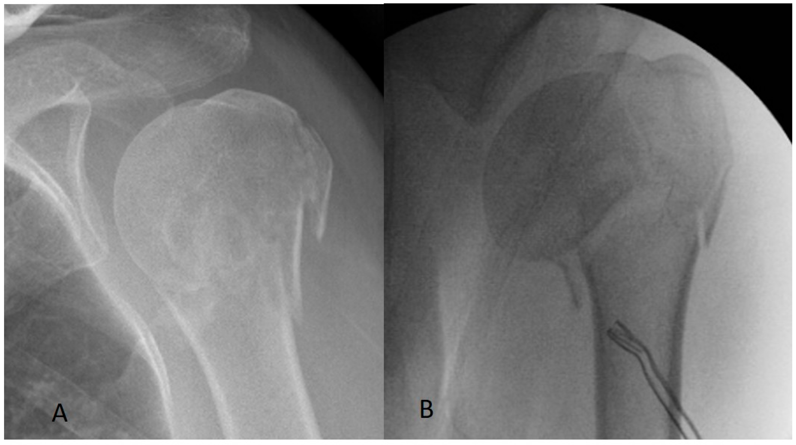
Figure 1. (A) Minimally displaced fracture of humeral head. (B) Unstable varus fractures with minimal disruption of the tuberosities of humeral head.
Std 14 00023 g001
Figure 2. (A) Stable valgus fracture of the proximal humerus. (B) Unstable varus fracture with significant disruption of the tuberosities of the proximal humerus.
Std 14 00023 g002
Figure 3. Neck–shaft angles of humeral head (α angle).
Std 14 00023 g003
Figure 4. Unstable varus fracture with minimal disruption of the tuberosities of humeral head treated with a nail.
Std 14 00023 g004
Figure 5. Unstable varus fracture with significant disruption of the tuberosities of the proximal humerus treated with a plate.
Std 14 00023 g005
Table 1. Comparison of the patients’ basic characteristics.
GROUPLocking-Plate Group (69)Intramedullary Nail Group (77)p-Value
AVERAGE AGE6.1 ± 3.2 years62.4 ± 2.9 yearsp = 0.42
SEX22 (M)–47 (F)28 (M)–49 (F)p = 0.35
BMI (kg/m2)24.2 ± 3.125.4 ± 2.1p = 0.43
PATIENTS WITH AT LEAST ONE RELEVANT COMORBIDITY3135p = 0.27
FRACTURE TYPE
Subgroup 149
Subgroup 2 45
Subgroup 32032
Table 2. Follow-up results of the intramedullary nail group and the locking-plate group.
PARAMETERLocking-Plate Group (69)Intramedullary Nail Group (77)p-Value
AVERAGE OPERATION TIME88.7 ± 10.5 min70.2 ± 8.3 minp = 0.03
HEALING TIME
(MONTHS)
8.7 ± 2.2 weeks7.9 ± 2.4 weeksp = 0.11
INITIAL NECK SHAFT
ANGLES (°)
134.6° ± 6.1°135.7° ± 5.6°p = 0.29
LAST NECK SHAFT
ANGLES (°)
132.5° ± 5.3°133.4° ± 6.4°p = 0.23
CONSTANT–MURLEY SCORE91.2 ± 6.790.5 ± 7.7p = 0.27
COMPLICATIONS16 patients (23.2%)7 patients (9.1%)p = 0.02
Table 3. Follow-up results of the intramedullary nail group and the locking-plate group in subgroup 3.
PARAMETERLocking-Plate Group (20)Intramedullary Nail Group (32)p-Value
AVERAGE OPERATION TIME89.5 ± 9.8 min70.9 ± 9.1 minp = 0.03
HEALING TIME
(MONTHS)
9.2 ± 2.6 weeks8.7± 2.2 weeksp = 0.09
INITIAL NECK SHAFT
ANGLES (°)
133.2° ± 5.6°134.9° ± 4.3°p = 0.32
LAST NECK SHAFT
ANGLES (°)
130.4° ± 4.6°132.1° ± 4.7°p = 0.25
CONSTANT–MURLEY SCORE89.3 ± 4.388.7 ± 5.9p = 0.19
COMPLICATIONS6 patients (30%)3 patients (9.3%)p = 0.01
Disclaimer/Publisher’s Note: The statements, opinions and data contained in all publications are solely those of the individual author(s) and contributor(s) and not of MDPI and/or the editor(s). MDPI and/or the editor(s) disclaim responsibility for any injury to people or property resulting from any ideas, methods, instructions or products referred to in the content.