Article

The Outcomes of Three Surgical Approaches for Acromioclavicular Dislocation Treatment: Findings from Vietnam

1
Department of Surgery, Hanoi Medical University, Hanoi 10000, Vietnam
2
Institute of Orthopaedics and Trauma Surgery, Viet Duc University Hospital, Hanoi 10000, Vietnam
*
Author to whom correspondence should be addressed.
Surg. Tech. Dev. 2022, 11(3), 105-113; https://doi.org/10.3390/std11030010
Submission received: 13 September 2022 / Revised: 1 November 2022 / Accepted: 8 November 2022 / Published: 15 November 2022

Abstract

:
Background: Acromioclavicular (AC) dislocation, one of the most common shoulder joint injuries, can be treated by several surgical methods. However, there are still few records about the treatment quality. This study aims to describe the outcomes of three surgical methods for acromioclavicular dislocation treatment at Viet Duc University Hospital, Vietnam. Methods: A cross-sectional study was conducted on 80 patients diagnosed with AC. We retrospectively collected data in the medical records and re-examined the patients. Results: There was no difference between the three groups of surgical approaches relating to the patient’s characteristics, except for the time from the accident to hospital admission. The median length of stay after surgery was highest in the Hook plate group (median (IQR) = 5(2) days), while it was lowest in the K-wire group (median (IQR) = 3(1) days) (p < 0.05). There is statistical significance in the difference of coracoclavicular distance between pre and post-operation in all three surgical method groups (p < 0.001). Conclusion: All of the methods—Hook plate, K-wire, and TightRope—were associated with optimistic outcomes and restored initial anatomy. While the three surgical methods are both safe and effective, the K-wire method is associated with a shorter length of stay and might be economical.

1. Introduction

Acromioclavicular (AC) joint injuries usually happen due to falling or accidents in sports, traffic, or other athletic-related activities. In particular, acromioclavicular injuries account for 40% of all shoulder injuries and 10% of sports injuries in high-energy contact sports, such as hockey or rugby [1,2]. A study at the United States Military Academy in 2012 showed that acromioclavicular dislocation is not a rare injury in sports; 162 cases were reported in 5 years and the incidence rate is 0.92% [3]. As reported in the study of the National Collegiate Athletic Association (NCAA—ISS), acromioclavicular dislocation occupies 41% of all shoulder injuries, and commonly happens in sports injuries, especially in rugby [4]. According to other authors, AC dislocation, accounting for 9% of all injuries, is one of the most common injuries [5].
The common mechanism for an acromioclavicular is a direct force onto the shoulder, with the arm in the adducted position, or a fall with an outstretched abducted hand [6]. AC dislocation causes pain and limited shoulder mobility, and if the patient is not treated in time, it can affect the patient‘s appearance, mentality, and life quality [7]. Conservative treatment could be applied for lower-grade dislocation; meanwhile, more severe cases require AC joint fixation or ligament repair. Although AC dislocation is a common injury, the number of studies describing clinical and paraclinical features and assessing the treatment result is still lacking. Realizing the necessity of assessing the result of AC dislocation surgical treatment, we carried out this study aimed to describe outcomes of three surgical methods for acromioclavicular dislocation treatment at Viet Duc University Hospital, Vietnam.

2. Materials and Methods

2.1. Participants and Selection Criteria

Eighty (80) patients diagnosed with type III AC dislocation or above according to Rockwood classification were treated from 07/2018 to 04/2021 at Viet Duc University Hospital.
Selection criteria: Type III AC dislocation or above according to Rockwood classification, treated with surgery at Viet Duc University Hospital.
Exclusion criteria: (1) Type I, II AC dislocation according to Rockwood; (2) ipsilateral traumatic arthrotomy of AC joint; (3) fracture of lateral end of the ipsilateral clavicle; (4) accompanying injuries, including acromion fracture, coracoid process fracture, and scapula fracture; and (5) inadequate or unclear information.

2.2. Study Design

A retrospective cross-sectional study was conducted.

2.3. Data Collection

The data are collected based on medical record samples, including:
General information and injury characteristics: Age, sex, side of injury, cause, time from accident to hospitalization clinical manifestation (shoulder pain; shoulder swelling; shoulder deformity; limited mobility), classification according to Rockwood based on X-ray imaging, [8] coracoclavicular distance before surgery.
Postoperative features: Postoperative coracoclavicular distance, hospitalization period, Constant–Murley score when re-examining, and postoperative complications. Constant–Murley score was identified through the Constant–Murley Score Scale, which is commonly used to evaluate overall shoulder function after an intervention. This scale is composed of 4 aspects: severity of pain, quality of daily activities, strength, and range of motion. The lowest score is 0—worst outcome, and the highest is 100—best outcome. The complications recorded include loss of reduction, hardware breakage/slippage, postoperative numbness, and subacromial bursa stenosis.
Process of carrying out the research: The list of the patients through file records and collected data based on medical record samples after we obtained permission from the hospital. The patients were selected with the above criteria. Then, we contacted the patients for re-examining, and assessed their recovery. Finally, the data were encoded and saved in the form of an Excel sheet (Figure 1).

Surgical Techniques and Postoperative Care

There are 3 surgical methods: K-wire, Hook plate, and TightRope. The general course for each surgical method is as follows:
1. K-wire:
The skin is incised about 6 cm from the acromion toward the sternoclavicular joint to reveal the AC joint and clean the injured area. After the manual repositioning of the AC joint, a hole is drilled on the clavicle, and steel thread is stitched afterward. Two wires are stuck from the acromion into the clavicle, then the steel thread is pressed to the wires’ tips. Before the skin closes, a negative pressure drain is placed. The patient must wear Desault’s bandage for 4–6 weeks and then practice rehabilitation exercises for a further 3 weeks. The steel thread is removed 1 year afterward (We believe that loss of reduction could happen if the thread is removed before 1 year).
2. Hook plate:
The skin is incised about 6 cm from the acromion toward the sternoclavicular joint to expose the AC joint and clean the injured area. After manual repositioning of the AC joint, a suitable plate, usually a 3-hole plate, is placed precisely under the acromion. Then, the plate is stuck to the lateral clavicular. The screws are used to fix the plate to the lateral clavicle. Before the skin closes, a negative pressure drain is placed. The patient must wear Desault’s bandage for 4–6 weeks and then practice rehabilitation exercises for a further 3 weeks. The Hook plate is removed 8–12 weeks afterward.
3. TightRope:
A 2 cm skin incision is made at the deltopectoral groove, then the coracoid process base is exposed by retracting the deltoid and the pectoralis major after ensuring hemostasis. A 2–3 cm skin incision at the superior area of the lateral clavicle is also made to reveal the lateral clavicle and AC joint. All the hematoma is debrided. A 4.5 mm drill bit is drilled through the lateral clavicle to the base of the coracoid. Then, the TightRope system is introduced through the tunnel, and the reduction and stabilization of the AC joint are performed. The TightRope system is fixed after checking the intraoperative C-arm. Finally, the AC joint’s capsule is sutured with nonabsorbable stitches before skin closing is performed. The patient must wear an arm sling or a Desault bandage after the surgery. The shoulder motion can be practiced 1 day after surgery.

2.4. Statistical Analysis

The data were transferred from Excel to Stata. We used Stata version 15.1 for data analysis. The mean, median, standard deviation, interquartile range of the quantitative variables, frequency, and proportion of the qualitative variables were calculated. The chi-square test and Fisher exact test were used to compare groups; meanwhile, the T-test and Mann–Whitney test were used for comparing the mean Constant–Murley score and mean hospitalization time and pre-/postoperative coracoclavicular distance. p-value < 0.05 is statistically significant.

2.5. Ethical Consideration

The Director Broad of Viet Duc University Hospital assessed and approved the study protocol. The patients’ information was kept confidential and only used for research.

3. Results

3.1. Patients’ Characteristics

A total of 80 patients were included in the study, of which the number of patients who received Hook plate, K-wire, and TightRope were 7, 41, and 32, respectively (Table 1). There is no difference between the three groups of surgical approaches relating to the patient’s characteristics, except for the time from the accident to hospital admission. The injury occurred mostly on the right side of the body and happened in men almost three times as much as in women. The main cause of this injury was traffic accidents, followed by sports injuries and falling accidents. All of the patients had significant manifestations: pain, shoulder deformity, and limited shoulder mobility. The most common classification was Type IIIb (Table 1).

3.2. Outcomes of Three Surgical Methods

The median length of stay after the surgery was highest in the Hook plate group (median (IQR) = 5(2) days), while it was lowest in the K-wire group (3(1) days). The difference was statistically significant (Table 2). The mean Constant–Murley score increased in the order of Hook plate, K-wire, and TightRope, but there was no statistically significant difference. There was also no difference in the complication rate after surgery (Table 2).
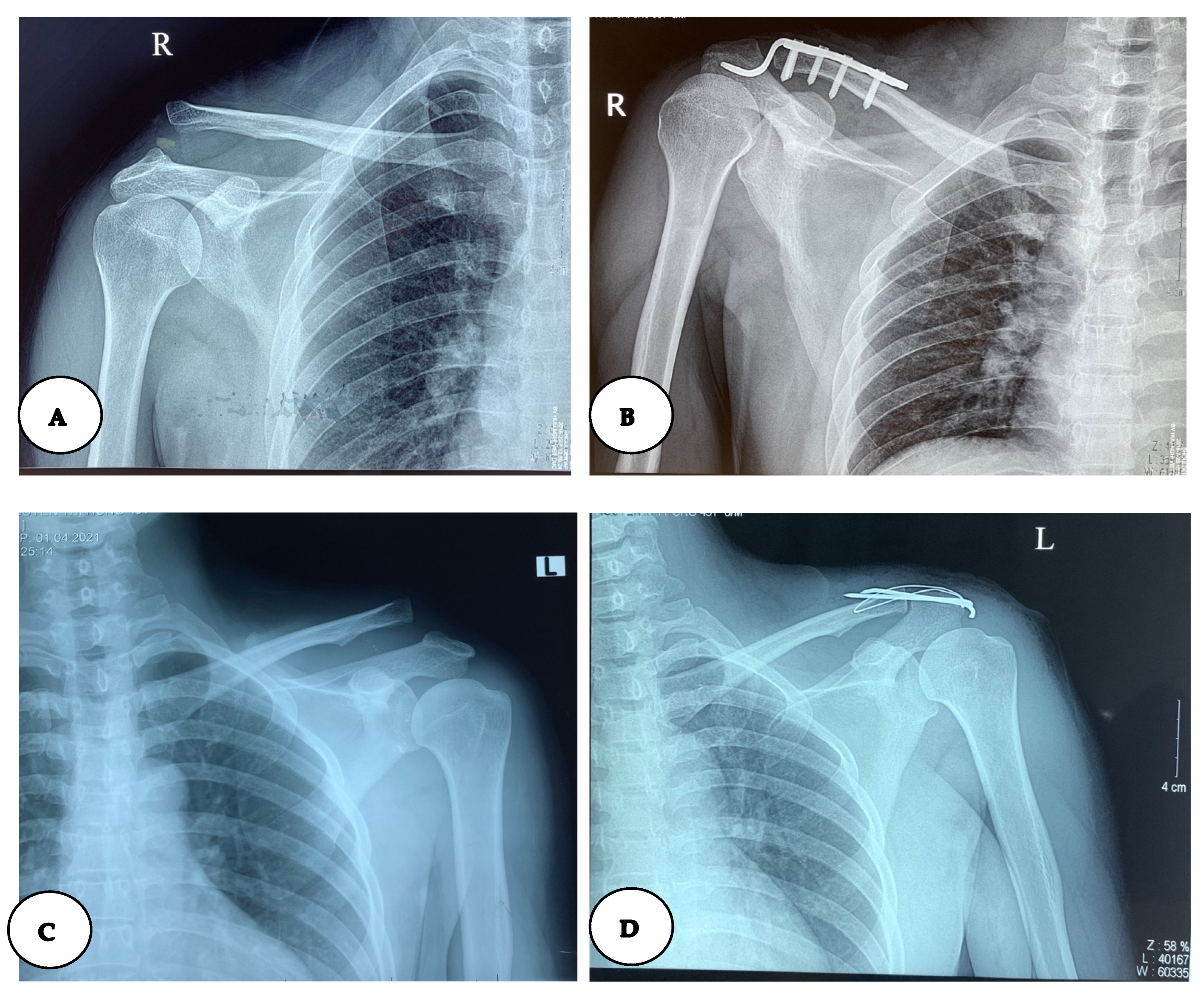
The preoperative coracoclavicular distance was highest in the Hook plate group, with the median (IQR) of 25.60 (9.86) mm, and lowest in the K-wire group (median (IQR): 19.35 (4.57) mm). There was a statistically significant (p = 0.04) difference in preoperative coracoclavicular distance between the three groups. (Table 3). After the surgery, the coracoclavicular distance was not different between the three groups. There is statistical significance in the coracoclavicular distance when comparing it pre-and post-operation (Table 3, Figure 2).

4. Discussion

This study examined surgical outcomes of three methods for acromioclavicular dislocation treatment. We found that three surgical methods have similar outcomes regarding bring the coracoclavicular distance to normal range after the surgery and other outcomes, such as Constant–Murley score and complications. K-wire was found to be a surgical method that corresponds to a shorter length of stay.
Regarding the patients’ characteristics, we found that acromioclavicular (AC) dislocation occurred mainly in men. This result is similar to the result of Chillemi’s research (2013) on 108 patients in Italy. Chillemi’s result revealed that 50.5% of the population was from 20 to 39 years old, and the sex ratio was 8.5 males per female [5]. However, in Chillemi’s research, the main cause was sports injury (42.9%); meanwhile the main cause in our study was traffic accidents (63.75%). Unlike developed countries, where sports injuries cause the most cases of AC dislocation, the burden of traffic accidents in Vietnam could explain the reason why this was the main cause, which shares the same result with other studies in Vietnam [1,5,9].
The majority of all AC dislocation patients at Viet Duc University Hospital are examined and treated early. The time of treatment is still controversial between Vietnamese authors and foreign authors. Vũ Xuân Thành (2019) [9] mentioned that there is no affection for anatomy or function whether the patient is treated before or after 3 months; however, according to Alexandre Lädermann (2021) [10], the rate of successful surgery in the patients admitted to the hospital less than 1 week from when the injury happened was higher than in the patients whose time was over 1 week. Regarding the clinical features, the number of patients having manifestations such as shoulder pain, swelling, shoulder deformity (moving superiorly), and limited mobility occupied nearly 100%, which is similar to the agreement of both foreign and Vietnamese authors. We found that the most common types of AC dislocation were type III, IV, and V. This shared the same result as other studies [9,10].
In the aspect of each surgical method, the Hook plate was the least common. Patients using Hook plates are recommended hardware removal after 8–12 weeks to avoid re-dislocation if the hardware is removed too soon, or shoulder stiffness in contrast [11]. Most patients using Hook plates stayed in the hospital for short-term inpatient treatment. The postoperative Constant–Murley score in our study is lower than Kienast et al.’s study in 2011, with the figure being 92.4 (p < 0.001), which shows a statistical significance [12]. Rehabilitation quality could be the cause of the difference in treatment outcomes. While our patients only received postoperative rehabilitative instructions within 3 weeks, the patients in Kienast et al.’s study had their limbs immobilized for 4–6 days after surgery then they received continuous rehabilitation within at least 16 weeks [7,12]. However, there was no difference in the percentage of complications in the Hook plate group between the two studies (p = 0.16) [12]. Furthermore, a small sample size of the Hook plate group can partly explain the above results.
The rate of postoperative complications in the K-wire-using group was 17.07%. A study by Young-Jun Kim and Yong-Min Chun (2016) showed the same result, with the figure for complications being 21.7% (including hardware breakage/slippage and loss of reduction after K-wire removal) [13]. This similarity revealed that treatment with K-wire brings the patient a positive outcome (the mean Constant–Murley score was 87.6) but the complications are popular. Our results are similar to ones of a study by Bernd A Leidel on 70 patients treated with K-wire (2009) in the aspect of mean Constant–Murley score (88 ± 10) [14].
All of the patients using TightRope had a successful repair, which means the rate of failure was 0%. This is a good result compared to Thiel’s report (2011) with the failure rate being 16.6% [15]. This difference could be explained by Thiel’s patient selection; the patients in Thiel’s study are more severe (Type IV and V), whereas those in ours are mainly classified as Type III (53.1%). Moreover, our research was carried out later than Thiel’s, when the technique was enhanced to prevent failure. The patients treated with TightRope in our study had good and excellent early outcomes, which was manifested through the mean postoperative Constant–Murley score (89.1). The postoperative complications rate of these patients is lower than the figure in Olivos-Meza’s report (8/52) with a p-value = 0.04 [16]. We assumed that the confidence level of our data is not high because our research is a retrospective study. In addition, TightRope has been recently applied at Viet Duc University Hospital, so it is difficult to assess the risks and complications comprehensively.
Through the study, both three methods brought positive outcomes of restoring anatomy and patient mobility. However, we still need more intensive studies to choose the best method. Our research had some limits. Firstly, this is a retrospective study, so it is difficult to control the patient’s adherence to treatment and follow up on the complications. Next is the small number of patients using Hook plates, which reduces the statistical significance. Finally, TightRope has just been applied for treatment at Viet Duc University Hospital in recent years, therefore a comprehensive assessment would require more time.

5. Conclusions

In conclusion, three surgical methods bring satisfactory outcomes in type III AC dislocation. The postoperative coracoclavicular distance became normal without severe complications. There was no failure at any surgery, and a low rate of complication. Hook plate, K-wire, and TightRope both gave the patients optimistic outcomes and restored initial anatomy.

Author Contributions

Conceptualization, N.X.T., N.M.T. and V.T.T.; methodology, V.T.T., P.V.H. and D.N.L.M.; software, N.M.T. and H.H.P.; validation, N.X.T., N.M.T., V.T.T., P.V.H., H.H.P., D.M.D., B.T.N., T.M.L.T. and D.N.L.M.; formal analysis, V.T.T.; investigation, P.V.H., H.H.P., D.M.D. and B.T.N.; resources, N.X.T. and V.T.T.; data curation, P.V.H., H.H.P., D.M.D., B.T.N., T.M.L.T. and D.N.L.M.; writing—original draft preparation, V.T.T.; writing—review and editing, N.X.T., N.M.T., V.T.T., P.V.H., H.H.P., D.M.D., B.T.N., T.M.L.T. and D.N.L.M.; visualization, V.T.T.; supervision, N.X.T.; project administration, V.T.T.; funding acquisition, N.M.T. All authors have read and agreed to the published version of the manuscript.

Funding

This research received no external funding.

Institutional Review Board Statement

The study was conducted in accordance with the Declaration of Helsinki, and approved by the Ethics Committee of Viet Duc University Hospital (protocol code 08.2019.NCVD on 16 October 2019).

Informed Consent Statement

Informed consent was obtained from all subjects involved in the study.

Data Availability Statement

The data that support the findings of this study are available from the corresponding author upon reasonable request.

Acknowledgments

We would like to thank Viet Duc Hospital for permitting us to conduct this study. Our gratitude goes to all participants who were willing to participate in this study and gave us their time.

Conflicts of Interest

The authors declare no conflict of interest.

References

  1. Sirin, E.; Aydin, N.; Mert Topkar, O. Acromioclavicular joint injuries: Diagnosis, classification and ligamentoplasty procedures. EFORT Open Rev. 2018, 3, 426–433. [Google Scholar] [CrossRef] [PubMed]
  2. Ruiz Ibán, M.A.; Sarasquete, J.; Gil de Rozas, M.; Costa, P.; Tovío, J.D.; Carpinteiro, E.; Hachem, A.I.; Perez España, M.; Asenjo Gismero, C.; Diaz Heredia, J.; et al. Low prevalence of relevant associated articular lesions in patients with acute III–VI acromioclavicular joint injuries. Knee Surg. Sport. Traumatol. Arthrosc. 2019, 27, 3741–3746. [Google Scholar] [CrossRef] [PubMed]
  3. Pallis, M.; Cameron, K.L.; Svoboda, S.J.; Owens, B.D. Epidemiology of acromioclavicular joint injury in young athletes. Am. J. Sports Med. 2012, 40, 2072–2077. [Google Scholar] [CrossRef] [PubMed]
  4. Dragoo, J.L.; Braun, H.J.; Bartlinski, S.E.; Harris, A.H. Acromioclavicular joint injuries in National Collegiate Athletic Association football: Data from the 2004–2005 through 2008–2009 National Collegiate Athletic Association Injury Surveillance System. Am. J. Sports Med. 2012, 40, 2066–2071. [Google Scholar] [CrossRef] [PubMed]
  5. Chillemi, C.; Franceschini, V.; Dei Giudici, L.; Alibardi, A.; Salate Santone, F.; Ramos Alday, L.J.; Osimani, M. Epidemiology of isolated acromioclavicular joint dislocation. Emerg. Med. Int. 2013, 2013, 171609. [Google Scholar] [CrossRef] [PubMed]
  6. Kiel, J.; Kaiser, K. Acromioclavicular Joint Injury; StatPearls: Treasure Island, FL, USA, 2022. [Google Scholar]
  7. Available online: http://hocvienquany.edu.vn/Tapchi_YDHQS/Portal/TrangChu.html (accessed on 27 January 2022).
  8. Rockwood, C.A., Jr.; Green, D.P.; Bucholz, R.W. Fractures in Adults, 6th ed.; Bucholz, R.W., Heckman, J.D., Court-Brown, C.M., Koval, K.J., Tornetta, P., III, Wirth, M.A., Eds.; Lippincott Williams & Wilkins: Philadelphia, PA, USA, 1936. [Google Scholar]
  9. XV, T. Study on the Results of Treatment of Acromioclavicular Joint Dislocation by Reconstructing the Clavicle Ligament. Doctoral Dissertation, University of Medicine and Pharmacy at Ho Chi Minh city, Ho Chi Minh City, Vietnam, 2020. [Google Scholar]
  10. Ladermann, A.; Denard, P.J.; Collin, P.; Cau, J.B.C.; Van Rooij, F.; Piotton, S. Early and delayed acromioclavicular joint reconstruction provide equivalent outcomes. J. Shoulder Elb. Surg. 2021, 30, 635–640. [Google Scholar] [CrossRef]
  11. Fraser-Moodie, J.A.; Shortt, N.L.; Robinson, C.M. Injuries to the acromioclavicular joint. J. Bone Joint Surg. Br. 2008, 90, 697–707. [Google Scholar] [CrossRef] [PubMed]
  12. Kienast, B.; Thietje, R.; Queitsch, C.; Gille, J.; Schulz, A.P.; Meiners, J. Mid-term results after operative treatment of rockwood grade III-V acromioclavicular joint dislocations with an AC-hook-plate. Eur. J. Med. Res. 2011, 16, 52–56. [Google Scholar] [CrossRef] [PubMed] [Green Version]
  13. Kim, Y.-J.; Chun, Y.-M. Treatment of Acute Acromioclavicular Joint Dislocation: Kirschner’s Wire Trans-acromial Fixation versus AO Locking Hook Plate Fixation. Clin. Shoulder Elb. 2016, 19, 149–154. [Google Scholar] [CrossRef] [Green Version]
  14. Leidel, B.A.; Braunstein, V.; Pilotto, S.; Mutschler, W.; Kirchhoff, C. Mid-term outcome comparing temporary K-wire fixation versus PDS augmentation of Rockwood grade III acromioclavicular joint separations. BMC Res. Notes 2009, 2, 84. [Google Scholar] [CrossRef] [PubMed]
  15. Thiel, E.; Mutnal, A.; Gilot, G.J. Surgical outcome following arthroscopic fixation of acromioclavicular joint disruption with the tightrope device. Orthopedics 2011, 34, e267–e274. [Google Scholar] [CrossRef] [PubMed]
  16. Olivos-Meza, A.; Almazan-Diaz, A.; Calvo, J.A.; Jimenez-Aroche, C.A.; Valdez-Chavez, M.V.; Perez-Jimenez, F.; Ibarra, C.; Cruz-Lopez, F. Radiographic displacement of acute acromioclavicular joint dislocations fixed with AC TightRope. JSES Int. 2020, 4, 49–54. [Google Scholar] [CrossRef] [PubMed]
Figure 1. Flow chart of study.
Std 11 00010 g001
Figure 2. The X-ray image of patients in three surgical method: (A) Hook plate before surgery; (B) Hook plate after surgery; (C) K-wire before surgery; (D) K-wire after surgery; (E) TightRope before surgery; (F) TightRope after surgery.
Std 11 00010 g002aStd 11 00010 g002b
Table 1. General information and injury characteristics of participants.
CharacteristicSurgical Methodsp-Value
(PA–B/PA–C/PB–C)
Hook Plate (A)
(n = 7)
K-Wire (B)
(n = 41)
TightRope (C)
(n = 32)
Age ( x ¯   ± SD)44.29 ± 9.5939.12 ± 11.1341.53 ± 9.720.26/0.50/0.34
Sex n (%)
Male6 (85.71)34 (82.93)20 (62.5)0.86/0.26/0.05
Female1 (14.29)7 (17.07)12 (37.5)
Injuried side of the body n (%)
Right4 (57.14)17 (41.46)14 (43.75)0.44/0.52/0.84
Left3 (42.86)24 (58.54)18 (56.25)0.52/0.52/0.99
Cause of injury n (%)
Traffic 3 (42,86)26 (63.41)22 (68.75)0.12/0.32/0.34
Domestic2 (28.57)13 (31.71)6 (18.75)
Sport1 (14.29)2 (4.88)3 (9.38)
Falling1 (14.29)0 (0)1 (3.13)
Time from accident to hospitalization (day)
median (IQR)
0 (0)3 (6)1 (6.5)0.001/0.009/0.32
Clinical manifestation n (%)
Shoulder pain7 (100)41 (100)32 (100)-
Swelling6 (85.71)40 (97.56)31 (96,88)0.15/0.22/0.86
Shoulder deformity7 (100)41 (100)32 (100)-
Limited mobility7 (100)41 (100)32 (100)-
Rockwood Classification n (%)
Type IIIb3 (42.86)30 (73.17)17 (53.13)0.20/0.63/0.20
Type IV0 (0)1 (2.44)2 (6.25)
Type V4 (57.14)10 (24.39)13 (40.63)
All7 (100)41 (100)32 (100)
Table 2. Comparison of outcomes between the 3 surgical methods.
CharacteristicsHook Plate (A)
(n = 7)
K-Wire (B)
(n = 41)
TightRope (C)
(n = 32)
p-Value
(PA–B/PA–C/PB–C)
Postoperative length
of stay (day)
median (IQR)
5 (2)3 (1)3.5 (2)0.007/0.15/0.033
Constant–Murley score
( x ¯   ± SD)
83.86 ± 6.1287.56 ± 5.67 89.06 ± 5.150.12/0.025/0.25
Postoperative complications
n (%)
Yes2 (28.57)7 (17.07)1 (3.13)0.48/0.06/0.09
No5 (71.43)34 (82.93)31 (96.88)
Table 3. Coracoclavicular distance between pre- and post-operation by surgical methods.
CharacteristicsHook Plate
(n = 7)
K-Wire
(n = 41)
TightRope
(n = 32)
Preoperative coracoclavicular distance (mm)
median (IQR)
25.60 (9.86)19.35 (4.57)20.58 (8.35)
Postoperative coracoclavicular distance (mm)
( x ¯   ± SD)
10.46 ± 0.3110.53 ± 0.2310.48 ± 0.27
p-value0.018<0.001<0.001
Publisher’s Note: MDPI stays neutral with regard to jurisdictional claims in published maps and institutional affiliations.