


Article

The CD70/CD27 Axis in Tyrosine Kinase Inhibitor-Treated Chronic Phase Chronic Myeloid Leukemia Cells Is Not an Achilles Heel

1
Paul O’Gorman Leukaemia Research Centre, School of Cancer Sciences, College of Medical, Veterinary and Life Sciences, University of Glasgow, 21 Shelley Road, Glasgow G12 0ZD, UK
2
Centre for Healthcare Randomised Trials (CHaRT), Aberdeen Centre for Evaluation, Health Sciences Building, Foresterhill, Aberdeen AB25 2ZD, UK
3
Scotland Institute, School of Cancer Sciences, College of Medical, Veterinary and Life Sciences, University of Glasgow, Switchback Road, Glasgow G61 1BD, UK
4
Medical Research Council Centre for Inflammation Research, Queen’s Medical Research Institute, 47 Little France Crescent, Edinburgh EH16 4TJ, UK
5
Scottish National Blood Transfusion Service, Jack Copland Centre, Currie, Edinburgh EH14 4BE, UK
6
argenx BV, Industriepark Zwijnaarde 7, Zwijnaarde, 9052 Ghent, Belgium
*
Author to whom correspondence should be addressed.
Hematol. Rep. 2026, 18(4), 59; https://doi.org/10.3390/hematolrep18040059
Submission received: 11 March 2026 / Revised: 17 July 2026 / Accepted: 4 August 2026 / Published: 17 August 2026

Abstract

Background/Objectives: Revolutionary small molecule, targeted, tyrosine kinase inhibitors (TKIs) in the clinic have engendered the perception that optimal treatment has already been established for diseases like chronic myeloid leukemia (CML). Addressing BCR::ABL1 oncokinase domain mutations in CML is unlikely to redress disease recrudescence owing to persistent leukemia stem cells (LSCs). Therefore, it is necessary to consider an alternative strategic approach: disrupting LSC interactions with the protective microenvironment and/or immune system. The interaction of the TNF-α superfamily member, CD27, with its upregulated ligand, CD70, initiates survival signaling specifically in CML cells when BCR::ABL1 is inhibited by TKIs and thus represents an attractive target. Previous studies modulating the expression of CD27 on LSCs in a mouse model of advanced phase CML were encouraging. In our study, we explored the CD70/CD27 axis as a therapeutic target in early-phase disease. Methods: Primitive CD34+ cells from treatment-naïve chronic phase (CP) CML patients were drug-exposed in an in vitro co-culture system; combination drug treatments of the therapeutic anti-CD70 antibody and TKIs, nilotinib, were assessed in our CP CML murine model. Results: The blockade of the CD70/CD27 axis in combination with TKIs did not result in greater LSC elimination than with nilotinib as a single agent in our CP CML models. Nonetheless, there was an observed reduction in CD70+ cells with combination treatment. Conclusion: Further preclinical study of the antibody as an adjunct in CD70-expressing hematological malignancy is perhaps warranted.

1. Introduction

Chronic myeloid leukemia (CML) is a relatively rare cancer with an annual incidence estimated at 1 in 100,000 in Western Europe. CML, being predominantly a disease of older age, significantly contributes to global morbidity and mortality, especially with the world’s aging population.
CML has a comparatively simple genetic landscape with the singular causative chromosomal aberration resulting from a reciprocal translocation between the long arms of chromosomes 9 and 22 ([t(9;22) (q34;q11)]) [1]. This event leads to the formation of the Philadelphia (Ph) chromosome and the BCR::ABL1 fusion oncogene as a result of the c-ABL proto-oncogene from chromosome 9 transferring to a 5.8 kb major breakpoint cluster region (M-BCR) on chromosome 22 [2]. BCR::ABL1 encodes a 210-kD phospho-oncoprotein that has constitutive tyrosine kinase activity that is uniquely necessary and sufficient for malignant transformation [3,4,5]. With advancing technologies, breakthroughs over the last couple of decades in the understanding of CML pathogenesis at a molecular level led to the development of molecularly targeted tyrosine kinase inhibitors (TKIs). The revolutionary introduction of TKIs into the clinic from the 2000s has perhaps engendered the perception that optimal treatment has already been established, but there are still many challenges to be faced in CML.
Hematopoietic stem cell transplantation (HSCT) prompted clinical interest in immunotherapy for CML in the 1970s as donor allogeneic T-cells were shown to exhibit a potent graft-versus-leukemia effect in comparison to syngeneic or T-cell-depleted grafts [6]. In recent years, it has become increasingly evident that the microenvironment and/or immune system may have a tumor-protective role. Therefore, a paradigm shift in the clinical approach to CML is now warranted to address the impact of leukemia stem cell (LSC) interactions. The development of increasingly potent TKIs is unlikely to redress the problems of resistance and recrudescence owing to the persistence of CML LSCs that are BCR::ABL1-kinase independent and therefore not killed by oncokinase inhibition [7,8].
CD27 is a TNF-receptor superfamily member expressed on subgroups of B, T and natural killer (NK) cells and has been shown to have a role in differentiation and expansion of effector T-cells [9]. Using high-density arrays to compare gene expression in murine HSCs and their mature progeny, Wiesmann et al. [10] showed that CD27 was also expressed by about 90% of purified hematopoietic stem cells (HSCs) from the bone marrow (BM). CD27 signaling on HSCs and early progenitors has been shown to provide a negative feedback signal to leukocyte differentiation, especially B cells. Nolte et al. stimulated progenitor cells, expressing the CD27 receptor, with its CD70 ligand, resulting in the inhibition of in vitro colony formation and in vivo lymphocyte outgrowth [11]. CD70 is a type II transmembrane glycoprotein and is a unique ligand for CD27 [12]. Upon immune activation, CD70 has been shown to be expressed on lymphocytes and subsets of dendritic cells [13].
Knowledge derived from studies in acute myeloid leukemia (AML) and CML [12,13,14] suggests that CD70/CD27 signaling can confer pro-survival and proliferative advantages to hematological malignant cells. In CML, BCR::ABL1-driven stabilization of beta-catenin results in constitutive Wnt signaling; TKI activity represses miR-29c downstream of both Wnt and BCR::ABL1 to directly upregulate SP1 while also downregulating DNMT1 through repression of miR-29a/b [13]. SP1 binds the promoter region of the CD70 gene. Therefore, when TKIs are present, CD70 promoter demethylation occurs in Ph+ CML cells, resulting in the availability of CD27 signaling for survival. Schürch et al. investigated modulation of the immune system through CD27 expression on LSCs in a mouse model of advanced-phase CML [12]. Nuclear localization of active beta-catenin and TRAF2 and NCK-interacting kinase (TNIK), consequent to binding of CD27 by its ligand, CD70, and expression of Wnt target genes, resulted in differentiation and increased proliferation of murine LSCs. Furthermore, blocking of LSC CD27 signaling prolonged survival.
In the present study, we explored the CD70/CD27 axis as a therapeutic target in early-phase disease, the stage when most CML patients present clinically. We examined the cell autonomous expression levels of CD70 and CD27 of primary chronic phase (CP) CML cells cultured in suspension. We also co-cultured CML cells, using the CD70-expressing HS-5 stromal cells to in vitro model physiological microenvironmental crosstalk and to trigger or boost CD70/CD27 signaling in the leukemia cells in the bicellular 2D model in the presence of TKI and/or CD70-blocking antibody to determine the effect of the combination on leukemia cell viability. Importantly, we also assessed the effect of the therapeutic blocking CD70 antibody on LSC persistence in a CP CML mouse model [15], as interactions with not only the tumor BM microenvironment but also accessory cells of the immune system may be critical for pathophysiological presentation of CD70 to its receptor, CD27.

2. Materials and Methods

2.1. Cell Lines and Primary Cell Isolation

Ethical approval for this study was obtained from the West of Scotland Research Ethics Committee, NHS Greater Glasgow and Clyde (United Kingdom; REC Reference 15_WS_0077). CML patients presenting in CP were consented to provide peripheral blood (PB) or leukapheresis samples from which stem/progenitor cells were enriched by positive magnetic selection (CliniMACS, Miltenyi Biotec, Bisley, UK) and cryopreserved at >90% CD34+ cells. Cryopreserved CML cells were recovered slowly from liquid nitrogen using a solution containing 100 U/mL DNase I (StemCell Technologies, Cambridge, UK), 20% (v/v) Human Serum Albumin (SNBTS, Glasgow, UK), 1 mM MgCl2 (Sigma-Aldrich, Gillingham, UK) and 115 mM trisodium citrate (Sigma-Aldrich) in 1× Dulbecco’s Phosphate Buffered Saline (DPBS) (Invitrogen, Paisley, UK). CML CD34+ cells were cultured in a growth factor-supplemented serum-free medium as previously described [8].
Human chronic myeloid leukemia (CML), K562, and KCL22 were purchased from the Leibniz Institute DSMZ—German Collection of Microorganisms and Cell Cultures GmbH, Braunschweig, Germany. The human stromal cell line, HS-5, was sourced from ATCC, Manassas, VA, USA.

2.2. Flow Cytometry Analysis

CML cells were stained at a concentration of 5 × 10⁵ cells/100 μL Hank’s Buffered Saline Solution (HBSS) in the presence of monoclonal antibodies directed against CD34 (PerCP; clone: 8G12; BDBiosciences, Wokingham, UK), CD27 (PE; clone: M-T271, BioLegend, London, UK), and CD70 (APC; clone: 113-16, BioLegend), and fluorescent events were acquired on a FACSCanto II analyzer (BDBiosciences). For detection of apoptosis, 5 × 10⁵ cells/100 μL HBSS were stained with Annexin V (FITC) and 7-AAD (BDBiosciences). Data analysis was performed with FlowJo v9 analysis software (Tree Star, Inc., Ashland, OR, USA). To distinguish leukemia cells from co-culture cell lines, the harvested cells were surface stained with anti-CD45 before testing apoptosis as described.

2.3. RNA Isolation, Quantitative Real-Time Polymerase Chain Reaction (qRT-PCR), and In Silico Analysis of Publicly Available Datasets

RNA was purified using the RNeasy Mini Kit (Qiagen, Hilden, Germany) and reverse transcribed to cDNA with SuperScript III Reverse Transcriptase (Invitrogen) according to the manufacturer’s instructions. Gene expression was analyzed by quantitative real-time PCR performed in a 7900HT Fast Real-Time PCR system (ThermoFisher Scientific, Birmingham, UK) using TaqMan Gene Expression Assays (ThermoFisher Scientific) for the following targets: CD27 molecule (CD27, Hs00386811m1), CD70 molecule (CD70, Hs00174297m1) and glucuronidase β (GUSB, Hs00939627m1).
Expression of SP1, which, according to the literature, should be downregulated when BCR::ABL1 or Wnt is active and conversely upregulated when BCR::ABL1 is inhibited by TKIs, was analyzed in a publicly available dataset hosted on the Stemformatics interface [16]. The Affymetrix Gene ST platform comprises a large array design made up of exon-based probe sets that are mapped to gene transcripts but does not include mismatch probe sets that are normally used as negative controls for assessing chip effects. Instead, antigenomic probe sets are included on the chip as essentially negative control probes. There should not be any hybridizations of gene products to these antigenomic probes. The median of antigenomic probe set expression determined the expression threshold for the array dataset.

2.4. In Vitro Assessment of CD70 Blocking Antibody Treatment of CML Cells

CD70-expressing HS-5 human stromal cells were used as a feeding signal to stimulate CD27 signaling in primary CML CD34+ cells rather than rely on auto-activation in leukemia cell monoculture. The use of a therapeutic antibody by blocking CD70/CD27 engagement and, hence, downstream Wnt signaling in the CD27-expressing LSC would be expected to impact LSC growth. CML CD34+ cells were seeded at a density of 2 × 10⁵/mL on a confluent human stromal monolayer of HS-5 cells in growth-factor-supplemented serum-free medium [8], in the presence of an anti-human CD70 blocking antibody (41D12 FcD; argenx BV, Gent, Belgium), over a time course and analyzed for apoptotic cell death by flow cytometry.

2.5. In Vivo Assessment of CD70 Blocking Antibody in a CP CML Mouse Model

All mice were maintained at the University of Glasgow Central Research Facilities under standard animal housing conditions in accordance with local and home-office regulations. The efficacy of anti-CD70 alone and in combination with TKIs was assessed in the SCL-tTA/BCR::ABL1 double transgenic (DTG) mouse model of CP CML, in which BCR::ABL1 is inducibly expressed in HSC, resulting in leukemia that closely resembles the human disease [15]. To synchronize leukemia development, pooled BM from CD45.2 donors was transplanted into CD45.1 hosts, following lethal irradiation. Confirmation of leukemia induction, on removal of tetracycline, was determined by elevated total white blood cell count. Thereafter, mice were treated, or not, for 4 weeks with 50 mg/kg nilotinib once daily, reconstituted in the vehicle 10% N-methyl-2-pyrrolidinone in PEG300, by oral gavage (standard dosing, according to [17]), 10 mg/kg CD70 blocking antibody (murine-specific clone FR-70 FcD; argenx BV) by IP injection every three days, or their combination. The FR-70 dosing schedule was adopted from [13]. At the end of the treatment period, a maximum of 6 mice from each cohort of 10 were sacrificed for immediate analysis, with the remainder followed for survival. The impact of the individual drugs and combinations on the survival of donor (BCR::ABL1+) Lin- Sca1+ c-kit+ (LSK), long-term HSCs and restricted progenitors (HPC1 and HPC2) was determined in PB, BM and spleen by flow cytometry as previously described [18]. Surface expression of CD70/27 on PB cells was analyzed post-mortem.

2.6. Statistical Methods

Ordinary one-way ANOVA was performed to compare the effect of an independent variable, such as drug treatment, on a dependent variable, such as cell surface protein expression. If the test revealed a statistically significant difference between at least two groups, Tukey’s honestly significant difference (HSD) post hoc test indicated where the differences lay across multiple comparisons.

3. Results

3.1. CML Cells Co-Express CD70/CD27 at Low Levels

To elucidate the presence of the target for the therapeutic anti-CD70 antibody, Q-PCR for CD70 and its receptor, CD27, as well as flow cytometric analysis of surface protein expression, was performed in leukemia cells. Ph+ cells, either CP CML CD34+ primary patient cells or human cell lines, expressed slightly lower levels of CD70 by relative quantification than primary chronic lymphocytic leukemia (CLL) cells (positive expression control) at the gene level, and CD27 was expressed at an even lower level, as indicated by a high positive ΔCt value (Figure 1A,B). This finding was corroborated by the presence of CD70 protein on the surface of primary CML cells and a lack of detectable CD27 surface protein expression.
SP1 is a transcription factor known to be regulated by BCR::ABL1 and/or Wnt that binds to the promoter region of the CD70 gene. Inactivation of BCR::ABL1 with TKIs is therefore expected to upregulate SP1. In an independent publicly available dataset generated from the culture of primary human CML CD34+ cells with or without mesenchymal stem cell (MSC) stromal support that protects against TKI killing, SP1 expression was calculated using the median of expression for the Gene ST array and compared against the median of above-threshold Log2 normalized probe expression values [19]. The detection threshold in the Zhang dataset was Log2 3.96; the overexpressed median was Log2 6.55 [19]. SP1 Log2 expression was in excess of eight and did not significantly differ between each scenario (plus/minus MSC, plus/minus imatinib) (Figure 1C).

3.2. Effect of Blocking CD70 in CML Cells In Vitro and In Vivo

In CML, the Wnt/β-catenin pathway that regulates stem cell self-renewal is constitutively active because of BCR::ABL1-mediated β-catenin stabilization [13]. Wnt signaling is inhibited by the miR-29 family (29a, 29b and 29c) [20]. miR-29b has been shown to downregulate DNA methyl transferase 1 (DNMT1) by targeting SP1, a known transactivating factor of DNMT1 [21]. The intention was to assay the on-target effects of blocking the CD70 antibody alone or in combination with the second-generation TKI, nilotinib, at the molecular level in human CML CD34+ cells. In keeping with the lack of discernible biological effect (e.g., enhanced cell death) of blocking CD70 antibody treatment plus TKIs in suspension culture in vitro, none of the molecular targets mentioned above were found to be modulated at the transcript level in our experiments. This result led us to consider in silico analysis of an independent dataset generated from the culture of primary human CML CD34+ cells with or without MSC stromal support, with or without first-generation TKIs, imatinib, that showed no modulation of SP1 under these in vitro conditions (Figure 1C) [19].
We reasoned there may be insufficient auto-activation of CD70/CD27 in suspension culture of CML CD34+ cells, as both CD70 and CD27 were expressed at low levels (Figure 1A,B). Therefore, CML CD34+ cells were cultured on CD70-expressing human stromal cell line, HS-5 (Figure 1D), to potentially activate trace CD27 on CML CD34+ cells through interaction with its ligand on the accessory (stromal) cells, in the presence of 30 nM nilotinib (approaching IC50 in suspension culture and previously demonstrated to inhibit BCR::ABL1 signaling without inducing excess cell death so combination effect(s) could be seen [22]) and increasing concentration of anti-CD70 antibody (10–300 μg/mL). This approach neither initiated non-canonical Wnt signaling activity, as indicated by a lack of modulation of target genes, nor improved CML CD34+ cell killing over and above nilotinib alone (Figure 1D). Notably, the CML CD34+ cells were more viable and proliferative on HS-5 compared to suspension culture, which suggested that local physical interactions and/or paracrine growth factor signaling were at play to counter the effects of BCR::ABL1 silencing by nilotinib. Therefore, we focused on the in vivo model of CP CML in which the accessory cells of the immune system could present CD70 to BCR::ABL1+ CD27+ LSCs in their natural environment.
Using the SCL-tTA/BCR::ABL1 double transgenic (DTG) mouse model of CP CML [15], in the absence of tetracycline, the tetracycline-controlled transactivator (tTA) protein binds the TRE promoter, hence inducibly expressing BCR::ABL1. The stem cell leukemia (SCL) promoter exclusively expressed in hematopoietic cells enables BCR::ABL1 expression in HSC and progenitors, resulting in leukemia approximately 3 weeks later that closely resembles the human disease (Figure 2A). The expected myeloproliferative response to the removal of tetracycline from the drinking water for 21 days prior to initiating treatment of DTG mice was confirmed by the high percentage of Gr1+ Mac1+ cells in the PB of vehicle-dosed animals. Myelocytosis was reverted by the FR-70 plus nilotinib combination (‘combo’) to a value lower than nilotinib monotherapy and the transplant control arm (‘ON Tet’) (Figure 2B; p < 0.05). Spleen cellularity across the treatment arms did not differ; however, splenomegaly of the vehicle-dosed animals was evident from the spleen-to-body weight ratio with respect to nilotinib and the combination (Figure 2C; p < 0.001). Using SLAM markers (CD150, CD48) (Figure 2D) to resolve distinct subpopulations within the LSK cells of the BM, the HSC (150+ 48−; Figure 2E) and HPC1 (150− 48+) or HPC2 (150+ 48+; Figure 2F) were not different across arms in this study. The bulk LSK population did not reveal any greater effect of the combination with respect to single agents (Figure 2G).
Secondary transplants may have revealed differences in leukemic re-populating potential between the treatment arms. Ordinary one-way ANOVA was performed to compare the effect of drug treatments on CD27 or CD70 surface protein expression. The test revealed that there was a statistically significant difference between at least two groups in CD27 (F(3,18) = 5.985, p = 0.0052) and CD70 (F(3,18) = 5.331, p = 0.0083). Tukey’s HSD test for multiple comparisons found that CD70 surface protein expression on CD45.2+ donor cells from primary host animals culled at their censor point showed significantly lower frequency with the combination treatment (average ± standard deviation; 1.77 ± 0.81%) than was seen with either FR70 antibody (7.99 ± 4.08%; p = 0.033, 95% C.I.= 0.4109 to 12.04 ), or vehicle (8.74 ± 4.12%; p = 0.011, C.I. = 1.431 to 12.52) (Figure 2H). Nilotinib monotherapy (4.28 ± 1.96%) CD70 frequency was numerically greater than combination treatment but did not reach statistical significance (Figure 2H). Further, the mean percentage of viable CD45.2+ donor cells that were CD27 positive with nilotinib monotherapy (2.57 ± 0.68%) was numerically greater than the combination (2.02 ± 0.75%), although not statistically significant (Figure 2H). Average CD27 frequency with nilotinib treatment was significantly greater than vehicle (1.04 ± 0.52%; p = 0.007, 95% C.I.= −2.671 to −0.3861; Figure 2H) suggesting possible accumulation or persistence of LSC-containing subpopulations, which we have previously ascribed to TKI activity [7,8].
Survival curves were generated for all arms of this study; a maximum of four animals from each cohort of 10 were followed on cessation of therapy at 49 days until the animals reached a humane endpoint (Figure 2I). No differences were noted between the experimental arms.

4. Discussion

This study aimed to propose a paradigm shift in the clinical approach to tyrosine kinase-driven stem cell diseases such as CML to address LSC interactions with the protective microenvironment and/or immune system rather than tumor bulk or cell-intrinsic properties of LSCs. The mainstay of treatment in CML remains oncoprotein-targeted TKI monotherapy, such as nilotinib. There are key areas of unmet clinical need in CML currently: CML LSC persistence despite inhibition of BCR::ABL1 signaling and the linked concept of inability to confidently attempt treatment-free remission for all; frank TKI resistance through point mutation where TKIs become ineffective; and progression to blast crisis where TKI monotherapy does not achieve disease control. TKIs are very well tolerated. Most commercial drug development programs, therefore, aim to combat TKI resistance by addressing BCR::ABL1 kinase domain mutations. However, the development of TKIs of increasing potencies is unlikely to redress the problems of resistance and disease recrudescence, owing to persistent LSCs that are BCR::ABL1-kinase independent and, therefore, not killed by TK inhibition. Indeed, BCR::ABL1 inhibition may invoke survival mechanisms such as CD70/27 signaling to activate Wnt. Hence an alternative strategy and/or complementary target must still be sought.
ARGX-110 (also known as JNJ-74494550 or cusatuzumab) is a defucosylated monoclonal IgG1 antibody capable of targeting and neutralizing the human TNFα-like ligand, CD70. Fully humanized, it is reported that this therapeutic antibody can lyse CD70+ tumor cells via complement-mediated cytotoxicity (CDC), antibody-dependent cellular phagocytosis (ADCP) or enhanced antibody-directed cellular cytotoxicity (ADCC) [23,24]. Interim results from a Phase 1b clinical trial of ARGX-110 as a single agent conducted in nine patients with CD70+ T-cell lymphoma (TCL) reported four treated patients with signs of clinical/biological anti-tumor activity, two of which had >90% reduction in the circulating malignant clone (Table 1; ClinicalTrials.gov Identifier: NCT01813539/EudraCT 2012-005046-38) [25]. The antibody’s direct tumor lysis mechanism of action through blockade of CD70 was attractive to test against CD70-expressing leukemia cells, such as CML. Our study aimed to address CML LSC persistence despite successful inhibition of BCR::ABL1 signaling by TKIs by selectively targeting the CD70/CD27 pathway that had previously been shown to be upregulated in CML cells upon BCR::ABL1-kinase inhibition and contributes to CML LSC survival [12,13]. However, in liquid suspension monoculture, we did not observe any direct tumor cell killing mediated through antibody binding to CD70+ primary CML CD34+ leukemia cells in response to the therapeutic antibody alone or in the presence of TKIs. Critically, the response of rare LSCs specifically and differentially marked from normal stem cells by CD26 [26], CD93 [27] or IL1-RAP [28], for example, and that comprise a small but significant proportion of the CD34+CD38- subpopulation that is presumed responsible for CML LSC persistence, likely was masked; fluorescence activated cell sorting of such LSCs from bulk CD34+ cells based on these putative markers is warranted but would have yielded too few cells to analyze. We attempted to improve activation of CD70/CD27 signaling in the LSC fraction through presentation of CD70 by HS-5 stromal cells to any CML CD34+ CD27+ cells contained in the co-culture, but the strategy did not improve efficacy; that may be explained by the low CD27 expression levels on CML CD34+ cells. Together with our suspicion that ‘auto-activation’ through unicellular co-expression of CD70 and CD27 may not be optimal and to overcome the limitations of non-physiological two-cell, 2D co-culture in vitro models, we opted to test the combination in an in vivo model with a competent immune system to encourage possible ADCP to occur. Preclinical data in an in vivo model of CML had suggested a potential breakthrough in dual targeting of BCR::ABL1 with TKIs and CD70/CD27 signaling with ARGX-110 in a transplant model of more advanced phase disease [13].
In their murine BM transduction model of CML, Schürch et al. showed that CD27 is expressed on murine BCR::ABL1+ leukemia stem/progenitor cells [12]. The binding of CD27 to its ligand, CD70, induced overexpression of Wnt/β-catenin target genes, leading to increased proliferation of BCR::ABL1+ LSCs. Furthermore, blocking CD70/CD27 signaling in LSCs resulted in delayed disease progression and prolonged survival of treated mice. Supporting data in CD27−/− mice showed improved survival. Important and encouraging data using primary human CML cells were generated in xenograft mice whereby recipients were treated within 1 week of transplant, which notably meant that short-term as opposed to long-term LSCs were studied [13]. However, the levels of engraftment and the clinical parameters displayed suggested that the human donor cells were from the more advanced phase of disease. Owing to low levels of engraftment by early CP CML cells (typically <5%), transplanted mice do not normally have overt signs and symptoms, e.g., leukocytosis and splenomegaly of CML, as observed by Riether et al. [13].

5. Conclusions

The blockade of the CD70/CD27 axis, a hypothesized survival signaling pathway in CML CD34+ cells when BCR::ABL1 signaling is inhibited by TKIs, did not result in greater LSC elimination than nilotinib as a single agent in our model of CP CML. Hence, further work is required to determine if a synergistic dose of both nilotinib and a blocking CD70 antibody for in vivo use could be found; thereafter toxicity to normal CD34+ cells should be assessed. Owing to negative findings in our in vivo model, the latter control toxicity experiment was not justifiable in this study, but non-specific toxicity was not observed. Although we could not demonstrate an overt therapeutic advantage to combining anti-CD70 with nilotinib over TKI monotherapy, we did see a reduction in CD70+ hematological cells in combination-treated animals. A fuller investigation of the putative role for the anti-CD70 molecule as an adjunct in overtly CD70-expressing chronic hematological malignancy, such as chronic lymphocytic leukemia, may be warranted.

Author Contributions

Conceptualization, H.G.J. and M.C.; Formal analysis, J.C., M.E.D., Y.-C.H., E.F., L.J., H.W. and H.G.J.; Funding acquisition, H.G.J.; Investigation, M.E.D., Y.-C.H., J.C., A.M., M.W.M. and H.G.J.; Methodology, J.C., C.J.C., M.A.E. and A.M.M.; Resources, J.J. and P.Z.; Supervision, H.G.J.; Writing—original draft, J.C., A.M., M.W.M., M.C., H.W., A.M.M. and H.G.J.; Writing—review and editing, J.C., M.E.D., Y.-C.H., C.J.C., E.F., L.J., M.A.E., J.J., P.Z., M.C., H.W., A.M.M. and H.G.J. All authors have read and agreed to the published version of the manuscript.

Funding

This research was funded by Cancer Research UK New Agents Committee—Preclinical Grants, Application Reference C45542/A21705 (Mark E. Drotar); Howat Foundation (flow cytometry); Friends of Paul O’Gorman and Beatson Cancer Charity (Michael W. Moles); and MRC DTP in Precision Medicine, University of Glasgow (Lorna Jackson).

Institutional Review Board Statement

This study was conducted in accordance with the Declaration of Helsinki and approved through the Integrated Research Application System (IRAS) in the UK by the West of Scotland Research Ethics Committee 4 (15_WS_0077 approved from 5 June 2015). The animal study protocol was approved by the local University of Glasgow Animal Welfare and Ethical Review Board and the UK Home Office (PPL: 60/4492, approved in March 2013).

Informed Consent Statement

Informed consent was obtained from all subjects involved in this study.

Data Availability Statement

Data is contained within the article.

Acknowledgments

The authors wish to thank all patients for donating samples to the hematological cell research bank; their clinical care teams for consenting patients and taking the samples; and colleagues at the Paul O’Gorman Leukaemia Research Centre who made significant technical contributions to this study, namely Karen Dunn (support of in vivo models), Heather Morrison (primer design, Fluidigm Q-PCR expertise), Vic Campbell and Gillian Horne (feeder layers, Fluidigm Q-PCR expertise), Alan Hair (cell bank), and Deborah Reilly (RNA bank). With thanks to Fraser Jørgensen for graphic communication advice. Special thanks to Luc van Rompaey (argenX) for initial communications and negotiations. Remembering Tessa L Holyoake, an inspirational leader, pre-eminent clinician, scientist and friend.

Conflicts of Interest

M.C. holds research funding from Cyclacel Ltd. and Incyte; advisory board member for Novartis, Incyte, Pfizer, Ascentage, Crossbow, Servier, Mendus B.V.; honoraria from Astellas, Jazz Pharmaceuticals, Novartis, Incyte, Pfizer, Servier, Azurity and Janssen. J.J. and P.Z. are employees of argenx BV.

Abbreviations

The following abbreviations are used in this manuscript:
AMLAcute myeloid leukemia
BMBone marrow
CLLChronic lymphocytic leukemia
CMLChronic myeloid leukemia
CMMLChronic myelomonocytic leukemia
CPChronic phase
DNMT1DNA methyltransferase 1
DTGDouble transgenic
HPCHematopoietic progenitor cell
HSCHematopoietic stem cell
HSCTHematopoietic stem cell transplant
LSCLeukemia stem cell
LSKLineage Sca1 c-kit
MDSMyelodysplastic syndrome
MPPMultipotent progenitor
MSCMesenchymal stem cell
PBPeripheral blood
PhPhiladelphia chromosome
TCLT-cell lymphoma
TKITyrosine kinase inhibitor
TNFαTumor necrosis factor alpha
TNIKTRAF2- and NCK-interacting kinase

References

  1. Rowley, J.D. A new consistent chromosomal abnormality in chronic myelogenous leukemia identified by quinacrine fluorescence and giemsa staining. Nature 1973, 243, 290–293. [Google Scholar] [CrossRef] [Scilit] [PubMed]
  2. Ben-Neriah, Y.; Daley, G.Q.; Mes-Masson, A.M.; Witte, O.N.; Baltimore, D. The chronic myelogenous leukemia-specific P210 protein is the product of the bcr/abl hybrid gene. Science 1986, 233, 212–214. [Google Scholar] [CrossRef] [Scilit] [PubMed]
  3. Fialkow, P.J.; Jacobson, R.J.; Papayannopoulou, T. Chronic myelocytic leukemia: Clonal origin in a stem cell common to the granulocyte, erythrocyte, platelet and monocyte/macrophage. Am. J. Med. 1977, 63, 125–130. [Google Scholar] [CrossRef] [Scilit] [PubMed]
  4. Franz, W.M.; Berger, P.; Wang, J.Y.J. Deletion of an N-terminal regulatory domain of the c-Abl tyrosine kinase activates its oncogenic potential. EMBO J. 1989, 8, 137–147. [Google Scholar] [CrossRef] [Scilit] [PubMed]
  5. Daley, G.Q.; Van Etten, R.A.; Baltimore, D. Induction of chronic myelogenous leukemia in mice by the P210bcr/abl gene of the Philadelphia chromosome. Science 1990, 247, 824–830. [Google Scholar] [CrossRef] [Scilit] [PubMed]
  6. Cayssials, E.; Guilhot, F. Beyond tyrosine kinase inhibitors: Combinations and other agents. Best Pract. Res. Clin. Haematol. 2016, 29, 271–283. [Google Scholar] [CrossRef] [Scilit] [PubMed]
  7. Graham, S.; Jørgensen, H.G.; Allan, E.; Pearson, C.; Alcorn, M.J.; Richmond, L.; Holyoake, T.L. Primitive, quiescent, Philadelphia-positive stem cells from patients with chronic myeloid leukemia are insensitive to STI571 in vitro. Blood 2002, 99, 319–325. [Google Scholar] [CrossRef] [Scilit] [PubMed]
  8. Hamilton, A.; Helgason, G.V.; Schemionek, M.; Zhang, B.; Myssina, S.; Allan, E.K.; Nicolini, F.E.; Müller-Tidow, C.; Bhatia, R.; Brunton, V.G.; et al. Chronic myeloid leukemia stem cells are not dependent on Bcr-Abl kinase activity for their survival. Blood 2012, 119, 1501–1510. [Google Scholar] [CrossRef] [Scilit] [PubMed]
  9. Borst, J.; Hendriks, J.; Xiao, Y. CD27 and CD70 in T cell and B cell activation. Curr. Opin. Immunol. 2005, 7, 275–281. [Google Scholar] [CrossRef] [Scilit] [PubMed]
  10. Wiesmann, A.; Phillips, R.L.; Mojica, M.; Pierce, L.J.; Searles, A.E.; Spangrude, G.J.; Lemischka, I. Expression of CD27 on murine hematopoietic stem and progenitor cells. Immunity 2000, 12, 93–199. [Google Scholar] [CrossRef] [Scilit] [PubMed]
  11. Nolte, M.A.; Arens, R.; van Os, R.; van Oosterwijk, M.; Hooibrink, B.; van Lier, R.A.W.; van Oers, M.H.J. Immune activation modulates hematopoiesis through interactions between CD27 and CD70. Nat. Immunol. 2005, 6, 412–418. [Google Scholar] [CrossRef] [Scilit] [PubMed]
  12. Schürch, C.; Riether, C.; Matter, M.S.; Tzankov, A.; Ochsenbein, A.F. CD27 signaling on chronic myelogenous leukemia stem cells activates Wnt target genes and promotes disease progression. J. Clin. Investig. 2012, 122, 624–638. [Google Scholar] [CrossRef] [Scilit] [PubMed]
  13. Riether, C.; Schürch, C.M.; Flury, C.; Hinterbrandner, M.; Drück, L.; Huguenin, A.-L.; Baerlocher, G.M.; Radpour, R.; Ochsenbein, A.F. Tyrosine kinase inhibitor—Induced CD70 expression mediates drug resistance in leukemia stem cells by activating Wnt signaling. Sci. Transl. Med. 2015, 7, 298ra119. [Google Scholar] [CrossRef] [Scilit] [PubMed]
  14. Riether, C.; Schürch, C.M.; Bührer, E.D.; Hinterbrandner, M.; Huguenin, A.-L.; Hoepner, S.; Zlobec, I.; Pabst, T.; Radpour, R.; Ochsenbein, A.F. CD70/CD27 signaling promotes blast stemness and is a viable therapeutic target in acute myeloid leukemia. J. Exp. Med. 2017, 214, 359–380. [Google Scholar] [PubMed]
  15. Koschmieder, S.; Göttgens, B.; Zhang, P.; Iwasaki-Arai, J.; Akashi, K.; Kutok, J.L.; Dayaram, T.; Geary, K.; Green, A.R.; Tenen, D.G.; et al. Inducible chronic phase of myeloid leukemia with expansion of hematopoietic stem cells in a transgenic model of BCR-ABL leukemogenesis. Blood 2005, 105, 324–334. [Google Scholar] [CrossRef] [Scilit] [PubMed]
  16. Wells, C.A.; Mosbergen, R.; Korn, O.; Choi, J.; Seidenman, N.; Matigian, N.A.; Vitale, A.M.; Shepherd, J. Stemformatics: Visualisation and sharing of stem cell gene expression. Stem Cell Res. 2013, 10, 387–395. [Google Scholar] [CrossRef] [Scilit] [PubMed]
  17. Scott, M.T.; Liu, W.; Mitchell, R.; Clarke, C.J.; Kinstrie, R.; Warren, F.; Almasoudi, H.; Stevens, T.; Dunn, K.; Pritchard, J.; et al. Activating p53 abolishes self-renewal of quiescent leukemic stem cells in residual CML disease. Nat. Commun. 2024, 15, 651. [Google Scholar] [CrossRef] [Scilit] [PubMed]
  18. Scott, M.T.; Korfi, K.; Peter Saffrey, P.; Hopcroft, L.E.M.; Kinstrie, R.; Pellicano, F.; Guenther, C.; Gallipoli, P.; Cruz, M.; Dunn, K.; et al. Epigenetic reprogramming sensitizes CML stem cells to combined EZH2 and tyrosine kinase inhibition. Cancer Discov. 2016, 6, 1248–1257. [Google Scholar] [CrossRef] [Scilit] [PubMed]
  19. Zhang, B.; Li, M.; McDonald, T.; Holyoake, T.L.; Holyoake, T.L.; Moon, R.T.; Campana, D.; Shultz, L.; Bhatia, R. Microenvironmental protection of CML stem and progenitor cells from tyrosine kinase inhibitors through N-cadherin and Wnt-β-catenin signaling. Blood 2013, 121, 1824–1838. [Google Scholar] [CrossRef] [Scilit] [PubMed]
  20. Tan, M.; Wu, J.; Cai, Y. Suppression of Wnt signaling by the miR-29 family is mediated by demethylation of WIF-1 in non-small-cell lung cancer. Biochem. Biophys. Res. Commun. 2013, 438, 673–679. [Google Scholar] [CrossRef] [Scilit] [PubMed]
  21. Garzon, R.; Liu, S.; Fabbri, M.; Liu, Z.; Heaphy, C.E.A.; Callegari, E.; Schwind, S.; Pang, J.; Yu, J.; Muthusamy, N.; et al. MicroRNA-29b induces global DNA hypomethylation and tumor suppressor gene reexpression in acute myeloid leukemia by targeting directly DNMT3A and 3B and indirectly DNMT1. Blood 2009, 113, 6411–6418. [Google Scholar] [CrossRef] [Scilit] [PubMed]
  22. Jørgensen, H.G.; Allan, E.K.; Jordanides, N.E.; Mountford, J.C.; Holyoake, T.L. Nilotinib exerts equipotent anti-proliferative effects to imatinib and does not induce apoptosis in CD34+ CML cells. Blood 2007, 109, 4016–4019. [Google Scholar] [CrossRef] [Scilit] [PubMed]
  23. Silence, K.; Dreier, T.; Moshir, M.; Ulrichts, P.; Gabriels, S.M.E.; Saunders, M.; Wajant, H.; Brouckaert, P.; Huyghe, L.; Van Hauwermeiren, T.; et al. ARGX-110, a highly potent antibody targeting CD70, eliminates tumors via both enhanced ADCC and immune checkpoint blockade. mAbs 2014, 6, 523–532. [Google Scholar] [CrossRef] [Scilit] [PubMed]
  24. Aftimos, P.; Rolfo, C.; Sylvie Rottey, S.; Offner, F.; Bron, D.; Maerevoet, M.; Soria, J.-C.; Moshir, M.; Dreier, T.; Van Rompaey, L.; et al. Phase I dose-escalation study of the anti-CD70 antibody ARGX-110 in advanced malignancies. Clin. Cancer Res. 2017, 23, 6411–6420. [Google Scholar] [CrossRef] [Scilit] [PubMed]
  25. Michot, J.-M.; Silence, K.; Maerevoet, M.; Aftimos, P.; Rottey, S.; Rolfo, C.; Offner, F.; Van Rompaey, L.; Moshir, M.; de Haard, H.; et al. A Phase I Study in T Cell Lymphoma Patients Treated with Anti-CD70 Simple Antibody™ ARGX-110. Haematologica 2016, 101, 105. [Google Scholar]
  26. Sicuranza, A.; Raspadori, D.; Bocchia, M. CD26/DPP-4 in Chronic Myeloid Leukemia. Cancers 2022, 14, 891. [Google Scholar] [CrossRef] [Scilit] [PubMed]
  27. Kinstrie, R.; Horne, G.A.; Morrison, H.; Irvine, D.; Munje, C.; Castañeda, E.G.; Moka, H.A.; Dunn, K.; Cassels, J.E.; Parry, N.; et al. CD93 is expressed on chronic myeloid leukemia stem cells and identifies a quiescent population which persists after tyrosine kinase inhibitor therapy. Leukemia 2020, 34, 1613–1625. [Google Scholar] [CrossRef] [Scilit] [PubMed]
  28. Järås, M.; Johnels, P.; Hansen, N.; Agerstam, H.; Tsapogas, P.; Rissler, M.; Lassen, C.; Olofsson, T.; Bjerrum, O.W.; Richter, J.; et al. Isolation and killing of candidate chronic myeloid leukemia stem cells by antibody targeting of IL-1 receptor accessory protein. Proc. Natl. Acad. Sci. USA 2010, 107, 16280–16285. [Google Scholar] [CrossRef] [Scilit] [PubMed]
  29. Riether, C.; Pabst, T.; Höpner, S.; Bacher, U.; Hinterbrandner, M.; Banz, Y.; Müller, R.; Manz, M.G.; Gharib, W.H.; Francisco, D.; et al. Targeting CD70 with cusatuzumab eliminates acute myeloid leukemia stem cells in patients treated with hypomethylating agents. Nat. Med. 2020, 26, 1459–1467. [Google Scholar] [CrossRef] [Scilit] [PubMed]
  30. Pabst, T.; Papayannidis, C.; Demirkan, F.; Doronin, V.; Fogliatto, L.M.; Guttke, C.; Gyan, E.; Hamad, N.; Herrera, P.; Hultberg, A.; et al. Cusatuzumab plus azacitidine in newly diagnosed acute myeloid leukaemia ineligible for intensive chemotherapy (CULMINATE): Part one of a randomised, phase 2, dose optimisation study. Lancet Haematol. 2023, 10, e902–e912. [Google Scholar] [CrossRef] [Scilit] [PubMed]
  31. Ikezoe, T.; Usuki, K.; Aida, K.; Hatayama, T.; Shirahase, T.; Yamauchi, T. Cusatuzumab plus azacitidine in Japanese patients with newly diagnosed acute myeloid leukemia ineligible for intensive treatment. Cancer Sci. 2023, 114, 1037–1044. [Google Scholar] [CrossRef] [Scilit] [PubMed]
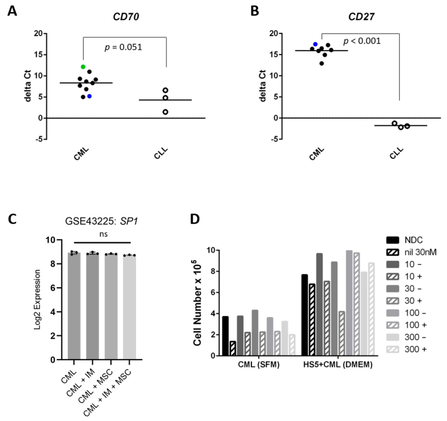
Figure 1. CD70-expressing HS-5 stromal cells support CML CD34+ cell growth and protect against TKIs, but an anti-CD70 antibody does not synergize with nilotinib in vitro. (A) To elucidate the presence of the target for the therapeutic antibody, Q-PCR for CD70 and its receptor CD27 was performed in leukemia cells. Higher mean CD70 mRNA expression was detected in CLL (open circles, n = 3 patients; positive control for expression) compared to CML CD34+ patient cells (black circles, n = 8 patients) or Ph+ human cell lines, K562 (blue circle) and KCL22 (green circle) using a relative quantification method whereby Ct (gene of interest) − Ct (reference gene) = ΔCt, so a negative value indicates relative higher expression. Unpaired student’s t-test performed for statistical significance. (B) Significantly higher CD27 mRNA was detectable in CLL (positive control for expression) than CML patient cells. (C) In silico analysis of SP1 expression, a transcription factor known to be regulated by BCR::ABL1 and/or Wnt that binds to the promoter region of the CD70 gene, in an independent dataset generated from the culture of primary human CML CD34+ cells with or without MSC stromal support, with or without imatinib (IM) [19]. Inactivation of BCR::ABL1 with TKIs was expected to upregulate SP1 [13]. (D) CML CD34+ cells grew better on CD70+ HS-5 MSC stromal support in HS-5 medium (HS5+CML (DMEM)) to mimic the BM microenvironment with respect to serum-free medium supplemented with physiological concentration growth factors (CML (SFM)). Stromal cells can offer protection against TKI killing [19]. Therefore, as predicted, CML CD34+ cells were less sensitive to a TKI (nilotinib; nil 30 nM) on CD70+ HS-5, but anti-CD70 (titrated from 10 to 300 μg/mL, presence indicated by ‘+’) did not augment the response to nilotinib (n = 1). Feeder cells were unaffected by treatments. NDC: no drug control. ns: not significant.
Hematolrep 18 00059 g001
Figure 2. A drug feeding experiment in a double transgenic (DTG) chronic phase (CP) CML mouse model showed no combined effect of anti-CD70 with nilotinib on stem/progenitor cell compartment subpopulations: (A) Schematic diagram of the transplantation and dosing schedule of the SCL-tTA/BCR::ABL1 DTG mouse model. In the absence of tetracycline, the tetracycline-controlled transactivator (tTA) protein binds the TRE promoter, hence inducibly expressing BCR::ABL1. The stem cell leukemia (SCL) promoter exclusively expressed in hematopoietic cells enables BCR::ABL1 expression in HSCs and progenitors, resulting in leukemia that closely resembles the human disease. Treatment with nilotinib (50 mg/kg) daily by oral gavage and/or FR-70 (10 mg/kg) every three days by intraperitoneal injection or vehicle (PBS) was begun 21 days after removal of tetracycline from the drinking water. (B) A myeloproliferative response as shown by the high percentage of Gr1+ Mac1+ cells in the PB of vehicle-dosed animals was observed, which was reverted by the combination (‘combo’) of nilotinib and FR70 (murine-specific anti-CD70) to normal values of the transplant controls kept on tetracycline (‘ON Tet’). (C) Splenomegaly of the vehicle-dosed animals was evident with respect to nilotinib and the combination. (D) Gating strategy to resolve distinct immature hematopoietic subpopulations: LSK; HPC1/2, hematopoietic progenitor cell; MPP, multipotent progenitor; HSC, hematopoietic stem cell. Using SLAM markers to resolve distinct subpopulations of (E) CD150+ CD48− (HSC), (F) CD150CD48+ (HPC1) and CD150+ CD48+ (HPC2), and (G) bulk LSK cells did not reveal any greater effect of the combination with respect to nilotinib as a single agent in these hematopoietic compartments. (H) Surface protein expression analysis by flow cytometry on CD45.2+ donor bone marrow cells from primary host animals culled at their censor point showed a significantly higher percentage of CD70+ cells in the vehicle and FR70 groups but not the nilotinib monotherapy with respect to the combination (‘combo’) treatment. The percentage of viable CD45.2+ CD27+ donor cells in the nilotinib monotherapy treatment group was only significantly greater than the vehicle, not the combination. Ordinary one-way analysis of variance (ANOVA) was performed to reveal any statistical significance between at least two groups; Tukey’s HSD post hoc test revealed the pair-wise differences. * p < 0.05; ** p < 0.01; **** p < 0.001, ns not significant. ON Tet was not included in the ANOVA table. (I) Animals from each cohort were followed through treatment from day 21 (‘Rx start’ indicated on Days off tet axis by arrow) and after stopping treatment at day 49 (‘Rx end’) until signs of morbidity (e.g., sustained weight loss) precipitated the decision to cull.
Hematolrep 18 00059 g002
Table 1. Clinical trials with therapeutic anti-CD70 antibody, cusatuzumab.
Study TitleTrial No.Drug Combination
Tested
Patient No.PhaseReference/Notes
A Study of ARGX-110 in Participants with Advanced MalignanciesNCT01813539cusatuzumab99I/IISilence et al., 2013. doi: 10.4161/mabs27398 [23]
A Study of ARGX-110 in Combination with Azacytidine in Participants with Newly Diagnosed AML or High-Risk MDSNCT03030612azacitidine +
cusatuzumab
38I/IIRiether et al., 2020. doi: 10.1038/s41591-020-0910-8 [29]
A Study of Cusatuzumab Plus Azacitidine in Participants with Newly Diagnosed AML Who Are Not Candidates for Intensive Chemotherapy (CULMINATE)NCT04023526azacitidine +
cusatuzumab
103IIPabst et al., 2023. doi:10.1016/S2352-3026(23)00207-7 [30]
A Study of Cusatuzumab Plus Azacitidine in Japanese Participants with Newly Diagnosed AML or High-Risk MDS Who Are Not Candidates for Intensive TreatmentNCT04241549azacitidine +
cusatuzumab
9IIkezoe et al. 2022. doi: 10.1111/cas.15663 [31]
Cusatuzumab in Combination with Background Therapy for the Treatment of Participants with AML (ELEVATE)NCT04150887azacitidine +
cusatuzumab +
venetoclax
61IActive, not recruiting.
Estimated study completion May 2027
Footnotes: cusatuzumab is the International Non-proprietary Name (INN) for the therapeutic anti-CD70 antibody also known as ARGX-110 or JNJ-74494550. Abbreviations: AML, acute myeloid leukemia; MDS, myelodysplastic syndrome.
Disclaimer/Publisher’s Note: The statements, opinions and data contained in all publications are solely those of the individual author(s) and contributor(s) and not of MDPI and/or the editor(s). MDPI and/or the editor(s) disclaim responsibility for any injury to people or property resulting from any ideas, methods, instructions or products referred to in the content.


