
Diagnostic Challenges in Acute Leukemia: From Dental Pain to Catastrophic Intracerebral Hemorrhage

 , and
, and 
Abstract
1. Introduction
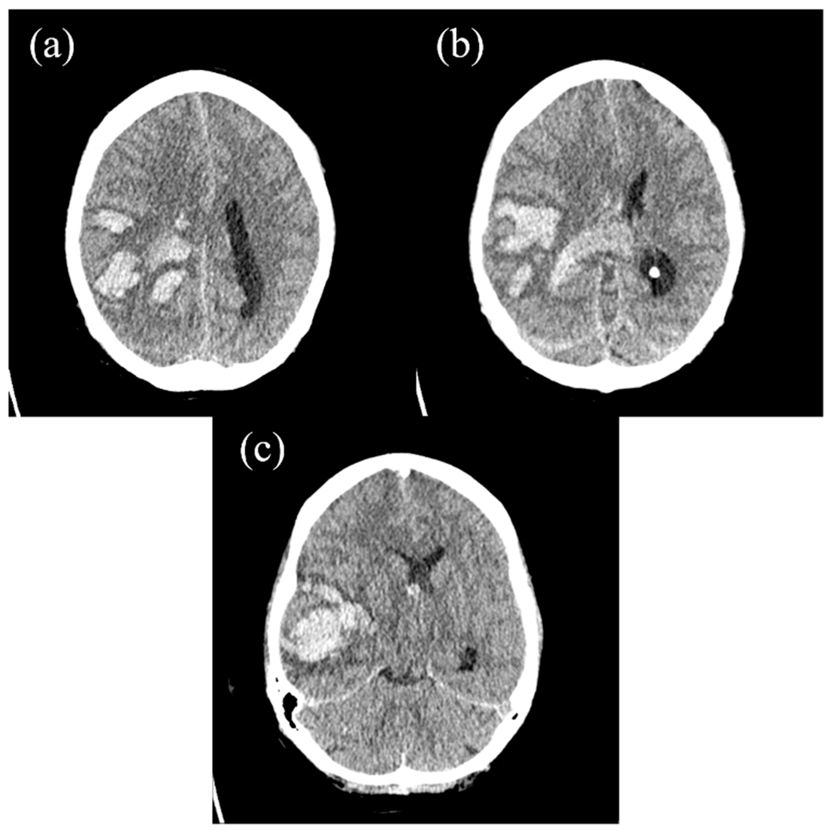
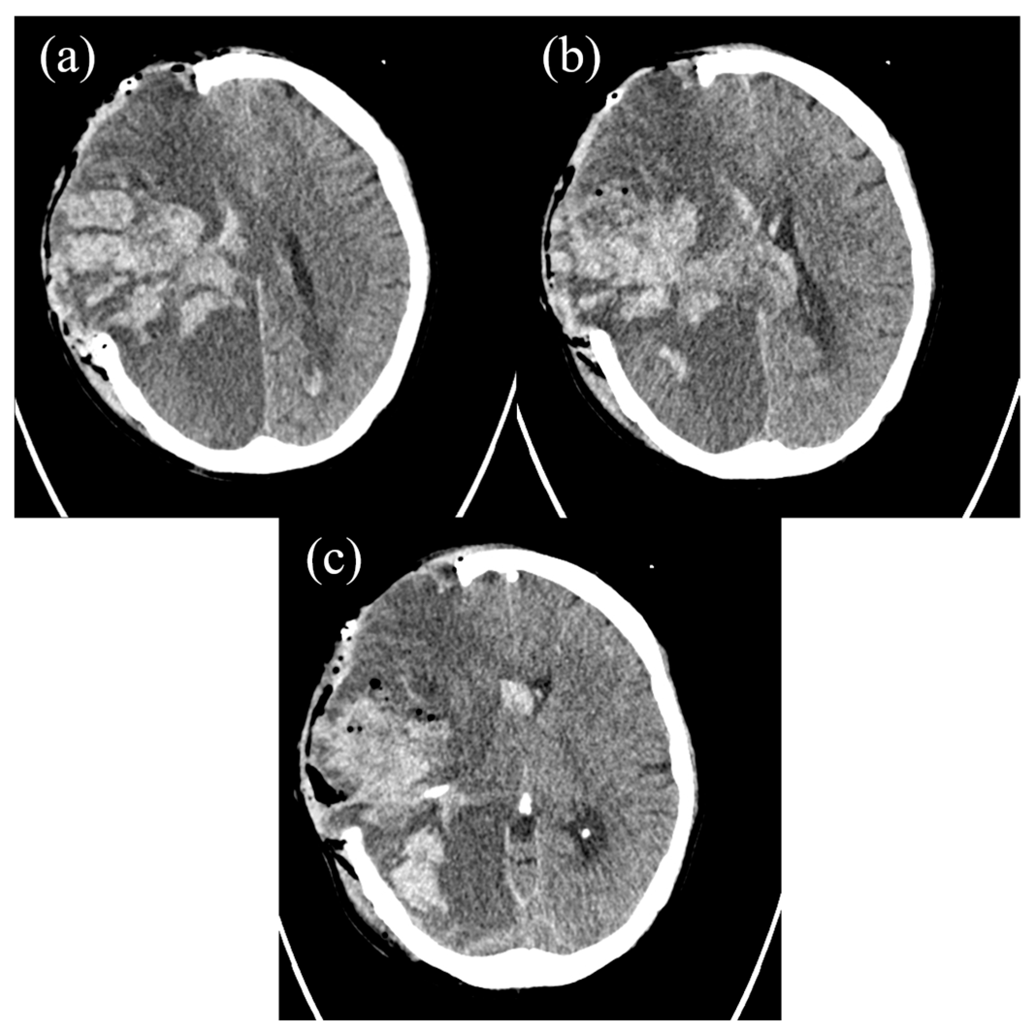
2. The Case Report
3. Discussion
4. Conclusions
Author Contributions
Funding
Institutional Review Board Statement
Informed Consent Statement
Data Availability Statement
Conflicts of Interest
Abbreviations
| AML | Acute Myeloid Leukemia |
| ALL | Acute Lymphoblastic Leukemia |
| DIC | Disseminated Intravascular Coagulopathy |
| APL | Acute Promyelocytic Leukemia |
| ICH | Intracerebral Hemorrhage |
| CT | Computed Tomography |
| CRP | C-Reactive Protein |
| ASAT | Aspartate Aminotransferase |
| GGT | Gamma-Glutamyl Transferase |
| INR | International Normalized Ratio |
| CBC | Complete Blood Count |
| CD | Cluster of Differentiation |
| PCC | Prothrombin Complex Concentrate |
| L | Liter |
References
- Fatahzadeh, M.; Krakow, A.M. Manifestation of acute monocytic leukemia in the oral cavity: A case report. Spec. Care Dent. 2008, 28, 190–194. [Google Scholar] [CrossRef]
- Reenesh, M.; Munishwar, S.; Rath, S.K. Generalised leukaemic gingival enlargement: A case report. J. Oral Maxillofac. Res. 2012, 3, e5. [Google Scholar] [CrossRef]
- Quispe, R.A.; Aguiar, E.M.; de Oliveira, C.T.; Neves, A.C.X.; Santos, P. Oral manifestations of leukemia as part of early diagnosis. Hematol. Transfus. Cell Ther. 2022, 44, 392–401. [Google Scholar] [CrossRef] [PubMed]
- Wu, J.; Fantasia, J.E.; Kaplan, R. Oral manifestations of acute myelomonocytic leukemia: A case report and review of the classification of leukemias. J. Periodontol. 2002, 73, 664–668. [Google Scholar] [CrossRef] [PubMed]
- Hou, G.L.; Tsai, C.C. Primary gingival enlargement as a diagnostic indicator in acute myelomonocytic leukemia. A case report. J. Periodontol. 1988, 59, 852–855. [Google Scholar] [CrossRef] [PubMed]
- Weckx, L.L.; Hidal, L.B.; Marcucci, G. Oral manifestations of leukemia. Ear Nose Throat J. 1990, 69, 341–342, 345–346. [Google Scholar]
- Bastos Silveira, B.; Di Carvalho Melo, L.; Amorim Dos Santos, J.; Ferreira, E.B.; Reis, P.E.D.; De Luca Canto, G.; Acevedo, A.C.; Massignan, C.; Guerra, E.N.S. Oral manifestations in pediatric patients with leukemia: A systematic review and meta-analysis. J. Am. Dent. Assoc. 2024, 155, 858–870.e830. [Google Scholar] [CrossRef]
- Suárez-Cuenca, J.A.; Arellano-Sánchez, J.L.; Scherling-Ocampo, A.A.; Sánchez-Hernández, G.; Pérez-Guevara, D.; Chalapud-Revelo, J.R. Rapidly progressing, fatal and acute promyelocytic leukaemia that initially manifested as a painful third molar: A case report. J. Med. Case Rep. 2009, 3, 102. [Google Scholar] [CrossRef]
- Chen, C.Y.; Tai, C.H.; Tsay, W.; Chen, P.Y.; Tien, H.F. Prediction of fatal intracranial hemorrhage in patients with acute myeloid leukemia. Ann. Oncol. 2009, 20, 1100–1104. [Google Scholar] [CrossRef]
- Koschade, S.E.; Stratmann, J.A.; Miesbach, W.; Steffen, B.; Serve, H.; Finkelmeier, F.; Brandts, C.H.; Ballo, O. Intracranial hemorrhage in newly diagnosed non-promyelocytic acute myeloid leukemia patients admitted for intensive induction chemotherapy. Eur. J. Haematol. 2022, 108, 125–132. [Google Scholar] [CrossRef]
- Rose-Inman, H.; Kuehl, D. Acute leukemia. Emerg. Med. Clin. N. Am. 2014, 32, 579–596. [Google Scholar] [CrossRef] [PubMed]
- Chen, C.Y.; Tai, C.H.; Cheng, A.; Wu, H.C.; Tsay, W.; Liu, J.H.; Chen, P.Y.; Huang, S.Y.; Yao, M.; Tang, J.L.; et al. Intracranial hemorrhage in adult patients with hematological malignancies. BMC Med. 2012, 10, 97. [Google Scholar] [CrossRef] [PubMed]
- Ho, C.; Lo, Y.Y.; Crawford, J.R. Intracerebral haemorrhage as an initial manifestation of T-cell acute lymphoblastic leukaemia in a paediatric patient. BMJ Case Rep. 2022, 15, e252594. [Google Scholar] [CrossRef] [PubMed]
- Michalak, E.; Dudzik, A.; Śręba, J.; Kęsek, B.; Darczuk, D. Oral manifestations of leukaemia: Cooperation between dentist and haematologist. Hematol. Clin. Pract. 2022, 13, 55–61. [Google Scholar] [CrossRef]
- Lim, H.C.; Kim, C.S. Oral signs of acute leukemia for early detection. J. Periodontal Implant. Sci. 2014, 44, 293–299. [Google Scholar] [CrossRef]
- Zhang, J.-Y.; Li, Y.; Ma, Y.-S.; Sun, X.-J.; Liu, Y.-Z.; Yin, Y.-K.; Hu, B.; Su, M.-H.; Li, Q.-L.; Mi, Y.-C.; et al. Clinical characteristics and prognostic factors in intracranial hemorrhage patients with hematological diseases. Ann. Hematol. 2022, 101, 2617–2625. [Google Scholar] [CrossRef]
- Estey, E.; Döhner, H. Acute myeloid leukaemia. Lancet 2006, 368, 1894–1907. [Google Scholar] [CrossRef]
- Wu, M.Y.; Lin, C.H.; Hou, Y.T.; Lin, P.C.; Yiang, G.T.; Tien, Y.C.; Yeh, H.C. Syncope as Initial Presentation in an Undifferentiated Type Acute Myeloid Leukemia Patient with Acute Intracranial Hemorrhage. Brain Sci. 2019, 9, 207. [Google Scholar] [CrossRef]
- Patil, S.; Nourbakhsh, A.; Thakur, J.D.; Khan, I.S.; Guthikonda, B. Fatal intracranial hemorrhage as the initial presentation of acute lymphocytic leukemia: A case report. Turk. Neurosurg. 2013, 23, 568–571. [Google Scholar] [CrossRef]
- Van de Louw, A. Effect of leukapheresis on blood coagulation in patients with hyperleukocytic acute myeloid leukemia. Transfus. Apher. Sci. 2017, 56, 214–219. [Google Scholar] [CrossRef]
- Kawanami, T.; Kurita, K.; Yamakawa, M.; Omoto, E.; Kato, T. Cerebrovascular disease in acute leukemia: A clinicopathological study of 14 patients. Intern. Med. 2002, 41, 1130–1134. [Google Scholar] [CrossRef]
- Zhang, Q.; Li, X.; Wei, Z.; Ye, X.; Zhu, L.; Xie, M.; Xie, W.; Zhu, J.; Li, L.; Zhou, D.; et al. Risk factors and clinical characteristics of non-promyelocytic acute myeloid leukemia of intracerebral hemorrhage: A single center study in China. J. Clin. Neurosci. 2017, 44, 203–206. [Google Scholar] [CrossRef]
- Yu, Z.; Zheng, J.; Guo, R.; Ma, L.; You, C.; Li, H. Prognostic impact of leukocytosis in intracerebral hemorrhage: A PRISMA-compliant systematic review and meta-analysis. Medicine 2019, 98, e16281. [Google Scholar] [CrossRef]
- Baharoglu, M.I.; Cordonnier, C.; Al-Shahi Salman, R.; de Gans, K.; Koopman, M.M.; Brand, A.; Majoie, C.B.; Beenen, L.F.; Marquering, H.A.; Vermeulen, M.; et al. Platelet transfusion versus standard care after acute stroke due to spontaneous cerebral haemorrhage associated with antiplatelet therapy (PATCH): A randomised, open-label, phase 3 trial. Lancet 2016, 387, 2605–2613. [Google Scholar] [CrossRef]
- Röllig, C.; Ehninger, G. How I treat hyperleukocytosis in acute myeloid leukemia. Blood 2015, 125, 3246–3252. [Google Scholar] [CrossRef]
- Koi, S.; Nogami, A.; Fujiwara, H.; Bando, K.; Saito, M.; Osada, Y.; Tanaka, K.; Sakashita, C.; Yoshifuji, K.; Okada, K.; et al. Acute promyelocytic leukemia with asymptomatic cerebral hemorrhage. Rinsho Ketsueki 2023, 64, 1426–1430. [Google Scholar]

Disclaimer/Publisher’s Note: The statements, opinions and data contained in all publications are solely those of the individual author(s) and contributor(s) and not of MDPI and/or the editor(s). MDPI and/or the editor(s) disclaim responsibility for any injury to people or property resulting from any ideas, methods, instructions or products referred to in the content. |
© 2025 by the authors. Licensee MDPI, Basel, Switzerland. This article is an open access article distributed under the terms and conditions of the Creative Commons Attribution (CC BY) license (https://creativecommons.org/licenses/by/4.0/).
Share and Cite
Pinchuk, A.; Roch, S.P.; Mawrin, C.; Behme, D.; Stein, K.-P.; Neyazi, B.; Mikusko, M.; Sandalcioglu, I.E.; Rashidi, A. Diagnostic Challenges in Acute Leukemia: From Dental Pain to Catastrophic Intracerebral Hemorrhage. Hematol. Rep. 2025, 17, 36. https://doi.org/10.3390/hematolrep17040036
Pinchuk A, Roch SP, Mawrin C, Behme D, Stein K-P, Neyazi B, Mikusko M, Sandalcioglu IE, Rashidi A. Diagnostic Challenges in Acute Leukemia: From Dental Pain to Catastrophic Intracerebral Hemorrhage. Hematology Reports. 2025; 17(4):36. https://doi.org/10.3390/hematolrep17040036
Chicago/Turabian StylePinchuk, Anatoli, Stefan P. Roch, Christian Mawrin, Daniel Behme, Klaus-Peter Stein, Belal Neyazi, Martin Mikusko, Ibrahim Erol Sandalcioglu, and Ali Rashidi. 2025. "Diagnostic Challenges in Acute Leukemia: From Dental Pain to Catastrophic Intracerebral Hemorrhage" Hematology Reports 17, no. 4: 36. https://doi.org/10.3390/hematolrep17040036
APA StylePinchuk, A., Roch, S. P., Mawrin, C., Behme, D., Stein, K.-P., Neyazi, B., Mikusko, M., Sandalcioglu, I. E., & Rashidi, A. (2025). Diagnostic Challenges in Acute Leukemia: From Dental Pain to Catastrophic Intracerebral Hemorrhage. Hematology Reports, 17(4), 36. https://doi.org/10.3390/hematolrep17040036