Article

VsAPX1 Is Up-Regulated by ABA and Heat Stress in Common Vetch (Vicia sativa)

by
Farah Abu Siam
1,
Saeid Abu-Romman
2,
Saja A. K. Al-Rubaye
2,
Ruba M. AL-Mohusaien
3 and
Monther T. Sadder
4,*
1
Department of Agricultural Biotechnology and Genetic Engineering, Faculty of Agricultural Technology, Al-Ahliyya Amman University, Amman 19111, Jordan
2
Department of Biotechnology, Faculty of Agricultural Technology, Al-Balqa Applied University, Al-Salt 19117, Jordan
3
Department of Plant Production, Smart and Sustainable Agriculture, Faculty of Agriculture, Ajloun National University, Ajloun 26810, Jordan
4
Plant Biotechnology Laboratory, Department of Horticulture and Crop Science, School of Agriculture, University of Jordan, Amman 11942, Jordan
*
Author to whom correspondence should be addressed.
Int. J. Plant Biol. 2026, 17(3), 16; https://doi.org/10.3390/ijpb17030016
Submission received: 12 January 2026 / Revised: 9 February 2026 / Accepted: 19 February 2026 / Published: 28 February 2026
(This article belongs to the Section Plant Response to Stresses)

Abstract

Ascorbate peroxidase (APX) is a heme-containing enzyme involved in hydrogen peroxide (H2O2) detoxification within the ascorbate–glutathione (AsA–GSH) cycle. In this study, the full-length genomic DNA and cDNA of an APX1 gene (VsAPX1) were cloned and characterized from Vicia sativa. The genomic sequence of VsAPX1 is 2425 bp in length and comprises 10 exons separated by nine introns, with the first intron located within the 5′ untranslated region (5′UTR). The corresponding cDNA is 1010 bp long and includes a 61 bp 5′UTR, a 753 bp open reading frame, and a 196 bp 3′UTR. VsAPX1 encodes a predicted cytosolic APX protein of 250 amino acids, with a molecular weight of 27.1 kDa and a theoretical isoelectric point (pI) of 5.60. Bioinformatics analysis revealed that the deduced VsAPX1 protein shares high sequence similarity with cytosolic APX1 proteins from other plant species, contains conserved APX domains, and clusters within the cytosolic APX clade in phylogenetic analysis. Quantitative real-time PCR analysis showed that VsAPX1 expression exhibits transient and moderate changes in response to abiotic stress and phytohormone treatments. Transcript levels increased at early time points following heat stress (42 °C), abscisic acid, and salicylic acid treatments, and after 4 h of jasmonic acid exposure, whereas hydrogen peroxide treatment resulted in a gradual down-regulation of expression. Overall, this study provides the first molecular and expression characterization of a cytosolic APX1 gene from Vicia sativa and establishes a foundation for future functional analyses of antioxidant genes in this species.

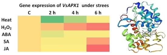
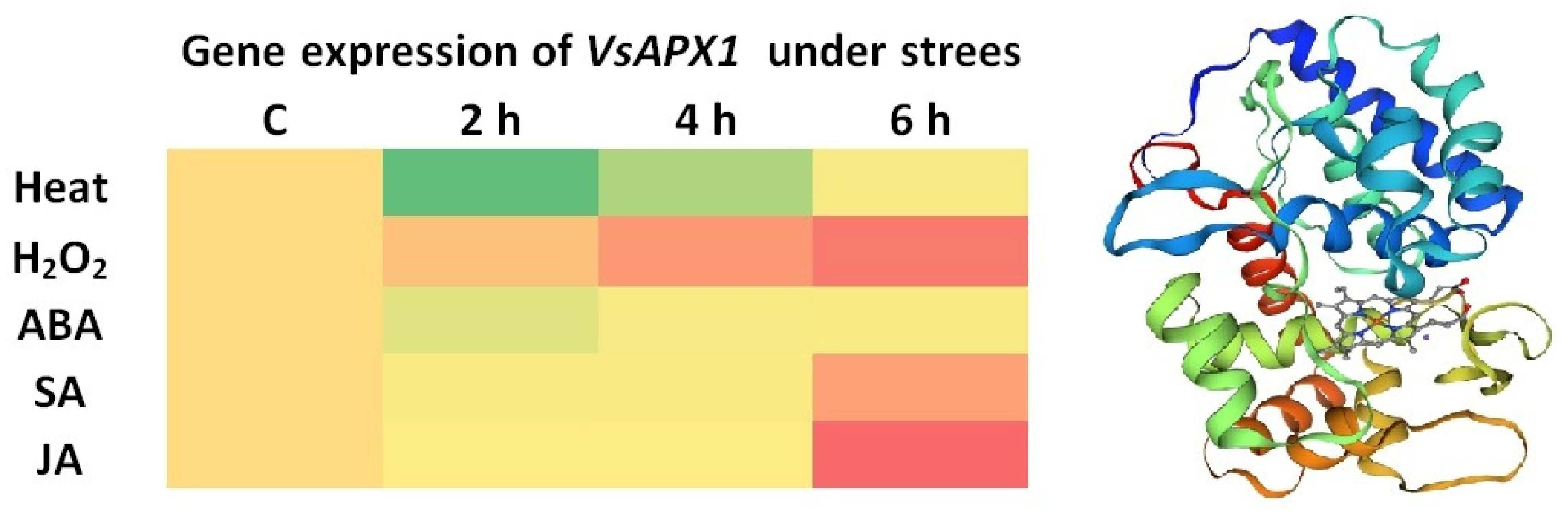
Graphical Abstract

1. Introduction

In nature, plants are exposed to various environmental stresses during their lifetime [1,2]. Global climatic change increases plants’ vulnerability to various abiotic stresses, including temperature, salinity, drought, flooding, high and low light intensity, nutrient deficiency, and chemical factors (e.g., pH and heavy metals) [3]. Plants also suffer from biotic stresses (e.g., pests and diseases) [4]. All these stresses cause morphological, physiological, and molecular changes [5,6] that adversely impact agricultural yield, production, and global food security. Furthermore, stress conditions induce the overproduction and accumulation of reactive oxygen species (ROS) in plant cells, including superoxide anion (O2•−), hydroxyl radical (OH), hydrogen peroxide (H2O2), and singlet oxygen (1O2) [7,8]. ROS are highly reactive and harmful by-products generated from different organelles (e.g., chloroplasts, peroxisomes, and mitochondria) that impose oxidative damage [9,10] to different cellular components, including lipids, proteins, carbohydrates, and nucleic acid, and may ultimately lead to apoptosis and senescence [7,11]. On the other hand, ROS, especially H2O2 at the steady-state level, acts as an essential cell signaling molecule and regulates many plant processes such as development, growth, reproduction, and responses to biotic and abiotic stresses [12].
In response to ROS overproduction, plants evolve a specific and flexible combination of signaling molecules and defense systems. These signaling molecules and defense systems are kinases/phosphatase signaling cascade, plant bioregulators (e.g., salicylic acid (SA), ethylene (ET), jasmonic acid (JA), abscisic acid (ABA), and auxin), defense, and antioxidant genes [3]. Therefore, plants can avoid and tolerate the adverse effects of oxidative stresses through the antioxidant defense system (ROS scavengers), which represents the first line of defense [13], including non-enzymatic antioxidants (e.g., flavonoids, carotenoids, ascorbic acid (AsA), glutathione (GSH), proline, and α-tocopherol) (3), and enzymatic antioxidants (e.g., superoxide dismutase (SOD), glutathione reductase (GR), glutathione S-transferase (GST), glutathione peroxidase (GPX), monodehydroascorbate reductase (MDHAR), dehydroascorbate reductase (DHAR), peroxidase (POX), catalase (CAT), and ascorbate peroxidase (APX)) [11]. Together, enzymatic and non-enzymatic antioxidants minimize oxidative stress injuries and maintain ROS homeostasis by rapidly detoxifying the excess ROS [12,14].
Ascorbate peroxidase (APX, EC: 1.11.1.11) is a class I heme peroxidase, and an important ROS-scavenging enzyme [3]. It has a high affinity to H2O2 and regulates and maintains cellular H2O2 content at a steady-state level [15,16]. Therefore, APX is considered as a key enzyme in the ascorbate–glutathione (AsA-GSH) cycle, reducing H2O2 to water (H2O) and oxygen (O2) by utilizing ascorbate as an electron donor [4]. Plants’ APXs are encoded by multigenic families and found as isoenzymes that are classified according to their subcellular localization as cytosolic, peroxisomal/glyoxysomal, mitochondrial, or chloroplastic (stromal sAPX and thylakoid tAPX) [16]. They have been cloned from different plant species [15,16]. Furthermore, APX activity and expression were reported to increase under different stresses [11], such as high light intensity, high and low temperature, drought, heavy metals, wounding, and pathogen infection [15]. Moreover, the overexpression of APXs improves transgenic plants’ tolerance to drought, heat, high light, and salinity [17].
Common vetch (Vicia sativa L.) is an annual leguminous herb that belongs to the Fabaceae family [18]; it is self-pollinated with 2n = 12 chromosomes [19]. It originated in arid regions of the Middle East [20], and is now widely distributed in Central Asia, North Asia, Europe, and North America [18]. Common vetch can grow in a wide range of soil and climatic conditions and can tolerate drought and cold conditions [21]. Common vetch is suitable for crop rotation, cultivated either as a monocrop or as an intercrop with cereals, which enhances forage harvesting, and yield [21]. Moreover, it is used as a cover crop not only for cereals but also for vegetables such as tomato and pepper [22]. Moreover, this plant reduces herbicides application by suppressing weed growth [22], due to its high content of polyphenols and flavonoids [23]. These chemicals have allelopathic properties that are released in the soil via leachates and root exudates [24]. In addition, V. sativa has a high capacity for nitrogen fixation [18], and therefore, it decreases utilization of fertilizer and pesticides [21], enhancing microbial community, and soil enzyme activities such as sucrase, urease, phosphatase, and catalase [18]. Furthermore, common vetch is easy to cultivate, has a high protein content, and grows rapidly; therefore, it can be used as a green manure, livestock feed as grain, and for silage and hay production [25].
Heat stress represents an increasingly important abiotic stress on cool-season legumes, including common vetch, particularly in the Mediterranean regions where rising temperatures negatively affect germination, vegetative growth, and forage yield. In addition, they lead to ROS production, reducing photosynthesis efficiency and cellular homeostasis, which requires efficient antioxidant mechanisms. Therefore, investigating the expression of VsAPX1 under heat stress may provide important insights into the thermotolerance in common vetch.
Recent studies characterized some of the antioxidant genes in V. sativa, such as catalase (VsCAT) and chloroplastic copper/zinc superoxide dismutase (VsCu/Zn-SOD) [26,27]. APX plays a central and isoform-specific role in ROS detoxification, stress signaling, and redox homeostasis. Although APX genes have been characterized in several species [28], APX from Vicia sativa has not been functionally examined, and its regulatory behavior under specific stresses remains unknown. Species-specific APX isoenzymes often differ in expression patterns, subcellular localization, kinetic properties, and stress responsiveness; thus, cloning and characterizing APX from V. sativa provides a first functional characterization. Moreover, our study integrates expression profiling, structural/sequence analysis, and stress-responsive induction patterns, which together would provide a comprehensive characterization.
Despite extensive studies on APX1 genes in several legumes, no molecular or expression-level characterization has been reported for V. sativa, limiting comparative and applied research in this species. Therefore, this study aims to characterize the APX gene from V. sativa, and perform bioinformatic analyses of the APX protein and investigate its structure and expression pattern in response to abiotic stresses.

2. Materials and Methods

2.1. Plant Growth and Treatments

Common vetch (V. sativa, Mahali) seeds were received from the National Agricultural Research Center (NARC), Jordan. Seeds were grown in peatmoss and perlite mix (2:1) in plastic pots (14 × 14 cm, three seeds per pot) in a greenhouse (16 h photoperiod and 55 μmol m−2 s−1 from white fluorescent lamps, around 60% relative humidity) and were irrigated on alternate days with tap water (100 mL). For gene expression purposes, one-month-old seedlings were subjected to different treatments for 6 h, including: heat stress (seedlings were exposed to 42 °C in incubator for 6 h), phytohormone treatment (seedlings were sprayed until dripping with 1 mM salicylic acid (SA), 100 μM abscisic acid (ABA), or 100 μM jasmonic acid (JA) one time), and hydrogen peroxide (H2O2) (seedlings were sprayed with 10 mM H2O2 one time). Leaflet tissues were collected after 0, 2, 4, and 6 h of treatment, from each treated seedling and control (at each time point). The harvested tissue samples were directly dipped in liquid nitrogen and stored at −20 °C for RNA extraction.

2.2. Gene Cloning

For gene cloning, total RNA was extracted from one-month-old vegetative tissues treated by 12% PEG (6000) by using IQeasyTM Plus Plant RNA Extraction Mini Kit (iNtRON Biotechnology, Gyeonggi-do, Republic of Korea) according to the manufacturer’s protocol, RNA integrity was verified by gel electrophoresis (Figure S5). A reverse transcription (RT) reaction was performed to synthesize the first-strand cDNA from 2 μg of total RNA by using the SCRIPT cDNA Synthesis Kit (Jena Bioscience, Jena, Germany), with oligo (dT)20 as a primer in a reaction volume of 20 μL. cDNA samples’ concentration and purity were measured by spectrophotometer at 260 and 280 nm (Biochrom, Cambridge, UK), and stored at −20 °C.
For identifying APX gene structure, genomic DNA was isolated from vegetative tissues of three-week-old seedlings. Collected tissues were harvested and dipped immediately in liquid nitrogen and the tissues were ground into a fine powder in liquid nitrogen using a micro pestle. Total genomic DNA was isolated by using GenEluteTM Plant Genomic DNA Miniprep Kit (Sigma, Steinheim, Germany) according to the manufacturer’s protocol. Genomic DNA quality was detected using agarose gel, and their concentration and purity were estimated by spectrophotometer based on absorbance at 260 and 280 nm (Biochrom, Cambridge, UK), and stored at −20 °C.
In silico sequence of VsAPX was retrieved from V. sativa RNAseq SRA database “https://www.ncbi.nlm.nih.gov/sra (accessed on 1 January 2025)” using APX orthologs from related legumes. Thereafter, specific primers spanning the entire cDNA (VsAPX-F: 5′-CTCGTGTCACTAGGGTTTATCT-3′) and (VsAPX-R: 5′-CAAATTAGCTGGGCATTACCAC-3′) were designed on Primer-BLAST “https://www.ncbi.nlm.nih.gov/tools/primer-blast/index.cgi (accessed on 1 January 2025)” and utilized in PCR amplification. The PCR reaction was performed using iNtRON i-MAX II system (iNtRON, Gyeonggi-do, Republic of Korea). cDNA PCR amplification was carried out as follows: Initial denaturation at 95 °C for 5 min, then 35 cycles of: 95 °C for 30 s, 56 °C for 30 s, and 72 °C for 1.5 min, and then final extension at 72 °C for 10 min. In case of using gDNA as a template, the following PCR amplification program was as follows: Initial denaturation at 95 °C for 5 min and then 35 cycles of: 95 °C for 30 s, 56 °C for 30 s, and 72 °C for 2.5 min, and then final extension at 72 °C for 10 min. PCR products were separated on 1% agarose gel and visualized under UV light. The single specific PCR product bands (around 1 kb in case of cDNA as a template and 2.5 kb in case of gDNA as a template) were cut from the gel by a razor blade and recovered as stated by Wizard® SV Gel and PCR Clean-Up System (Promega, Madison, WI, USA). The purified single specific PCR product was ligated into the pGEM®-T Easy Vector (Promega, Madison, WI, USA), and then transformed into Escherichia coli JM109 competent cells (Promega, Madison, WI, USA) by the heat shock method. The recombinant plasmid DNA was extracted by using E.Z.N.A. TM Plasmid Miniprep Kit I (Omega Bio-tek, Inc., Norcross, GA, USA) according to the manufacturer’s instructions. Positive clones were verified using PCR (either using VsAPX-specific primers or universal primers (T7p and SP6)) and sequenced (Macrogen, Seoul, Republic of Korea).

2.3. VsAPX Sequence Analysis and Gene Structure

The result of bidirectional sequencing of cDNA-APX was assembled in a contig by using BioEdit software, and the vector backbone was removed by VecSecreen “https://www.ncbi.nlm.nih.gov/tools/vecscreen/ (accessed on 1 January 2025)”. The open reading frame was detected by using the ORF finder of NCBI “https://www.ncbi.nlm.nih.gov/orffinder/ (accessed on 1 January 2025)” and BLAST with SRA data (https://blast.ncbi.nlm.nih.gov/). For gDNA-APX, the received sequence results were used as a template to design a pair of internal primers (VsAPX-Fc: 5′-TAACAGCGGTCTTGATATTGC-3′) and (VsAPX-Rc: 5′-AATGGTGTGACCACCAGATAG-3′), to cover the whole internal region of the gene. All gDNA sequences were assembled in one contig, and the genomic structure (exon/intron organization) of VsAPX gene was detected by pairwise alignment with cDNA using the BLAST algorithm, Splign-NCBI “https://www.ncbi.nlm.nih.gov/sutils/splign/splign.cgi (accessed on 1 January 2025)”, and Gene Structure Display Server 2.0 “http://gsds.gao-lab.org/ (accessed on 1 January 2025)”. Gene structure was constructed by using BioRender “https://biorender.com/ (accessed on 1 January 2025)”. The VsAPX1 promoter (1500 bps upstream) was retrieved from chromosome 1 of the reference genome sequence for V. sativa at the Genbank. PlantPAN 4.0 was used to find promoter TFBS and related TFs “https://plantpan.itps.ncku.edu.tw/plantpan4/index.html (accessed on 1 January 2025)”.

2.4. Bioinformatic Analysis

The full-length nucleotide sequence was translated by ExPASY Translate tool “https://web.expasy.org/translate/ (accessed on 1 January 2025)”, and the physical and chemical parameters of the protein were obtained by using the ProtParam tool “https://web.expasy.org/protparam/ (accessed on 1 January 2025)”. Subcellular localization of VsAPX was predicted by TargetP 2.0 “https://services.healthtech.dtu.dk/services/TargetP-2.0/ (accessed on 1 January 2025)”, ProtComp 9.0 online tool http://www.softberry.com/cgi-bin/programs/proloc/protcomppl.pl (accessed on 1 January 2025)”, and DeepLoc 2.0 https://services.healthtech.dtu.dk/services/DeepLoc-2.0 (accessed on 1 January 2025)”. The conserved protein domains were identified by NCBI Conserved Domain Database https://www.ncbi.nlm.nih.gov/Structure/cdd/cddsrv.cgi (accessed on 1 January 2025)” and InterPro 91.0 https://www.ebi.ac.uk/interpro/ (accessed on 1 January 2025)”. Clustal Omega was performed for multiple sequence alignment https://www.ebi.ac.uk/Tools/msa/clustalo/ (accessed on 1 January 2025)”. Protein 3D structure was predicted using SWISS-MODEL https://swissmodel.expasy.org/ (accessed on 1 January 2025)”. Phylogenetic analysis of APX proteins was carried out by a neighbor-joining algorithm using the MEGA 11 program after bootstrap re-sampling analysis with 1000 replicates to assess branch support.

2.5. VsAPX Gene Expression Analysis

Expression of VsAPX gene was quantified in V. sativa seedlings under different treatment conditions at different time points by using quantitative real-time PCR (qRT-PCR). Total RNA was isolated from the treated leaflet tissues and their control by using Hybrid-RTM (GeneALL, Seoul, Republic of Korea), according to the manufacturer’s protocol. One hundred nanograms of total RNA were used to synthesize the first strand of cDNA using EasyScript® First-Strand cDNA synthesis Super Mix Kit (TransGen Biotech, Beijing, China). The relative expression pattern of the VsAPX gene was assayed using a pair of specific primers designed by Primer3 input online tool, (VsAPXq-F: 5′-AGCAGTTCCCTATTGTGAGCT-3′) and (VsAPXq-R: 5′-GCCCCATAGCTTTTCCAAACA-3′), that results in 195 bp amplicon, whereas the V. sativa Actin gene was used as an internal control (GenBank accession No. GU946218) and amplified using specific primers (VsActinqF: 5′-CAATCCAGGCCGTCTTGTCTC-3′; VsActinqR: 5′-TCTGTTAAATCACGCCCAGCA-3′), resulting in 157 bp amplicon [26]. The qRT-PCR reaction mix contained 10 μL of 2xTransScript® Green qPCR SuperMix, 1 μL of each specific primer (10 μM), 3 μL of cDNA template, and 5 μL of nuclease-free water to make up a total volume of 20 μL. The amplification reaction was carried out using a CFX96 Real-Time PCR Detection System (Bio-Rad, Hercules, CA, USA) with two steps, initial denaturation at 95 °C for 10 min, followed by 45 cycles of denaturation at 95 °C for 10 s then annealing and extension at 60 °C for 30 s at which florescent was acquired. Following qRT-PCR, to confirm primer stringency, melting curve from 65 °C to 95 °C was used; increment 0.5 °C each 5 s at which florescent was acquired. The fold change in transcript level (stresses compared to control) was achieved using the 2−ΔΔCT method [29] according to the following equation: fold change in expression = 2−ΔΔCT, while ΔΔCT = ((CT VsAPX − CT VsAct)Treated − (CT VsAPX − CT VsAct)Control). All qRT-PCR experiments were performed with three biological replicates. The gene expression results are expressed as fold change, and the experiment was a completely randomized design, and subjected to one-way analysis of variance (ANOVA) using SAS 9.4. Differences between interval times of treatment means were determined by least significant difference (LSD) test at 95% confidence interval, and a significance level less than 0.05 (p ≤ 0.05).

3. Results

3.1. Cloning and Sequence Analysis of V. sativa APX Gene

As the GenBank was lacking any entry related to APX under V. sativa, the SRA database was searched for RNAseq data. V. sativa RNAseq (SRX6435314) was selected and assembled using CLC Genomic Workbench (Version 9.1). Assembly was BLAST-searched using APX orthologous sequences with complete cDNA entries of related legumes (Pisum sativum). Based on BLAST results, a pair of gene-specific primers (VsAPX-F and VsAPX-R) were designed to amplify the APX gene from V. sativa. The cDNA was generated from total RNA extracted from one-month-old V. sativa seedlings subjected to 12% PEG for 24 h. The amplified PCR product (around 1 kb) was cloned into the pGEM®-T Easy Vector then sequenced. The full-length APX cDNA was 1010 bp, including 753 bp of ORF flanked by 61 bp of 5′-UTR and 196 bp of 3′-UTR. The ORF of V. sativa APX encodes 250 amino acid residues, with a molecular weight of 27.09862 kDa and a theoretical isoelectric point (pI) of 5.60. The V. sativa APX gene was designated as VsAPX1, and its nucleotide sequence was submitted to GenBank under the accession number OR842549.
The subcellular localization of the VsAPX1 protein was predicted using online bioinformatic tools: TargetP 2.0, ProtComp 9.0, and DeepLoc 2.0 (Figure S1). TargetP 2.0 excludes the probability of VsAPX1 being localized in the mitochondria or chloroplast, while the highest value was recorded for “other” cellular locations. Therefore, ProtComp 9.0 was checked, and its result showed that VsAPX1 is most likely predicted to be cytoplasmic. On the other hand, DeepLoc 2.0 confirms the previous results and showed the lack of localization signals in the VsAPX1 protein.
BLASTp search against protein sequence database showed that the VsAPX1 sequence shared high similarity to known APX homologs from other plant species (Figure S2), including Lens culinaris subsp. culinaris AXI69835.1 (97%), Pisum sativum XP_050898393.1 (98%), Medicago truncatula XP_003606510.1 (97%), Cicer arietinum XP_004505943.1 (95%), Medicago sativa AIY27528.1 (94%), Trifolium pratense PNX98620.1 (96%), Glycine max NP_001237785.1 (96%), Vigna unguiculata AAB03844.1 (96%), Solanum lycopersicum NP_001234782.1 (92%), Zea mays NP_001152746.1 (92%), Arabidopsis thaliana BAA03334.1 (91%), and Hordeum vulgare subsp. vulgare CAA06996.1 (89%).
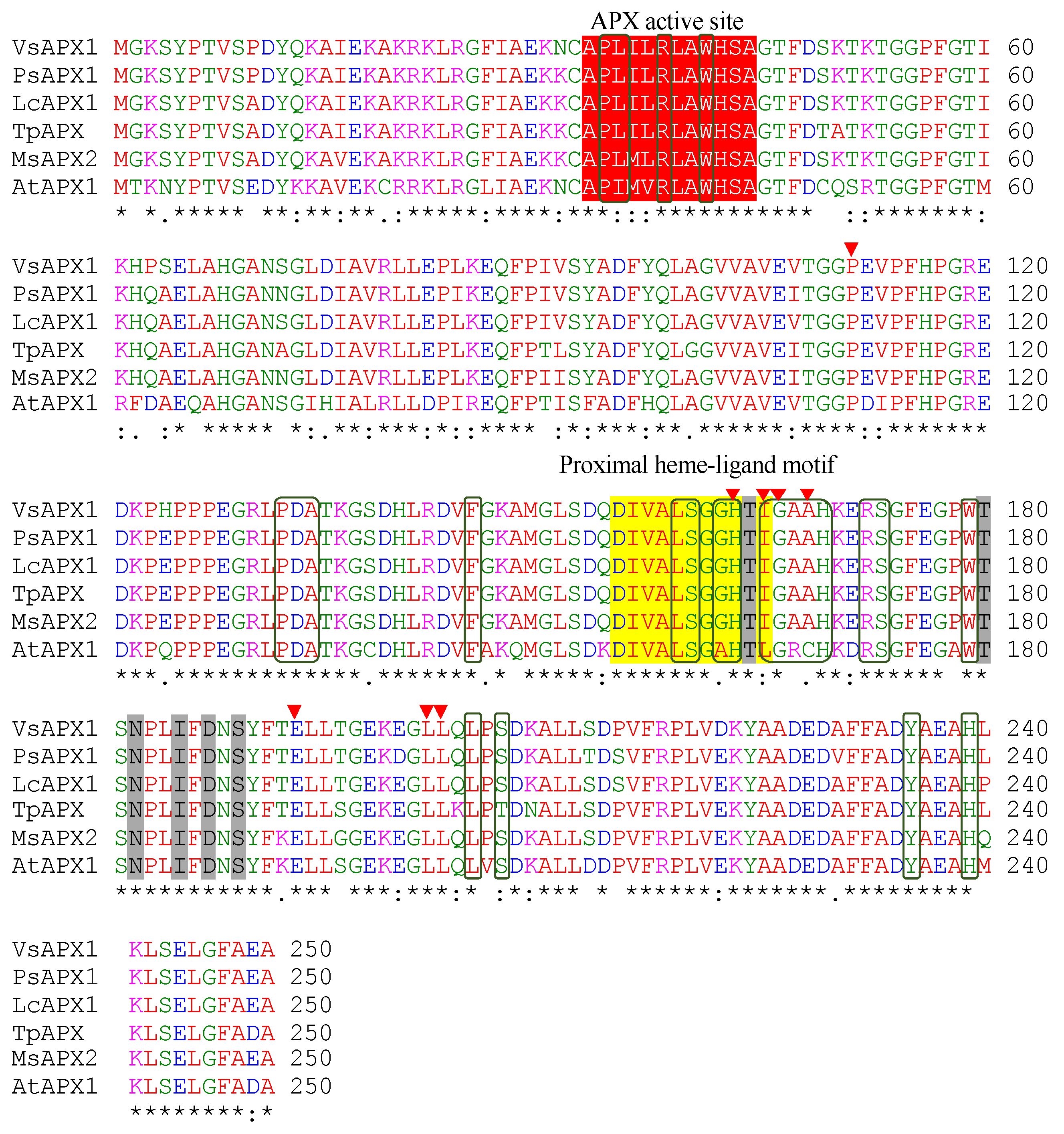
Multiple sequence alignment (Figure 1) was performed by using Clustal Omega between the VsAPX1 protein and other legumes cytosolic APX homologs: Pisum sativum (PsAPX1, AAA33645.1), Lens culinaris subsp. culinaris (LcAPX1, AXI69835.1), Trifolium pratense (TpAPX, PNX98620.1) Medicago sativa (MsAPX2, AIY27528.1) and Arabidopsis thaliana (AtAPX1, BAA03334.1). The alignment showed the presence of conserved amino acid residues. The conserved domains of the VsAPX1 protein were predicted using NCBI Conserved Domains (CDDs) and InterPro. InterPro analysis revealed conserved APX active site (APLILRLAWHSA, 33-44 aa), proximal heme-ligand motif (DIVALSGGHTI, 155-165 aa) and other conserved sites including six K+ binding sites (T164, T180, N182, I185, D187, S189), eight substrate binding sites (P111, H163, I165, G166, A168, E193, L202, L203), and 24 heme binding sites (P34, L35, R38, W41, P132, D133, A134, F145, L159, S160, G162, H163, I165, G166, A167, A168, H169, R172, S173, W179, L205, S207, Y235, H239) (Figure 1). Furthermore, CDD results confirmed the presence of previously conserved sites and demonstrated that the VsAPX1 protein belongs to a plant-peroxidase-like superfamily (cl00196), member (PLN02364).
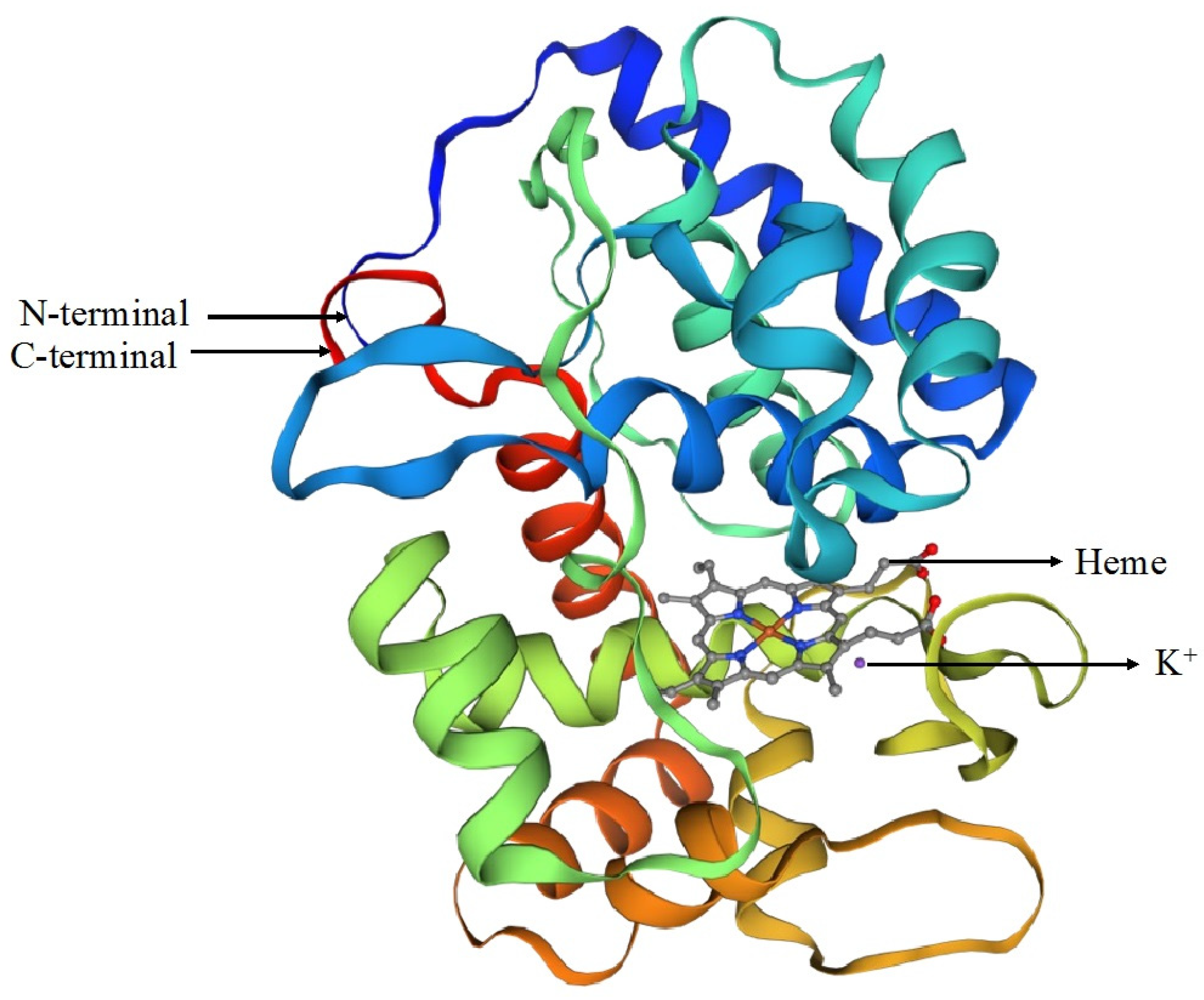
Homology modeling of the VsAPX1 protein was performed using SWISS-MODEL (Figure 2). The best structural template identified was the cytosolic ascorbate peroxidase 1APX (PDB: 1apx.1.A), an X-ray structure of 2.2 Å resolution, showing 95.18% sequence identity, 0.98 coverage, and a GMQE score of 0.98. The predicted quaternary structure was supported by a QSQE value of 0.66, consistent with the dimeric organization of the template.
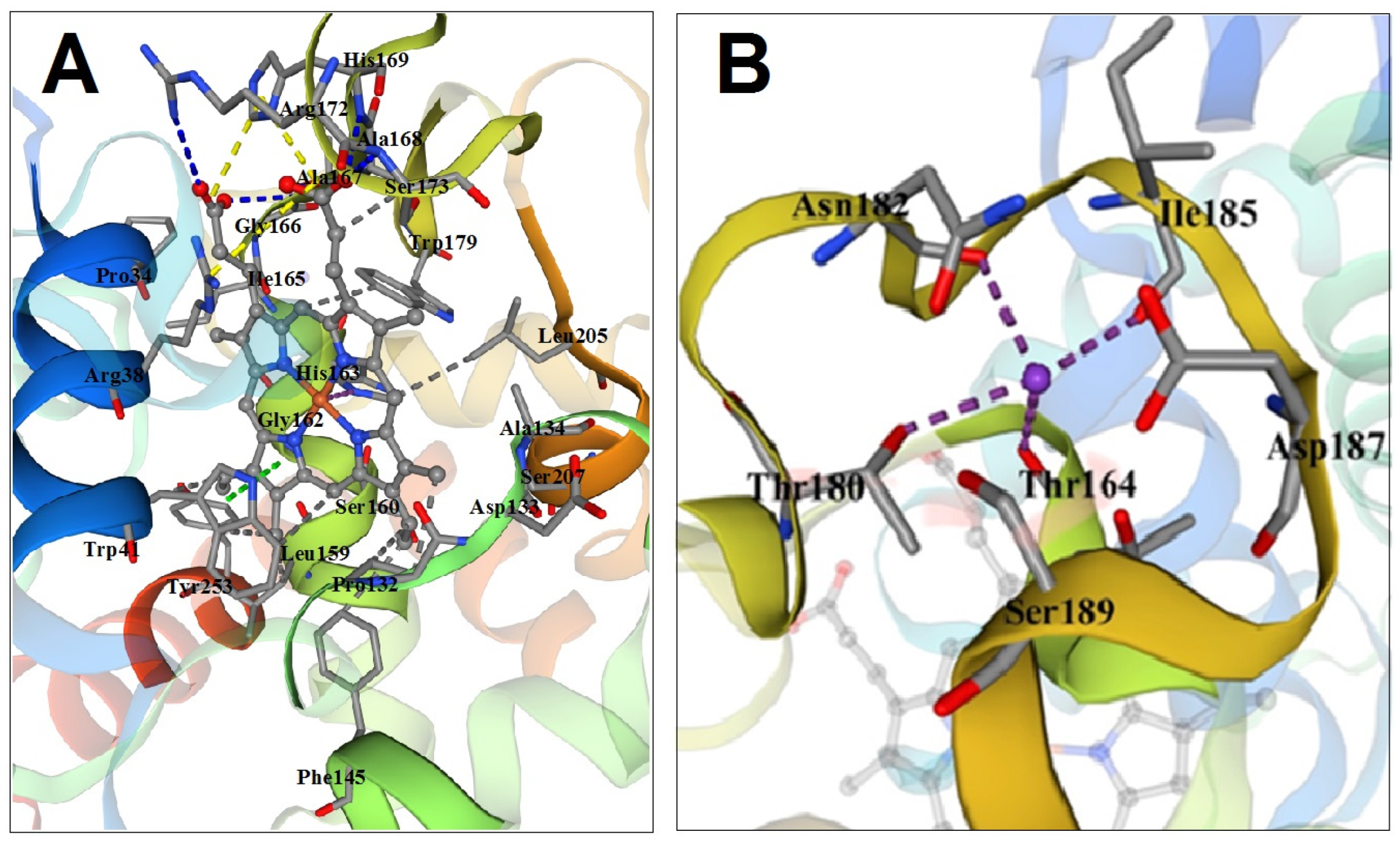
The resulting 3D model correctly positioned the heme prosthetic group and the nearby potassium-binding region (Figure 3). The amino acids shown surrounding these sites represent predicted spatial proximities from the homology model, not experimentally verified contacts. Therefore, these features should be interpreted as structural hypotheses. Experimental validation, such as mutagenesis or biochemical assays, will be required to confirm the contribution of individual residues.
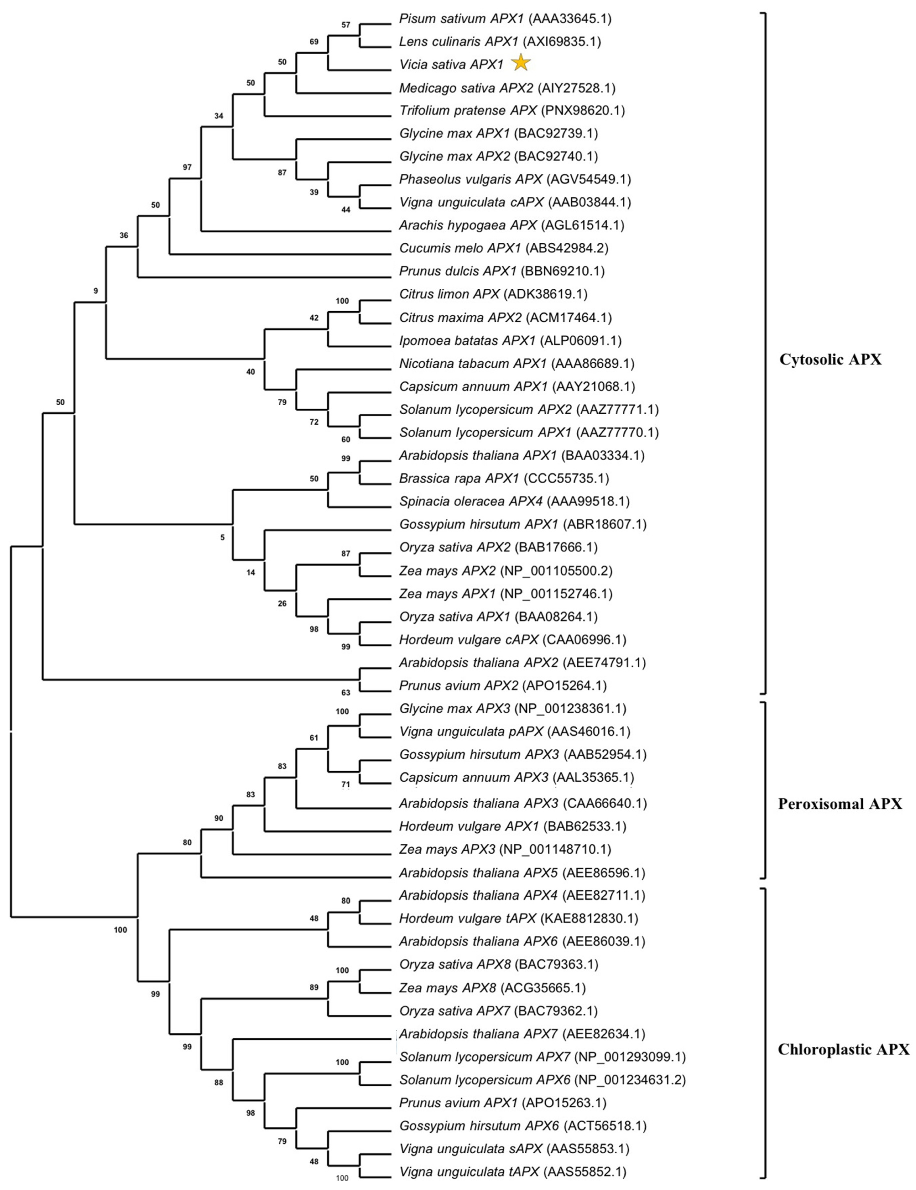
To study the phylogeny of VsAPX1 and plant homologs, APX proteins were retrieved from GenBank and aligned by ClustalW. Based on sequence similarity, a phylogenetic tree was constructed by a neighbor-joining method using the MEGA 11 program with bootstrap re-sampling analysis with 1000 replicates. The phylogenetic tree showed that APX proteins clearly clustered into three clades according to ascorbate peroxidase subcellular localization: cytosolic, peroxisomal, and chloroplast. The VsAPX1 protein clustered with the cytosolic APX clade and was closely related to other legumes like pea and lentil (Figure 4).

3.2. Ascorbate Peroxidase Gene Structure

To determine VsAPX1 gene structure, APX gDNA was amplified by PCR using the same specific primers (VsAPX-F and VsAPX-R). The amplified PCR resulted in around 2.5 kb of product, which was cloned into pGEM®-T Easy Vector and was sequenced. To complete the waking primer sequencing and cover the entire internal region of the gene, a pair of internal primers (VsAPX-Fc and VsAPX-Rc) designed based on the received sequence were used as a template. The VsAPX1 gene structure was determined by comparison of the APX cDNA with the gDNA sequence, using the BLAST algorithm, Splign-NCBI, and Gene Structure Display Server 2.0. The full-length VsAPX1 gDNA nucleotide sequence was 2425 bp, and was submitted to GenBank under the accession number OR756531, and the comparison in Figure S3 shows that the VsAPX1 gene contains 10 exons separated by nine introns. All introns contain the GT-AG dinucleotide at both ends, and the first intron was localized within the 5′UTR closer to the start codon (ATG). Introns of the VsAPX1 gene were determined according to the intron’s position relative to the reading frame of the gene. Intron 1 is located in the 5′UTR, introns 3, 4, 6, and 8 are located between two codons (phase 0 intron), intron 5 is located between the first and second nucleotide of a codon (phase 1 intron), and introns 2, 7, and 9 are located between the second and third nucleotide of a codon (phase 2 intron).
A comparison between the VsAPX1 genomic structure (exon/intron organization) and cytosolic APX of other plants (P. sativum APX1 (M93051.1), A. thaliana APX1a (D14442.1), A. thaliana APX1b (X80036.1), and F. ananassa APX (AF158654.1)) was performed to study structural changes (Figure S4). The results revealed that the VsAPX1 gene structure is similar to that of PsAPX1 and FaAPX and has a conserved exon/intron order, including exons with the same length as well as conserved intron phases, while VsAPX1 is different from AtAPX1b as it lacks the 5′UTR intron. In addition, exons 5 and 6 of VsAPX1 may have been merged to form exon 5 in AtAPX1a. However, this hypothesis needs further investigation using synteny analysis.
The promoter region (1500 bps) of VsAPX1 revealed multiple transcription factor binding sites (TFBS) for major TFs including MYB/SANT, WRKY and NAC NAM (Figure 5).

3.3. Expression Pattern Analysis of VsAPX1 Gene

The differential expression of the VsAPX1 gene was studied in one-month-old V. sativa seedlings by using qRT-PCR in response to heat stress, hydrogen peroxide, and phytohormonal (ABA, SA, and JA) stimuli at different time points (0, 2, 4, and 6 h).
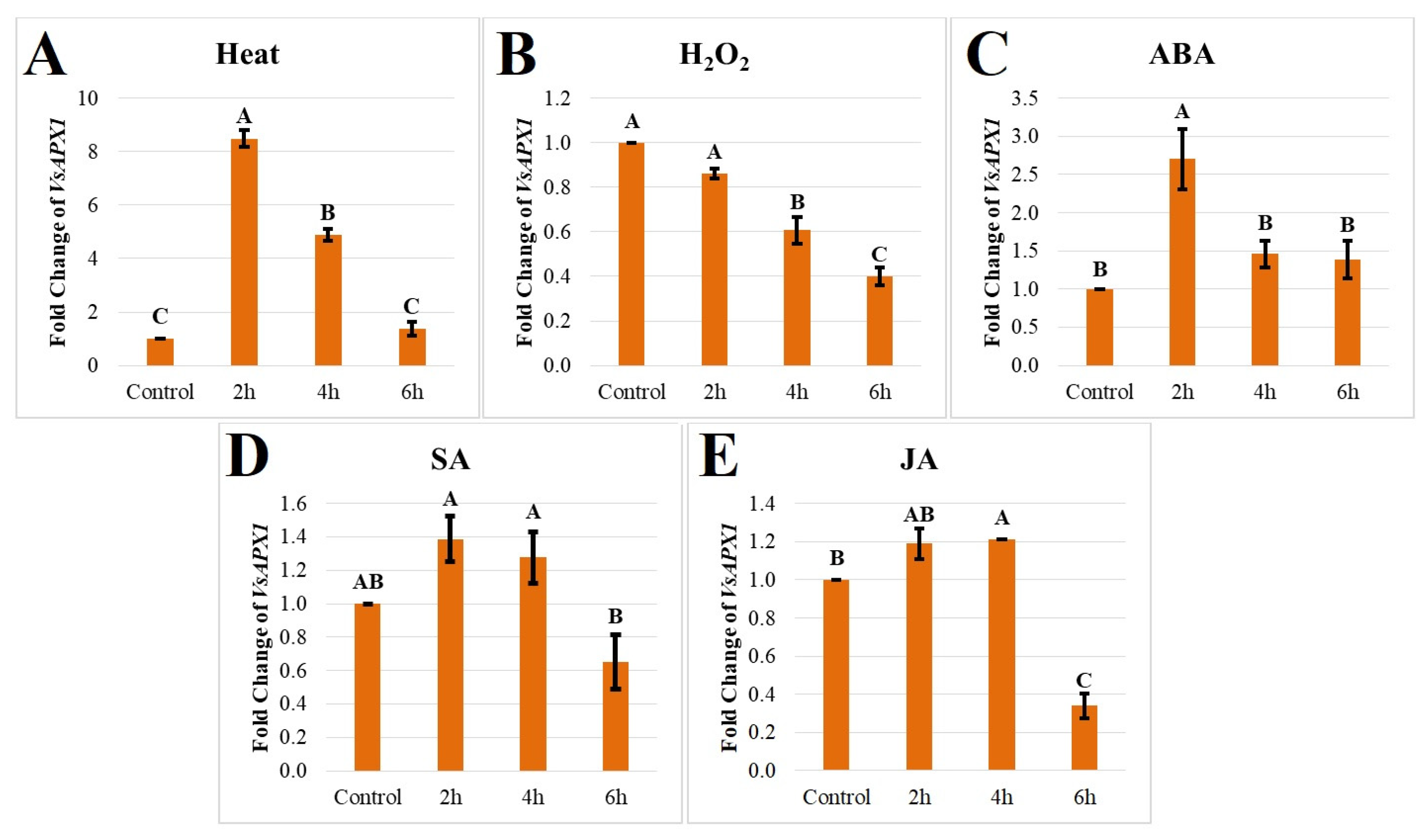
To examine VsAPX1 transcript level under abiotic stresses, seedlings were exposed to heat stress (42 °C); the expression of VsAPX1 compared to the control was highly up-regulated by 8.4-fold after 2 h then decreased sharply to 4.8-fold after 4 h and to 1.36-fold after 6 h, which reflects a transient stress shock response (Figure 6A). The VsAPX1 transcript was down-regulated gradually over time by 0.86, 0.60, and 0.39-fold after 2 h, 4 h, and 6 h, respectively, in response to hydrogen peroxide (10 mM) compared to the control (Figure 6B). Although the 10 mM H2O2 treatment exceeds endogenous physiological concentrations, this level is widely used in plant molecular studies to induce reproducible oxidative stress and trigger antioxidant gene responses (e.g., 5–20 mM in leaf and callus assays). During the exposure period used here, the tissues remained visually healthy, with no necrosis or collapse observed. While electrolyte leakage or additional physiological viability indicators were not measured, the transcriptional responses and absence of tissue damage suggest that the treatment elicited oxidative signaling rather than extensive toxicity.
The VsAPX1 transcript levels showed modest responses to phytohormone treatments. Following ABA (100 μM) treatment, transcript abundance increased slightly to 2.7-fold after 2 h, before decreasing to 1.4- and 1.3-fold at 4 h and 6 h, respectively (Figure 6C). SA (1 mM) treatment resulted in a small increase to 1.38-fold after 2 h, which then decreased slightly to 1.27-fold at 4 h and further declined to 0.65-fold at 6 h (Figure 6D). JA (100 μM) treatment caused a modest rise to 1.18-fold at 2 h, peaking at 1.2-fold at 4 h, and subsequently decreasing to 0.34-fold at 6 h compared to the control (Figure 6E). These results indicate that VsAPX1 transcript levels exhibit only slight and transient changes in response to the tested phytohormones.

4. Discussion

4.1. Sequence Analysis of VsAPX1 Gene and Protein

APXs are important H2O2 scavenger antioxidant enzymes, which maintain H2O2 homeostasis under normal and stressful conditions. The cytosolic APX isoenzyme is the most studied among other isoforms, due it being moderately responsive and induced under different stresses [30], as well as its role in different defense processes, and in plant homeostasis redox regulation [31]. Guo et al. (2020) proved the role of cytosolic APX1 in maintaining redox balance to regulate cotton photosynthetic rate and yield [32]. Also, in Arabidopsis thaliana, APX1 protected chloroplast under high light intensity [30]. Therefore, the present study characterized the full-length gDNA and cDNA cytosolic ascorbate peroxidase gene from the V. sativa plant. The VsAPX1 cDNA sequence contains an in-frame start site (ATG, 62 bp) and stop site (TAG, 812 bp); these signals suggest that the VsAPX1 cDNA is full length. Furthermore, a putative polyadenylation signal site (AAATAA) was detected in the VsAPX1 sequence at a 159 bp downstream stop codon; this signal site was previously reported in other legumes [28]. Different studies have cloned and characterized cytosolic APX cDNA from different plant species such as pea [33], sweet potato [34], potato [35], and spinach [36].
The ORF of VsAPX1 encodes 250 amino acid residues (Figure 1), with a molecular weight of 27.1 kDa. APX isoenzymes are different in molecular weight, for example, in rice chloroplast (thylakoidal APX (~51 kDa), stromal APX (~33-38 kDa)), peroxisomal APX (~32 kDa), and cytosolic APX (~27 kDa) [37], while in potato mitochondrial APX they are ~31 kDa [38]. VsAPX1 amino acid residues lack localization signals, an N-terminal transit peptide sequence and C-terminal transmembrane domains, according to the protein targeting analysis. This confirms the bioinformatic results that the VsAPX1 protein is cytosolic ascorbate peroxidase. APX can be localized to different cellular compartments which perform distinct antioxidant functions. However, the cytosolic VsAPX1 acts as a central hub for maintaining whole-cell redox balance and integrating stress and hormone signaling pathways. Therefore, the predicted cytosolic localization suggests a broader regulatory role in coordinating cellular antioxidant responses rather than organelle-specific protection.
Sequence similarity analysis of VsAPX1 with closely related proteins from different plant species was investigated using BLASTp (Figure S2). The results showed that the deduced VsAPX1 amino acid sequence was highly similar to other cytosolic APX proteins; this could indicate that the cloned gene encodes cytosolic APX.
Ascorbate peroxidase protein, in its active form, is a dimer (containing two identical subunits) distinguished by two domains, C-terminal and N-terminal, surrounded by the heme, and specified by its residues that are essential for the activity [39]. Ascorbate, heme (iron), and potassium ions are critical for APX activity [40]. In the present study, the multiple sequence analysis of the VsAPX1 primary structure (Figure 1) revealed fundamental and functional conserved motifs, domains and residues that are essential for APX protein structure and function. These include the APX active site, proximal heme-ligand motif, K+ binding site, substrate binding sites, and heme binding sites. Several studies have identified the proximal His-163 in the heme binding site and Arg-38 which are essential for heme binding. The distal His-42 and Arg-38 in the active site are responsible for the heterolytic cleavage of H2O2. The hydrogen bonding between His-163, Asp-208, and Trp-179 forms the active site structure [41]. The substrate ascorbate binds to the active site by four hydrogen bonds with Lys-30, Arg-172, Cys-32, and the heme moiety. At the proximal domain, K+ ions bind to K+ binding sites (Thr-164, Thr-180, Asp-187) that are required for APX activity [39]. Phosphorylation of Thr-59 and Thr-164 residues was reported to increase tomato APX enzyme activity, and S-nitrosylation of Cys-32 enhances APX enzyme catalytic activity [42]. APX class I is distinguished from other classes by Trp-41 and Trp-179 instead of Phe-41 and Phe-179 [39]. Moreover, cytosolic APX is differentiated by Phe-175 instead of Trp-175 found in the chloroplast, and Ser-43, Phe-57, and Thr-59 are replaced by Asp-43, Asn-57, and Ser-59 in other APX isoforms [40]. These data suggest that VsAPX1 belongs to the class I cytosolic ascorbate peroxidase.
Molecular phylogenetics deals with evolutionary relationships based on different macromolecules (DNA, RNA, and protein) [43]. This study clarifies the phylogenetic relationship of VsAPX1 with APX orthologs from other plant species; for this purpose, a neighbor-joining phylogenetic tree was constructed (Figure 4). The resulting tree showed that APX proteins were clearly separated into three groups based on their subcellular localization: cytosolic, peroxisomal, and chloroplastic, and the VsAPX1 protein was clustered within the cytosolic clade which is closely related to the leguminous species Pisum sativum and Lens culinaris, indicating a close relationship between these APX proteins. This result is consistent with the phylogenetic tree generated in the study of 44. This apparent divergence between orthologs was revealed in previous studies indicating that cytosolic and peroxisomal APX isoenzymes were generated by a duplication event of a non-chloroplastic ancestral gene [37]. According to Qu et al. (2020), results of APX genes’ phylogenetic tree and exons’ structure revealed that the ancestors of monocots and dicots underwent genome duplication [16]. Ozyigit et al.’s (2016) APX phylogenetic tree of 18 plant species showed segmental and tandem duplications in some APX genes [15].

4.2. Ascorbate Peroxidase Gene Structure

The identification of the VsAPX1 gene structure provides insights into the organization of exons and introns in this gene, which can aid in further genetic studies and manipulation of the gene. To investigate the structure of VsAPX1, the exon/intron organization was constructed and compared with other plant cytosolic APX genes. In the present study, the VsAPX1 gene contains 10 exons interspaced by nine introns (Figure S3). Comparing the VsAPX1 gene structure with other plant species (Figure S4), the length of VsAPX1 exons of ORF 9 exons is highly identical to pea APX1 (which is the only gene available with details for APX in legumes) [28], and strawberry [44]. Moreover, similar results were found in other plant APX genes such as tomato [45], rice [37], maize [16,46], and wild watermelon [47]. Whereas there are differences in intron length and nucleotide sequences, the intron number and intron phases are conserved [48]. This suggests a conserved cytosolic APX1 gene architecture in higher plants [16,37]. Likewise, APX genes’ promoter cis-elements, exon–intron organization, and number were studied in Populus trichocarpa [49]. On the other hand, Arabidopsis thaliana APX1a, APX1b had nine exons and eight introns [50]. A major difference appears in exon 5 of AtAPX1a, which is separated by an 86 bp intron to exon 5 and exon 6 in the VsAPX1 gene. The first intron was located within 5′UTR. A similar observation was made in those of other plant species except for the A. thaliana APX1b (Supplementary Figure S4), which lacks the 5′UTR intron, the gene encoding a second family of cytosolic APX; and a similar observation was made in Dancy’ tangerine (Citrus reticulata Blanco) [50,51]. Mittler and Zilinskas (1992) detected a part of the GPEI enhancer (TGATTCAG) sequence in the 5′UTR intron, which is a regulator element for glutathione transferase P that regulates gene transcription by interaction with transcription factors and RNA II polymerase [28]. Some studies discussed the effect of the 5′UTR intron, which may interact with other elements located in the promoter that regulate APX1 gene expression; in fact, the leader intron of the APX20 gene in tomato increased expression of a reporter gene in leaves but it was absent in roots [46]. The gain of an intron in the 5′-untranslated region and in exon number five of VsAPX1 causes exon fission but does not cause a shift in reading frame, unlike the exon loss/gain that can cause a shift in reading frame [52]. The intron insertion at this location may be due to the presence of proto-splice sites (G|G, and MAG|R; M: A or C, R: A or G) that increase the chance of intron insertion at this position [53].

4.3. Expression Pattern Analysis of VsAPX1 Gene in Response to Stresses

Plants have complex defense systems that protect themselves from environmental changes. These defense systems are augmented and activated by the perception of stress signaling molecules (phytohormones and ROS (H2O2)), which induce signal transduction cascades and then regulate gene expression of the stress-responsive genes [11]. Antioxidant enzymes scavenge the excess ROS and maintain cellular homeostasis from oxidative damage [13]. APX is one of the crucial enzymes in the AsA-GSH cycle and can scavenge excess H2O2 [54]. Recent studies showed up-regulation of APX gene expression under different abiotic stresses and stress-response chemicals, which could be due to the presence of stress-response cis-acting elements in the promoter that activate APX gene expression [49]. These elements include phytohormone-responsive, abiotic stress-responsive, and growth and development-responsive elements. Furthermore, several TFs were found to bind TFBS on the VsAPX1 promoter (Figure 5), including major abiotic stress TFs including WRKY and NAC.
Generally, the cAPX 5′ regulatory region has a heat shock-responsive element and anti-peroxidative element (ARE) that might aid in H2O2 scavenging. The present study investigated the expression pattern of the APX gene in V. sativa under phytohormone treatments (ABA, SA, and JA) and abiotic stresses (42 °C and H2O2) using qRT-PCR, to understand and define the possible involvement of the VsAPX1 gene to stresses.
The stress-induced expression patterns observed for VsAPX1 are consistent with reports from other legumes, supporting the conserved role of cytosolic APX1 in ROS detoxification. While no novel functional properties were identified, the present data establish VsAPX1 as a canonical APX1 homolog in V. sativa, thereby extending the comparative framework of antioxidant gene regulation within legumes.
The increased global temperature is a critical climate-change problem, resulting in an increasing rate of evaporation and dehydration in plants and soil [55]. Heat stress resulted in the deactivation of enzymes, protein misfolding, disturbance of cell metabolism, increased fluidity of the membrane lipid, and increased production and accumulation of ROS (e.g., oxidative damage) [56]. Recent studies demonstrated that some APX genes are heat inducible [1]. Under heat stress, the increasing level of cellular H2O2 acts as a signaling molecule that induces heat stress signal transduction components, including the heat stress transcription factor, which binds to the heat shock element (HSE) in the promoter of the APX gene and controls its expression [57]. This explains the quick response of the APX gene to heat stress [58]. HSE has been determined in the APX promoter of pea [28] and strawberry [45]. In the present study, after exposing seedlings to 42 °C, the expression of VsAPX1 scored an early increase after 2 h (Figure 6A). Other studies found that heat stress increased APX gene expression in a variety of plant species, including pea [28], rice [59], alfalfa [60], Arabidopsis thaliana APX2 [61], and sweet potato APX1 [34]. Moreover, heat stress increased the activity of the cucumber cAPX enzyme [62]. The overexpression of pea cAPX enhanced heat tolerance in transgenic tomato [63]. Furthermore, the heat tolerance of Arabidopsis thaliana was increased by the overexpression of the cabbage APX gene [56].
H2O2 is an important non-radical ROS, generated from normal cellular metabolism as a harmful by-product that causes damage and inactivation for cellular components [64,65]. At basal levels, H2O2 acts as a regulatory signal for different physiological processes including photosynthesis, photorespiration, stomatal closure, growth, cell cycle, development, and senescence [66]. Furthermore, under stress conditions, H2O2 acts as a signaling transduction molecule, due to being highly stable and diffusible, and can pass through the plasma membrane via aquaporins and diffuse from different organelles and transport the signal to the nucleus through redox reactions, integrated with the MAPK pathway [67], which are involved in the regulation of nuclear gene expression of many transcription factors, and the up-regulation of antioxidative enzymes [62].
Interestingly, exogenous H2O2 treatment did not result in sustained induction of VsAPX1 expression (Figure 6B), but instead a reduction in expression was observed over time. In fact, similar responses have been reported for several antioxidant genes and may reflect the complex dynamics of ROS signaling. APX genes are often rapidly and transiently induced during the early phase of oxidative stress, with expression peaks occurring within minutes to 1 h, followed by feedback repression once cellular redox homeostasis is restored. Moreover, high concentrations of exogenously applied H2O2 can impose oxidative damage on cellular components and transiently inhibit transcriptional processes. Moskova et al. (2009) reported that exogenous application of H2O2 decreased APX enzyme activity in pea [68]. In contrast to other studies, the exogenous application of H2O2 increased gene expression in sweet potato [34], and cucumber cAPX enzyme activity [62]. In addition, H2O2 treatment was reported to induce APX gene expression in cultured soybean cells [69]. This is possibly due to the presence of an anti-peroxidative element (ARE) in the APX promoter, which could be involved in the regulation of the APX gene and responsible for the H2O2-induced response [28,69]. It is also possible that regulation occurs predominantly at post-transcriptional or enzymatic levels, or that other APX isoforms and antioxidant systems compensate for ROS detoxification. Therefore, the observed decline in VsAPX1 transcript levels likely reflects tight redox-dependent feedback control rather than reduced involvement in oxidative stress defense.

4.4. Expression Analysis of VsAPX1 Gene in Response to Phytohormones

Plant bioregulators are chemicals found in small quantities that have a large influence on regulating plant growth, development, and yield under normal and stressful conditions [70,71]. The plant bioregulators SA, ABA, and JA were selected because they are key regulators in stress signaling. ABA mediates abiotic stress responses such as drought, salinity, and heat, while JA and SA are central components of defense and stress-adaptation interacting with ROS signaling [72]. Therefore, evaluating VsAPX1 expression under these treatments enables assessment of its potential involvement in hormone-mediated stress responses and signaling crosstalk. According to previous studies, the cis-elements were detected in APX genes that contribute to the phytohormones’ stress responses, mainly the abscisic acid-responsive elements, MeJA-responsive element and salicylic acid-responsive element [73]. These elements were detected in different plant species’ APX promotor such as peanut AhAPX genes [73], Ammopiptanthus nanus [74], Triticum aestivum [75] and Brassica spp [76]. Furthermore, the exogenous application of phytohormones at a suitable dose enhanced plant tolerance to abiotic stress conditions [77].
ABA acts as a stress-responsive hormone that plays a crucial role in plant adaptation [78]. Its biosynthesis increases under stress conditions and regulates the expression of different stress-responsive genes and protective proteins like antioxidant enzymes, late embryogenesis abundant proteins, and dehydrins [79]. Therefore, exogenous ABA treatment was reported to increase the expression and activity of ascorbate peroxidase in different plant species, which is possibly due to the presence of the APX promoter cis-elements antioxidant-responsive element and ABA-responsive element [30]. In the peanut AhAPX1 promoter, an abscisic acid-responsive element (ABRE) was recorded, which was supported by up-regulation of AhAPX1 expression when peanut tissues were exposed to ABA [73]. In this study, the foliar application of ABA markedly increased the VsAPX1 expression after 2 h; this indicates that ABA has a signaling effect on VsAPX1 expression. Similar observations were reported after ABA treatment in pea [28], maize [47], and sweet potato [34].
The distinct temporal expression profiles of VsAPX1 observed under different phytohormone treatments suggest that this gene is regulated through hormone-specific signaling pathways rather than through a generalized stress response. In particular, the earlier transcriptional peak detected after ABA treatment (2 h) compared with the delayed induction under JA (2–4 h) likely reflects differences in the kinetics of their respective signaling cascades. ABA signaling is typically mediated through rapid activation of ABRE-dependent transcription factors, including AREB/ABF and ABI5 [80], which directly bind ABA-responsive elements in promoters of stress-associated genes and promote early transcriptional activation of antioxidant defenses. Such fast ABA-mediated responses are consistent with the role of ABA as a primary regulator of drought, heat, and oxidative stress adaptation. In contrast, JA signaling proceeds through the COI1–JAZ–MYC2 pathway [81], where degradation of JAZ repressors is required prior to activation of MYC2-dependent transcription, a multistep process that may account for the relatively delayed VsAPX1 induction observed following JA application. Therefore, the different response kinetics likely reflect intrinsic differences in hormone perception, signal transduction, and transcriptional regulation.
SA acts as a growth-regulating and protector molecule that improves crop plant tolerance under stress conditions [70], and induces the gene expression of antioxidants, HSPs, chaperones, and genes involved in the biosynthesis of secondary metabolites [79]. Foliar application of SA under abiotic stresses enhances the activity of antioxidant enzymes and reduces oxidative stress impacts such as lipid peroxidation and membrane injury [82]. The current study showed a slight rise in the VsAPX1 transcript level after 2 h of foliar SA treatment. Some studies reported that SA application increased endogenous H2O2 accumulation and increased the expression and activity of antioxidant enzymes [83]. The SA application increases the transcript level of sweet potato cytosolic ascorbate peroxidase [34], and pea APX enzyme activity [84].
JA aids in plant reproduction, development, tendril coiling, fruit ripening, chlorosis, allelopathy, production of secondary metabolites, flower and seed development, wounding and herbivory, and leaf senescence [77]. Whereas, under a stressful environment, JA acts as a signaling molecule and stress-responsive hormone [85]. JA was reported to interact with other plant hormones, transcription factors, and enhance the expression of JA-associated genes and stress-responsive genes and increase the activity of the antioxidant defense system [72]. VsAPX1 transcript level was highly accumulated after 4 h of JA application. A similar observation was recorded in rice OsAPX1 [86], and sweet potato [34]. The foliar application of JA alone or in combination with salinity on bitter melon seedlings decreased the activity of the APX enzyme [85].
In the present study, the expression profiling of VsAPX1 was conducted primarily in leaf tissues, as leaves represent the major site of photosynthesis and consequently the principal source of reactive oxygen species (ROS) production under both normal and stress conditions. Chloroplast electron transport, photorespiration, and environmental stresses such as heat and exogenous hormone treatments are known to markedly enhance H2O2 generation in photosynthetically active tissues, necessitating strong antioxidant defenses. Cytosolic APX isoforms have been widely reported to exhibit higher activity and functional relevance in leaves [87], where they cooperate with chloroplastic antioxidant systems to maintain cellular redox homeostasis. Therefore, focusing on leaf tissues allowed us to capture the most physiologically relevant and dynamic transcriptional responses of VsAPX1 to oxidative and hormonal stimuli. Nevertheless, antioxidant genes often display tissue-specific regulation depending on metabolic activity and stress perception. Future investigations examining roots and stems under similar stress conditions will provide a more comprehensive understanding of the spatial regulation and whole-plant physiological roles of VsAPX1.
It should be noted that we presented VsAPX1 expression, which may not correlate with enzymatic activity. APX proteins are frequently regulated at post-transcriptional and post-translational levels, including protein stability, redox modifications, and cofactor availability. Therefore, measurements of enzyme activity and protein accumulation will be required in future studies to fully elucidate the functional contribution of VsAPX1 to oxidative stress tolerance.

4.5. Redox Reaction and VsAPX1

Plant APX is known to have the strongest affinity for H2O2 scavenging within the AsA–GSH cycle compared to other peroxidases in plant cells, which is critical for maintaining cellular homeostasis under stress conditions [88]. Furthermore, stress-related genes interact extensively through crosstalk during stress perception and response, ultimately inducing the expression of tolerance and resistance pathways.
In cotton, silencing the cytosolic GhAPX1 gene led to reduced photosynthesis, impaired growth and development, and increased water loss, whereas its overexpression enhanced tolerance of cotton fibers to oxidative stress [32]. Similarly, overexpression of the Chinese cabbage APX gene in Arabidopsis thaliana increased heat tolerance in transgenic lines, resulting in higher chlorophyll content, improved germination rates, and lower MDA and H2O2 levels compared to non-transgenic plants [56].
We focused specifically on APX1 rather than the entire gene family due to its relative importance. Chloroplastic and mitochondrial APXs are highly sensitive to ascorbate deficiency and have short half-inactivation times (less than 30 s), in contrast to cytosolic and peroxisomal isoenzymes, which remain active for about an hour or more [16]. Among the APX isoforms, cytosolic APX is the most extensively studied because it is strongly induced under diverse stresses, is more resistant to ascorbate depletion, and is involved in multiple defense processes and redox homeostasis regulation [31]. It is also considered the most responsive isoform to environmental constraints [30]. In addition, overexpression or suppression of APX1 regulates redox balance and thereby influences cotton photosynthetic rate and yield [32].
A recent genome-wide study in common bean (Phaseolus vulgaris) identified 101 APX family members and demonstrated strong tissue-specific and stress-inducible expression patterns under drought and salinity, highlighting the central role of APX genes in oxidative stress mitigation and abiotic stress tolerance. These findings further support the conserved involvement of cytosolic APX isoforms in regulating H2O2 homeostasis across legumes [89]. Similarly, a cytosolic APX gene (LcAPX1) cloned from lentil (Lens culinaris) showed conserved structural features and stress-responsive expression, with significant induction under salinity, H2O2, ABA, and JA treatments. These findings further support the conserved role of legume APX1 isoforms in hormone-mediated antioxidant defense and oxidative stress regulation [90]. Transcriptomic analysis of common vetch under salt stress also revealed enhanced activation of ROS-scavenging pathways, including significant up-regulation of APX4, with stronger induction in the salt-tolerant genotype. These findings highlight the importance of APX-mediated antioxidant defenses in stress adaptation and further support the conserved role of APX genes in oxidative stress regulation in Vicia species [91].

5. Conclusions

In this study, we cloned and characterized the full-length VsAPX1 gene from the forage legume Vicia sativa. Sequence and bioinformatic analyses indicate that VsAPX1 encodes a cytosolic ascorbate peroxidase and is closely related to APX1 homologs from other legumes. Expression analyses under heat and phytohormone treatments revealed transient and moderate changes in transcript abundance, indicating that VsAPX1 transcription is responsive to environmental and hormonal cues.
While these expression patterns are consistent with the conserved involvement of APX1 genes in stress-related processes, the present data do not allow direct conclusions regarding the functional contribution of VsAPX1 to stress tolerance or adaptation. Rather, this work provides a species-specific molecular and expression framework for VsAPX1 in Vicia sativa. Further studies, including promoter analyses and functional characterization, will be required to more precisely elucidate the physiological role and regulatory control of VsAPX1 under stress conditions.

Supplementary Materials

The following supporting information can be downloaded at: https://www.mdpi.com/article/10.3390/ijpb17030016/s1, Figure S1: Subcellular localization of VsAPX1 polypeptide as predicted using online bioinformatic tools; TargetP 2.0 (A), ProtComp 9.0 (B), and DeepLoc 2.0 (C). Figure S2: Sequence similarities between APXs of different plant species. The values show the percentage of similar amino acids. Figure S3: VsAPX1 gene structure. Schematic alignment of VsAPX1 gDNA and cDNA sequences indicating the location of intron–exon boundaries, constructed by the BioRender online tool. The sizes of the exon and intron are drawn using the scale at the bottom; each 1 cm equals 50 bp. Exons are shown in boxes, introns in black lines, the coding region shaded in purple, and UTRs are shaded in gray. Numbers indicate the first and last nucleotides of the exons. Numbers at the middle of introns and exons indicate their length. The intron phases are shown on the bottom of each exon boundary. Figure S4 shows a comparison of gene structure (exon/intron organization) between VsAPX1 and APX of other plant species constructed by the BioRender online tool. The sizes of the exon and intron are drawn using the scale at the bottom; each 1 cm equals 50 bp. Exons are shown in boxes, introns in black lines, the coding region is shaded in yellow, and UTRs are shaded in gray. Numbers at the middle of introns and exons indicate their length. The intron phases are shown on the bottom of each exon boundary. The sequences selected are P. sativum APX1 (M93051.1), A. thaliana APX1a (D14442.1), A. thaliana APX1b (X80036.1), and F. ananassa APX (AF158654.1). Figure S5: Agarose gel electrophoresis of total RNA extraction from treated V. sativa seedlings. Three microliters of RNA sample mixed with 4 µL loading dye (Thermos Fisher Scientific, USA) and lauded in the well. Analyzed in 1% agarose gel with 0.5X TBE buffer and stained with ethidium bromide, after running the the bands were separated. Lane M: Marker (1 kb ladder (Thermos Fisher Scientific, USA)); lanes 1-4 RNA samples.

Author Contributions

Conceptualization, S.A.-R.; methodology, S.A.K.A.-R.; software, F.A.S.; validation, S.A.K.A.-R.; formal analysis, F.A.S.; investigation, M.T.S.; resources, S.A.-R.; data curation, R.M.A.-M. and M.T.S.; writing—original draft preparation, all authors; writing—review and editing, M.T.S.; visualization, F.A.S.; supervision, M.T.S.; project administration, S.A.-R.; funding acquisition, S.A.-R. All authors have read and agreed to the published version of the manuscript.

Funding

This research received no external funding.

Data Availability Statement

The original contributions presented in this study are included in this article/Supplementary Material. Further inquiries can be directed to the corresponding author.

Acknowledgments

The authors are grateful to Al-Balqa Applied University and University of Jordan for support in conducting this study.

Conflicts of Interest

The authors declare no conflicts of interest.

Abbreviations

The following abbreviations are used in this manuscript:
qRT-PCRQuantitative real time polymerase chain reaction
PEGPolyethylene glycol
SRASequence Read Archive
BLASTBasic Local Alignment Search Tool
NCBINational Center for Biotechnology Information
TFBSTranscription factor binding sites

References

  1. Sadder, M.T.; Alshomali, I.; Ateyyeh, H.; Musallam, A. Physiological and molecular responses of common fig (Ficus carica L.) to long term salinity stress. Physiol. Mol. Biol. Plants 2021, 27, 107–1173. [Google Scholar]
  2. Sadder, M.T.; Alsadon, A.A.; Wahb-Allah, M.A. Transcriptomic analysis of tomato lines reveals putative stress-specific biomarkers. Turk. J. Agric. For. 2014, 38, 700–715. [Google Scholar] [CrossRef]
  3. Sachdev, S.; Ansari, S.A.; Ansari, M.I.; Fujita, M.; Hasanuzzaman, M. Abiotic stress and reactive oxygen species: Generation, signaling, and defense mechanisms. Antioxidants 2021, 10, 277. [Google Scholar] [CrossRef]
  4. Saini, P.; Gani, M.; Kaur, J.J.; Godara, L.C.; Singh, C.; Chauhan, S.S.; Francies, R.M.; Bhardwaj, A.; Kumar, N.B.; Ghosh, M.K. Reactive oxygen species (ROS): A way to stress survival in plants. In Abiotic Stress-Mediated Sensing and Signaling in Plants: Omics Perspective; Springer: Berlin/Heidelberg, Germany, 2018; pp. 127–153. [Google Scholar]
  5. Alsadon, A.A.; Ibrahim, A.A.; Wahb-Allah, M.A.; Ali, A.A.M.; Sadder, M.T. Tomato under salinity stress: Correlation between growth and yield components and responsive genes. Acta Hortic. 2015, 1081, 111–119. [Google Scholar] [CrossRef]
  6. AL-Kiyyam, M.; Shibli, R.A.; Zatimeh, A.; Tahtamouni, R.W.; AlQudah, T.S.; Hassan, S. Evaluation of genetic diversity in Jordanian Solanum nigrum plants and genetic stability of in vitro grown plant using (AFLP) technique. Jordan J. Biol. Sci. 2024, 17, 123–128. [Google Scholar]
  7. Kumari, A.; Bhinda, M.S.; Sharma, S.; Chitara, M.K.; Debnath, A.; Maharana, C.; Parihar, M.; Sharma, B. ROS regulation mechanism for mitigation of abiotic stress in plants. In Reactive Oxygen Species; IntechOpen: London, UK, 2021; pp. 1–42. [Google Scholar]
  8. Tahtamouni, R.W.; Shibli, R.A.; Al-Qudah, T.S.; Azzam, H.; Saifan, S. Chiliadenus montanus (Vahl.) Brullo which grows wild in the Jordanian environment shows distinguished Terpinen-4-ol levels and antibacterial powers. Jordan J. Biol. Sci. 2024, 17, 507–512. [Google Scholar]
  9. Abu-Romman, S. Genotypic response to heat stress in durum wheat and the expression of small HSP genes. Rend. Lincei 2016, 27, 261–267. [Google Scholar] [CrossRef]
  10. Huang, H.; Ullah, F.; Zhou, D.X.; Yi, M.; Zhao, Y. Mechanisms of ROS regulation of plant development and stress responses. Front. Plant Sci. 2019, 10, 800. [Google Scholar] [CrossRef] [PubMed]
  11. Nadarajah, K.K. ROS homeostasis in abiotic stress tolerance in plants. Int. J. Mol. Sci. 2020, 21, 5208. [Google Scholar] [CrossRef]
  12. Kumar, A.; Guleria, S.; Ghosh, D.; Dogra, V.; Kumar, S. Managing reactive oxygen species–some learnings from high altitude extremophytes. Environ. Exp. Bot. 2021, 189, 104525. [Google Scholar] [CrossRef]
  13. Rajput, V.D.; Singh, R.K.; Verma, K.K.; Sharma, L.; Quiroz-Figueroa, F.R.; Meena, M.; Gour, V.S.; Minkina, T.; Sushkova, S.; Mandzhieva, S. Recent developments in enzymatic antioxidant defence mechanism in plants with special reference to abiotic stress. Biology 2021, 10, 267. [Google Scholar] [CrossRef]
  14. Abu-Romman, S.; Shatnawi, M. Isolation and expression analysis of chloroplastic copper/zinc superoxide dismutase gene in barley. S. Afr. J. Bot. 2011, 77, 328–334. [Google Scholar] [CrossRef]
  15. Ozyigit, I.I.; Filiz, E.; Vatansever, R.; Kurtoglu, K.Y.; Koc, I.; Öztürk, M.X.; Anjum, N.A. Identification and comparative analysis of H2O2-scavenging enzymes (ascorbate peroxidase and glutathione peroxidase) in selected plants employing bioinformatics approaches. Front. Plant Sci. 2016, 7, 301. [Google Scholar] [CrossRef]
  16. Qu, C.; Wang, L.; Zhao, Y.; Liu, C. Molecular evolution of maize ascorbate peroxidase genes and their functional divergence. Genes 2020, 11, 1204. [Google Scholar] [CrossRef]
  17. He, M.; He, C.Q.; Ding, N.Z. Abiotic stresses: General defenses of land plants and chances for engineering multistress tolerance. Front. Plant Sci. 2018, 9, 1771. [Google Scholar] [CrossRef]
  18. Wang, Q.; Zhang, C.; Li, J.; Wu, X.; Long, Y.; Su, Y. Intercropping Vicia sativa L. improves the moisture, microbial community, enzyme activity and nutrient in rhizosphere soils of young kiwifruit plants and enhances plant growth. Horticulturae 2021, 7, 335. [Google Scholar] [CrossRef]
  19. Mikić, A.; Mihailović, V.; Ćupina, B.; Vasiljević, S.; Milošević, B.; Katanski, S.; Matić, R.; Radojević, V.; Kraljević-Balalić, M. Agronomic characteristics related to grain yield and crude protein content in common vetch (Vicia sativa) accessions of diverse geographic origin. N. Z. J. Agric. Res. 2013, 56, 297–308. [Google Scholar] [CrossRef]
  20. Huang, Y.F.; Gao, X.L.; Nan, Z.B.; Zhang, Z.X. Potential value of the common vetch (Vicia sativa L.) as an animal feedstuff: A review. J. Anim. Physiol. Anim. Nutr. 2017, 101, 807–823. [Google Scholar] [CrossRef]
  21. Córdoba, E.M.; Fernández-Aparicio, M.; González-Verdejo, C.I.; López-Grau, C.; Muñoz-Muñoz, M.D.V.; Nadal, S. Search for resistant genotypes to cuscuta campestris infection in two legume species, Vicia sativa and Vicia ervilia. Plants 2021, 10, 738. [Google Scholar] [CrossRef] [PubMed]
  22. Mennan, H.; Jabran, K.; Zandstra, B.H.; Pala, F. Non-chemical weed management in vegetables by using cover crops: A review. Agronomy 2020, 10, 257. [Google Scholar] [CrossRef]
  23. Magalhaes, S.C.; Taveira, M.; Cabrita, A.R.; Fonseca, A.J.; Valentão, P.; Andrade, P.B. European marketable grain legume seeds: Further insight into phenolic compounds profiles. Food Chem. 2017, 215, 177–184. [Google Scholar] [CrossRef] [PubMed]
  24. Ch, K.; Sturm, D.J.; Varnholt, D.; Walker, F.; Gerhards, R. Allelopathic effects and weed suppressive ability of cover crops. Plant Soil Environ. 2016, 62, 60–66. [Google Scholar]
  25. Tigka, E.; Beslemes, D.; Kakabouki, I.; Pankou, C.; Bilalis, D.; Tokatlidis, I.; Vlachostergios, D.N. Seed rate and cultivar effect on contribution of Vicia sativa L. green manure to soil amendment under mediterranean conditions. Agriculture 2021, 11, 733. [Google Scholar] [CrossRef]
  26. Abu-Romman, S. Molecular characterization of a catalase gene (VsCAT) from Vicia sativa. Int. J. Biol. 2016, 8, 66–76. [Google Scholar] [CrossRef]
  27. Abu-Romman, S. Molecular cloning and gene expression analysis of chloroplastic copper/zinc superoxide dismutase gene in Vicia sativa L. Res. Crops 2019, 20, 215–222. [Google Scholar]
  28. Mittler, R.; Zilinskas, B.A. Molecular cloning and characterization of a gene encoding pea cytosolic ascorbate peroxidase. J. Biol. Chem. 1992, 267, 21802–21807. [Google Scholar] [CrossRef] [PubMed]
  29. Livak, K.J.; Schmittgen, T.D. Analysis of relative gene expression data using real-time quantitative PCR and the 2−ΔΔCT method. Methods 2001, 25, 402–408. [Google Scholar] [CrossRef]
  30. Saxena, S.C.; Salvi, P.; Kamble, N.U.; Joshi, P.K.; Majee, M.; Arora, S. Ectopic overexpression of cytosolic ascorbate peroxidase gene (Apx1) improves salinity stress tolerance in Brassica juncea by strengthening antioxidative defense mechanism. Acta Physiol. Plant. 2020, 42, 45. [Google Scholar] [CrossRef]
  31. Caverzan, A.; Jardim-Messeder, D.; Paiva, A.L.; Margis-Pinheiro, M. Ascorbate peroxidases: Scavengers or sensors of hydrogen peroxide signaling? In Redox Homeostasis in Plants; Springer Nature: Cham, Switzerland, 2019; pp. 85–115. [Google Scholar]
  32. Guo, K.; Li, Z.; Tian, H.; Du, X.; Liu, Z.; Huang, H.; Wang, P.; Ye, Z.; Zhang, X.; Tu, L. Cytosolic ascorbate peroxidases plays a critical role in photosynthesis by modulating reactive oxygen species level in stomatal guard cell. Front. Plant Sci. 2020, 11, 446. [Google Scholar] [CrossRef] [PubMed]
  33. Mittler, R.; Zilinskas, B.A. Molecular cloning and nucleotide sequence analysis of a cDNA encoding pea cytosolic ascorbate peroxidase. Fed. Eur. Biochem. Soc. 1991, 289, 257–259. [Google Scholar] [CrossRef]
  34. Park, S.Y.; Ryu, S.H.; Jang, I.C.; Kwon, S.Y.; Kim, J.G.; Kwak, S.S. Molecular cloning of a cytosolic ascorbate peroxidase cDNA from cell cultures of sweet potato and its expression in response to stress. Mol. Genet. Genom. 2004, 271, 339–346. [Google Scholar] [CrossRef]
  35. Kawakami, S.; Matsumoto, Y.; Matsunaga, A.; Mayama, S.; Mizuno, M. Molecular cloning of ascorbate peroxidase in potato tubers and its response during storage at low temperature. Plant Sci. 2002, 163, 829–836. [Google Scholar] [CrossRef]
  36. Ishikawa, T.; Sakai, K.; Takeda, T.; Shigeoka, S. Cloning and expression of cDNA encoding a new type of ascorbate peroxidase from spinach. Fed. Eur. Biochem. Soc. 1995, 367, 28–32. [Google Scholar] [CrossRef]
  37. Teixeira, F.K.; Menezes-Benavente, L.; Margis, R.; Margis-Pinheiro, M. Analysis of the molecular evolutionary history of the ascorbate peroxidase gene family: Inferences from the rice genome. J. Mol. Evol. 2004, 59, 761–770. [Google Scholar] [CrossRef]
  38. De Leonardis, S.; Dipierro, N.; Dipierro, S. Purification and characterization of an ascorbate peroxidase from potato tuber mitochondria. Plant Physiol. Biochem. 2000, 38, 773–779. [Google Scholar] [CrossRef]
  39. Dąbrowska, G.; Kata, A.; Goc, A.; Szechyńska-Hebda, M.; Skrzypek, E. Characteristics of the plant ascorbate peroxidase family. Acta Biol. Cracoviensia Ser. Bot. 2007, 49, 7–17. [Google Scholar]
  40. Jespersen, H.M.; Kjærsgård, I.V.; Østergaard, L.; Welinder, K.G. From sequence analysis of three novel ascorbate peroxidases from Arabidopsis thaliana to structure, function and evolution of seven types of ascorbate peroxidase. Biochem. J. 1997, 326, 305–310. [Google Scholar] [CrossRef]
  41. Lazzarotto, F.; Menguer, P.K.; Del-Bem, L.E.; Zámocký, M.; Margis-Pinheiro, M. Ascorbate peroxidase neofunctionalization at the origin of APX-R and APX-L: Evidence from basal archaeplastida. Antioxidants 2021, 10, 597. [Google Scholar] [CrossRef]
  42. Ravi, B.; Foyer, C.H.; Pandey, G.K. The integration of reactive oxygen species (ROS) and calcium signalling in abiotic stress responses. Plant Cell Environ. 2023, 46, 1985–2006. [Google Scholar] [CrossRef] [PubMed]
  43. Bogusz, M.; Whelan, S. Phylogenetic tree estimation with and without alignment: New distance methods and benchmarking. Syst. Biol. 2017, 66, 218–231. [Google Scholar] [CrossRef]
  44. Kim, I.J.; Chung, W.I. Isolation of genomic DNA containing a cytosolic ascorbate peroxidase gene (ApxSC) from the strawberry (Fragaria × ananassa). Biosci. Biotechnol. Biochem. 1998, 62, 1358–1363. [Google Scholar] [CrossRef]
  45. Gadea, J.; Conejero, V.; Vera, P. Developmental regulation of a cytosolic ascorbate peroxidase gene from tomato plants. Mol. Gen. Genet. 1999, 262, 212–219. [Google Scholar] [CrossRef]
  46. Liu, Y.J.; Yuan, Y.; Liu, Y.Y.; Liu, Y.; Fu, J.J.; Zheng, J.; Wang, G.Y. Gene families of maize glutathione–ascorbate redox cycle respond differently to abiotic stresses. J. Plant Physiol. 2012, 169, 183–192. [Google Scholar] [CrossRef]
  47. Malambane, G.; Tsujimoto, H.; Akashi, K. The cDNA structures and expression profile of the ascorbate peroxidase gene family during drought stress in wild watermelon. J. Agric. Sci. 2018, 10, 56–71. [Google Scholar] [CrossRef]
  48. Patthy, L. Intron-dependent evolution: Preferred types of exons and introns. Fed. Eur. Biochem. Soc. 1987, 214, 1–7. [Google Scholar] [CrossRef] [PubMed]
  49. Leng, X.; Wang, H.; Zhang, S.; Qu, C.; Yang, C.; Xu, Z.; Liu, G. Identification and characterization of the APX gene family and its expression pattern under phytohormone treatment and abiotic stress in Populus trichocarpa. Genes 2021, 12, 334. [Google Scholar] [CrossRef]
  50. Santos, M.; Gousseau, H.; Lister, C.; Foyer, C.; Creissen, G.; Mullineaux, P. Cytosolic ascorbate peroxidase from Arabidopsis thaliana L. is encoded by a small multigene family. Planta 1996, 198, 64–69. [Google Scholar] [CrossRef] [PubMed]
  51. Kunta, M.; Del Rio, H.S.; Skaria, M.; Louzada, E.S. Isolation and molecular characterization of a putative ascorbate peroxidase gene from citrus. Int. J. Fruit Sci. 2010, 10, 1–15. [Google Scholar] [CrossRef]
  52. Xu, G.; Guo, C.; Shan, H.; Kong, H. Divergence of duplicate genes in exon–intron structure. Proc. Natl. Acad. Sci. USA 2012, 109, 1187–1192. [Google Scholar] [CrossRef]
  53. Nguyen, H.D.; Yoshihama, M.; Kenmochi, N. Phase distribution of spliceosomal introns: Implications for intron origin. BMC Evol. Biol. 2006, 6, 69. [Google Scholar] [CrossRef] [PubMed]
  54. Hasanuzzaman, M.; Bhuyan, M.H.M.; Anee, T.I.; Parvin, K.; Nahar, K.; Mahmud, J.A.; Fujita, M. Regulation of ascorbate-glutathione pathway in mitigating oxidative damage in plants under abiotic stress. Antioxidants 2019, 8, 384. [Google Scholar] [CrossRef]
  55. Hassan, M.U.; Chattha, M.U.; Khan, I.; Chattha, M.B.; Barbanti, L.; Aamer, M.; Iqbal, M.M.; Nawaz, M.; Mahmood, A.; Ali, A.; et al. Heat stress in cultivated plants: Nature, impact, mechanisms, and mitigation strategies a review. Plant Biosyst. 2020, 155, 211–234. [Google Scholar] [CrossRef]
  56. Chiang, C.M.; Chien, H.L.; Chen, L.F.O.; Hsiung, T.C.; Chiang, M.C.; Chen, S.P.; Lin, K.H. Overexpression of the genes coding ascorbate peroxidase from Brassica campestris enhances heat tolerance in transgenic Arabidopsis thaliana. Biol. Plant. 2015, 59, 305–315. [Google Scholar] [CrossRef]
  57. Panchuk, I.I.; Volkov, R.A.; Schöffl, F. Heat stress-and heat shock transcription factor-dependent expression and activity of ascorbate peroxidase in Arabidopsis. Plant Physiol. 2002, 129, 838–853. [Google Scholar] [CrossRef]
  58. Banerjee, A.; Roychoudhury, A. Abiotic stress tolerance in plants by priming and pretreatment with hydrogen peroxide. In Priming and Pretreatment of Seeds and Seedlings; Springer: Singapore, 2019; pp. 417–426. [Google Scholar]
  59. Sato, Y.; Murakami, T.; Funatsuki, H.; Matsuba, S.; Saruyama, H.; Tanida, M. Heat shock-mediated APX gene expression and protection against chilling injury in rice seedlings. J. Exp. Bot. 2001, 52, 145–151. [Google Scholar] [CrossRef] [PubMed]
  60. Li, W.; Wei, Z.; Qiao, Z.; Wu, Z.; Cheng, L.; Wang, Y. Proteomics analysis of alfalfa response to heat stress. PLoS ONE 2013, 8, e82725. [Google Scholar] [CrossRef] [PubMed]
  61. Wang, L.; Ma, K.B.; Lu, Z.G.; Ren, S.X.; Jiang, H.R.; Cui, J.W.; Chen, G.; Teng, N.J.; Lam, H.M.; Jin, B. Differential physiological, transcriptomic and metabolomic responses of Arabidopsis leaves under prolonged warming and heat shock. BMC Plant Biol. 2020, 20, 86. [Google Scholar] [CrossRef]
  62. Song, X.S.; Hu, W.H.; Mao, W.H.; Ogweno, J.O.; Zhou, Y.H.; Yu, J.Q. Response of ascorbate peroxidase isoenzymes and ascorbate regeneration system to abiotic stresses in Cucumis sativus L. Plant Physiol. Biochem. 2005, 43, 1082–1088. [Google Scholar] [CrossRef]
  63. Wang, Y.; Wisniewski, M.; Meilan, R.; Cui, M.; Fuchigami, L. Transgenic tomato (Lycopersicon esculentum) overexpressing cAPX exhibits enhanced tolerance to UV-B and heat stress. J. Appl. Hortic. 2006, 8, 87–90. [Google Scholar] [CrossRef]
  64. Das, K.; Roychoudhury, A. Reactive oxygen species (ROS) and the response of antioxidants as ROS-scavengers during environmental stress in plants. Front. Environ. Sci. 2014, 2, 53. [Google Scholar] [CrossRef]
  65. Mittler, R. ROS are good. Trends Plant Sci. 2017, 22, 11–19. [Google Scholar] [CrossRef]
  66. Hasanuzzaman, M.; Bhuyan, M.H.M.; Zulfiqar, F.; Raza, A.; Mohsin, S.M.; Mahmud, J.A.; Fujita, M.; Fotopoulos, V. Reactive oxygen species and antioxidant defense in plants under abiotic stress: Revisiting the crucial role of a universal defense regulator. Antioxidants 2020, 9, 681. [Google Scholar] [CrossRef]
  67. Singh, A.; Kumar, A.; Yadav, S.; Singh, I.K. Reactive oxygen species-mediated signaling during abiotic stress. Plant Gene 2019, 18, 100173. [Google Scholar] [CrossRef]
  68. Moskova, I.; Todorova, D.; Alexieva, V.; Ivanov, S.; Sergiev, I. Effect of exogenous hydrogen peroxide on enzymatic and nonenzymatic antioxidants in leaves of young pea plants treated with paraquat. Plant Growth Regul. 2009, 57, 193–202. [Google Scholar] [CrossRef]
  69. Lee, S.C.; Kang, B.G.; Oh, S.E. Induction of ascorbate peroxidase by ethylene and hydrogen peroxide during growth of cultured soybean cells. Mol. Cells 1999, 9, 166–171. [Google Scholar] [CrossRef]
  70. Alhaithloul, H.A.S.; Abu-Elsaoud, A.M.; Soliman, M.H. Abiotic stress tolerance in crop plants: Role of phytohormones. In Abiotic Stress in Plants; IntechOpen: London, UK, 2020; pp. 1–23. [Google Scholar]
  71. Hasanuzzaman, M.; Raihan, M.; Hossain, R.; Masud, A.A.C.; Rahman, K.; Nowroz, F.; Rahman, M.; Nahar, K.; Fujita, M. Regulation of reactive oxygen species and antioxidant defense in plants under salinity. Int. J. Mol. Sci. 2021, 22, 9326. [Google Scholar] [CrossRef]
  72. Wang, J.; Song, L.; Gong, X.; Xu, J.; Li, M. Functions of jasmonic acid in plant regulation and response to abiotic stress. Int. J. Mol. Sci. 2020, 21, 1446. [Google Scholar] [CrossRef] [PubMed]
  73. Raza, A.; Sharif, Y.; Chen, K.; Wang, L.; Fu, H.; Zhuang, Y.; Chitikineni, A.; Chen, H.; Zhang, C.; Varshney, K.R.; et al. Genome-wide characterization of ascorbate peroxidase gene family in peanut (Arachis hypogea L.) revealed their crucial role in growth and multiple stress tolerance. Front. Plant Sci. 2022, 13, 962182. [Google Scholar] [CrossRef] [PubMed]
  74. Wang, Y.; Cao, S.; Sui, X.; Wang, J.; Geng, Y.; Gao, F.; Zhou, Y. Genome-wide characterization, evolution, and expression analysis of the ascorbate peroxidase and glutathione peroxidase gene families in response to cold and osmotic stress in Ammopiptanthus nanus. J. Plant Growth Regul. 2023, 42, 502–522. [Google Scholar] [CrossRef]
  75. Tyagi, S.; Verma, P.C.; Singh, K.; Upadhyay, S.K. Molecular characterization of ascorbate peroxidase (APX) and APX-related (APX-R) genes in Triticum aestivum L. Genomics 2020, 112, 4208–4223. [Google Scholar] [CrossRef]
  76. Verma, D.; Upadhyay, S.K.; Singh, K. Characterization of APX and APX-R gene family in Brassica juncea and B. rapa for tolerance against abiotic stresses. Plant Cell Rep. 2022, 41, 571–592. [Google Scholar] [CrossRef] [PubMed]
  77. Awan, S.A.; Khan, I.; Rizwan, M.; Zhang, X.; Brestic, M.; Khan, A.; El-Sheikh, M.; Alyemeni, M.; Ali, S.; Huang, L. Exogenous abscisic acid and jasmonic acid restrain polyethylene glycol-induced drought by improving the growth and antioxidative enzyme activities in pearl millet. Physiol. Plant. 2021, 172, 809–819. [Google Scholar] [CrossRef] [PubMed]
  78. Gong, Z.; Xiong, L.; Shi, H.; Yang, S.; Herrera-Estrella, L.R.; Xu, G.; Chao, D.Y.; Li, J.; Wang, P.Y.; Qin, F.; et al. Plant abiotic stress response and nutrient use efficiency. Sci. China Life Sci. 2020, 63, 635–674. [Google Scholar] [CrossRef]
  79. Wani, S.H.; Kumar, V.; Shriram, V.; Sah, S.K. Phytohormones and their metabolic engineering for abiotic stress tolerance in crop plants. Crop J. 2016, 4, 162–176. [Google Scholar] [CrossRef]
  80. Liu, X.; Cheng, R.; Chen, Y.; Wang, S.; Qin, F.; Wang, D.; Liu, Y.; Hu, L.; Meng, S. Identification and characterization of the AREB/ABF/ABI5 gene family in sandalwood (Santalum album L.) and its potential role in drought stress and ABA treatment. Forests 2023, 14, 1691. [Google Scholar] [CrossRef]
  81. Chini, A.; Boter, M.; Solano, R. Plant oxylipins: COI1/JAZs/MYC2 as the core jasmonic acid-signalling module. FEBS J. 2009, 276, 4682–4692. [Google Scholar] [CrossRef] [PubMed]
  82. Pourghasemian, N.; Moradi, R.; Naghizadeh, M.; Landberg, T. Mitigating drought stress in sesame by foliar application of salicylic acid, beeswax waste and licorice extract. Agric. Water Manag. 2020, 231, 105997. [Google Scholar] [CrossRef]
  83. Dikilitas, M.; Simsek, E.; Roychoudhury, A. Modulation of abiotic stress tolerance through hydrogen peroxide. In Protective Chemical Agents in the Amelioration of Plant Abiotic Stress: Biochemical and Molecular Perspectives; Wiley Online Library: Hoboken, NJ, USA, 2020; pp. 147–173. [Google Scholar]
  84. Yadu, S.; Dewangan, T.L.; Chandrakar, V.; Keshavkant, S. Imperative roles of salicylic acid and nitric oxide in improving salinity tolerance in Pisum sativum L. Physiol. Mol. Biol. Plants 2017, 23, 43–58. [Google Scholar] [CrossRef]
  85. Alisofi, S.; Einali, A.; Sangtarash, M.H. Jasmonic acid-induced metabolic responses in bitter melon (Momordica charantia) seedlings under salt stress. J. Hortic. Sci. Biotechnol. 2020, 95, 247–259. [Google Scholar] [CrossRef]
  86. Wang, Y.; Wu, J.; Choi, Y.W.; Jun, T.H.; Kwon, S.W.; Choi, I.S.; Kim, Y.C.; Gupta, R.; Kim, S.T. Expression analysis of Oryza sativa ascorbate peroxidase 1 (OsAPx1) in response to different phytohormones and pathogens. J. Life Sci. 2015, 25, 1091–1097. [Google Scholar] [CrossRef]
  87. Ribeiro, C.W.; Korbes, A.P.; Garighan, J.A.; Jardim-Messeder, D.; Carvalho, F.E.L.; Sousa, R.H.V.; Caverzan, A.; Teixeira, F.K.; Silveira, J.A.G.; Margis-Pinheiro, M. Rice peroxisomal ascorbate peroxidase knockdown affects ROS signaling and triggers early leaf senescence. Plant Sci. 2017, 263, 55–65. [Google Scholar] [CrossRef] [PubMed]
  88. Anjum, N.A.; Sharma, P.; Gill, S.S.; Hasanuzzaman, M.; Khan, E.A.; Kachhap, K.; Mohamed, A.A.; Thangavel, P.; Devi, G.D.; Vasudhevan, P.; et al. Catalase and ascorbate peroxidase—Representative H2O2-detoxifying heme enzymes in plants. Environ. Sci. Pollut. Res. 2016, 23, 19002–19029. [Google Scholar] [CrossRef] [PubMed]
  89. Tüfekçi, E.D.; Tellioğlu, B.; Aygören, A.S.; Yaprak, E.; Ilhan, E. Genome-wide characterization of ascorbate peroxidase (APX) gene family in Phaseolus vulgaris L. of response to multiple abiotic stresses. S. Afr. J. Bot. 2025, 177, 429–444. [Google Scholar] [CrossRef]
  90. Ramadan, H.R.; Abu-Romman, S.; Odat, N. Isolation and expression analysis of ascorbate peroxidase gene from lentil. Plant Omics 2025, 17, 33–47. [Google Scholar]
  91. Sun, Y.; Zhao, N.; Sun, H.; Xu, S.; Lu, Y.; Xi, H.; Guo, Z.; Shi, H. Transcriptome profiling reveals molecular responses to salt stress in common vetch (Vicia sativa L.). Plants 2024, 13, 714. [Google Scholar] [CrossRef]
Figure 1. Multiple sequence alignment of V. sativa APX protein sequence with related cytosolic APX proteins from different plant species. Red (background) box indicates conserved APX active site, yellow (background) box is a proximal heme-ligand motif, and gray (background) box is K+ binding site, arrowheads indicate substrate binding sites, and green boxes heme binding sites. Conserved amino acid residues are indicated by asterisks below the sequence, and numbers to the right indicate the amino acid positions.
Ijpb 17 00016 g001
Figure 2. Theoretical three-dimensional structure modeling of the VsAPX1 protein using SWISS-MODEL online tool. Predicted VsAPX1 protein structure based on the crystal structure of pea APX protein, each helix was given a different color for clarity.
Ijpb 17 00016 g002
Figure 3. Predicted structural environment of the heme group (A) and potassium-binding region (B) in VsAPX1 based on the SWISS-MODEL homology model. Shown residues represent predicted contacts derived from the structural template (1APX) and do not constitute experimentally validated interactions.
Ijpb 17 00016 g003
Figure 4. Phylogenetic analysis of V. sativa APX protein (yellow star) with other plant species. APX proteins were aligned by ClustalW, and the phylogenetic tree was constructed using the neighbor-joining method with 1000 bootstrap replicates by MEGA11. GenBank accession numbers are indicated in parentheses.
Ijpb 17 00016 g004
Figure 5. Predicted transcription factor binding sites on the promoter region of VsAPX1. Key fro color shapes is located below the figure.
Ijpb 17 00016 g005
Figure 6. Relative expression pattern of VsAPX1 in V. sativa seedlings exposed to (A) heat stress, (B) H2O2, (C) ABA, (D) SA, and (E) JA as measured by qRT-PCR. The transcript level of VsAPX1 was normalized to VsActin (GU946218) as a housekeeping gene and expressed as a ratio relative to the control which was set at 1. Each value represents the average of three biological replicates; the error bars indicate the standard error (±SE). Different letters indicate significant differences according to the LSD test at p ≤ 0.05.
Ijpb 17 00016 g006
Disclaimer/Publisher’s Note: The statements, opinions and data contained in all publications are solely those of the individual author(s) and contributor(s) and not of MDPI and/or the editor(s). MDPI and/or the editor(s) disclaim responsibility for any injury to people or property resulting from any ideas, methods, instructions or products referred to in the content.