Article

Enhancing Stress Resilience in a Drought-Tolerant Zea mays Cultivar by Integrating Morpho-Physiological and Proteomic Characterization

by
Rotondwa Rabelani Sinthumule
,
Charlie Sithole
,
Joseph Lesibe Gaorongwe
,
Kegomoditswe Martha Matebele
,
Oziniel Ruzvidzo
and
Tshegofatso Bridget Dikobe
*
Unit for Environmental Sciences and Management, Department of Botany, North-West University, Mmabatho 2735, South Africa
*
Author to whom correspondence should be addressed.
Int. J. Plant Biol. 2025, 16(4), 133; https://doi.org/10.3390/ijpb16040133
Submission received: 17 September 2025 / Revised: 28 October 2025 / Accepted: 10 November 2025 / Published: 21 November 2025

Abstract

Maize is not only a staple across the sub-Saharan Africa (SSA) region but also a substantially economically valuable cereal crop. As a seasonal crop, its successful cultivation depends on favorable rainfall patterns and climatic conditions. However, environmental stresses such as drought limit its productivity. Enhancing stress resilience requires understanding the morphological, physiological, and proteomic response mechanisms that contribute to drought tolerance. Hence, it is critical to understand its adaptive capacity at the protein level to achieve improved stress-tolerant cultivars and increased yields in the future. Our study investigated drought stress responses in a drought-tolerant maize cultivar subjected to polyethylene glycol (PEG)-induced water deficit, combining one-dimensional gel electrophoresis (1DE) with LC-MS/MS to profile the leaf proteome. From the analysis, 50 of the 439 identified maize leaf proteins were further studied due to their significant differential expression and functional relevance, revealing the interconnection between the proteomic patterns as well as the morphological and physiological responses that enable drought resilience. These insights provide a foundation for improving stress-tolerant maize cultivars through integrative characterization approaches.

1. Introduction

Recently, climate change has intensified food insecurity across sub-Saharan Africa (SSA) with long-term negative macroeconomic effects, particularly on economic growth and poverty [1]. The agricultural sector supports over 70% of South African regions; however, feeding approximately 9.9 billion people has become a challenging task [2,3,4]. Drought has been a prevalent problem inhibiting crop production for years, affecting agricultural yields globally and particularly in semi-arid areas [5]. Drought is a climatic phenomenon defined as a sufficiently long period of imbalance between precipitation and evaporation that can increase soil water scarcity and cause water stress in plants, limiting their development and/or production [5,6]. However, drought can also occur as a result of high light intensity, high temperature, and dry winds [7]. Drought affects morphology, physiology, and molecular processes, such as delayed flowering, decreased dry matter accumulation and distribution, decreased photosynthetic rate, metabolic limitations, and oxidative chloroplast damage [5]. Nonetheless, plants have developed coping mechanisms such as osmotic adjustment, changes in photosynthetic pigments, leaf reduction, hormone stimulation, and accumulation of antioxidant scavengers [8]. Hence, being mindful of plants such as halophytes, gypsophytes, and xerophytes that are naturally adapted to grow under stressful conditions, research indicates that natural plant species have a greater tolerance and faster mechanisms to adapt or endure biotic and abiotic stresses than cultivated plant species [9].
On the contrary, Lippmann [10] states that, unlike natural plant species that rely on natural selection, species selection by scientists and breeders greatly improves crop resilience and resistance to extreme conditions; thus, the generation of artificially selected climate change-resilient crops by plant breeding is a realistic goal. To meet food demands, plant scientists and breeders are under pressure to improve and develop high-yielding crops that are resilient and resistant to extreme conditions.
The application of “omics” technologies such as genomics, transcriptomics, proteomics, and metabolomics to agriculture ensures consistency and predictability in plant breeding processes and produces high-quality food crops of nutritional value that are resistant to both biotic and abiotic stresses [11]. Proteomics is a cutting-edge technology that focuses on the qualitative and quantitative study of proteins, specifically their composition, structures, functions, and interactions with other proteins that control cellular activities [12,13]. Proteomics analysis is significant for various applications such as (i) better understanding of biology at the whole plant level; (ii) food analysis and safety issues; (iii) promoting molecular markers development for plant improvement support using modern technologies and breeding programs; and (iv) biomarker development for food security and human health [14].
While there are economically viable methods to increase crop production under harsh conditions, the production of drought-tolerant maize plants is a potential strategy to meet the growing food demand in both developed and developing countries. However, an understanding of the physiological and genetic mechanisms in the developmental stages is essential [5]. Hence, this study investigated the morphological, physiological, and molecular mechanisms regulating drought resistance in a Z. mays (CAP 311) cultivar exposed to drought stress.

2. Materials and Methods

2.1. Plant Material, Sterilization, Growth, and Treatment Conditions

The Z. mays cultivar CAP 311 was carefully chosen for this study. The seeds were obtained from Capstone Seeds Potchefstroom, RSA. The seeds were selected for uniformity based on size and physical appearance for each pot. The seeds were surface-sterilized in a 50 mL Falcon tube with 70% (v/v) ethanol and vortexed for a minute, followed by further decontamination with 1.25% (v/v) sodium hypochlorite solution (bleach) for 10 min. Subsequently, sterilized seeds were washed three times with 3 mL of sterile distilled water. The washed seeds were allowed to imbibe in sterile distilled water at room temperature for 20 min to break seed dormancy and promote germination. Five sterilized seeds were spread over sterile wet vermiculite in each of 12 Petri dishes of 90 mm × 15 mm size. The whole set was placed in a growth chamber system (Lab Companion, Model GC-300TL, Jeio Tech, Daejeon, Republic of Korea) with bright diffused light, 70–80% relative humidity, and 23 °C temperature for 7 days. On the 8th day, the sprouts were transplanted into 12 plastic plant pots (20 cm diameter). Each pot was filled with a 3:2 (v/v) mixture of sterile potting soil (Culterra professional potting mix, Builders, Park Rynie, South Africa) and vermiculite (Sanscape vermiculite, Builders, Park Rynie, South Africa). All seedlings were watered with 200 mL of sterilized tap water at an interval of 2 days for 20 days of long days (16 h) and short nights (8 h) under greenhouse conditions. After 20 days of seedling growth, maize plants were split into three independent groups (4 plants per treatment) in a random selection to eliminate any variation due to environmental conditions. Treatment of plants was carried out, where the control plants (untreated) were irrigated with sterilized tap water only, and experimental plants (2 treatment groups) were irrigated with either 10% (w/v) or 25% (w/v) polyethylene glycol (PEG) (6000) solutions at an interval of 2 days for 20 days under greenhouse conditions. The plants were closely monitored until the treatment was stopped. The plant tissues (leaves, roots, and shoots) were collected considering four biological replicates in each treatment group for morphological, physiological, and molecular analysis. The harvested samples were frozen in liquid nitrogen to stop any enzymatic activity. The tissue samples were ground in liquid nitrogen using a mortar and pestle and stored at −80 °C until further processing.

2.2. Morphological Measurements

Growth Functional Traits

Height for all plant treatments was measured in centimeters (cm). The number of leaves per pot plant was counted manually. The length (cm) for randomly selected shoots and roots from each treatment group was measured to assess the plants’ compatibility with the varying treatments. The leaf area measurements were obtained according to Vázquez-Arellano [15] with the formula: LA = 0.75 × L × W, where L and W are the length and width of the maize leaf, respectively, not considering leaves touching the ground. The leaf and root fresh weights (FWs) were immediately weighed after harvesting using an electronic weighing balance (Radwag, model # PS 750/C/2, Lasec, Midrand, RSA) in grams. The leaf turgid weight (TW) was recorded after soaking in water overnight. The leaf dry weight (DW) was recorded after drying in an oven (NWU Instrument Makery, model#AA838MB-AT, Potchefstroom, RSA) at 80 °C for 24 h. The relative water content (RWC) was then determined as leaf RWC (%) = [(FW − DW)/(TW − DW)] × 100, where FW is fresh weight, TW is turgid weight, and DW is dry weight [16]. All plant growth parameters were measured in three independent biological replicates.

2.3. Physiological Measurements

2.3.1. Gas Exchange Parameters

The effect of drought stress was assayed by measuring the photosynthetic rate (A) (µmol. m−2.s−1), transpiration rate (E) (mol. m−2.s−1), and stomatal water conductance (gs) (mol. m−2.s−1) using a portable gas analyzer LCpro-SD (ADC BioScientific, Hertfordshire, UK) on fully developed plant attached leaves. The respiration rate was recorded as the negative values of transpiration against time. The measurements were taken for 3 min at 10 s intervals under ambient temperature (25–27 °C).

2.3.2. Leaf Stomatal Density

To assess the variation in stomatal number, the abaxial and adaxial surfaces of selected leaves were carefully smeared with a thin layer of clear nail polish and allowed to dry for 3–5 min. Using clear tape (16 mm × 20 mm), thin imprints were peeled off from both leaf surfaces, mounted on a microscope slide, and smoothed to avoid air bubbles according to Wu and Zhao [17]. Field-of-view (FOV) micrographs per slide were captured at 400x magnification using a Primo Star light microscope (Carl Zeiss Microscopy, Jena, Germany) connected to a digital camera (Axiocam 208 color, Zeiss, Jena, Germany). Image analysis was performed using ImageJ 1.x software (https://imagej.net/ij/) to measure the length and width of the stomatal opening in µm. To estimate the stomatal density, the number of stomata per FOV was divided by the area of each field, i.e., stomatal density = stomata/area of FOV, where FOV = field number/magnification number and area of FOV =   π r 2 [18].

2.3.3. Protein Extraction and Quantification

Protein was extracted from the leaves of five biological replicates of the control (non-treated) and experimental groups (10 and 25% PEG treated). The protein crude extraction was prepared using a NucleoSpin® TriPrep kit (Catalog# 740966, Macherey-Nagel, Düren, Germany) by adding 350 µL lysis buffer (RP1) and 3.5 µL reducing agent (β-mercaptoethanol) to the ground leaf tissues, followed by centrifugation of the homogenate for 1 min at 11,000× g in a NucleoSpin® Filter. About 350 µL of 70% ethanol was added to the flow-through in a sterile Eppendorf tube and vortexed twice for 5 s. A total of 700 µL of the recovered flow-through was transferred into a fresh sterile Eppendorf tube and one volume of protein precipitator (PP) was added to it, followed by vigorous mixing. The cocktail mixture was incubated at room temperature for 10 min to increase protein yield, followed by centrifugation for 5 min at 11,000× g. Subsequently, the obtained supernatant was discarded, while the precipitate was washed with 500 µL of 50% ethanol and centrifuged for 1 min at 11,000× g. The pellet was air-dried at room temperature for 10 min. Two sets of the dried pellets were stored in separate 2 mL Eppendorf tubes (one set for 1 SDS-PAGE and the other set for liquid chromatography mass spectrometry analysis) at −20 °C.
The total protein concentrations were measured from all sets of pellets for each treatment condition using a 2000 Nanodrop spectrophotometer (Thermo Scientific Inc., Santa Clara, CA, USA). A set of known protein concentration per sample (about 40 μL) was kept aside for sodium dodecyl-sulfate polyacrylamide gel electrophoresis (SDS-PAGE) analysis to determine its purity and quality. The protein concentration was calculated from an absorbance of 280 nm through a NanoDrop following a modified Beer’s Law equation with an E1% of 6.7 [19].

2.4. SDS-PAGE-Based Protein Profiling and Mass Spectrometric Identification (LC-MS/MS)

SDS-PAGE was prepared according to the Laemmli [20] method, consisting of 12% (v/v) running and 5% (v/v) stacking gels. One set of the stored pellets was thawed on ice to evaluate the quality of varying protein extracts. About 20 µL of protein-solubilizing buffer containing TCEP, PSB-TCEP (Macherey-Nagel, Düren, Germany) was added to the dried pellet and incubated in a D1100 AccuBlock digital dry bath (model # 1660562; Bio-Rad Laboratories, Berkeley, CA, USA) for 5 min at 95 °C for protein denaturation. The samples were allowed to cool to room temperature and centrifuged at 11,000× g for 5 min. About 3 µL of unstained protein marker (Catalog# P7704S New England Biolabs Inc., Ipswich, MA, USA) was run alongside 10 µg of the extracted leaf protein samples from the control and experiments (10 and 25% PEG treated), then electrophoresed at 200 volts until the dye front had reached the end of the gel. Protein gel was stained for 30 min with Coomassie brilliant blue R-250 (Sigma), followed by de-staining (100% acetic acid, ethanol, methanol, and sterile distilled water) for 45 min on an ultra-rocker (Bio-Rad Laboratories Inc., Berkeley, CA, USA). The gel was rinsed with sterile distilled water and visually inspected for osmotic stress-responsive proteins. The developed gel was photographed with a Chemi DOCTM Imaging system (Bio-Rad Laboratories Inc., Berkeley, CA, USA) using the Bio-Image LabTM software, V 3.0.1.
The label-free shotgun proteomics approach was used to measure the peptides present in a sample and determine the abundance of proteins from different samples [21]. A total of 15 pelleted protein samples (5 biological replicates) were sent for LC-MS/MS analysis at the Centre for Proteomic and Genomic Research (CPGR, Woodstock, Cape Town, South Africa).
The 15 protein pellets were resuspended and denatured by adding 2% sodium dodecyl sulphate (SDS, Sigma) and 50 mM triethylammonium bicarbonate (TEAB, Sigma, Montréal, Canada) at 95 °C for 10 min. Subsequently, all the denatured proteins were centrifuged at 10,000× g for 5 min. Protein concentration was determined using the QuantiPro BCA assay kit (Sigma) following the manufacturer’s instructions. HILIC beads (ReSyn Biosciences, Gauteng, RSA) were aliquoted into a new tube and the shipping solution was removed in preparation for the HILIC magnetic bead workflow. The beads were washed for one minute with 250 µL wash buffer (15% acetonitrile (ACN), 100 mM ammonium acetate (Sigma), pH 4.5), followed by resuspension to a concentration of 2.5 mg/mL in loading buffer (30% ACN, 200 mM ammonium acetate, pH 4.5). A total of 20 µg was transferred from each sample to a protein LoBind plate (Merck, Darmstadt, Germany). The lysate was reduced with 20 mM dithiothreitol (DTT; Sigma) and alkylated with 30 mM iodoacetamide (IAA; Sigma) at 95 °C for 10 min. HILIC magnetic beads were added at an equal volume to the sample and at a 5:1 total protein ratio. The plate was then shaken at 900 rpm for 30 min at room temperature to allow the protein to bind to the beads. After binding, the beads were washed four times in 500 µL of 95% ACN for one minute. Trypsin (Promega, Madison, NY, USA) made up in 50 mM triethylammonium bicarbonate (TEAB) was added at a ratio of 1:20 total protein, and LysC (Pierce Thermo Scientific Inc., Rockford, IL, USA) was added at a ratio of 1:250 total protein for digestion. At 45 °C, the plate was incubated for two hours, followed by the removal of the peptide-containing supernatant and dried. Samples were then resuspended in LC loading buffer: 0.1% formic acid (FA), 2.5% ACN.
LCMS analysis was conducted on a Q-Exactive quadrupole-Orbitrap mass spectrometer (Thermo Fischer Scientific, Waltham, MA, USA) coupled with a Dionex Ultimate 3000 nano-UPLC system. Peptides were solubilized in 0.1% FA (Sigma), supplemented with 2% ACN (Burdick and Jackson), followed by loading onto a C18 trap column (300 μm × 5 mm × 5 μm). The trapped samples were washed for 3 min before the peptides were eluted onto the analytical column. Chromatographic separation was performed with a Waters nanoEase (Zenfit) M/Z Peptide CSH C18 column (75 μm × 25 cm × 1.7 μm). Peptides were eluted for analysis from solution A: LC water (Burdick and Jackson, BJLC365), 0.1% FA and solvent B: ACN, 0.1% FA. The multi-step gradient for peptide separation was generated at 300 nL/min. The gradient was then held at 80% solvent B for 10 min before returning it to 2% solvent B for 15 min. The mass spectrometer was operated in positive ion mode with a capillary temperature of 320 °C at 1.95 kV electrospray voltage.
Relative quantification was conducted using Progenesis QI for Proteomics v2.0.5556.29015 (Nonlinear Dynamics, Newcastle upon Tyne, UK). Data processing included peak picking, run alignment, and normalization (singly charged spectra were removed from the processing pipeline). Protein quantitation was performed using the “relative quantitation method with non-conflicting peptides”. PCA plots were generated showing the grouping for various treatments compared with one another.

2.5. Bioinformatics Analysis for Functional Annotation, Gene Mapping, and Protein–Protein Interaction (PPI) Network Construction

To assign functional descriptions to Z. mays sequences, a database search was performed with Byonic Software v3.8.13 (Protein Metrics, Cupertino, CA, USA) using the maize reference proteome sourced from UniProt (www.uniprot.org) dated 12 June 2021. All valid and regulated proteins were received in Excel spreadsheet files. The identified proteins were assigned names, subcellular localization, biological process, cellular component, and molecular functions using in silico bioinformatics tools, i.e., Venny 2.1 (https://bioinfogp.cnb.csic.es/tools/venny/index.html, accessed on 30 June 2021). Gene Ontology, Panther, EXPASY, and literature sources were used for protein comparison within treatments.
UniProt accession numbers corresponding to the 50 key DEPs were cross-referenced to maize gene loci using UniProtKB, MaizeGDB, and NCBI. In case of accessions that lacked a direct UniProt locus cross-reference, the protein sequences were retrieved from UniProt (https://www.uniprot.org/uniprotkb, accessed on 18 October 2025) and BLASTP (https://www.uniprot.org/blast, accessed on 18 October 2025) searched against the Zea mays B73 RefGen_v5 protein set (EnsemblPlants/MaizeGDB). The matching B73 gene model with the highest identity and coverage was recorded as the corresponding maize gene locus (LOCxxxxx/Zm00001ebXXXXX). In addition, all the locus mappings are indicated in Table 1 and Table 2.
To explore potential protein–protein associations and identify functional clusters, a protein–protein interaction (PPI) analysis was performed using STRING version 12.0 (https://string-db.org/, accessed on 25 October 2025) restricted to the Zea mays taxonomic database and a confidence score threshold of ≥0.4. The resulting interaction datasets were imported into Cytoscape (v3.9.1) for visualization and network topology analysis, enabling identification of major hubs and biological pathways associated with osmotic stress adaptation.

2.6. Statistical Analysis and Data Integration

All experiments were performed in five independent biological replicates. A one-way analysis of variance (ANOVA) test was performed to compare the differences among treatments for the morphological, physiological, and proteomic data. In cases where ANOVA indicated significant effects, mean comparisons were carried out using the Tukey–Kramer post hoc test at a 5% significance level (p < 0.05).

3. Results

3.1. Morphological and Growth Responses of Zea mays

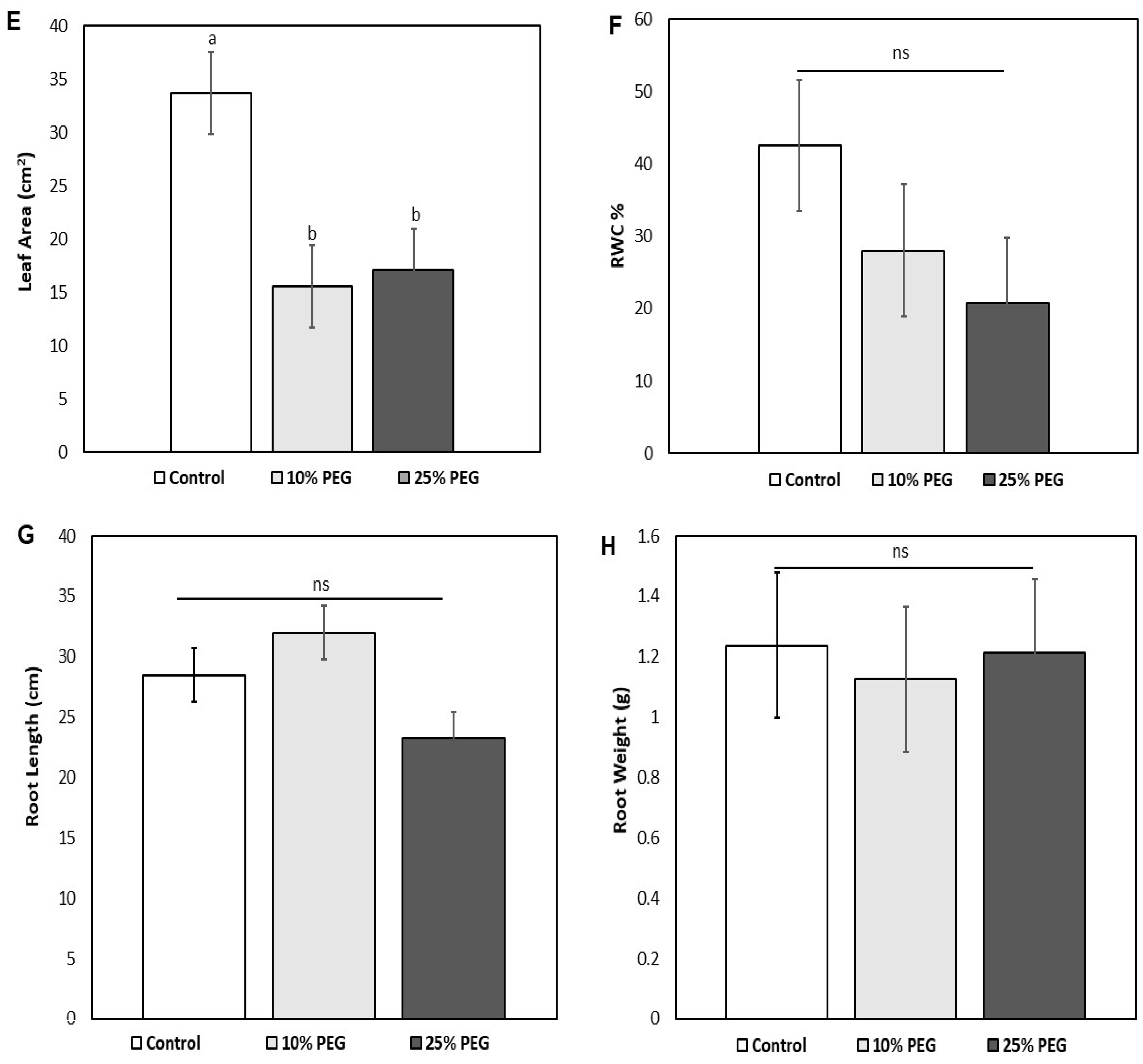
Maize plants were morphologically evaluated for the effects of polyethylene glycol (PEG)-induced osmotic stress. Different growth parameter changes, including plant height, leaf length, leaf number, leaf area, leaf fresh weight, leaf turgid weight, leaf dry weight, root length, and root weight, were measured and recorded for the three groups. Among the morphological traits, leaf color did not show any significant changes between the control (green) and 10% PEG-treated plants (green with brown apex); however, 25% PEG-treated plants showed thinner and dead (brown) leaves. There was no significant difference in plant heights for the 10% PEG-treated seedlings (p = 0.329) in comparison with the control (untreated) plants (Figure 1A). The 10% PEG treatment did not impact the height of maize plants. However, a significant decrease from 42.5 to 34.75 cm in plant height of the 25% PEG-treated Z. mays seedlings (p < 0.05) was evident (Figure 1A). The leaf lengths of different treatments showed a significant decrease (p = 0.013) as compared to the non-treated seedlings from 29.25 to 18.5 cm for the 10% PEG treatment and 19 cm for the 25% PEG treatment (Figure 1A). The PEG treatment demonstrated the negative impact of osmotic stress on the leaf lengths of Z. mays seedlings. However, no significant differences (p > 0.05) in leaf length between the two treatments (10 and 25% PEG) were observed. A significant decrease (p = 0.274) in the number of leaves was observed for the 25% PEG treatment (Figure 1C). In comparison, the 10% PEG treatment showed a decrease in leaf count from 17 to 13 leaves. Interestingly, an increase was noted in the leaf number for the 10% PEG-treated seedlings compared to the control (Figure 1C).
The results further indicated that PEG treatment had no significant effect on leaf fresh weight with an increase in PEG concentration (p = 0.894). Similarly, the leaf fresh weight for the 25% PEG treatment showed no apparent effect as compared to the 10% PEG treatment (Figure 1D). The leaf dry weights across all treatments displayed no apparent statistical differences (p = 0.61) (Figure 1D). However, the leaf turgid weights for the PEG treatments displayed a decrease compared to the control (p = 0.02) from approximately 0.135 to 0.079 and 0.081 g, respectively (Figure 1D). The effect of PEG on the leaf area showed a significant statistical difference (p = 0.004) between the varying treatments (Figure 1E). The leaf areas of the 10 and 25% PEG were significantly reduced compared to the control (non-treated) seedlings from 33.72 to 15.5 and 17.08 cm2, respectively. Nonetheless, no noticeable significance between the leaf area of 10 and 25% PEG-treated seedlings (p > 0.05) was indicated.
A highly significant difference (p < 0.05) was observed between the control (non-treated) and 25% PEG-treated plants with respect to the relative water content (RWC). The RWC of the control was ~42.5%, while osmotic stress decreased the RWC of the treated plants (10 and 25% PEG). Though the RWC for the 10% PEG-treated plants was ~28.03% and declined for the 25% PEG-treated plants to ~20.73%, no significant differences (p = 0.30) were observed between the two treatments (Figure 1F). Further morphological evaluations were conducted between the control (A) 10% (B) and (C) 25% PEG-treated roots. Maize seedlings that were treated with 10% PEG developed slightly longer roots compared to the 25% PEG treatment. However, seedlings grown under 25% PEG treatment (23.25 cm) showed a significant decrease (p < 0.05) in root length compared to the control (28.5 cm) and 10% PEG-treated seedlings (32 cm) (Figure 1G). As the stress concentration increased, less dense roots developed. Osmotically stressed plants demonstrated a partial higher average root length (Figure 1G) compared to the control plants. In general, there was no significant effect on the root weights within any of the treatments (p = 0.967) (Figure 1H).

3.2. Effects of Osmotic Stress on the Physiological Traits of Z. mays

3.2.1. Evaluation of Osmotic Stress on the Stomatal Morphology, Number, and Density

To evaluate the physiological changes induced by osmotic stress, stomatal analysis was carried out. Stomatal micrographs for all the treatments were taken for morphological observation and comparison. Morphological observations displayed turgid guard cells and open stomata for the control, while 10% and 25% PEG presented flaccid guard cells and closed stomata. Furthermore, the stomatal pore, complex length, and width were measured in micrometers (µm), wherein the 10% PEG-treated plants showed a large pore size (69.18 µm) and a longer width (28.16 µm) and length (66.19 µm) compared to the control pore (20 µm), width (28.19 µm), and length (22.80 µm). The 25% PEG-treated plants had a pore size of 29.55 µm, width of 19.31 µm, and length of 36.40 µm. Additional assessments were carried out to evaluate the stomatal number and density from both leaf surfaces (abaxial and adaxial) for all plant groups (control 10% and 25% PEG). The stomatal number for the upper surface treated with 25% PEG (~6) noticeably decreased in comparison to the control (~7.75) and 10% PEG treatment (~8.25). However, the 10% PEG treatment showed no difference from the control (p > 0.05), yet there was a slight increase in comparison to the 25% PEG treatment (p < 0.05). The lower surface stomatal numbers for the 10 and 25% PEG treatments (~13.75 and 13.25) were not statistically different from the control (~14.75), nor were they different from each other (p > 0.05). Similarly, the stomatal density for the upper surface of the 25% PEG treatment (3157.9 stomata/mm2) noticeably decreased in comparison to the control (4078.95 stomata/mm2) and 10% PEG treatment (4342.13 stomata/mm2). However, the 10% PEG treatment displayed no difference from the control (p > 0.05), yet there was a slight increase in comparison to the 25% PEG treatment (p < 0.05). The lower surface stomatal densities for the 10 and 25% PEG treatments were not statistically different from the control, nor were they different from each other (p > 0.05).

3.2.2. Effects of Osmotic Stress on the Leaf Gaseous Exchange Parameters

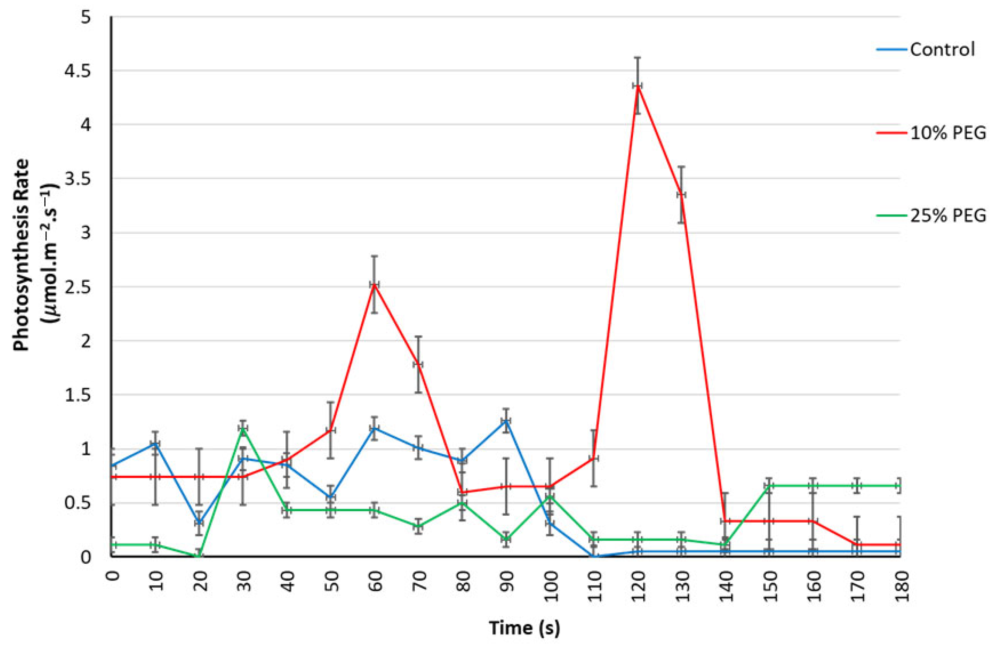
The photosynthetic rates of the three treatments showed an unstable trend. The 10% PEG treatment fluctuated repeatedly, whereby at the first 60 s the photosynthetic rate was strongly elevated, followed by a sharp decline at 80 s. It then reached a maximum photosynthetic rate at 120 s, followed by an ultimate decrease at 140 s. The control and 25% PEG treatments maintained moderate photosynthetic rates from 0 to 100 s, followed by a total inhibition from 110 s to the endpoint (Figure 2).
The stomatal conductance for the control significantly increased over time, particularly after 100 s. The 10% PEG treatment constantly remained moderate and stable to the end after the initial pick up at 60 s (Figure 3). Stomatal conductance for the 25% PEG treatment remained flat throughout the measurement period. Generally, PEG treatment strongly inhibited stomatal conductance (Figure 3).

3.2.3. Respiration and Transpiration Rates Under Osmotic Stress

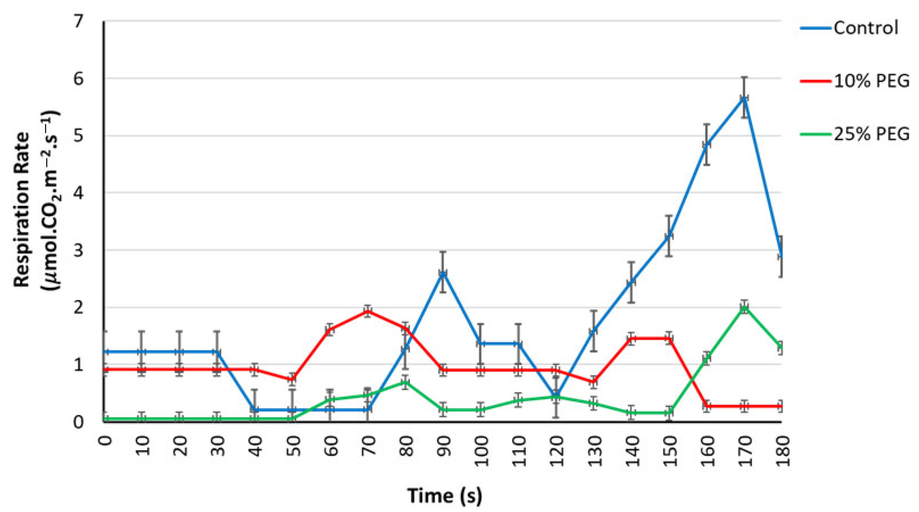
The control reached the highest rate of respiration in comparison with the PEG treatments at 170 s. The 10% PEG treatment maintained a moderate trend, though it was lower than the control; however, the 25% PEG treatment maintained the lowest respiration rate and only peaked at 170 s (Figure 4).
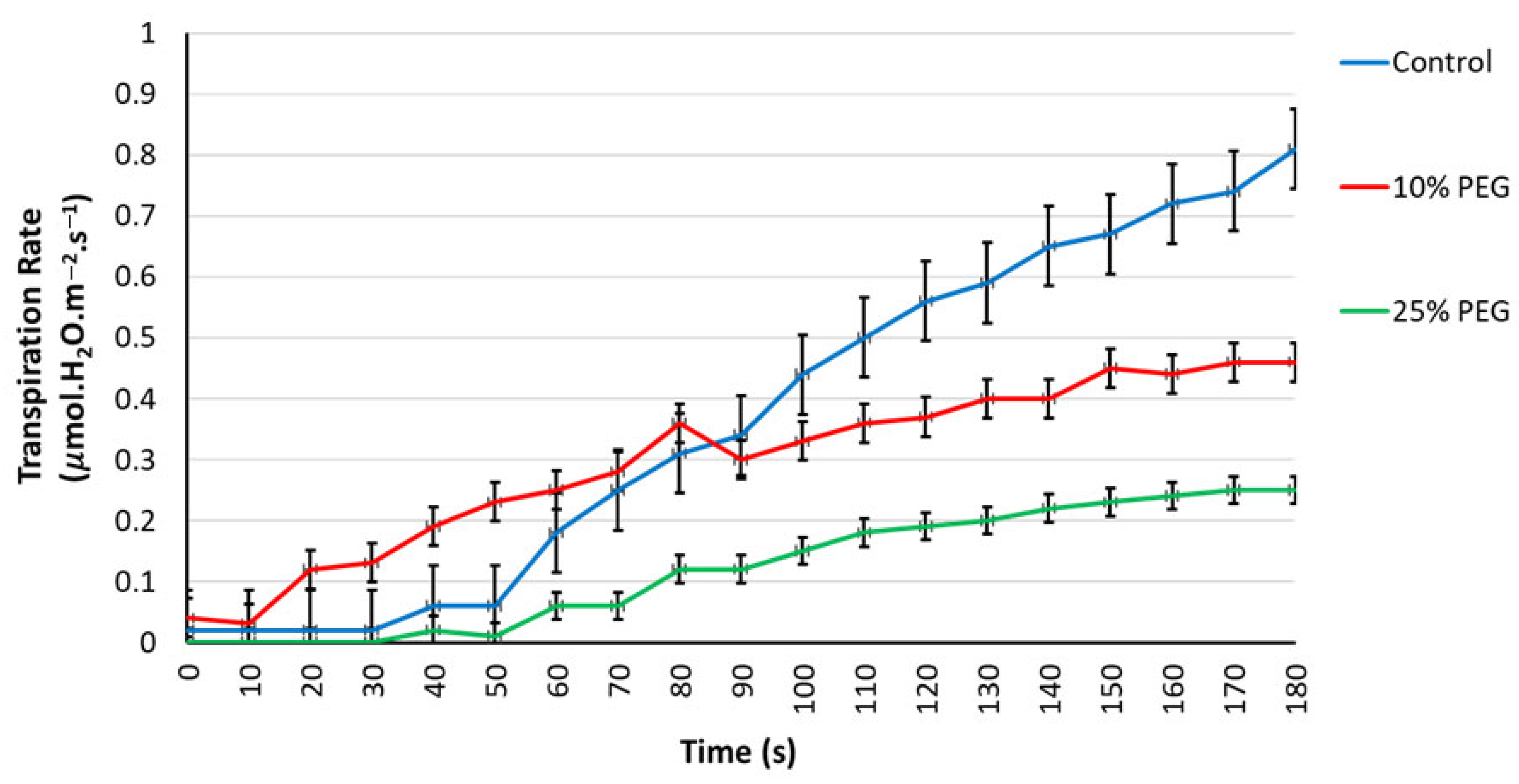
Transpiration rates showed a similar trend across the three treatments, in which there was a gradual increase over time (Figure 5). An increase in PEG treatment concentration showed inhibition in transpiration rate. The control and 10% PEG groups reached their maximum transpiration rates at 180 s (Figure 5).

3.3. Assessment of Maize Protein Expressional Profiles in Response to PEG-Induced Osmotic Stress via One-Dimensional Gel Electrophoresis (1DE)

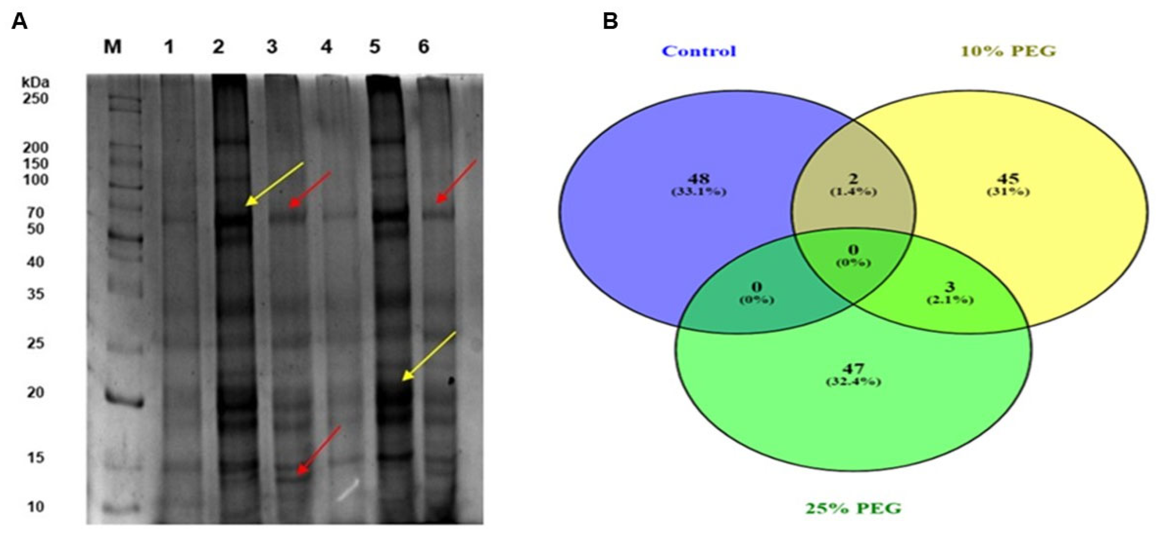
To evaluate the quality and loading quantities of protein extracts, one-dimensional gel electrophoresis (1DE) was carried out prior to label-free mass spectrometry analysis. Coomassie brilliant blue-stained maize leaf protein bands (in three replicates) were visible (Figure 6A). Numerous osmotic stress-induced proteins were expressed with molecular weights (MW) ranging from 10 to 250 kDa. The protein samples for each treatment showed similarity in protein expression, banding patterns, and abundance, suggesting that protein loading was relatively uniform. The results further showed that the protein extracts from all treatments covered the MW range between 10 and 200 kDa, wherein some bands were more highly expressed than others. The control samples 1 and 4 (lanes 2 and 5) demonstrated partial expression of protein bands, whilst the 10% PEG-treated samples 2 and 5 (lanes 3 and 6) displayed a higher expression of the protein bands indicated by yellow arrows (70 and 20 kDa). The 10% PEG treatment group also uniquely showed an expression of large mass protein between 250 and 200 kDa whilst the 25% PEG-treated samples 3 and 6 (lanes 4 and 7) displayed a greater mass of protein between 70 and 50 kDa (~68 kDa) as indicated by the red arrows, at the same time, showing a uniquely expressed protein between 15 and 10 kDa (~14 kDa) (Figure 6A).

3.4. Bioinformatic Analysis and Functional Annotation of Osmotic Stress Responsive Proteins in Maize Leaves

Approximately 439 differentially expressed valid proteins were identified using LC-MS/MS. Of the 439 proteins, 50 for each treatment were further investigated based on their consistent differential expression (≥2-fold change with statistical significance, p < 0.05) under stress conditions compared to controls. Figure 6B, based on the 50 selected DEPs, reveals that no single protein, 0 (0%), was commonly and significantly differentially expressed across all the three treatments; however, 2 (1.4%) common proteins were expressed between the control and 10% PEG treatment. On the other hand, 10% and 25% PEG treatments had 3 (2.1%) common proteins. The differentially expressed proteins decreased to 45 (31%) at a 10% PEG concentration and 47 (32.4%) at a 25% PEG concentration when compared to 48 (33.1%) in the control (Figure 6B).

3.5. Functional Classification and Expression Pattern Analysis of the Osmotic Stress Responsive Proteins

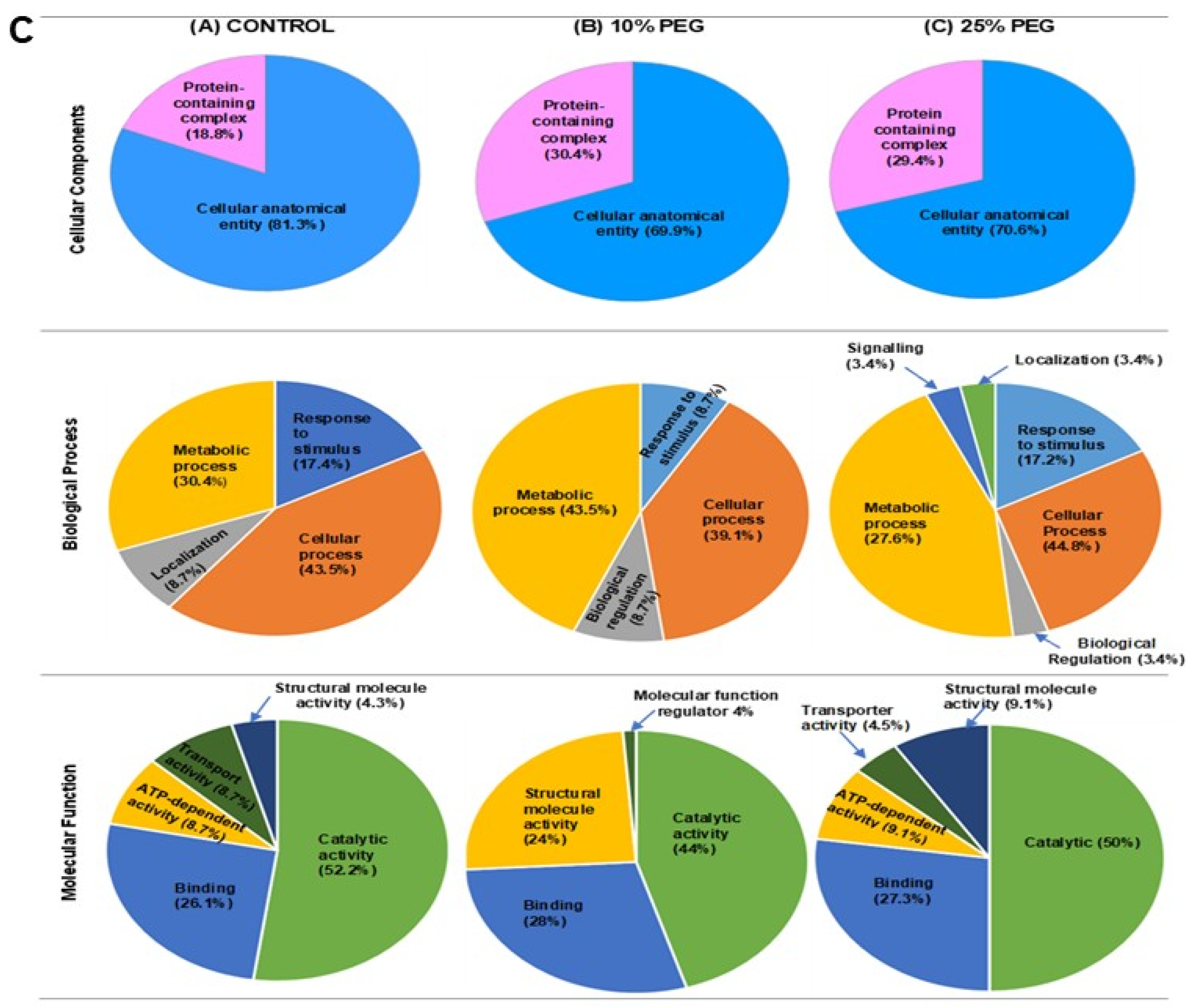
The proteins were further classified, and the results are tabulated in Table 1 and Table 2, showing protein names, subcellular locations, biological processes, molecular functions, and theoretical Mr/pI. The results revealed that the largest number of proteins were localized in the cytoplasm (28 proteins), while varying proteins were located in the chloroplast (19 proteins), extracellular region (15 proteins), membrane (12 proteins), and ribosome (9 proteins). The remaining proteins were localized to the plastid (8), thylakoid (8), cytosol (6), etc. (Table 1 and Table 2). The functional classifications of these proteins in Figure 6C revealed that at all functional classification levels, large proportions of these proteins were involved in cellular anatomical entities and catalytic activities across all treatments. Followed by cellular and metabolic processes, whilst other functional categories included proteins that are involved in protein-containing complexes, response to stimulus, localization, biological regulation, signaling, binding, transport, structural molecule activity, ATP-dependent activity, and molecular function regulation (Figure 6C). At the cellular component level, proteins were overrepresented in cellular anatomical entities, with the control showing the expression of 13 (81.3%) proteins found at the cell junction, cell periphery, cytosol, chromatin, cytoplasm, endomembrane system, external encapsulating structure, intracellular anatomical structure, membrane and organelle (Figure 6C). The 10% PEG treatment had 16 (69.6%) and the 25% PEG had 12 (70.6%) proteins found at the same location as in the control (Table 1 and Table 2). For the protein-containing complex, the control expressed 3 (18.8%) proteins, i.e., K7VN08, Q9M640, and C4JA45. The 10% PEG treatment group had 6 (30.4%) proteins, i.e., B4FCX3, P06589, C0HFM4, P06586, B4FW06, and P27723. The 25% PEG treatment group had 5 (29.4%) proteins, i.e., C0P455, B4FWJ8, A0A1D6JW41, K7VNQ7, and K7UDG5 (Table 1 and Table 2).
The biological process showed a higher representation of metabolic process proteins for the 10% PEG (10; 43.5%) [i.e., B7ZXQ3, P09315, B4FRC4, C0HFM4, C4IZ94, B4F833, C0P5A9, B4F8L7, K7V2Z8, and P27723] (Table 1) compared to the control (7; 30.4%) [i.e., A0A1D6L6U6, K7WFV1, C4IZ94, B4F833, K7TXI5, C0PAU7, and P93805], whilst in the 25% PEG these proteins were less represented (8; 27.6%) [i.e., B4F938, B4FWJ8, B4FFZ2, B4FAL9, B4FAD4, B4FYH2, K7UDG5, and B6THZ8] in comparison to the control. The cellular process for the control showed 10 (43.5%) expressed proteins, i.e., Q9M640, A0A1D6L6U6, A0A1D6MUE8, C4JA45, K7WFV1, C4IZ94, B4F833, K7TXI5, C0PAU7, and C4J410 (Table 2). In addition, the 10% PEG treatment group had 9 (39.1%) proteins [i.e., P06589, B7ZXQ3, B4FRC4, B6TNT5, C0HFM4, C4IZ94, B4F833, K7V2Z8, and P27723]. The 25% PEG treatment group had 13 (44.8%) proteins involved in cellular processes, i.e., B4F938, B4FWJ8, B4FFZ2, K7VJF3, B4FAL9, B4FAD4, B4FYH2, B6SN61, K7VNQ7, K7UDG5, B4F9E8, B6THZ8, and B4F9K4 (Figure 6C; Table 1 and Table 2).
Proteins responsible for response to stimulus demonstrated a decrease from the control (4; 17.4%) [i.e., A0A1D6MUE8, K7TXI5, A0A1D6LE55, and C4J410] to 10% PEG (2; 8.7%) [i.e., B4FRD6 and B6TNT5] (Figure 6C; Table 1); however, not much difference was observed in 25% PEG (5; 17.2%) as compared to the control [B4FWJ8, K7VJF3, B6SN61, B4F9E8, and B4F9K4] (Table 2). The control had no biological regulation proteins, while the 10% PEG treatment group had 2 (8.7%), i.e., C0HFM4 and C0P5A9, and the 25% PEG treatment group expressed 1 (3.4%), i.e., B4FWJ8. On the contrary, the control had 2 (8.7%) localization proteins, i.e., Q9M640 and K7WFV1, whilst the 10% PEG treatment group had none. The 25% PEG treatment group had 1 (3.4%) localization protein [i.e., K7VNQ7], whilst uniquely showing 1 (3.4%) signaling protein, i.e., B4FWJ8 (Figure 6C; Table 1).
At the molecular function level, the majority of proteins were represented in the catalytic activity category (Figure 6C). The control had 12 proteins (52.2%) responsible for hydrolase, isomerase, ligase, oxidoreductase, and transferase activities. The 10% PEG treatment group had 11 proteins (44.0%), i.e., B4FRD6, B4FRH1, B7ZXQ3, P09315, B8A2X5, B4FRC4, B6TNT5, C0P3R8, C4IZ94, B4F833, and B4F8L7. While 25% PEG had 11 (50.0%), i.e., B4F938, B4FWJ8, B4FFZ2, K7VJF3, C0P3R8, B4FAL9, B4FAD4, B4FYH2, B6SN61, K7UDG5, and B6THZ8 (Table 2). For the binding process, the control had 6 (26.1%) proteins, i.e., B4F8V5, A0A1D6MUE8, C4JA45, C0PAU7, Q94F78, and C4J410 (Table 1). The 10% PEG treatment group had 7 (28.0%), i.e., B4F9N8, P09315, B8A2X5, B4FRC4, C0HFM4, B4FW06, and B4F8L7, while the 25% PEG treatment group had 6 (27.3%) proteins, i.e., C0P455, B4FWJ8, K7VJF3, A0A1D6L210, B4F9E8, and B4F9K4. For transport activity proteins, the control had 2 (8.7%) proteins, i.e., B4FBF6 and Q6R9G1, while the 25% PEG treatment group had 1 (4.5%), i.e., K7VNQ7 (Table 2). The control had 2 (8.7%) ATP-dependent activity proteins, i.e., A0A1D6MUE8 and C4J410, whilst the 25% PEG treatment group had 2 (9.1%) proteins, i.e., K7VJF3 and B4FWJ8. The control had 1 (4.3%) protein for structural molecule activity (C4JA45); for the 10% PEG treatment group, there were 6 (24.0%), i.e., P06589, B4G1J8, C0HFM4, P06586, B4FW06, and P27723; and for the 25% PEG treatment group, there were 2 (9.1%) proteins, i.e., C0P455 and A0A1D6JW41. The 10% PEG treatment group had 1 (4.0%) unique molecular function regulator protein, i.e., B8A2X5 (Figure 6C and Table 1).

3.6. Protein–Protein Interaction Network of Osmotic Stress-Responsive Proteins

To further investigate the functional interrelationships among the identified DEPs, a protein–protein interaction (PPI) analysis was performed using the STRING 12.0 database. The resulting interaction networks were visualized and analyzed in Cytoscape (v3.9.1) to identify clusters, hub proteins, and biological pathways potentially contributing to osmotic stress regulation in Zea mays.
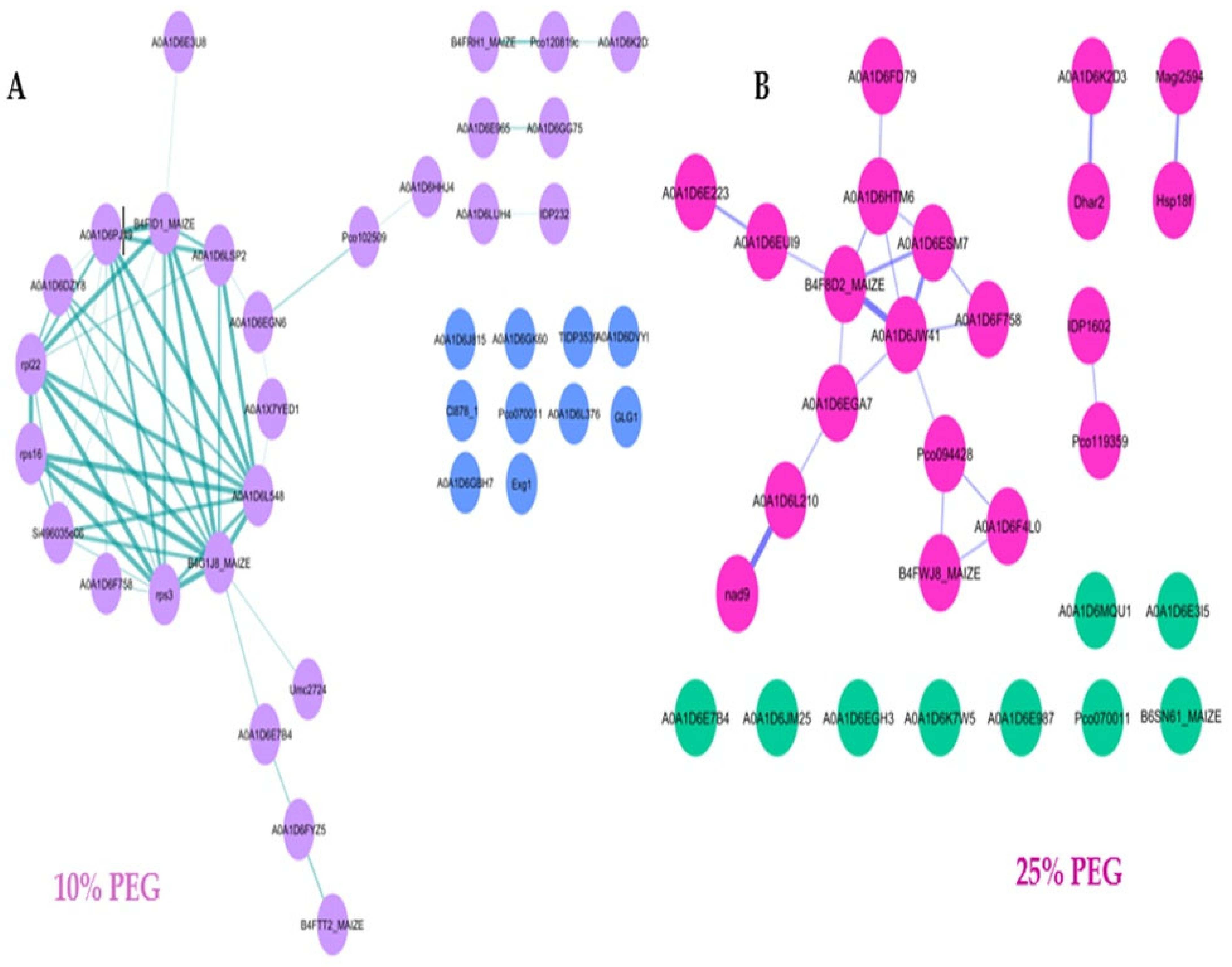
Under the 10% PEG treatment (Figure 7A), the PPI network displayed a dense and highly interconnected cluster, reflecting strong coordination among proteins involved in translation, protein folding, and energy metabolism. Notably, ribosomal subunits (40S S10-1, 40S S17-1, 60S L13a-1, 50S, and 30S proteins), elongation factor γ1, and methionine aminopeptidase formed the core of this network, indicating enhanced regulation of protein synthesis. Peptidyl-prolyl cis–trans isomerase (PPIase) and other folding-related proteins supported proteostasis maintenance. At the same time, ornithine carbamoyl transferase and pectin esterase were associated with amino acid metabolism and cell wall remodeling, respectively. Proteins linked to organellar translation, such as polyribonucleotide nucleotidyl transferase 2 (mitochondrial), and signal recognition particle 54 kDa protein (chloroplastic), highlighted coordinated protection of translational and metabolic functions. Overall, these interactions demonstrate an adaptive proteomic response involving translational reprogramming and metabolic adjustment to sustain cellular homeostasis under mild osmotic stress.
In contrast, the 25% PEG network (Figure 7B) exhibited reduced connectivity and fragmented clusters, indicating weakened interaction networks under severe osmotic stress. Enzymes such as pyruvate kinase and fructose-bisphosphate aldolase were associated with energy metabolism, whereas proline- and valine-tRNA ligases suggested restricted translational efficiency. Although ribosomal proteins (40S S4-3, S20-1, 60S L4-1, and ribosomal S4e domain-containing protein) were still detected, their limited connections implied impaired coordination of protein biosynthesis. The presence of heat shock 70 kDa protein 5, binding protein homolog 2, and T-complex protein 1 subunit zeta pointed to activation of chaperone systems in a less organized manner. Mitochondrial proteins, including aconitate hydratase 3 and NADH dehydrogenase subunits, indicated sustained but stressed respiratory activity. Overall, the 25% PEG treatment reflects a shift from coordinated metabolic regulation toward fragmented and energy-depleting stress responses, revealing the metabolic strain imposed by severe osmotic stress.

4. Discussion

Rapid population growth has led to an increased interest in maize cultivation for food and non-food purposes [22]. However, drought-induced osmotic stress restricts plant growth and development by impairing physiological and biochemical processes. Osmotic stress is perceived by the cell as turgor loss and triggers various signaling cascades for osmotic adjustment [23,24,25]. Proteomic approaches provide insight into plant stress-responsive proteins that underpin adaptive mechanisms. Our study investigated the effects of osmotic stress on the morphology, physiology, and proteomic profiles of maize (Zea mays) seedlings. Polyethylene glycol (PEG)-induced osmotic stress has been widely used to simulate drought stress under controlled conditions [26,27]. In this study, maize seedlings exposed to 10% (w/v) and 25% (w/v) PEG displayed distinct morphological, physiological, and proteomic responses that together illustrated adaptive and maladaptive mechanisms of drought response.
Plants undergo morphological changes when subjected to drought stress, including reduced plant height, leaf area, and biomass [28,29]. After 20 days, PEG treatments significantly affected leaf morphology, particularly leaf number, length, and area.
At the morphological level, mild osmotic stress (10% PEG) induced a slight reduction in leaf size but promoted additional leaf formation and longer roots, suggesting a compensatory growth mechanism. In contrast, severe stress at 25% PEG caused chlorosis and drastically decreased leaf area, senescence, and root proliferation, indicating growth inhibition resulting from dehydration-induced energy limitation [30,31]. These morphological changes demonstrate adaptive strategies, where leaf death under severe stress reduces water loss. Proteomic shifts corresponded with these morphological responses.
Various proteins including those involved in energy metabolism and stress-association were differentially expressed, supporting the observed morphological patterns in response to osmotic stress. Proteomic analysis revealed an overrepresentation of peptidyl-prolyl cis–trans isomerases (PPIases), which are involved in protein folding and stabilization under cellular stress. Even though these enzymes are widely recognized as general stress proteins, their abundance alongside reduced relative water content (RWC) suggests their relation to impaired ion homeostasis [32]. This concurs with reports that PPIase accumulation reflects disturbances in protein folding and ion flux under abiotic stress [33].
Additionally, various proteins related to sugar metabolism and the Krebs cycle were upregulated, highlighting an activation of core carbon metabolism pathways. Upregulation of glycolysis and TCA cycle enzymes under mild stress supports adaptive growth, whereas their suppression under severe stress reflects growth arrest [34,35]. Thus, in our study the enrichment of these pathways under 10% PEG corresponds with maintained root elongation and partial photosynthetic function, whereas their suppression at 25% PEG coincided with growth arrest confirming a dose-dependent metabolic adaptation.
We further focused on evaluating key leaf traits including length, number, weight, area, and plant height to associate morphological changes with overall growth performance (Figure 1).
During mild stress (10% PEG), plant height was maintained, while severe stress (25% PEG) reduced it due to early leaf senescence along with a decline in cell division [30]. Severe drought led to significant damage to maize growth. The leaf lengths for both the 10% and 25% PEG treatments decreased compared to the control, from 29.25 cm to 18.5 cm and 19 cm, respectively. Likewise, mild stress (10% PEG) increased the leaf number and root length, which could be due to hormonal regulation, while severe stress (25% PEG) reduced leaf size and number, reflecting a maladaptive response. Such responses have been observed in plants where low levels of abiotic stress enhance growth parameters [36,37]. Water deficit has been noted to have a negative influence not only on leaf size but also on the number of leaves [38].
The fresh and dry leaf weights showed no significant changes for either treatment compared to the control. Nonetheless, the leaf turgid weights for the 10% and 25% PEG treatments displayed a significant decrease compared to the control (Figure 1D). Weight decrease is a common unfavorable influence of drought stress in plants [39]. Ultimately, the leaf areas for both treatments significantly decreased. To adapt to drought, plant leaves generally adopt smaller leaf areas due to the decrease in leaf turgor pressure, canopy temperature, and the availability of photoassimilates [29]. Allakhverdiev [40] indicated that insufficient leaf area reduces sunlight use efficiency. Equally, the decrease in photosynthesis and canopy structure under drought stress may result in a drop in the total fresh weight of a plant. To a large extent, plant size is determined by the strong interrelation between the number of leaves, their sizes, photosynthesis capacity, and functional traits [26,32]. Generally, the increase in PEG concentration in this study resulted in a significant decrease in all of the morphological traits of leaves.
Under 10% PEG treatment, root proliferation decreased while root length increased (Figure 1G). Zulfqar [41] emphasized that it is common to observe increased root lengths across stressed plant species. This notion was supported by Luo [42]’s observation of longer roots during the initial stages of drought stress in cotton plants. Deeper roots are a desirable trait for water extraction from deeper layers as an adaptation mechanism against drought stress [31]. On the contrary, in 25% PEG-treated maize seedlings, the roots were thinner and had less proliferation and decreased length (Figure 1G). Robin [27] demonstrated similar findings in castor bean, where PEG-induced osmotic stress reduced the length, density, and diameter of root hairs. Yang [29] viewed such root system configurations as enablers for water deficiency in a plant, whereby the reduction in root length could be linked to the drop of relative turgidity and dehydration of the protoplasm, which is associated with turgor loss and reduced cell division as an osmotic stress coping mechanism. When the availability of water is limited, there is a reduction in osmotic pressure resulting in a plant water deficit [27,42,43]. Additionally, the root weights demonstrated no significant change across all of the varying treatment conditions (Figure 1H). Chen [44] found a strong correlation between root weight and plant height and no significant correlation between root depth and plant height in wheat under drought stress. However, in our study, the root weight and plant height for both treatments showed no correlation, whilst there was a correlation between the root length and plant height for the 25% PEG treatment. Genetic and environmental factors such as experimental conditions and stress duration and intensity affect root growth, as well as response to drought [45]. Root morphological changes may be a result of stress resistance or the stimulation of root growth under low-moisture conditions [46]. Overall, our study has demonstrated that all drought-induced treatments influenced the root growth and morphology, though there were differences in the response to root growth.
The observed increase in root length under mild stress (10% PEG) coincided with a higher normalized abundance of energy metabolism enzymes such as pyruvate kinase and glyceraldehyde-3-phosphate dehydrogenase (GAPDH) (Table 2). While statistical correlation coefficients were not calculated, this pattern suggests a potential association between enhanced energy metabolism and adaptive root elongation under mild stress conditions. Similar associations have been reported in previous studies, where elevated glycolytic enzyme activity supported root growth under osmotic or drought stress in maize and other crops [34,41,47]. In contrast, reduced proliferation under severe stress (25% PEG) reflects metabolic downregulation and energy depletion. These findings, therefore, provide a qualitative association between proteomic shifts in central carbon metabolism and root morphological responses to PEG-induced osmotic stress. Similarly, the abundance of antioxidant-related proteins, including glutathione peroxidase, late embryogenesis abundant (LEA) proteins, and heat shock proteins (HSP70), underscores an integrated defense system that mitigates oxidative stress and prevents protein misfolding.
Apart from the external form, drought stress has different degrees of influence on physiological and biochemical parameters [26,28,29]. Hence, we further assessed the physiological parameters of the Z. mays seedlings. When plants experience drought stress, their stomatal response is probably the first line of defense against dehydration as it is quicker than other changes such as pigmentation, proteins, leaf area, chloroplast ultrastructure, and root growth [48]. In addition to metabolic pathway alterations, complex signaling networks involving abscisic acid (ABA) and numerous systems co-participate in stomatal regulation to restrict excess water loss [7,49].
We further explored the water status of the plant tissues and their ability to survive stressful conditions via relative water content (RWC) [24,50]. RWC is a critical parameter for plant growth and development, as it is directly related to the ability of plants to absorb nutrients, photosynthesize, and maintain their structural integrity [30,51]. It further reflects the incorporated effects of morphological and physiological changes that plants experience to prevent water loss and maintain water balance [52]. As PEG concentration increased in our study, a significant decrease in RWC was noted in all of the treatments, with a decline from ~42.5% (control) to ~28.03% and ~20.73% (10 and 25% PEG), respectively. Our results correlate with the previous studies that have reported similar results, including maize [30,39] and wheat [50]. Several factors contribute to the decrease in RWC in addition to water reduction in the soil. These include enhanced transpiration and reduced osmotic potential in stressed leaves, as well as alterations in cellular structure [53].
When further evaluating the influence of PEG-induced osmotic stress on the physiology of Z. mays seedlings, it was considered that the net photosynthetic rate and leaf stomatal conductance could be affected by the induced stress; thus, these factors were thoroughly studied. Our results demonstrated that the photosynthetic rate for the 10% PEG-treated plants increased to a moderate rate at 60 s, reached its maximum potential at 120 s, and then dropped. Our observation supports that these high rates and drops could possibly be a rapid osmotic stress response [54], whilst the 25% PEG-treated plants unrestrainedly photosynthesized at lower rates than the control. Relatedly, a study on ferns, gymnosperms, and angiosperms found that the maximum photosynthetic rate is positively correlated with stomatal density; however, it had no relation with stomatal size [55]. While Zulfqar [41] put forward that leaf area and photosynthetic rate are directly proportional, we could deduce based on our results that low adaxial stomatal density, as well as low leaf area, are indeed correlated with low photosynthetic rates.
The data presented in Figure 3 demonstrate that PEG-induced osmotic stress strongly inhibited stomatal aperture to reduce water loss through transpiration. The control suggests a healthy physiological activity [56]. The 10% PEG treatment indicates partial stomatal closure, reflecting a mild osmotic stress response with the plant conserving water while still allowing limited gas exchange. Such mild stress triggers adaptive mechanisms without fully inhibiting stomatal function [36]. The 25% PEG treatment remained flat, reflecting severe stomatal closure due to high osmotic stress caused by PEG, reducing water availability and prompting the plant to minimize transpiration. The complete suppression of stomatal conductance is consistent with the findings of [5] that high osmotic stress leads to stomatal closure, reduced CO2 uptake, and impaired photosynthesis. According to Wu [55], stomatal conductance usually increases under mild drought stress. In our study, the gs of the drought-stressed seedlings showed little to no stomatal activity. Low gs reduces water loss through transpiration and can hinder the improvement of photosynthetic efficiency for a good crop yield [57,58]. Changes in stomatal conductance depend on RWC and can occur rapidly; however, plants do not show large decreases in stomatal conductance when experiencing drought stress gradually [59,60]. Therefore, in our study, we inferred that the PEG-induced stress was rapid and that drought stress decreased photosynthesis by lowering stomatal conductance to carbon dioxide.
In addition to photosynthesis and stomatal conductance, respiration and transpiration rates were also evaluated. The control reached the highest rate of respiration in comparison with the PEG treatments at 170 s. The 10% PEG treatment maintained a moderate trend, though it was lower than the control. This trend could be an indication of the adjustment in the metabolic activity of the plant to cope with the stress [61]. However, the 25% PEG treatment maintained the lowest respiration rate and only peaked at 170 s. The gaseous exchange, as well as transpiration rates, are controlled by the stomata [62]. Zulfqar [41] demonstrated that in the sugarcane crop, when the water content decreased, the CO2 uptake also decreased as the stomata closed.
Transpiration rates showed a similar trend across the three treatments, in which there was a gradual increase over time. Similar to stomatal conductance, transpiration rates increased under mild water stress, whilst severe stress caused a decrease in rates as a reflex to stimuli [30,63]. An increase in PEG treatment concentration showed inhibition in transpiration rates, i.e., the 25% PEG treatment showed lower transpiration rates than the other treatments. Sikuku [64] identified that the decrease in transpiration, as well as photosynthesis, in rice was due to reduced stomatal conductance. Likewise, similar results were noted in maize [39]. Since our results mirrored a gradual increase, Marchin [65] suggests that these patterns occur either due to water competition driving plants to maintain high rates of transpiration until a level of water depletion or by stomatal closure.
The physiological impairments observed in photosynthesis and stomatal conductance under PEG-induced osmotic stress correspond with the proteomic expression changes detected in this study. The upregulation of chlorophyll a–b binding protein (tr|K7TXI5) and ATP synthase subunit gamma (tr|K7WFV1) suggests an adaptive mechanism to sustain photosynthetic electron transport and ATP generation under dehydration. On the other hand, chlorophyll a–b binding proteins stabilize photosystem II and delay chlorophyll degradation, maintaining light-harvesting efficiency during moderate stress [66,67], while enhanced ATP synthase activity supports energy supply for repair and osmolyte synthesis [68,69]. These proteomic adjustments explain the transient maintenance of photosynthetic activity observed under 10% PEG and its collapse under 25% PEG, reflecting the threshold at which cellular energy regulation can no longer compensate for severe osmotic inhibition.
The composition of minerals, such as antioxidants and proteins, is also affected by drought. Plants display various responses at the cellular level, which are related to their biochemistry [70]. Proteomic evaluation to identify differentially regulated proteins has been conducted in response to osmotic stress. Drought-stressed plants tolerate stress by adjusting their structural and biochemical form through the accumulation of proteins [71]. Proteins are the “workhorses” of cells in living organisms, responsible for a wide range of functions and modifications to the internal and/or external cellular conditions that cause temporal changes in the proteome expression, thus affecting their growth and are encoded in leaf growth regulatory genes of varying functional classes that regulate cellular processes [71,72]. Changes in proteomic expression in drought-affected plants such as wheat [73] and rice [74,75] have been observed before. The proteomic analysis highlights differentially expressed proteins associated with significant biological processes, molecular processes, and several metabolic pathways [70].
Various studies use gel-based proteomic separation to make sense of abiotic stress-responsive mechanisms. Likewise, in our study, we used the same technique to analyze the fractions of soluble leaf extracts by 1DE with Coomassie brilliant blue for staining. We observed a similarity in protein expression, banding patterns, and abundance. The protein profile for the control (lanes 2 and 5) exhibited partial expression, whilst the 10% PEG (lanes 3 and 6) protein profile exhibited less abundant proteins at 70 and 20 kDa. Additionally, newly pronounced proteins were observed between 250 and 200 kDa in the 10% PEG protein profile. The 25% PEG protein profile presented less abundant proteins between 70 and 50 kDa (~68 kDa), simultaneously presenting uniquely pronounced proteins 15 and 10 kDa (~14 kDa) (Figure 6A). Our results coincide with those of Lukhele [76], who found that higher protein abundance is not always found in non-stressed plants than in stressed ones. Similarly, our protein expression profiles resulted in both less and more abundant proteins. Under PEG-mediated osmotic stress, Thamaga [77] noted a decrease in protein synthesis and total soluble proteins in Z. mays leaves. In contrast, our results demonstrate that PEG-induced osmotic stress stimulated the abundance of several proteins in Z. mays leaves.
We further performed functional annotation and subcellular localization for 50 proteins of the 439 differentially identified proteins, in which the majority were related to photosynthesis, ATP synthesis, and protein biosynthesis (Figure 6C). The subcellular localization further revealed that most of the proteins were actively synthesized in the chloroplast and cytoplasm. Essential proteins such as ATP synthase were exhibited in all of the treatments’ protein profiles. The control protein profile exhibited stress-response heat shock 70 kDa proteins (tr|C4J410; tr|A0A1D6MUE8), which are predicted to prevent aggregation and regulate processes important for plant survival, assist in the folding of proteins and promote the anti-stress ability of the cell [70,78]. It is common for stressed plants to accumulate ROS in their cells, which leads to oxidative stress. Antioxidant enzymes are then induced to prevent excess ROS accumulation and contribute to cell survival [38,79]. In our study, Calvin cycle enzymes that play major roles in carboxylation, such as fructose biphosphate aldolase (tr|B4FAL9) and hydrogen peroxide (tr|B4FRD6), were identified in the 10% PEG treatment protein profile. These enzymes are vital for carboxylation; therefore, their downregulation resulted in reduced photosynthesis rates [70]. The 10% PEG protein profile also exhibited peptide-methionine (tr|B6TNT5), which produces polyamines and ethylene for cell division, morphogenesis, and plant senescence; pyruvate kinase (PK) (tr|B4FYH2), which catalyzes ADP into ATP under stress and is recognized to yield more energy for tolerance; glyceraldehyde-3-phosphate dehydrogenase (GAPDH) (tr|B4F8L7), which alleviates ROS-induced cell damage under stress; and adenosyl homocysteinase (tr|A0A804NLS4), which regulates intracellular methylation and improves drought tolerance by slowing membrane damage [79,80].
To strengthen the relevance of our proteomic findings, the differentially expressed proteins (DEPs) were cross-referenced with their corresponding gene loci in the maize genome using UniProt, MaizeGDB, and NCBI databases. Various key proteins identified, including LEA (tr|B4F9K0), HSP70 (tr|C4J410), GAPDH (tr|B4F8L7), and pyruvate kinase (tr|B4FYH2), correspond to genes previously associated with drought tolerance in maize, sorghum, and rice through transcriptomic and QTL mapping studies [81,82,83,84,85,86]. For instance, the ZmLEA3 and ZmHSP70-1 genes have been reported to enhance osmotic adjustment and protect cellular proteins under dehydration stress [81,82], while ZmGAPDH contributes to ROS detoxification and energy balance during drought spells [83]. Similarly, pyruvate kinase genes (ZmPK1) have been associated with improved carbon metabolism and drought adaptation [84]. These associations affirm that the proteomic responses observed in this study are underpinned by stress-responsive genes already recognized for their functional role in drought tolerance. Despite the present study focusing primarily on protein identification and functional characterization, future integrative analyses linking DEPs with genomic markers, promoter regions, and SNP variations will be essential to translate these molecular signatures into breeding tools for marker-assisted selection and genetic improvement of drought-tolerant maize cultivars [85,86].
The 25% PEG protein profile exhibited LEA protein (tr|B4F9K0), which is a water-soluble protein synthesized in high desiccation-tolerant plants, as well as HSPs: 17.5 kDa class II HSP (tr|B4f9k4) and 17.4 kDa class III HSP (tr|B4F9K4), which are components for stress response and ATP binding, hydrolysis activity, and dependent protein folding [84,87]. Soluble proteins are linked with amino acids that are produced in response to heat or drought stress to maintain the cell’s normal functionality [88]. In sunflowers, osmotic stress similarly induced low-MW HSPs [89]. The 10% and 25% PEG treatments both expressed glutathione peroxidase (GPX) (tr|COP3R8), which is responsible for the oxidative stress response. By increasing glutathione content under stress, wheat revealed that it could withstand drought stress, and this response was deemed common even in barley [40,57]. Under unfavorable conditions, GPX acts synergistically with other antioxidant enzymes to eliminate harmful free radicals [80]. Therefore, its identification in our study provides insight that it is crucial for ROS-scavenging in stressed seedlings to confer tolerance. These observations suggest that drought stress primarily affects proteins involved in metabolism and stress adaptation. This descriptive functional profiling provides an overview of stress-responsive pathways. In future studies, more detailed enrichment or abundance-based analyses are required to support these patterns.
To further understand the functional interactions and coordination among these proteins under osmotic stress, we performed protein–protein interaction (PPI) network analysis which revealed insights into the molecular mechanisms of maize adaptation to varying osmotic stress levels [90]. Under mild stress conditions (10% PEG) (Figure 7A), the formation of a dense and highly interconnected PPI network suggests a well-coordinated cellular response. Key proteins involved in translation, protein folding, and primary metabolism form central hubs, indicating that maize maintains cellular homeostasis through enhanced protein synthesis and stability. This observation aligns with the findings of Li et al. [67], who reported that drought-tolerant maize cultivars exhibit robust translational machinery and proteostasis mechanisms under stress conditions. Conversely, under severe osmotic stress (25% PEG) (Figure 7B), the fragmented PPI network reflects disrupted protein coordination and reduced translational capacity. The limited connectivity of proteins involved in energy metabolism and stress-response pathways suggests a compromised ability to sustain cellular functions under high stress. This is consistent with a study by Zhao et al. [91], who observed that maize subjected to combined drought and heat stress exhibited a disorganized PPI network, correlating with decreased stress tolerance. Moreover, the central role of molecular chaperones and heat shock proteins in the PPI network underscores their importance in maintaining protein integrity and function during stress. These findings are supported by Palotai et al. [92], who highlighted the critical role of chaperones as integrators of cellular networks, particularly under stress conditions. Collectively, the PPI network analysis reveals that maize employs a coordinated and robust molecular strategy to cope with mild osmotic stress, characterized by enhanced protein synthesis and stability. However, under severe stress, the disruption of this network indicates a threshold beyond which maize’s adaptive capacity is compromised. These insights contribute to our understanding of the molecular basis of drought tolerance in maize and can inform strategies for developing more resilient cultivars. Overall, the proteomic adjustments observed in maize seedlings provide a molecular association between the morphological reductions in leaf and root growth and the physiological impairments in photosynthesis, stomatal conductance, and RWC, highlighting an integrated drought stress response. Our findings demonstrate that maize seedlings employ a multi-layered drought response, integrating morphological adjustments, metabolic reprogramming, and chaperone-mediated stress protection. The overrepresentation of PPIases and metabolic enzymes adds mechanistic relevance to the proteomic findings, linking molecular and phenotypic resilience to underlying biochemical pathways. This integrative understanding enhances the relevance of the results and provides a framework for future research on the development of drought-tolerant maize varieties.

5. Conclusions

The responses of plants to drought are complex and can be observed at the morphological, physiological, and molecular levels in relation to stress duration and severity. This study evaluated the morphological, physiological, and proteomic responses of Z. mays to PEG-induced osmotic stress. Exposure to drought-induced osmotic stress hampered the morpho-physiological growth of the Z. mays seedlings. Our study successfully linked the Z. mays seedling response to the interactive damage for all studied traits, such as plant height leaf and root growth parameters, as well as stomatal morphology, relative water content (RWC) and leaf gaseous exchange parameters. The results fairly demonstrated variations in the response of the treated plants when compared to the control plants, thus reflecting the wider range of adaptation capabilities that maize uses to survive stress. Additionally, proteomic analysis carried out in maize leaves to monitor the osmotic stress-responsive proteins and a variety of proteins were positively identified. Among them, the most differentially expressed response proteins were related to cellular components (cellular anatomical entity), molecular function (catalytic activity), and biological processes (metabolic and cellular processes), respectively. Our overall analyses of Z. mays revealed the negative effect of osmotic stress on plant growth and development, along with novel insights into the physiological and proteomic level response mechanisms. Our findings could thus be used as a tool to bridge the gap in future studies to improve osmotic stress tolerance in maize. Additionally, the cross-referencing of drought-responsive proteins with their encoding genes provides a functional basis for understanding the genetic control of stress adaptation. Future studies combining proteomic data with genomic and marker-based analyses will be critical for translating these molecular signatures into practical tools for maize breeding and drought-resilient cultivar development.

Author Contributions

T.B.D. and O.R. conceived the idea of this project, R.R.S. performed the experiments and results analysis, C.S., J.L.G. and K.M.M. advised on proteomics analysis, and R.R.S. and T.B.D. wrote the manuscript. All authors have read and agreed to the published version of the manuscript.

Funding

The authors extend their appreciation to the Department of Botany, North-West University (Mahikeng Campus), and the National Research Foundation (NRF) of South Africa Grant No. 150868 for funding this research work.

Data Availability Statement

The original contributions presented in this study are included in the article. Further inquiries can be directed to the corresponding author.

Conflicts of Interest

The authors declare no conflicts of interest.

References

  1. Baptista, D.M.S.; Farid, M.M.; Fayad, D.; Kemoe, L.; Lanci, L.S.; Mitra, M.P.; Muehlschlegel, T.S.; Okou, C.; Spray, J.A.; Tuitoek, K.; et al. Climate Change and Chronic Food Insecurity in Sub-Saharan Africa; International Monetary Fund: Washington, DC, USA, 2022. [Google Scholar]
  2. Cammarano, D.; Valdivia, R.O.; Beletse, Y.G.; Durand, W.; Crespo, O.; Tesfuhuney, W.A.; Jones, M.R.; Walker, S.; Mpuisang, T.N.; Nhemachena, C.; et al. Integrated assessment of climate change impacts on crop productivity and income of commercial maize farms in northeast South Africa. Food Secur. 2020, 12, 659–678. [Google Scholar] [CrossRef]
  3. Nawaz, M.; Wang, X.; Saleem, M.H.; Khan, M.H.U.; Afzal, J.; Fiaz, S.; Ali, S.; Ishaq, H.; Khan, A.H.; Rehman, N.; et al. Deciphering Plantago ovata forsk leaf extract mediated distinct germination, growth and physio-biochemical improvements under water stress in maize (Zea mays L.) at early growth stage. Agronomy 2021, 11, 1404. [Google Scholar] [CrossRef]
  4. Population Reference Bureau. World Population Data Sheet; Population Reference Bureau: Washington, DC, USA, 2020; Available online: https://www.prb.org/wp-content/uploads/2020/07/letter-booklet-2020-world-population.pdf (accessed on 15 June 2021).
  5. Farooq, M.; Wahid, A.; Lee, D.J.; Ito, O.; Siddique, K.H. Advances in drought resistance of rice. Crit. Rev. Plant Sci. 2009, 28, 199–217. [Google Scholar] [CrossRef]
  6. Cavatte, P.C.; Martins, S.C.; Morais, L.E.; Silva, P.E.; DaMatta, F.M. The Physiology of Abiotic Stresses. In Plant Breeding for Abiotic Stress Tolerance; Springer: Berlin/Heidelberg, Germany, 2012; pp. 21–51. [Google Scholar]
  7. Salehi-Lisar, S.Y.; Bakhshayeshan-Agdam, H. Drought Stress in Plants: Causes, Consequences, and Tolerance. In Drought Stress Tolerance in Plants Vol 1: Physiology and Biochemistry; Springer: Cham, Switzerland, 2016; pp. 1–16. [Google Scholar]
  8. Yasmeen, A. Exploring the Potential of Moringa (Moringa oleifera) Leaf Extract as a Natural Plant Growth Enhancer. Ph.D. Thesis, Department of Agronomy, Faculty of Agriculture, University of Agriculture, Faisalabad, Pakistan, 2011. [Google Scholar]
  9. Vicente, O.; Al Hassan, M.; Boscaiu, M. Contribution of Osmolyte Accumulation to Abiotic Stress Tolerance in Wild Plants Adapted to Different Stressful Environments. In Osmolytes and Plants’ Acclimation to Changing Environment: Emerging Omics Technologies; Springer: Berlin/Heidelberg, Germany, 2016; pp. 13–25. [Google Scholar]
  10. Lippmann, R.; Babben, S.; Menger, A.; Delker, C.; Quint, M. Development of wild and cultivated plants under global warming conditions. Curr. Biol. 2019, 29, R1326–R1338. [Google Scholar] [CrossRef]
  11. Ghatak, A.; Chaturvedi, P.; Weckwerth, W. Cereal crop proteomics: Systemic analysis of crop drought stress responses towards marker-assisted selection breeding. Front. Plant Sci. 2017, 8, 757. [Google Scholar] [CrossRef] [PubMed]
  12. Mustafa, G.; Komatsu, S. Plant proteomic research for improvement of food crops under stresses: A review. Mol. Omics 2021, 17, 860–880. [Google Scholar] [CrossRef]
  13. Agrawal, G.K.; Sarkar, A.; Righetti, P.G.; Pedreschi, R.; Carpentier, S.; Wang, T.; Barkla, B.J.; Kohli, A.; Ndimba, B.K.; Bykova, N.V.; et al. A decade of plant proteomics and mass spectrometry: Translation of technical advancements to food security and safety issues. Mass Spectrom. Rev. 2013, 32, 335–365. [Google Scholar] [CrossRef] [PubMed]
  14. Okoli, E.; Nworji, M.; Okoronkwo, C. Potentials of proteomics in plant improvement. Int. J. Sci. Adv. 2021, 2, 10. [Google Scholar] [CrossRef]
  15. Vázquez-Arellano, M.; Reiser, D.; Paraforos, D.S.; Garrido-Izard, M.; Griepentrog, H.W. Leaf area estimation of reconstructed maize plants using a time-of-flight camera based on different scan directions. Robotics 2018, 7, 63. [Google Scholar] [CrossRef]
  16. González, L.; González-Vilar, M. Determination of relative water content. In Handbook of Plant Ecophysiology Techniques; Springer: Dordrecht, The Netherlands, 2001; pp. 207–212. [Google Scholar]
  17. Wu, S.; Zhao, B. Using Clear Nail Polish to Make Arabidopsis Epidermal Impressions for Measuring the Change of Stomatal Aperture Size in Immune Response. In Plant Pattern Recognition Receptors: Methods and Protocols; Springer: New York, NY, USA, 2017; pp. 243–248. [Google Scholar]
  18. Volenikova, M.; Ticha, I. Insertion profiles in stomatal density and sizes in Nicotiana tabacum L. plantlets. Biol. Plant. 2001, 44, 161–165. [Google Scholar] [CrossRef]
  19. Desjardins, P.; Hansen, J.B.; Allen, M. Microvolume protein concentration determination using the NanoDrop 2000c spectrophotometer. J. Vis. Exp. 2009, 33, e1610. [Google Scholar]
  20. Laemmli, U.K. Cleavage of structural proteins during the assembly of the head of bacteriophage T4. Nature 1970, 227, 680–685. [Google Scholar] [CrossRef]
  21. Domon, B.; Aebersold, R. Options and considerations when selecting a quantitative proteomics strategy. Nat. Biotechnol. 2010, 28, 710–721. [Google Scholar] [CrossRef]
  22. Žydelis, R.; Weihermüller, L.; Herbst, M.; Klosterhalfen, A.; Lazauskas, S. A model study on the effect of water and cold stress on maize development under nemoral climate. Agric. For. Meteorol. 2018, 263, 169–179. [Google Scholar] [CrossRef]
  23. Lozano-Juste, J.; Alrefaei, A.F.; Rodriguez, P.L. Plant osmotic stress signaling: MAPKKKs meet SnRK2s. Trends Plant Sci. 2020, 25, 1179–1182. [Google Scholar] [CrossRef]
  24. Imran, M.; Latif Khan, A.; Shahzad, R.; Aaqil Khan, M.; Bilal, S.; Khan, A.; Kang, S.M.; Lee, I.J. Exogenous melatonin induces drought stress tolerance by promoting plant growth and the antioxidant defence system of soybean plants. AoB Plants 2021, 13, plab026. [Google Scholar] [CrossRef] [PubMed]
  25. Valentovic, P.; Luxova, M.; Kolarovic, L.; Gasparikova, O. Effect of osmotic stress on compatible solutes content, membrane stability and water relations in two maize cultivars. Plant Soil Environ. 2006, 52, 184. [Google Scholar] [CrossRef]
  26. Razavizadeh, R.; Farahzadianpoor, F.; Adabavazeh, F.; Komatsu, S. Physiological and morphological analyses of Thymus vulgaris L. in vitro cultures under polyethylene glycol (PEG)-induced osmotic stress. In Vitr. Cell. Dev. Biol. Plant 2019, 55, 342–357. [Google Scholar] [CrossRef]
  27. Robin, A.H.K.; Ghosh, S.; Shahed, M.A. PEG-induced osmotic stress alters root morphology and root hair traits in wheat genotypes. Plants 2021, 10, 1042. [Google Scholar] [CrossRef]
  28. Matsunami, M.; Toyofuku, K.; Kimura, N.; Ogawa, A. Osmotic stress leads to significant changes in rice root metabolic profiles between tolerant and sensitive genotypes. Plants 2020, 9, 1503. [Google Scholar] [CrossRef]
  29. Yang, X.; Lu, M.; Wang, Y.; Wang, Y.; Liu, Z.; Chen, S. Response mechanism of plants to drought stress. Horticulturae 2021, 7, 50. [Google Scholar] [CrossRef]
  30. Rajasekar, M.; Hussainy, S.A.; Karthik, A. Effect of moisture deficit conditions on the performance of maize (Zea mays): A review. Int. J. Chem. Stud. 2020, 8, 2603–2609. [Google Scholar] [CrossRef]
  31. Mahmood, T.; Khalid, S.; Abdullah, M.; Ahmed, Z.; Shah, M.K.N.; Ghafoor, A.; Du, X. Insights into drought stress signaling in plants and the molecular genetic basis of cotton drought tolerance. Cells 2019, 9, 105. [Google Scholar] [CrossRef] [PubMed]
  32. Huang, W.; Ratkowsky, D.A.; Hui, C.; Wang, P.; Su, J.; Shi, P. Leaf fresh weight versus dry weight: Which is better for describing the scaling relationship between leaf biomass and leaf area for broad-leaved plants? Forests 2019, 10, 256. [Google Scholar] [CrossRef]
  33. Bissoli, G.; Niñoles, R.; Fresquet, S.; Palombieri, S.; Bueso, E.; Rubio, L.; García-Sánchez, M.J.; Fernández, J.A.; Mulet, J.M.; Serrano, R. Peptidyl-prolyl cis-trans isomerase ROF2 modulates intracellular pH homeostasis in Arabidopsis. Plant J. 2012, 70, 704–716. [Google Scholar] [CrossRef]
  34. Hussain, H.A.; Men, S.; Hussain, S.; Zhang, Q.; Ashraf, U.; Anjum, S.A.; Ali, I.; Wang, L. Maize tolerance against drought and chilling stresses varied with root morphology and the antioxidative defense system. Plants 2020, 9, 720. [Google Scholar] [CrossRef]
  35. Chevilly, S.; Dolz-Edo, L.; Morcillo, L.; Vilagrosa, A.; López-Nicolás, J.M.; Yenush, L.; Mulet, J.M. Identification of distinctive physiological and molecular responses to salt stress among tolerant and sensitive cultivars of broccoli (Brassica oleracea var. Italica). BMC Plant Biol. 2021, 21, 488. [Google Scholar] [CrossRef]
  36. Hasanuzzaman, M.; Nahar, K.; Alam, M.M.; Roychowdhury, R.; Fujita, M. Physiological, biochemical and molecular mechanisms of heat stress tolerance in plants. Int. J. Mol. Sci. 2013, 14, 9643–9684. [Google Scholar] [CrossRef] [PubMed]
  37. Kibria, M.G. Physiological and biochemical responses of salt-sensitive and salt-tolerant rice genotypes to salt stress. Master’s Thesis, Bangladesh Agricultural University, Bangladesh, India, 2015. [Google Scholar]
  38. Ali, A.E.E. Comparative molecular, physiological and proteomic analyses of maize and sorghum subjected to water deficit stress. Master’s Thesis, University of Western Cape, Cape Town, South Africa, 2019. [Google Scholar]
  39. Anjum, S.A.; Xie, X.; Wang, L.C.; Saleem, M.F.; Man, C.; Lei, W. Morphological, physiological and biochemical responses of plants to drought stress. Afr. J. Agric. Res. 2011, 6, 2026–2032. [Google Scholar]
  40. Allakhverdiev, S.I. Optimising photosynthesis for environmental fitness. Funct. Plant Biol. 2020, 47, iii–vii. [Google Scholar] [CrossRef]
  41. Zulfqar, M.; Siddique, S.; Sehar, U.; Mustafa, H.S.B.M.; Hasan, E.; Sadaqat, H.A. Effects of climate change on field crops in the scenario of food security. Nat. Sci. J. 2016, 14, 17–33. [Google Scholar]
  42. Luo, H.H.; Tao, X.P.; Hu, Y.Y.; Zhang, Y.L.; Zhang, W.F. Response of cotton root growth and yield to root restriction under various water and nitrogen regimes. J. Plant Nutr. Soil Sci. 2015, 178, 384–392. [Google Scholar] [CrossRef]
  43. Koevoets, I.T.; Venema, J.H.; Elzenga, J.T.M.; Testerink, C. Roots withstanding their environment: Exploiting root system architecture responses to abiotic stress to improve crop tolerance. Front. Plant Sci. 2016, 7, 1335. [Google Scholar] [CrossRef] [PubMed]
  44. Chen, X.; Zhu, Y.; Ding, Y.; Pan, R.; Shen, W.; Yu, X.; Xiong, F. The relationship between characteristics of root morphology and grain filling in wheat under drought stress. Peer J. 2021, 9, 12015. [Google Scholar] [CrossRef]
  45. Genc, Y.; Huang, C.Y.; Langridge, P. A study of the role of root morphological traits in growth of barley in zinc-deficient soil. J. Exp. Bot. 2007, 58, 2775–2784. [Google Scholar] [CrossRef]
  46. Zhou, G.; Zhou, X.; Nie, Y.; Bai, S.H.; Zhou, L.; Shao, J.; Cheng, W.; Wang, J.; Hu, F.; Fu, Y. Drought-induced changes in root biomass largely result from altered root morphological traits: Evidence from a synthesis of global field trials. Plant Cell Environ. 2018, 41, 2589–2599. [Google Scholar] [CrossRef]
  47. Hosseini, N.S.; Ghasimi Hagh, Z.; Khoshghalb, H. Morphological, antioxidant enzyme activity and secondary metabolites accumulation in response to polyethylene glycol-induced osmotic stress in embryo-derived plantlets and callus cultures of Salvia leriifolia. Plant Cell Tissue Organ Cult. (PCTOC) 2020, 140, 143–155. [Google Scholar] [CrossRef]
  48. Yordanov, I.; Velikova, V.; Tsonev, T. Plant responses to drought, acclimation, and stress tolerance. Photosynthetica 2000, 38, 171–186. [Google Scholar] [CrossRef]
  49. Eisele, J.F.; Fäßler, F.; Bürgel, P.F.; Chaban, C. A rapid and simple method for microscopy-based stomata analyses. PLoS ONE 2016, 11, e0164576. [Google Scholar] [CrossRef]
  50. Karimpour, M. Effect of drought stress on RWC and chlorophyll content on wheat (Triticum durum L.) genotypes. World Essays J. 2019, 7, 52–56. [Google Scholar]
  51. Levinsh, G. Water content of plant tissues: So simple that almost forgotten? Plants 2023, 12, 1238. [Google Scholar] [CrossRef]
  52. Sapes, G.; Sala, A. Relative water content consistently predicts drought mortality risk in seedling populations with different morphology, physiology and times to death. Plant Cell Environ. 2021, 44, 3322–3335. [Google Scholar] [CrossRef]
  53. Schoeman, S.P. Physiological Measurements of Daily Daylight Fertigated Citrus Trees. Ph.D. Thesis, Stellenbosch University, Stellenbosch, South Africa, 2002. [Google Scholar]
  54. Gavito, M.E.; Jakobsen, I.; Mikkelsen, T.N.; Mora, F. Direct evidence for modulation of photosynthesis by an Arbuscular mycorrhiza-induced carbon sink strength. New Phytol. 2019, 223, 896–907. [Google Scholar] [CrossRef]
  55. Wu, J.; Wang, J.; Hui, W.; Zhao, F.; Wang, P.; Su, C.; Gong, W. Physiology of plant responses to water stress and related genes: A review. Forests 2022, 13, 324. [Google Scholar] [CrossRef]
  56. Xiong, D.; Douthe, C.; Flexas, J. Differential coordination of stomatal conductance, mesophyll conductance, and leaf hydraulic conductance in response to changing light across species. Plant Cell Environ. 2018, 41, 436–450. [Google Scholar] [CrossRef] [PubMed]
  57. Chaves, M.M.; Pereira, J.S.; Maroco, J. Understanding plant responses to drought from genes to the whole plant. Funct. Plant Biol. 2003, 30, 239–264. [Google Scholar] [CrossRef] [PubMed]
  58. Sallam, A.; Alqudah, A.M.; Dawood, M.F.; Baenziger, P.S.; Börner, A. Drought stress tolerance in wheat and barley: Advances in physiology, breeding and genetics research. Int. J. Mol. Sci. 2019, 20, 3137. [Google Scholar] [CrossRef] [PubMed]
  59. Wang, Y.; Wang, Y.; Tang, Y.; Zhu, X.G. Stomata conductance as a goalkeeper for increased photosynthetic efficiency. Curr. Opin. Plant Biol. 2022, 70, 102310. [Google Scholar] [CrossRef] [PubMed]
  60. Chen, Z.L.; Wang, X.F. Color change analysis and water content inversion of young sandalwood in multi-angle under water stress. J. Appl. Ecol. 2019, 30, 2639–2646. [Google Scholar]
  61. Lawlor, D.W.; Tezara, W. Causes of decreased photosynthetic rate and metabolic capacity in water-deficient leaf cells: A critical evaluation of mechanisms and integration of processes. Ann. Bot. 2009, 103, 561–579. [Google Scholar] [CrossRef]
  62. Ribas-Carbo, M.; Taylor, N.L.; Giles, L.; Busquets, S.; Finnegan, P.M.; Day, D.A.; Lambers, H.; Medrano, H.; Berry, J.A.; Flexas, J. Effects of water stress on respiration in soybean leaves. Plant Physiol. 2005, 139, 466–473. [Google Scholar] [CrossRef] [PubMed]
  63. Pautov, A.; Bauer, S.; Ivanova, O.; Krylova, E.; Yakovleva, O.; Sapach, Y.; Pautova, I. Influence of stomatal rings on movements of guard cells. Trees 2019, 33, 1459–1474. [Google Scholar] [CrossRef]
  64. Sikuku, P.A.; Netondo, G.W.; Onyango, J.C.; Musyimi, D.M. Effects of water deficit on physiology and morphology of three varieties of NERICA rainfed rice (Oryza sativa L.). ARPN J. Agric. Biol. Sci. 2010, 5, 23–28. [Google Scholar]
  65. Marchin, R.M.; Backes, D.; Ossola, A.; Leishman, M.R.; Tjoelker, M.G.; Ellsworth, D.S. Extreme heat increases stomatal conductance and drought-induced mortality risk in vulnerable plant species. Glob. Change Biol. 2022, 28, 1133–1146. [Google Scholar] [CrossRef]
  66. Anderson, J.M.; Chow, W.S.; Park, Y.-I. The grand design of photosynthesis: Acclimation of the photosynthetic apparatus to environmental cues. Photosynth. Res. 2008, 98, 89–102. [Google Scholar] [CrossRef] [PubMed]
  67. Li, H.; Yang, M.; Zhao, C.; Wang, Y.; Zhang, R. Physiological and proteomic analyses revealed the response mechanisms of two different drought-resistant maize varieties. BMC Plant Biol. 2021, 21, 513. [Google Scholar] [CrossRef] [PubMed]
  68. Zhao, Y.; Wang, Y.; Yang, H.; Wang, W.; Wu, J.; Hu, X. Quantitative proteomic analyses identify ABA-related proteins and signal pathways in maize leaves under drought conditions. Front. Plant Sci. 2016, 7, 1827. [Google Scholar] [CrossRef]
  69. Zenda, T.; Liu, S.; Wang, X.; Jin, H.; Liu, G.; Duan, H. Comparative proteomic and physiological analyses of two divergent maize inbred lines provide more insights into drought-stress tolerance mechanisms. Int. J. Mol. Sci. 2018, 19, 3225. [Google Scholar] [CrossRef]
  70. Das, A.; Eldakak, M.; Paudel, B.; Kim, D.W.; Hemmati, H.; Basu, C.; Rohila, J.S. Leaf proteome analysis reveals prospective drought and heat stress response mechanisms in soybean. BioMed Res. Int. 2016, 2016, 6021047. [Google Scholar] [CrossRef]
  71. Ngara, R.; Ndimba, B.K. Understanding the complex nature of salinity and drought-stress response in cereals using proteomics technologies. Proteomics 2014, 14, 611–621. [Google Scholar] [CrossRef]
  72. Burgess, A.J.; Masclaux-Daubresse, C.; Strittmatter, G.; Weber, A.P.; Taylor, S.H.; Harbinson, J.; Yin, X.; Long, S.; Paul, M.J.; Westhoff, P.; et al. Improving crop yield potential: Underlying biological processes and future prospects. Food Energy Secur. 2023, 12, e435. [Google Scholar] [CrossRef]
  73. Kang, G.; Li, G.; Xu, W.; Peng, X.; Han, Q.; Zhu, Y.; Guo, T. Proteomics reveals the effects of salicylic acid on growth and tolerance to subsequent drought stress in wheat. J. Proteome Res. 2012, 11, 6066–6079. [Google Scholar] [CrossRef]
  74. Muthurajan, R.; Shobbar, Z.S.; Jagadish, S.V.K.; Bruskiewich, R.; Ismail, A.; Leung, H.; Bennett, J. Physiological and proteomic responses of rice peduncles to drought stress. Mol. Biotechnol. 2011, 48, 173–182. [Google Scholar] [CrossRef] [PubMed]
  75. Hamzelou, S.; Kamath, K.S.; Masoomi-Aladizgeh, F.; Johnsen, M.M.; Atwell, B.J.; Haynes, P.A. Wild and cultivated species of rice have distinctive proteomic responses to drought. Int. J. Mol. Sci. 2020, 21, 5980. [Google Scholar] [CrossRef]
  76. Lukhele, P.T.; Thamaga, L.; Ruzvidzo, O.; Dikobe, T.B. Morphological and proteomic analyses of Zea mays in response to water stress. Afr. J. Biotechnol. 2020, 19, 223–230. [Google Scholar] [CrossRef]
  77. Thamaga, L.; Ruzvidzo, O.; Dikobe, T.B. Morphological and proteomic evaluation of Zea mays in response to osmotic stress. Open Biotechnol. J. 2021, 15, 19. [Google Scholar] [CrossRef]
  78. Wang, X.; Zenda, T.; Liu, S.; Liu, G.; Jin, H.; Dai, L.; Dong, A.; Yang, Y.; Duan, H. Comparative proteomics and physiological analyses reveal important maize filling-kernel drought-responsive genes and metabolic pathways. Int. J. Mol. Sci. 2019, 20, 3743. [Google Scholar] [CrossRef] [PubMed]
  79. Wang, B.; Liu, C.; Zhang, D.; He, C.; Zhang, J.; Li, Z. Effects of maize organ-specific drought stress response on yields from transcriptome analysis. BMC Plant Biol. 2019, 19, 335. [Google Scholar] [CrossRef]
  80. Xu, C.; Sibicky, T.; Huang, B. Protein profile analysis of salt-responsive proteins in leaves and roots in two cultivars of creeping bentgrass differing in salinity tolerance. Plant Cell Rep. 2010, 29, 595–615. [Google Scholar] [CrossRef]
  81. Liu, Y.; Wang, L.; Xing, X.; Sun, L.; Pan, J.; Kong, X.; Zhang, M.; Li, D. ZmLEA3, a multifunctional group 3 LEA protein from maize (Zea mays L.), is involved in biotic and abiotic stresses. Plant Cell Physiol. 2013, 54, 944–959. [Google Scholar] [CrossRef] [PubMed]
  82. Hu, X.; Wu, L.; Zhao, F.; Zhang, D.; Li, N.; Zhu, G.; Li, C.; Wang, W. Phosphoproteomic analysis of the response of maize leaves to drought, heat, and their combination stress. Plant Physiol. Biochem. 2010, 48, 873–880. [Google Scholar] [CrossRef]
  83. Li, Y.H.; Cui, J.Y.; Zhao, Q.; Yang, Y.Z.; Wei, L.; Yang, M.D.; Liang, F.; Ding, S.T.; Wang, T.C. Physiology and proteomics of two maize genotypes with different drought resistance. Biol. Plant. 2019, 63, 519–528. [Google Scholar] [CrossRef]
  84. Zeng, W.; Peng, Y.; Zhao, X.; Wu, B.; Chen, F.; Ren, B.; Zhuang, Z.; Gao, Q.; Ding, Y. Comparative proteomics analysis of the seedling root response of drought-sensitive and drought-tolerant maize varieties to drought stress. Int. J. Mol. Sci. 2019, 20, 2793. [Google Scholar] [CrossRef]
  85. Zhang, R.; Zhang, C.; Yu, C.; Dong, J.; Hu, J. Integration of multi-omics technologies for crop improvement: Status and prospects. Front. Bioinform. 2022, 2, 1027457. [Google Scholar] [CrossRef]
  86. Li, R.; Wang, Y.; Li, D.; Guo, Y.; Zhou, Z.; Zhang, M.; Zhang, Y.; Würschum, T.; Liu, W. Meta-quantitative trait loci analysis and candidate gene mining for drought tolerance-associated traits in maize (Zea mays L.). Int. J. Mol. Sci. 2024, 25, 4295. [Google Scholar] [CrossRef]
  87. Ahmad, P.; Abdel Latef, A.A.; Rasool, S.; Akram, N.A.; Ashraf, M.; Gucel, S. Role of proteomics in crop stress tolerance. Front. Plant Sci. 2016, 7, 1336. [Google Scholar] [CrossRef] [PubMed]
  88. Zia, R.; Nawaz, M.S.; Siddique, M.J.; Hakim, S.; Imran, A. Plant survival under drought stress: Implications, adaptive responses, and integrated rhizosphere management strategy for stress mitigation. Microbiol. Res. 2021, 242, 126626. [Google Scholar] [CrossRef]
  89. Zhu, J.K.; Hasegawa, P.M.; Bressan, R.A.; Bohnert, H.J. Molecular aspects of osmotic stress in plants. Crit. Rev. Plant Sci. 1997, 16, 253–277. [Google Scholar] [CrossRef]
  90. Shannon, P.; Markiel, A.; Ozier, O.; Baliga, N.S.; Wang, J.T.; Ramage, D.; Amin, N.; Schwikowski, B.; Ideker, T. Cytoscape: A software environment for integrated models of biomolecular interaction networks. Genome Res. 2003, 13, 2498–2504. [Google Scholar] [CrossRef] [PubMed]
  91. Zhao, F.; Zhang, D.; Zhao, Y.; Wang, W.; Yang, H.; Tai, F.; Li, C.; Hu, X. The difference of physiological and proteomic changes in maize leaves adaptation to drought, heat, and combined both stresses. Front. Plant Sci. 2016, 7, 1471. [Google Scholar] [CrossRef] [PubMed]
  92. Palotai, R.; Szalay, M.S.; Csermely, P. Chaperones as integrators of cellular networks: Changes of cellular integrity in stress and diseases. IUBMB Life 2007, 59, 10–14. [Google Scholar] [CrossRef] [PubMed]
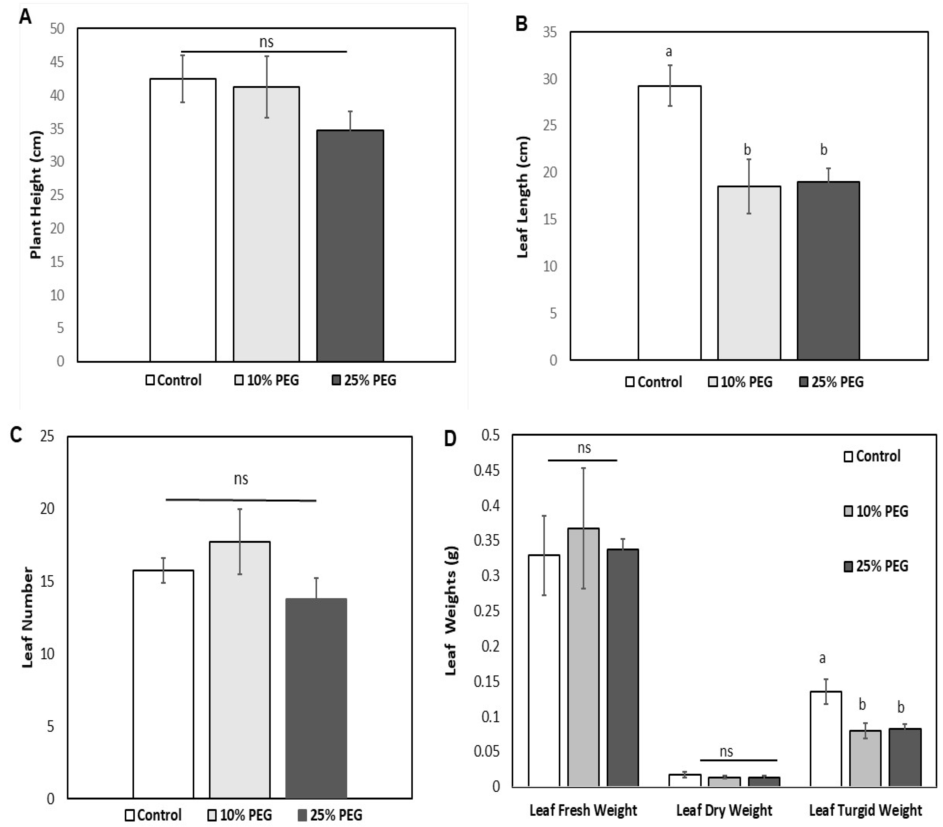
Figure 1. PEG-induced osmotic stress effect on maize: (A) Plant height (cm). (B) Leaf length (cm). (C) Leaf number. (D) Leaf weights (g) treated with water only (control) and varying PEG concentrations (10 and 25%). (E) Leaf area (cm2) treated with water only (control) and varying PEG concentrations (10 and 25%). (F) Relative water content of maize plants irrigated with water only (control) and experiments treated with varying PEG concentrations (10 and 25%). (G) Average root length (cm) and (H) root weight (g) of Z. mays for varying treatment conditions (control, 10% and 25% PEG). The error bars are representative of the mean (± standard error) of 3 independent seedling experiments (n = 3) from all the plant groups.
Ijpb 16 00133 g001aIjpb 16 00133 g001b
Figure 2. Effects of PEG osmotic stress on the photosynthesis rate of maize seedlings over time. Total photosynthesis rates were recorded after the treatment period between the non-treated (control) and treated (10 and 25% PEG). The error bars are representative of the means (± standard error) of 3 independent seedling experiments (n = 3).
Ijpb 16 00133 g002
Figure 3. Effects of PEG osmotic stress on the stomatal conductance rate of maize seedlings over time. Stomatal conductance rates were recorded after the treatment period between the non-treated (control) and treated (10 and 25% PEG). The error bars are representative of the means (± standard error) of 3 independent seedling experiments (n = 3).
Ijpb 16 00133 g003
Figure 4. Influence of PEG osmotic stress on leaf respiration efficiency of maize seedlings over time. Total respiration rates were recorded after the treatment period between the non-treated (control) and treated (10 and 25% PEG) seedlings. The error bars are representative of the means (± standard error) of 3 independent seedling experiments (n = 3).
Ijpb 16 00133 g004
Figure 5. Influence of PEG osmotic stress on leaf transpiration efficiency of maize seedlings over time. Total transpiration rates were recorded after the treatment period between the non-treated (control) and treated (10 and 25% PEG) seedlings. The error bars are representative of the means (± standard error) of 3 independent seedling experiments (n = 3).
Ijpb 16 00133 g005
Figure 6. (A) Comparative expression profile of maize leaf proteins in response to osmotic stress. An SDS-PAGE of the expressed protein fractions under osmotic stress, where M (lane 1) is the molecular weight marker. Lanes 2 and 5 are the water only (control), lanes 3 and 6 represent the 10% PEG treatment, and lanes 4 and 7 represent the 25% PEG treatment. (B) Venn diagram of the 50 selected differentially expressed proteins between the control and 10% and 25% PEG treatment groups. (C) Representation of functional classification and annotation distribution for the randomly selected 50 proteins categorized into diverse cellular components and biological and molecular functions in Z. mays under osmotic stress.
Ijpb 16 00133 g006aIjpb 16 00133 g006b
Figure 7. STRING based protein–protein interactions networks of differentially expressed proteins in maize under (A) 10% PEG and (B) 25% PEG treatments. Interconnected clusters (round purple nodes) in (A) represent coordinated responses associated with stress adaptation, while less interconnected clusters (pink nodes) in (B) represent disrupted protein coordination under high osmotic stress.
Ijpb 16 00133 g007
Table 1. Differentially expressed proteins (DEPs) identified in maize seedlings under control and 10% PEG-.
Control10% PEG
Accession NumberProtein NameGene/
Locus ID
Subcellular LocationBiological ProcessesMolecular FunctionPIMrAccession NumberProtein NameGene/
Locus ID
Subcellular LocationBiological ProcessesMolecular FunctionPIMr
tr|B4F833Diaminopimelate epimeraseLOC100191234CytosolLysine biosynthetic process via diaminopimelate Diaminopimelate epimerase activity 5.9637,799.11tr|B4F833Diaminopimelate epimeraseLOC100191234CytosolLysine biosynthetic process via diaminopimelateDiaminopimelate epimerase activity 5.9637,799.11
tr|C4IZ94Ornithine carbamoyltransferaseLOC100284885 Intracellular membraneArginine biosynthetic process via ornithineAmino acid binding6.7640,286.51tr|C4IZ94Ornithine carbamoyltransferaseLOC100284885 Intracellular membraneArginine biosynthetic process via ornithineAmino acid binding6.7640,286.51
tr|A0A1D6HM49Subtilisin-like protease SBT1.4LOC100279566None predictedProteolysisSerine-type endopeptidase activity 5.3578,345.99sp|P21569Peptidyl-prolyl cis–trans isomeraseCYPCytoplasmProtein folding; protein peptidyl-prolyl isomerizationCyclosporin A binding 8.9118,348.99
tr| B4F8V5NADH dehydrogenase [ubiquinone] iron–sulfur protein 1 mitochondrialLOC100191453Membrane ATP synthesis coupled electron transport4 iron, 4 sulfur cluster binding6.1080,679.62tr|C0P3R8Glutathione peroxidaseLOC100272844None predictedResponse to oxidative stressGlutathione peroxide activity 9.5524,993.61
tr|B4FYD3TOM1-like protein 2LOC100282255MembraneProtein transportPhosphatidylinositol binding; ubiquitin binding 4.5843,475.92tr|A0A804PHY0Peptidyl-prolyl cis–trans isomeraseLOC103627222None predictedProtein foldingPeptidyl-prolyl cis–trans isomerase activity9.0316,744.15
tr| K7WFV1ATP synthase subunit gammaZEAMMB73_
Zm00001d048091
Mitochondrial proton-transporting ATP synthase complex, catalytic sector F1F0Proton motive force-driven ATP synthesisProton-transporting ATP synthase activity, rotational mechanism9.1235,714.94tr| B6TQ06AminomethyltransferaseLOC100279332Mitochondrion MethylationAminomethyltransferase activity8.3644,034.54
tr| B5AMJ8Alpha-1,4 glucan phosphorylaseLOC100285259None predictedCarbohydrate metabolic processGlycogen phosphorylase activity6.8694,452.82tr|A0A804NZC5Glutamyl-tRNA(Gln) amidotransferase subunit B, chloroplastic/mitochondrialGATBChloroplastProtein biosynthesis; mitochondrial translation ATP binding6.1759,841.51
tr|B4FBF6Mitochondrial dicarboxylate/tricarboxylate transporter DTCLOC100274318Membrane
Transport Transmembrane transporter activity 9.7233,147.50tr|B7ZXQ3Cysteine proteinases superfamily proteinLOC100283689Lysosome Proteolysis involved in protein catabolic process Cysteine-type endopeptidase activity
tr|C4J410Heat shock 70 kDa proteinLOC100501536Cytoplasm Stress responseATP binding; ATP hydrolysis activity; ATP-dependent protein folding chaperone; heat shock protein binding; misfolded protein binding; protein folding chaperone; unfolded protein binding5.0770,882.41tr|A0A804NLS4AdenosylhomocysteinaseLOC100282150None predictedOne-carbon metabolismHydrolase activity 5.6348,377.59
tr| C4JA4560S ribosomal protein L5-1 homolog bZEAMMB73_
Zm00001d012161
Cytosolic large ribosomal subunit; nucleus Translation;
ribosomal large subunit assembly
5S rRNA binding; structural constituent of ribosome9.3434,3377.83tr|C0P5A9Optic atrophy 3 protein (OPA3)LOC100276616Mitochondrion Regulation of lipid metabolic process 10.0419,103.23
tr| K7VA9960S ribosomal protein L5-1 homolog bZEAMMB73_
Zm00001d012161
Cytoplasm; nucleusTranslation Structural constituent of ribosome5.8666,539.76tr|B4F8L7Glyceraldehyde-3-phosphate dehydrogenaseLOC542290Stromule Response to sucrose; response to light stimulus; response to coldmRNA, NAD and NADP binding5.9547,179.90
sp|P93805Phosphoglucomutase, cytoplasmic 2 (PGM 2) (Glucose phosphomutase 2)None retrievedCytoplasmCarbohydrate metabolism;
Glucose metabolism
Magnesium ion binding; phosphoglucomutase activity 5.4763,041.31tr|A0A1D6LSP5Elongation factor gamma1ZEAMMB73_
Zm00001d036959
None predictedProtein biosynthesis Elongation factor5.9143,457.95
tr|A0A804UKU7CN hydrolase domain-containing proteinLOC100279870None predictedHydrolase activity Hydrolase activity, acting on carbon–nitrogen (but not peptide) bonds, in linear amides 8.6240,920.58tr|K7V2Z8Carbamoyl-phosphate synthase (glutamine-hydrolyzing)LOC103636016Cytoplasm Pyrimidine biosynthesisATP binding; metal ion binding5.4912,7286.83
tr|A0A1D6KEP0Serine hydroxymethyltransferase ZEAMMB73_
Zm00001d030859
None predictedOne-carbon metabolismGlycine hydroxymethyltransferase activity; methyltransferase activity; pyridoxal phosphate binding7.6258,546.94tr|A0A804PU27Ribosomal_S4e domain-containing proteinNone retrievedRibosome Translation rRNA binding; ribosomal protein5.7928,447.42
tr|A0A804U9Z6Glutamate--cysteine ligase LOC542026 Plastid, chloroplast Glutathione biosynthesisATP binding; glutamate-cysteine ligase activity 6.4656,928.23tr|A0A804NG48GTP-binding nuclear proteinLOC100282843Nucleus Protein transport;
Nucleocytoplasmic transport
GTP binding 9.3419,867.92
tr|A0A804QKD5Glutathione hydrolaseLOC103634830MembraneGlutathione catabolic processAcyltransferase activity; glutathione hydrolase activity5.9566,539.76sp|P09315Glyceraldehyde-3-phosphate dehydrogenase A, chloroplasticGAPAChloroplast Glucose metabolic process; reductive pentose-phosphate cycle; response to light stimulus NAD and NADP binding 6.2536,096.26
tr|K7VN08ATP synthase B chainLOC100282566MembraneHydrogen ion transportProton transmembrane transporter activity 5.4822,715.27tr|A0A804MAM1Methionine aminopeptidaseLOC100192977None predictedProtein initiator methionine removal; proteolysis Metal ion binding; metalloaminopeptidase activity 6.8637,094.53
tr|B4FBC9Patellin-1LOC100192106CytoplasmCell cycle; cell divisionLipid binding4.5868,035.27tr|B4FRH1Thioredoxin M1 chloroplasticLOC100272895Cytoplasm None predictedOxidoreductase activity; protein-disulfide reductase activity8.6319,560.59
tr|C0PAU7Glucose-6-phosphate isomerase LOC100382805CytosolGlycolysis Carbohydrate derivative binding; glucose-6-phosphate isomerase activity; monosaccharide binding5.5667,431.16tr|B4FTT2Regulator of chromosome condensation2LOC100273233Chromatin; cytosol; nucleus Response to UV-B;
entrainment of circadian clock
Chromatin binding; photoreceptor activity; protein homodimerization activity5.3847,158.43
tr|B6UBZ9Cytochrome b-c1 complex subunit 7LOC100383274Mitochondrion inner membraneElectron transport; respiratory chain 9.6414,801.20tr|B4FU5350S ribosomal protein L9, chloroplastic (CL9)
LOC100285012Ribosome Translation Structural constituent of ribosome9.8121,660.30
tr|Q2MJJ9Putative RH2 proteinRH2None predictedNone predictedATP binding; hydrolase activity; RNA binding; RNA helicase activity 5.9845,945.84sp|P0658630S ribosomal protein S3, chloroplastic
rps3Chloroplast Translation rRNA binding9.7625,916.37
tr|B4FPJ2U6 snRNA-associated Sm-like protein LSm3LOC100194361NucleusmRNA processing; mRNA splicingRNA binding4.7811,235.91tr|B4F8M8Exoglucanase1
LOC100191390Extracellular region Carbohydrate metabolic process Hydrolase activity, hydrolyzing O-glycosyl compounds 6.5464,583.72
tr|C0P7G8Photosystem II repair protein PSB27-H1 chloroplasticLOC100282043Chloroplast thylakoid lumenPhotosystem II assembly and repairNone predicted9.8617,757.36tr|K7VDJ240S ribosomal protein S17-4
LOC100282953Ribonucleoprotein complex;
ribosome
Translation Structural constituent of ribosome 10.1416,443.05
tr|Q9M640Coatomer subunit deltaLOC541810Cytoplasm; Golgi membraneER–Golgi transport;
protein transport
Retrograde Golgi-to-ER transport of dilysine-tagged proteins5.5457,498.47tr| B8A2X5PectinesteraseLOC100280285None predictedCell wall modification; pectin catabolic processAspartyl esterase activity; pectinesterase activity8.7631,742.89
tr|K7TXI5Chlorophyll a-b binding protein, chloroplasticLOC542321Plastid, chloroplast thylakoid membranePhotosynthesisChlorophyll binding 7.9026,586.76tr|A0A1D6HT50Polyribonucleotide nucleotidyltransferaseLOC103631988None predictedmRNA catabolic process; RNA processing Polyribonucleotide nucleotidyltransferase activity5.6995,115.02
tr|B6TCK3NADH-cytochrome b5 reductaseLOC103649684None predictedNone predictedCytochrome-b5 reductase activity, acting on NAD(P)H9.0333,845.03tr|B4FRD6PeroxidaseLOC100281280Plant-type cell wall; extracellular regionHydrogen peroxideHeme binding; peroxidase activity7.1233,116.72
tr|B4FT19Oxygen evolving enhancer protein 3 containing proteinLOC100273120thylakoidPhotosynthesisCalcium ion binding 7.6525,342.86tr|B4FW0640S ribosomal protein S10-1LOC100273505Cytosolic small ribosomal subunit None predictedRNA binding; structural constituent of ribosome9.7819,938.47
tr|A0A1D6MUE8Heat shock 70 kDa protein 6 chloroplasticLOC103650436Chloroplast Stress response ATP binding; ATP-dependent protein folding chaperone; unfolded protein binding 5.0874,493.16tr| Q947B9Glucose-1-phosphate adenylyltransferaseGLG1Chloroplast Starch biosynthesis ATP binding; glucose-1-phosphate adenylyltransferase activity6.4856,482.41
sp|P69388Cytochrome b559 subunit alpha (PSII reaction center subunit V)psbEPlastid, chloroplast thylakoid membranePhotosynthetic electron transport chainElectron transfer activity; heme binding; iron ion binding 4.649444.60sp|P0658950S ribosomal protein L22, chloroplastic
rpl22Chloroplast Translation; ribosome assemblyrRNA binding; structural constituent of ribosome10.8217,655.21
tr|C0P3S5Fumarylacetoacetate (FAA) hydrolase familyZEAMMB73_
Zm00001d034739
Mitochondrion None predictedAcetylpyruvate hydrolase activity 5.4124,731.55tr|B6TGG73-oxoacyl-[acyl-carrier-protein] synthase
LOC100279917None predictedFatty acid biosynthetic process3-oxoacyl-[acyl-carrier-protein] synthase activity 7.5748,864.83
tr|A0A1D6H5M2Chalcone-flavonone isomerase family proteinLOC100285485None predictedNone predictedIntramolecular lyase activity 7.7229,540.85tr|B4FCX3Proteasome subunit beta
LOC100192865Cytoplasm;
nucleus
Proteasomal protein catabolic processThreonine-type endopeptidase activity 7.7728,912.76
tr|A0A804R1C2Starch synthase, chloroplastic/
amyloplastic
None retrievedPlastid, chloroplastStarch biosynthesis Glycogen (starch) synthase activity 5.6168,900.48tr|B6TNT5Peptide-methionine (S)-S-oxide reductase LOC100283982CytoplasmCellular response to oxidative stressL-methionine-(S)-S-oxide reductase activity; peptide-methionine (S)-S-oxide reductase activity 5.8520,504.80
tr|B4FQ59PhosphoribulokinaseLOC100282845Cytoplasm Phosphorylation ATP binding; phosphoribulokinase activity5.8444,749.89tr| P55240Glucose-1-phosphate adenylyltransferase small subunitGLG1Chloroplast; amyloplastStarch biosynthesisATP bindingN/AN/A
tr|A0A804NUX2Phosphoribulokinase None retrievedNone predictedReductive pentose-phosphate cycle ATP binding; phosphoribulokinase activity9.5127,481.52tr|A0A804P7H7Ribose-5-phosphate isomeraseNone retrievedNone predictedPentose-phosphate shunt, non-oxidative branchRibose-5-phosphate isomerase activity5.4431,500.71
sp|P33488Auxin-binding protein 4 (ABP)ABP4Endoplasmic reticulum lumen Auxin signaling pathway Receptor 6.1418,583.08tr|B4FRC4Serine-tRNA ligase
LOC541989Cytosol Seryl-tRNA aminoacylationAminoacyl-tRNA synthetase6.2751,663.79
tr|A0A1D6LE55PeroxidaseLOC103628960Extracellular regionHydrogen peroxide catabolic processLactoperoxidase activity; heme binding 5.2534,847.89tr|A0A804MJA1Fn3_like domain-containing proteinNone retrievedExtracellular region Xylan catabolic process Xylan 1,4-beta-xylosidase activity 8.5282,057.02
tr|A0A1X7YHG9ATP synthase subunit alpha, chloroplasticLOC118474820Plastid, chloroplast thylakoid membraneATP synthesis;
Hydrogen ion transport
ATP binding; proton-transporting ATP synthase activity, rotational mechanism; proton-transporting ATPase activity, rotational mechanism 5.8755,690.85tr|A0A1D6L55460S ribosomal protein L13a-1LOC100191912Large ribosomal subunitTranslation Structural constituent of ribosome10.3521,187.48
tr|A0A804LI62PsbP domain-containing proteinLOC103634474Extrinsic component of membranePhotosynthesis Calcium ion binding 7.5926,587.16tr| B4FAM660S ribosomal protein L13a-1LOC100191912Large ribosomal subunitTranslation Lyase activity; magnesium ion binding; thiamine pyrophosphate binding10.2023,600.17
sp|P48186ATP synthase subunit b, chloroplastic (ATP synthase F(0) sector subunit b) (ATP
se subunit I)
atpFPlastid, chloroplast thylakoid membraneATP synthesisATP binding; proton-transporting ATP synthase activity, rotational mechanism 9.2720,981.12tr|K7UMB3Signal recognition particle 54 kDa protein chloroplasticLOC103654352Signal recognition particle, endoplasmic reticulum targetingSRP-dependent cotranslational protein targeting to membrane 7S RNA binding; GTP binding; GTPase activity 9.5659,737.25
tr|A0A804NI62LegumainLOC542609Cellular anatomical entityProteolysis involved in protein catabolic processCysteine-type endopeptidase activity5.0744,767.98tr| B4FYT5Protein Kinase C630.09cLOC100274052None predictedPhosphorylationATP binding; kinase activity 5.6247,498.13
tr|A0A1D6E958Protease Do-like 8 chloroplasticLOC100272554Chloroplast thylakoid lumen Proteolysis; photosystem II repair Serine-type endopeptidase activity9.0751,190.49sp|P2772330S ribosomal protein S16, chloroplasticrps16Chloroplast TranslationStructural constituent of ribosome 10.3310,089.90
tr|A0A3L6EGC3Germin-like proteinOs08g0459700_1Secreted, extracellular space, apoplastNone predictedManganese ion binding 5.8920,101.08tr|A0A804UA42PRK domain-containing proteinLOC100274052Integral component of membraneATP binding; kinase activity DNA binding6.6158,115.71
tr|A0A096S0784-coumarate--CoA ligase-like 7LOC100191850MembranePhenylpropanoid metabolic process4-coumarate-CoA ligase activity7.0053,052.88tr|B8A1H0Glucose-6-phosphate 1-epimeraseLOC100273735Cytoplasm Carbohydrate metabolic process Carbohydrate binding 5.6834,240.88
tr|A0A804N9X8PectinesteraseNone retrievedNone predictedCell wall modification;
Pectin catabolic process
Aspartyl esterase activity; pectinesterase activity6.1954,997.66tr|B4G1J850S ribosomal protein L3-1 chloroplasticLOC100193892Ribonucleoprotein complex; ribosomeTranslation mRNA binding10.5927,867.52
tr|Q94F78Nucleosome/chromatin assembly factor Anfa104Cytoplasm; chromatin; nucleus Nucleus assembly; double-stand break repair Chromatin binding; histone binding 4.0829,331.84tr|B4FGN450S ribosomal protein L3-1 chloroplasticLOC100193892RibosomeTranslation Structural constituent of ribosome 7.6324,415.30
tr|A0A1D6L6U6Glutathione transferase LOC542734cytoplasmGlutathione metabolic processGlutathione transferase activity6.2025,258.20tr|C0HFM460S ribosomal protein L13a-1LOC100283655Cytosolic large ribosomal subunit; ribosome Negative regulation of translation; translation mRNA binding; structural constituent of ribosome 10.2223,629.10
tr|A0A1D6N672Photosystem II subunit PsbS1LOC542126Membrane; plastid Nonphotochemical quenching; response to high light intensityNone predicted9.0728,452.15tr|B4F9N8RNA-binding (RRM/RBD/RNP motifs) family proteinLOC100191663Ribonucleoprotein complex None predictedRNA binding 4.9951,279.59
tr|A0A804LJD2Guanylate kinaseLOC100283207None predictedPhosphorylationATP binding; guanylate kinase activity 9.1832,388.47tr|A0A804RFJ4Gp_dh_N domain-containing proteinNone retrievedNone predictedNone predictedNAD binding6.5128,369.38
tr|Q6R9G1NAD(P)H dehydrogenase subunit Hnad7Mitochondrion; thylakoid Mitochondrial electron transportNAD binding; NADH dehydrogenase (ubiquinone) activity; quinone binding6.7444,280.13tr|A0A804PBS5Inorganic diphosphatase LOC103626225CytoplasmPhosphate-containing compound metabolic processInorganic diphosphate phosphatase activity; magnesium ion binding 5.9920,994.02
tr|A0A804UKI0CN hydrolase domain-containing proteinLOC100279870None predictedHydrolase activity Hydrolase activity, acting on carbon–nitrogen (but not peptide) bonds, in linear amides 8.8929,227.53tr|A0A804NZ53SRP54 domain-containing proteinLOC103654352CytoplasmSRP-dependent cotranslational protein targeting to membraneRNA-binding;
GTPase activity
9.3656,091.99
Note: “None retrieved” indicates that no corresponding gene or locus ID could be retrieved from NCBI, UniProt, or EnsemblPlants databases for the listed protein accession. “None predicted” denotes that subcellular localization or annotation information was unavailable in the referenced databases at the time of analysis.
Table 2. Differentially expressed proteins (DEPs) identified in maize seedlings under control and 25% PEG.
Control 25% PEG
Accession NumberProtein NameGene/
Locus ID
Subcellular LocationBiological Processes
Molecular Function
PIMrAccession NumberProtein NameGene/
Locus ID
Subcellular LocationBiological Processes
Molecular Function
PIMr
TR|B4F833Diaminopimelate epimeraseLOC100191234CytosolLysine biosynthetic process via diaminopimelateDiaminopimelate epimerase activity 5.9637,799.11sp|P21569Peptidyl-prolyl cis–trans isomeraseCYPCytoplasm
Protein folding; protein peptidyl-prolyl isomerizationCyclosporin A binding8.9118,348.99
TR|C4IZ94Ornithine carbamoyltransferaseLOC100284885Intracellular membraneArginine biosynthetic process via ornithineAmino acid binding6.7640,286.51tr|C0P3R8Glutathione peroxidaseLOC100272844None predictedResponse to oxidative stressGlutathione peroxide activity 9.5524,993.61
TR|A0A1D6HM49Subtilisin-like protease SBT1.4LOC100279566None predictedProteolysisSerine-type endopeptidase activity 5.3578,345.99tr|A0A804PHY0Peptidyl-prolyl cis–trans isomeraseLOC103627222None predictedProtein foldingPeptidyl-prolyl cis–trans isomerase activity9.0316,744.15
TR| B4F8V5NADH dehydrogenase [ubiquinone] iron–sulfur protein 1 mitochondrialLOC100191453Membrane ATP synthesis coupled electron transport4 iron, 4 sulfur cluster binding6.1080,679.62tr|C0P4P5Non-reducing end alpha-L-arabinofuranosidase LOC100193890None predictedCellular carbohydrate catabolic process; L-arabinose metabolic process Alpha-L-arabinofuranosidase activity 5.0569,535.87
TR|B4FYD3TOM1-like protein 2LOC100282255MembraneProtein transportPhosphatidylinositol binding; ubiquitin binding 4.5843,475.92tr|K7VNQ7Mitochondrial import receptor subunit TOM40-1LOC110120361Integral component of membraneProtein import into mitochondrial matrixProtein transmembrane transporter activity6.0137,345.36
TR| K7WFV1ATP synthase subunit gammaLOC100274172
Mitochondrial proton-transporting ATP synthase complex, catalytic sector F1F0 Proton motive force-driven ATP synthesisProton-transporting ATP synthase activity, rotational mechanism9.1235,714.94tr|B4FMF7Aldose 1-epimeraseLOC100217298None predictedHexose metabolic processAldose 1-epimerase activity8.8936,388.96
TR| B5AMJ8Alpha-1,4 glucan phosphorylaseLOC100285259
None predictedCarbohydrate metabolic processGlycogen phosphorylase activity6.8694,452.82tr|C0P5Y35-methyltetrahydropteroyltriglutamate--homocysteine S-methyltransferaseLOC541942None predictedMethionine biosynthetic process; methylation 5-methyltetrahydropteroyltriglutamate-homocysteine S-methyltransferase activity; zinc ion binding 5.5484,492.46
TR|B4FBF6Mitochondrial dicarboxylate/tricarboxylate transporter DTCLOC100274318Membrane
Transport Transmembrane transporter activity 9.7233,147.50tr|Q6RW09Allene-oxide cyclaseaocChloroplastJasmonic acid biosynthetic processAllene-oxide cyclase activity9.0525,777.29
TR|C4J410Heat shock 70 kDa proteinLOC100501536Cytoplasm Stress responseATP binding; ATP hydrolysis activity; ATP-dependent protein folding chaperone; heat shock protein binding; misfolded protein binding; protein folding chaperone; unfolded protein binding5.0770,882.41tr|A0A804MQI0Valine-tRNA ligaseLOC100383209None predictedPost-embryonic development]; reproductive structure development; valyl-tRNA aminoacylationAminoacyl-tRNA editing activity; ATP binding; valine-tRNA ligase activity6.6011,6740.55
TR| C4JA4560S ribosomal protein L5-1 homolog bLOC10028491Cytosolic large ribosomal subunit; nucleus Translation;
ribosomal large subunit assembly
5S rRNA binding; structural constituent of ribosome9.3434,3377.83tr|B4FYH2Pyruvate kinase
LOC100273990Chloroplast stroma; cytoplasmGlycolytic process; photosynthesis ATP binding; kinase activity; magnesium ion binding; potassium ion binding; pyruvate kinase activity6.2661,479.03
TR| K7VA9960S ribosomal protein L5-1 homolog bLOC100284917Cytoplasm; nucleusTranslation Structural constituent of ribosome5.8666,539.76tr|B4FEA2Mitochondrial carnitine/acylcarnitine carrier-like protein
LOC118476033MembraneNitrogen compound transport; organic anion transport; organophosphate ester transport; transmembrane transport None predicted9.5430,155.95
SP|P93805Phosphoglucomutase, cytoplasmic 2 (PGM 2) (Glucose phosphomutase 2)LOC542358CytoplasmCarbohydrate metabolism;
Glucose metabolism
Magnesium ion binding; phosphoglucomutase activity 5.4763,041.31tr|A0A804R5D8Ribosomal_S4e domain-containing proteinNone retrievedRibosomeTranslationStructural constituent of ribosome6.8845,232.88
TR|A0A804UKU7CN hydrolase domain-containing proteinLOC100279870None predictedHydrolase activity Hydrolase activity, acting on carbon–nitrogen (but not peptide) bonds, in linear amides 8.6240,920.58tr|O2245340S ribosomal protein S4rps4RibosomeTranslation RNA binding10.2030,171.37
TR|A0A1D6KEP0Serine hydroxymethyltransferase LOC100381786None predictedOne-carbon metabolismGlycine hydroxymethyltransferase activity; methyltransferase activity; pyridoxal phosphate binding7.6258,546.94tr|C0P45560S ribosomal protein L4-1LOC100382075Cytosolic large ribosomal subunitTranslation RNA binding10.6544,292.54
TR|A0A804U9Z6Glutamate--cysteine ligase LOC542026 Plastid, chloroplast Glutathione biosynthesisATP binding; glutamate-cysteine ligase activity 6.4656,928.23tr|B4FWJ8Binding protein homolog2LOC732809
Cytoplasm Protein refolding Heat shock protein binding 5.1070,546.74
TR|A0A804QKD5Glutathione hydrolaseLOC103634830MembraneGlutathione catabolic processAcyltransferase activity; glutathione hydrolase activity5.9566,539.76tr|A0A1D6L210NADH dehydrogenase [ubiquinone] iron–sulfur protein 1 mitochondrial
LOC100280404Membrane ATP synthesis coupled electron transport4 iron, 4 sulfur cluster binding; NADH dehydrogenase6.0180,717.71
TR|K7VN08ATP synthase B chainLOC100282566MembraneHydrogen ion transportProton transmembrane transporter activity 5.4822,715.27tr|A0A804MU73T-complex protein 1 subunit zeta LOC100193477Cytoplasm ATP binding 6.8253,590.56
TR|B4FBC9Patellin-1LOC100192106CytoplasmCell cycle; cell divisionLipid binding4.5868,035.27tr|B6THZ8Threonine synthaseLOC100283397Cytoplasm Cysteine biosynthetic processPyridoxal phosphate binding6.4457,614.35
TR|C0PAU7Glucose-6-phosphate isomerase LOC100382805CytosolGlycolysis Carbohydrate derivative binding; glucose-6-phosphate isomerase activity; monosaccharide binding5.5667,431.16tr|B4FAL9Fructose-bisphosphate aldolase LOC542261Cytosol Fructose 1,6-bisphosphate metabolic process; glycolytic process Fructose-bisphosphate aldolase activity 7.5238,590.16
TR|B6UBZ9Cytochrome b-c1 complex subunit 7LOC100383274Mitochondrion inner membraneElectron transport; respiratory chainNone predicted9.6414,801.20tr|K7UDG5Proline--tRNA ligaseLOC100272402Cytoplasm Prolyl-tRNA aminoacylationATP binding6.7158,133.54
TR|Q2MJJ9Putative RH2 protein LOC732749None predictedNone predictedATP binding; hydrolase activity; RNA binding; RNA helicase activity 5.9845,945.84tr|C0PFN4Glucose-6-phosphate 1-epimeraseLOC100282752None predictedCarbohydrate metabolic process Carbohydrate binding; glucose-6-phosphate 1-epimerase activity 6.2636,652.84
TR|B4FPJ2U6 snRNA-associated Sm-like protein LSm3
LOC100194361NucleusmRNA processing; mRNA splicingRNA binding4.7811,235.91tr|B4F938Coproporphyrinogen oxidaseLOC100500945Cytoplasm Protoporphyrinogen IX biosynthetic process Coproporphyrinogen oxidase activity 8.1944,112.77
TR|C0P7G8Photosystem II repair protein PSB27-H1 chloroplasticLOC100282043Chloroplast thylakoid lumenPhotosystem II assembly and repairNone predicted9.8617,757.36tr|B4F871Protein DJ-1 homolog D (YLS5)LOC100280536None predictedNone predictedGlyoxalase III activity5.3741,209.28
TR|Q9M640Coatomer subunit deltaLOC541810Cytoplasm; Golgi membraneER-Golgi transport;
Protein transport
Retrograde Golgi-to-ER transport of dilysine-tagged proteins5.5457,498.47tr|B4FIH9Xylose isomeraseLOC100194385None predictedD-xylose metabolic processMetal ion binding; xylose isomerase activity5.4051,045.66
TR|K7TXI5Chlorophyll a-b binding protein, chloroplasticLOC542321Plastid, chloroplast thylakoid membranephotosynthesisChlorophyll binding 7.9026,586.76tr|B4G1K3Calcyclin-binding protein
Prolyl aminopeptidase)
SGT1
LOC100282523None predictedNone predictedS100 protein binding; tubulin binding; ubiquitin protein ligase binding8.6924,644.44
TR|B6TCK3NADH-cytochrome b5 reductaseLOC103649684None predictedNone predictedCytochrome-b5 reductase activity, acting on NAD(P)H9.0333,845.03tr|B4FR32Glyceraldehyde-3-phosphate deHaseN1 (NADP-dependent glyceraldehyde-3-phosphate dehydrogenase)LOC542583None predictedNone predictedOxidoreductase activity, acting on the aldehyde or oxo group of donors, NAD or NADP as acceptor6.8053,284.58
TR|B4FT19Oxygen evolving enhancer protein 3 containing proteinLOC100273120Thylakoid Photosynthesis Calcium ion binding 7.6525,342.86tr|B4FFZ2Ketol-acid reductoisomerase (EC 1.1.1.86) (Acetohydroxy-acid reductoisomerase) (Alpha-keto-beta-hydroxylacyl reductoisomerase)LOC100193695None predictedIsoleucine biosynthetic process; valine biosynthetic process.Isomerase activity; ketol-acid reductoisomerase activity; metal ion binding 6.3163,002.80
TR|A0A1D6MUE8Heat shock 70 kDa protein 6 chloroplasticLOC103650436Chloroplast Stress response ATP binding; ATP-dependent protein folding chaperone; unfolded protein binding 5.0874,493.16tr|B4FAD4Isocitrate dehydrogenase [NAD] catalytic subunit 5 mitochondrialLOC100191841Mitochondrion Isocitrate metabolic process; tricarboxylic acid cycle Isocitrate dehydrogenase (NAD+) activity; magnesium ion binding; NAD binding.6.5239,724.58
SP|P69388Cytochrome b559 subunit alpha (PSII reaction center subunit V)psbEPlastid, chloroplast thylakoid membranePhotosynthetic electron transport chainElectron transfer activity; heme binding; iron ion binding 4.649444.60tr|B4F9K0Late embryogenesis abundant protein group 2LOC100191638None predictedResponse to desiccation None predicted4.9235,274.07
TR|C0P3S5Fumarylacetoacetate (FAA) hydrolase family Mitochondrion None predictedAcetylpyruvate hydrolase activity 5.4124,731.55tr|A0A1D6JYM4Glutathione S-transferase L2 chloroplasticLOC100282747None predictedResponse to chemical Glutathione transferase activity6.1533,860.53
TR|A0A1D6H5M2Chalcone-flavonone isomerase family proteinLOC100285485None predictedNone predictedIntramolecular lyase activity 7.7229,540.85tr|Q6R9L0NADH dehydrogenase subunit 9nad9Mitochondrial respiratory chain complex I None predictedNADH dehydrogenase (ubiquinone) activity 8.5122,594.41
TR|A0A804R1C2Starch synthase, chloroplastic/amyloplasticLOC100283765Plastid, chloroplastStarch biosynthesis Glycogen (starch) synthase activity 5.6168,900.48tr|B4FT31Dehydroascorbate reductase DHAR3None predictedAscorbate glutathione cycleGlutathione transferase activity5.5423,355.73
TR|B4FQ59Phosphoribulokinase LOC100282845Cytoplasm Phosphorylation ATP binding; phosphoribulokinase activity5.8444,749.89tr|B4F9K417.5 kDa class II heat shock proteinLOC542723None predictedStress responseProtein self-association5.9517,868.64
TR|A0A804NUX2Phosphoribulokinase None retrievedNone predictedReductive pentose-phosphate cycle ATP binding; phosphoribulokinase activity9.5127,481.52tr|B4F9E817.4 kDa class III heat shock proteinLOC100191598None predictedProtein complex Oligomerization; protein folding; response to heat; response to hydrogen peroxide; response to salt stress Protein self-association; unfolded protein binding6.6018,347.76
SP|P33488Auxin-binding protein 4 (ABP)ABP4Endoplasmic reticulum lumen Auxin signaling pathway Receptor 6.1418,583.08tr|B4FQC9Proline iminopeptidase (PIP) LOC100272740CytoplasmAminopeptidase activity Proteolysis5.2436,602.16
TR|A0A1D6LE55PeroxidaseLOC103628960Extracellular region Hydrogen peroxide catabolic processLactoperoxidase activity; heme binding 5.2534,847.89tr|A0A804NGN9SGT1LOC100282745Plastid;
Thylakoid
None predictedChaperone binding 5.0240,229.33
TR|A0A1X7YHG9ATP synthase subunit alpha, chloroplasticLOC118474820Plastid, chloroplast thylakoid membraneATP synthesis;
Hydrogen ion transport
ATP binding; proton-transporting ATP synthase activity, rotational mechanism; proton-transporting ATPase activity, rotational mechanism 5.8755,690.85tr|B6SN61Grx_C2.1-glutaredoxin subgroup ILOC100303864Cytoplasm Cellular response to oxidative stress Glutathione disulfide oxidoreductase activity; glutathione oxidoreductase activity; protein-disulfide reductase (glutathione) activity7.7113,932.16
TR|A0A804LI62PsbP domain-containing proteinLOC103634474Extrinsic component of membranePhotosynthesis Calcium ion binding 7.5926,587.16tr|A0A1D6MNJ01-deoxy-D-xylulose-5-phosphate reductoisomeraseLOC542023None predictedIsopentenyl diphosphate biosynthetic process, methylerythritol 4-phosphate pathway1-deoxy-D-xylulose-5-phosphate reductoisomerase activity; isomerase activity; metal ion binding; NADPH binding6.4451,257.01
SP|P48186ATP synthase subunit b, chloroplastic (ATP synthase F(0) sector subunit b) (ATP
se subunit I)
atpFPlastid, chloroplast thylakoid membraneATP synthesisATP binding; proton-transporting ATP synthase activity, rotational mechanism 9.2720,981.12tr|A0A804PDW7Luminal-binding protein 5LOC732808Endoplasmic reticulumNone predictedATP binding; ATP-dependent protein folding chaperone5.0569,401.51
TR|A0A804NI62LegumainLOC542609Cellular anatomical entityproteolysis involved in protein catabolic processCysteine-type endopeptidase activity5.0744,767.98tr|A0A804LN04Branched-chain amino acid aminotransferase LOC100191754None predictedBranched-chain amino acid biosynthetic process; cellular amino acid biosynthetic process Branched-chain amino acid transaminase activity 5.8043,223.76
TR|A0A1D6E958Protease Do-like 8 chloroplasticLOC100272554Chloroplast thylakoid lumen Proteolysis; photosystem II repair Serine-type endopeptidase activity9.0751,190.49tr|A0A804MU73T-complex protein 1 subunit zetaLOC100193477Cytoplasm None predictedATP binding; ATP hydrolysis activity; ATP-dependent protein folding chaperone; unfolded protein binding 6.8253,590.56
TR|A0A3L6EGC3Germin-like proteinLOC100191976Secreted, extracellular space, apoplastNone predictedManganese ion binding 5.8920,101.08tr|A0A804RJL5Aconitate hydratase LOC100281040None predicted4 iron, 4 sulfur cluster binding; hydro-lyase activity; metal ion bindingOxoacid metabolic process 6.01966,932.89
TR|A0A096S0784-coumarate--CoA ligase-like 7LOC100191850MembranePhenylpropanoid metabolic process4-coumarate-CoA ligase activity7.0053,052.88tr|A0A1D6JW4140S ribosomal protein S20-1LOC103633583Cytosolic small ribosomal subunit; small ribosomal subunit Translation Structural constituent of ribosome 9.5113,824.16
TR|A0A804N9X8PectinesteraseLOC103650921None predictedCell wall modification;
Pectin catabolic process
Aspartyl esterase activity; pectinesterase activity6.1954,997.66tr|B6U237Heat shock 70 kDa protein 14 (Heat shock 70 kDa protein 4)LOC100285213None predictedATP binding; ATP-dependent protein folding chaperone None predicted5.2893,569.13
TR|Q94F78Nucleosome/chromatin assembly factor A nfa104Cytoplasm; chromatin; nucleus Nucleus assembly; double-stand break repair Chromatin binding; histone binding 4.0829,331.84tr|A0A1D6JW4140S ribosomal protein S20-1LOC103633583Component cytosolic small ribosomal subunitTranslation Ribosomal protein9.5113,824.16
TR|A0A1D6L6U6Glutathione transferase LOC542734Cytoplasm Glutathione metabolic processGlutathione transferase activity6.2025,258.20tr|B4F9K417.5 kDa class II heat shock proteinLOC542723None predictedProtein self-association; unfolded protein binding Protein complex oligomerization; protein folding; response to heat; response to hydrogen peroxide; response to salt stress 5.9517,868.64
TR|A0A1D6N672Photosystem II subunit PsbS1LOC542126Membrane; plastid Nonphotochemical quenching; response to high light intensityNone predicted9.0728,452.15tr|A0A804RJL5Aconitate hydrataseLOC100281040None predictedOxoacid metabolic processMetal ion binding 6.0196,932.89
TR|A0A804LJD2Guanylate kinaseLOC100283207None predictedPhosphorylationATP binding; guanylate kinase activity 9.1832,388.47tr|B6T8Q5T-complex protein 1 subunit zetaLOC100193477Cytoplasm None predictedATP binding6.2359,087.02
TR|Q6R9G1NAD(P)H dehydrogenase subunit Hnad7Mitochondrion; thylakoid Mitochondrial electron transportNAD binding; NADH dehydrogenase (ubiquinone) activity; quinone binding6.7444,280.13tr|B4FT31Dehydroascorbate reductaseDHAR3None predictedGlutathione dehydrogenase (ascorbate) activity; glutathione transferase activity Ascorbate glutathione cycle
TR|A0A804UKI0CN hydrolase domain-containing proteinLOC100279870None predictedHydrolase activity Hydrolase activity, acting on carbon–nitrogen (but not peptide) bonds, in linear amides 8.8929,227.53tr|K7VJF3Heat shock 70 kDa protein 5LOC100272911Cytoplasm ATP binding; ATP hydrolysis activity; ATP-dependent protein folding chaperone; heat shock protein binding; misfolded protein binding; protein folding chaperone; unfolded protein binding Cellular response to unfolded protein; chaperone cofactor-dependent protein refolding; protein refolding 5.2271,502.04
Note: “None retrieved” indicates that no corresponding gene or locus ID could be retrieved from UniProt, EnsemblPlants, and NCBI databases for the listed protein accession. “None predicted” denotes that subcellular localization or annotation information was unavailable in the referenced databases at the time of analysis.
Disclaimer/Publisher’s Note: The statements, opinions and data contained in all publications are solely those of the individual author(s) and contributor(s) and not of MDPI and/or the editor(s). MDPI and/or the editor(s) disclaim responsibility for any injury to people or property resulting from any ideas, methods, instructions or products referred to in the content.