Article

In Vitro Antagonism of Two Isolates of the Genus Trichoderma on Fusarium and Botryodiplodia sp., Pathogenic Fungi of Schizolobium parahyba in Ecuador

by
Carlos Belezaca-Pinargote
1,*,
Bélgica Intriago-Pinargote
1,
Brithany Belezaca-Pinargote
1,
Edison Solano-Apuntes
1,
Ricardo Arturo Varela-Pardo
2 and
Paola Díaz-Navarrete
3,*
1
Facultad de Ciencias Agrarias y Forestales, Universidad Técnica Estatal de Quevedo, Quevedo 120302, Ecuador
2
Departamento de Ciencias Agropecuarias y Acuícolas, Facultad de Recursos Naturales, Universidad Católica de Temuco, P.O. Box 15-D, Temuco 4780000, Chile
3
Departamento de Ciencias Veterinarias y Salud Pública, Facultad de Recursos Naturales, Universidad Católica de Temuco, Temuco 4780000, Chile
*
Authors to whom correspondence should be addressed.
Int. J. Plant Biol. 2025, 16(3), 85; https://doi.org/10.3390/ijpb16030085 (registering DOI)
Submission received: 16 June 2025 / Revised: 19 July 2025 / Accepted: 22 July 2025 / Published: 1 August 2025
(This article belongs to the Section Plant–Microorganisms Interactions)

Abstract

A newly emerging disease affecting Schizolobium parahyba (commonly known as pachaco), termed “decline and dieback,” has been reported in association with the fungal pathogens Fusarium sp. and Botryodiplodia sp. This study assessed the antagonistic potential of two Trichoderma sp. isolates (CEP-01 and CEP-02) against these phytopathogens under controlled laboratory conditions. The effects of three temperature regimes (5 ± 2 °C, 24 ± 2 °C, and 30 ± 2 °C) on the growth and inhibitory activity of two Trichoderma spp. isolates were evaluated using a completely randomized design. The first experiment included six treatments with five replicates, while the second comprised twelve treatments, also with five replicates. All assays were conducted on PDA medium. No fungal growth was observed at 5 ± 2 °C. However, at 24 ± 2 °C and 30 ± 2 °C, both isolates reached maximum growth within 72 h. At 24 ± 2 °C, both Trichoderma spp. isolates exhibited inhibitory activity against Fusarium sp. FE07 and FE08, with radial growth inhibition percentages (RGIP) ranging from 37.6% to 44.4% and 52,8% to 54.6%, respectively. When combined, the isolates achieved up to 60% inhibition against Fusarium sp., while Botryodiplodia sp. was inhibited by 40%. At 30 ± 2 °C, the antagonistic activity of Trichoderma sp. CEP-01 declined (25.6–32.4% RGIP), whereas Trichoderma sp. CEP-02 showed increased inhibition (60.3%–67.2%). The combination of isolates exhibited the highest inhibitory effect against Fusarium sp. FE07 and FE08 (68.4%–69.3%). Nonetheless, the inhibitory effect on Botryodiplodia sp. BIOT was reduced under elevated temperatures across all treatments. These findings reinforce the potential of Trichoderma spp. isolates as a viable and eco-friendly alternative for the biological control of pathogens affecting S. parahyba, contributing to more sustainable disease management practices. The observed inhibitory capacity of Trichoderma sp., especially under optimal temperature conditions, highlights its potential for application in integrated disease management programs, contributing to forest health and reducing reliance on chemical products.

1. Introduction

Schizolobium parahyba Vell. Blake (pachaco) is a fast-growing forest species with useful characteristics for the Ecuadorian plywood industry. Its wood, valued for its strength and durability, is ideal for construction and interior applications [1,2]. The species is notable for its rapid growth, making it a promising candidate for sustainable forestry practices [3,4]. Despite its potential, S. parahyba faces significant challenges, particularly its susceptibility to pests and diseases, which may compromise its long-term commercial viability [4]. Since the 1990s, this species has been severely affected by phytosanitary issues, especially by fungi of the genus Ceratocystis, which cause a disease known as “stem rot and progressive dieback,” resulting in high mortality rates among young trees in several Ecuadorian provinces [5]. This disease has led to substantial economic losses in commercial plantations and agroforestry systems, decreasing the commercial value of the wood and threatening the sustainable development of this species in Ecuador and other Latin American countries [2,6]. In Brazil, recent studies report that vascular and dieback diseases in S. parahyba can affect over 40% of seedlings in nurseries and 25% of trees in the field [7], highlighting the severity of the issue at the regional level. Furthermore, recent monitoring of young plantations has revealed a new symptomatology distinct from previously documented cases, characterized by sudden wilting, foliar necrosis, and shoot dieback. Current research by Belezaca-pinagorte et al. [2] has identified fungi of the genera Fusarium sp. and Botryodiplodia sp. as pathogens associated with this emerging condition, referred to as “decline and dieback,” thereby broadening the range of potential etiological agents. Sudden decline syndrome is a dangerous disease affecting various plant species [8,9]. In several cases, Fusarium spp. have been identified as the primary causal agents [10]. In particular, F. solani and F. oxysporum are associated with dry rot, leading to wilting and decay in trees exposed to biotic and abiotic stress, and resulting in reduced vigor [11,12,13]. Given the sanitary and economic impact of this emerging disease, the development of sustainable management strategies targeting the causal pathogens is urgently needed [14]. Environmentally friendly, cost-effective, and sustainable methods, especially those based on biological control, represent promising alternatives [15,16,17]. In this context, antagonistic microorganisms such as species of the genus Trichoderma have shown high potential in the suppression of phytopathogens. Trichoderma spp. is a fungal genus in the family Hypocreaceae, which is found in the soil, rotting wood, plants, and the ocean. Many species are characterized as opportunistic, avirulent, and symbiotic and can be used as biological control agents against important plant pathogenic fungi [18,19,20,21]. Trichoderma spp. act through multiple mechanisms: nutrient competition, by outcompeting pathogens for essential resources [22,23,24]; production of secondary metabolites [25], such as antimicrobials and hydrolytic enzymes, that effectively inhibit the growth of various phytopathogens [21,23,26,27]; mycoparasitism, where Trichoderma spp. parasitizes and directly attacks other fungi [28]; induction of systemic resistance [29]; and plant growth promotion [30], as Trichoderma enhances nutrient uptake and overall plant development, thereby improving crop yields [31,32].
This study aimed to evaluate the antagonistic activity of Trichoderma spp. isolates against fungal pathogens, specifically Fusarium sp. and Botryodiplodia sp., isolated from S. parahyba trees. This research addresses the growing phytosanitary threat that compromises the productivity and sustainability of this forest species. In this context, biocontrol using antagonistic microorganisms is proposed as a promising, sustainable, and environmentally friendly alternative.

2. Materials and Methods

2.1. Experimental Site and Conditions

Our experimental work was conducted at the Environmental and Plant Microbiology Laboratory of the Universidad Técnica Estatal de Quevedo (UTEQ), and at the Biotechnology Laboratory of Plantabal S.A., Ecuador.

2.2. Experimental Design

Two Trichoderma spp. isolates from distinct ecological environments were used in this study. Trichoderma sp. CEP-01 was obtained from a Capsicum annuum L. (pepper) crop in Portoviejo, Manabí Province, while Trichoderma sp. CEP-02 was isolated from Tectona grandis (teak) plantations at the Tropical Experimental Station “Pichilingue” (SETP), part of the National Institute of Agricultural Research (INIAP), located in Quevedo, Los Ríos Province. The phytopathogenic fungi used in this research included Fusarium sp. FE07, Fusarium sp. FE08, and Botryodiplodia sp. BIOT, all isolated from S. parahyba trees exhibiting symptoms of decline and dieback. These fungal isolates were previously obtained and taxonomically identified by our research team in earlier studies [2,5], and are currently preserved in the Microbiology Laboratory of UTEQ.

2.3. Activation of Trichoderma spp. Isolates and Phytopathogenic Fungi

Both Trichoderma spp. and phytopathogenic fungi isolates were reactivated separately on Petri dishes with an 8.5 cm diameter containing approximately 10 mL of potato dextrose agar (PDA) medium. Cultures were incubated at room temperature (24 ± 2 °C) for eight days to allow sufficient mycelial development for subsequent experiments [33].

2.4. Temperature Effects on the Growth of Trichoderma spp. Isolates

Following incubation, fungal plugs with a 0.5 cm diameter were excised from the margins of the colonies using a sterile cork borer. These plugs were transferred to the center of fresh PDA plates and incubated at three different temperatures: 5 ± 2 °C, 24 ± 2 °C, and 30 ± 2 °C. Radial mycelial growth of Trichoderma sp. isolates CEP-01 and CEP-02 colonies was measured at 24, 48, 72, and 96 h post-inoculation using a digital caliper (Multicomp PRO MP012475). A completely randomized design (CRD) was employed, consisting of six treatments with five replicates each (Petri dishes containing PDA medium), as detailed in Table 1.

2.5. In Vitro Antagonistic Capacity of Trichoderma spp. Isolates

Previously reactivated fungal colonies of CEP-01, CEP-02, and the phytopathogens FE07, FE08, and BIOT were sectioned into discs with a 0.5 cm diameter using a sterile cork borer to ensure clean, uniform inoculum. Antagonistic activity was assessed using the dual culture technique described by [34]. In this method, mycelial disks of Trichoderma spp. and the phytopathogens were placed on opposite ends of 8.5 cm Petri dishes containing 10 mL of PDA. To facilitate competitive interaction, the phytopathogenic fungi were inoculated 24 h prior to the introduction of Trichoderma spp., allowing the latter to interact with actively growing pathogenic colonies on a shared medium [35]. The co-cultures were incubated under three temperature conditions (5 ± 2 °C, 24 ± 2 °C, and 30 ± 2 °C) and monitored for seven days. Radial mycelial growth was measured every 24 h for both antagonists and pathogens. Observations were terminated when the Trichoderma colony fully overgrew and reached the opposite edge of the Petri dish, covering the phytopathogen. When applicable, inhibition zones were also recorded. The mean values of the Percentage of Inhibition of Radial Growth (RGIP) were calculated using the formula proposed by [15] (Equation (1)).
RGIP = R1 − R2/R1 × 100
where R1 is the maximum radial growth (radius of the control phytopathogen colony), and R2 is the reduced radial growth (radius of the phytopathogen colony in the presence of the antagonist).

2.6. Microscopic Analysis and Antagonism Scoring

Hyphal samples were collected from the interaction zones between Trichoderma spp. and phytopathogenic colonies and prepared for optical microscopy. The purpose was to observe hyphal interactions indicative of mycoparasitic behavior. The degree of antagonism was determined using the scale developed by Bell et al. (1982), as adapted by [34], presented in Table 2.
The assay followed a completely randomized design (CRD) consisting of 12 experiments, including various combinations of Trichoderma ssp. isolates and phytopathogenic fungi, along with their respective controls. Each treatment was replicated five times using Petri dishes containing PDA medium. The treatments are outlined in Table 3.

2.7. Statistical Analysis

Quantitative data were analyzed using descriptive statistics, including the mean, standard deviation, standard error, and coefficient of variation. To test for statistically significant differences among treatments, an analysis of variance (ANOVA) was performed at a 95% confidence level (p < 0.05), following verification of normality and homogeneity of variances. The least significant difference (LSD) test was then applied for pairwise comparisons, also at a significance level of 95% (p < 0.05). Statistical analyses were conducted using SAS software, version 9.0, for Windows.

3. Results

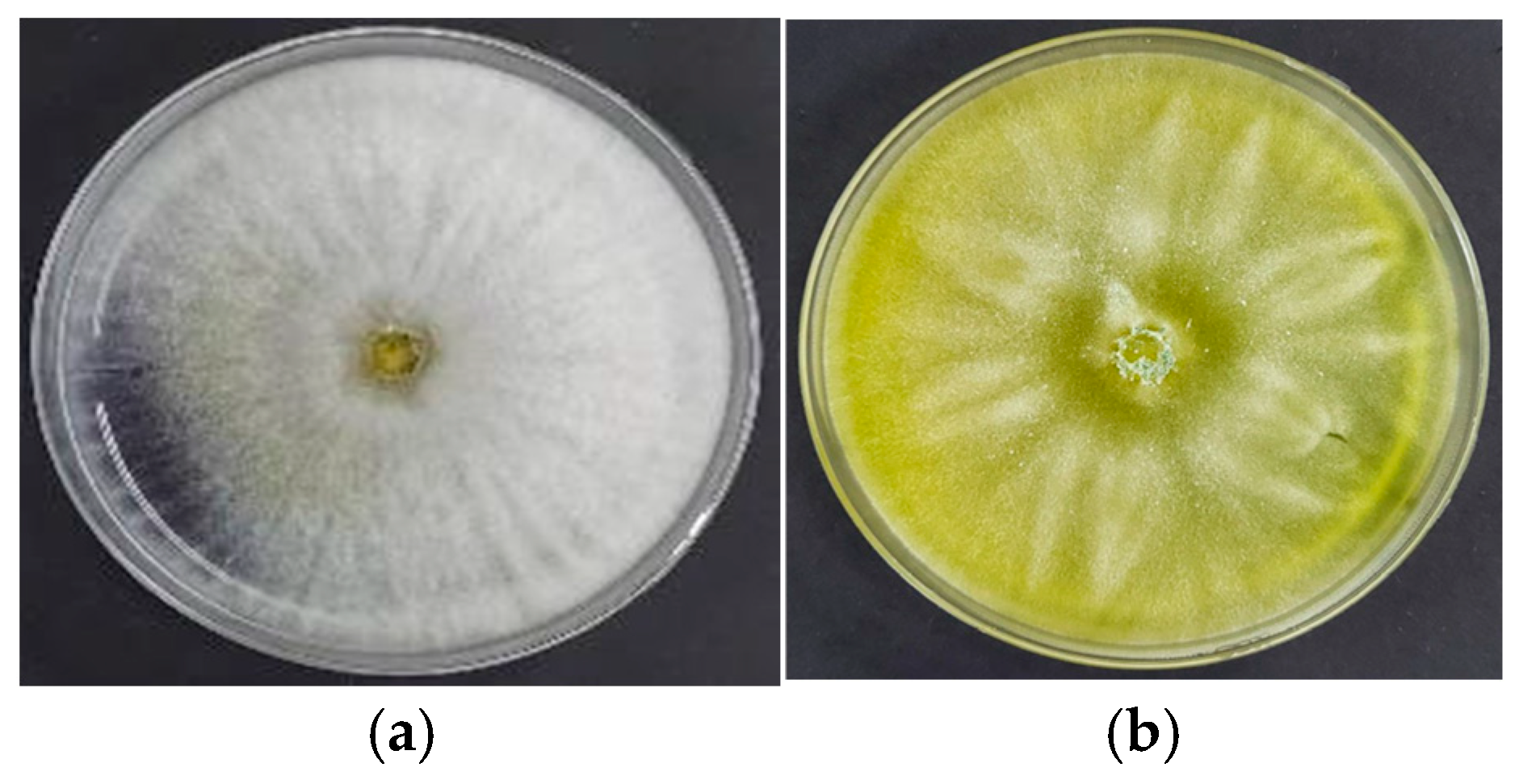
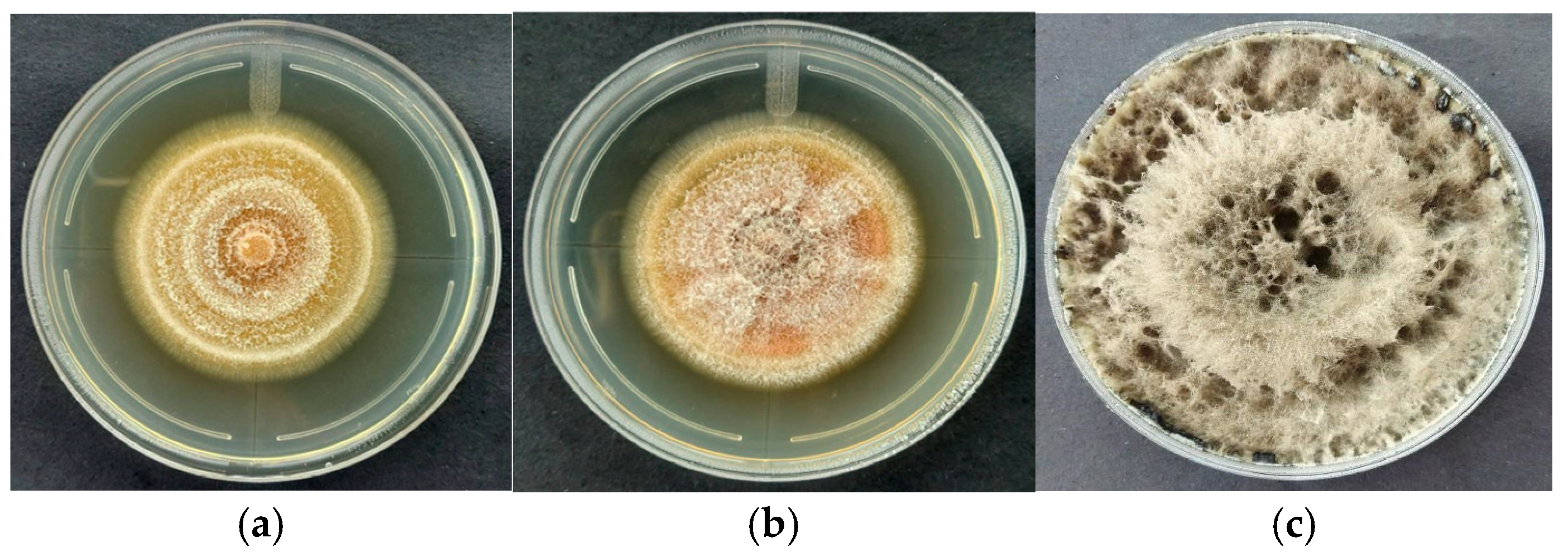
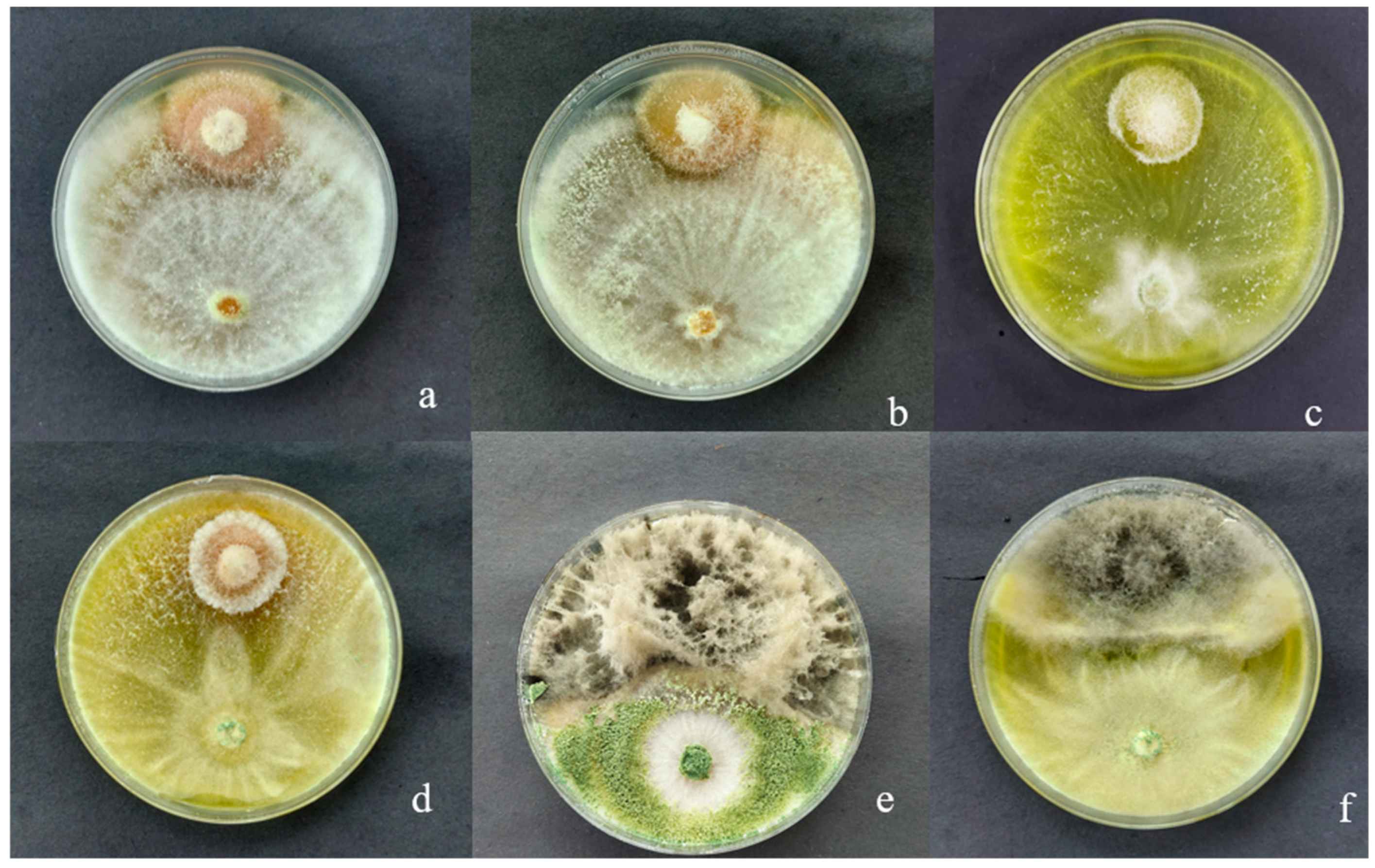
3.1. Macroscopic Characteristics of Trichoderma sp. CEP-01, CEP-02 and S. Parahyba Phytopathogens

Distinct macroscopic morphological features were observed among the fungal isolates at incubation temperatures of 24 ± 2 °C and 30 ± 2 °C. Trichoderma sp. CEP-01 completely colonized the PDA medium within 72 h at both temperatures, forming white, cottony colonies with abundant aerial mycelium and no concentric rings. In contrast, Trichoderma sp. CEP-02 also achieved full colonization at 72 h but developed light green to yellowish, powdery colonies with visible concentric rings. Notably, incubation at 30 ± 2 °C stimulated conidial production in Trichoderma sp. CEP-02 (Figure 1).
Similarly, Figure 2 illustrates the morphological characteristics and growth performance of the phytopathogens at different temperatures. The isolate FE07 exhibited average colony diameters of 5.3 cm and 5.7 cm after 168 h of incubation at 24 ± 2 °C and 30 ± 2 °C, respectively. The colonies were cottony white with concentric rings, white mycelia, wine-colored conidia, and a light wine-colored reverse. Fusarium FE08 produced slightly larger colonies (6.26 cm at 24 ± 2 °C and 5.8 cm at 30 ± 2 °C), with a similar cottony texture, orange conidia, and a reverse coloration ranging from orange to yellowish (Figure 2a,b). Botryodiplodia sp. displayed the fastest growth, reaching diameters of 8.4 cm and 8.5 cm within 72 h at 24 ± 2 °C and 30 ± 2 °C, respectively. The colonies were dark (black), with dense cottony mycelium, abundant dark pycnidia, and conidia that were hyaline when immature and dark when mature (Figure 2c). At 5 ± 2 °C, no fungal growth was observed, likely due to the adaptation of the species to stable tropical temperature conditions, typically around 24 °C.

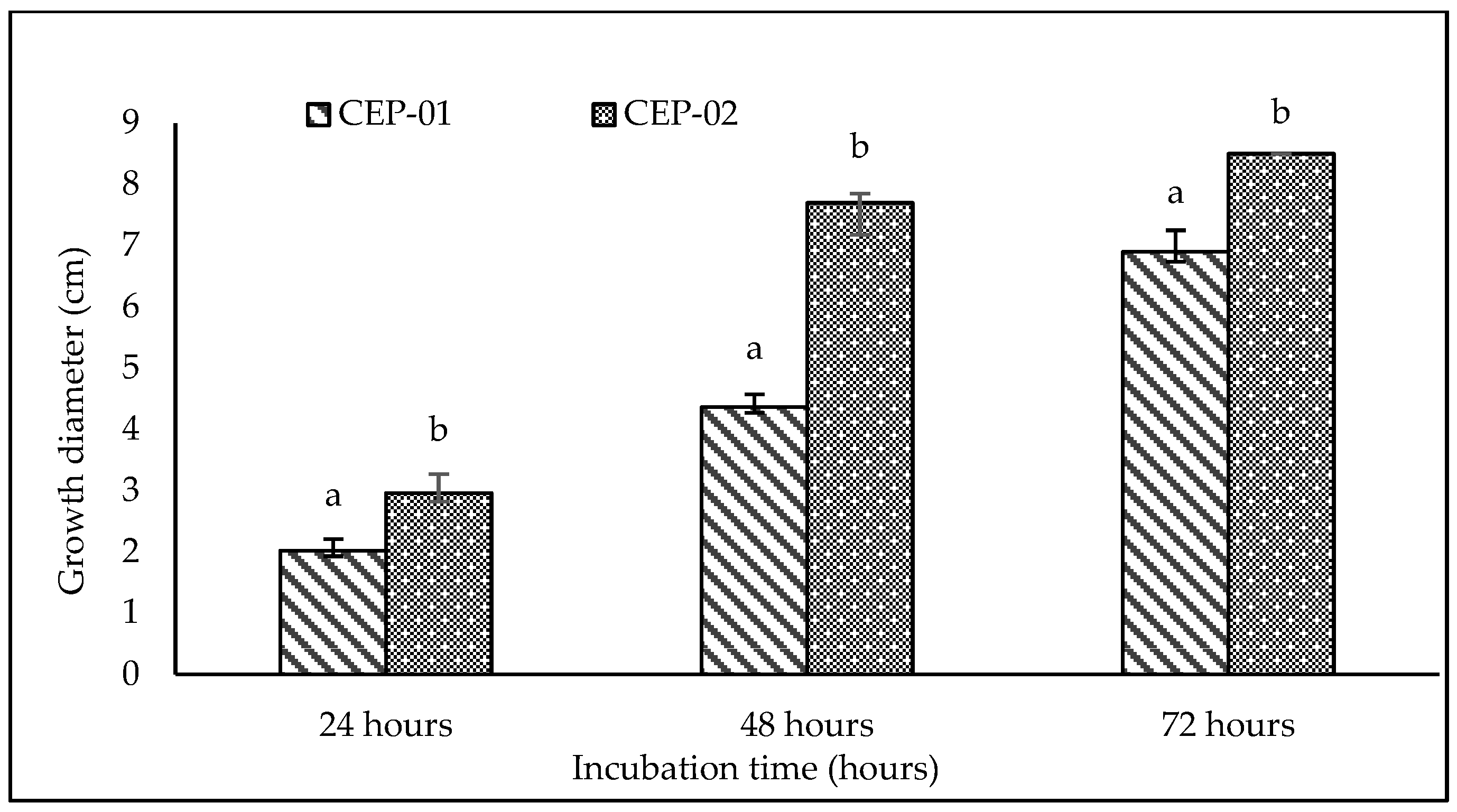
3.2. Effect of Incubation Temperature on Trichoderma sp. CEP-01 and Trichoderma sp. CEP-02 Growth Rates

Neither Trichoderma spp. isolates exhibited growth at 5 ± 2 °C. At 24 ± 2 °C, both Trichoderma sp. CEP-01 and CEP-02 achieved full plate colonization (8.5 cm) by 72 h. After 24 and 48 h, Trichoderma sp. CEP-01 reached diameters of 2.2 cm and 6.3 cm, while Trichoderma sp. CEP-02 showed faster growth with 2.7 cm and 8.2 cm, respectively (Figure 3). At 30 ± 2 °C, Trichoderma sp. CEP-01 displayed diameters of 2.0 cm, 4.4 cm, and 6.9 cm at 24, 48, and 72 h, while Trichoderma sp. CEP-02 fully colonized the medium by 72 h (Figure 4).

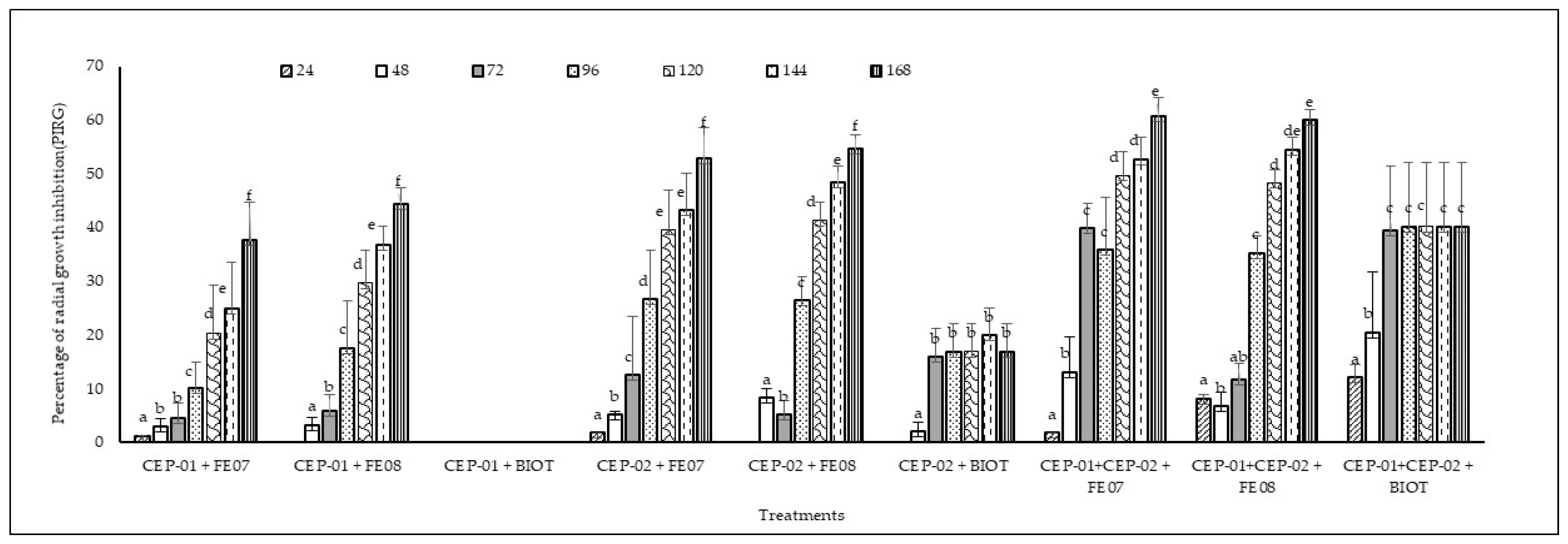
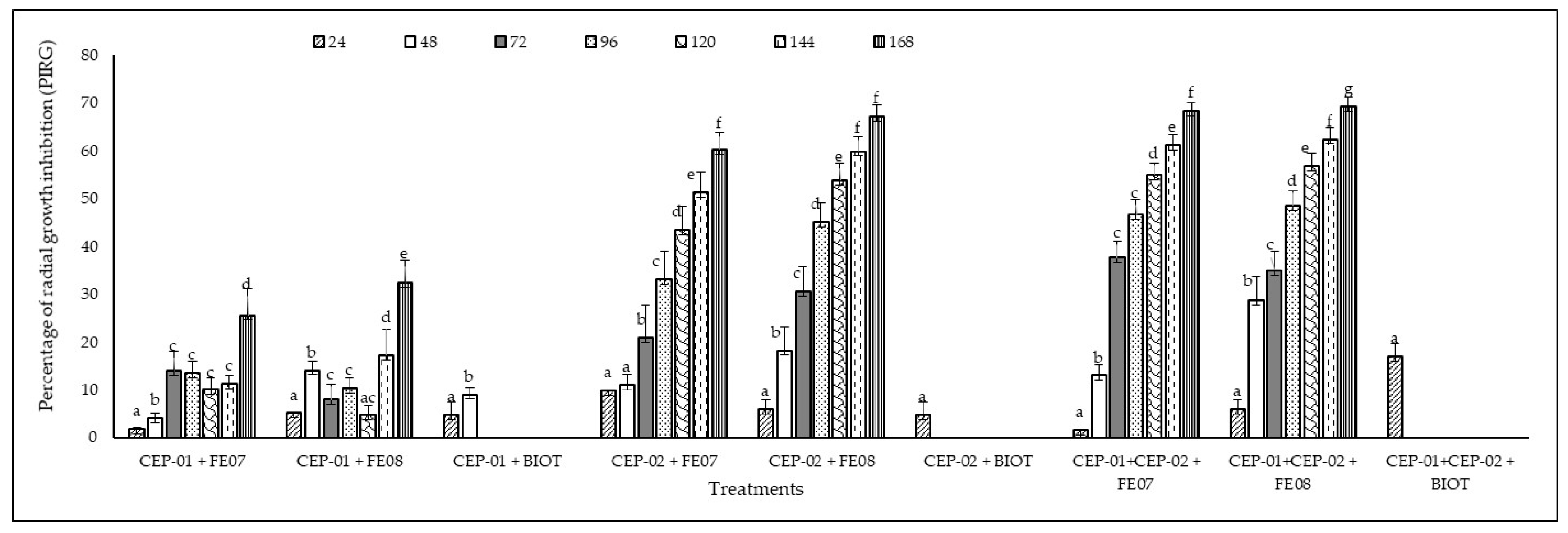
3.3. In Vitro Antagonistic Capacity of Native Trichoderma spp. CEP-01 and CEP-02 Isolates Against Phytopathogenic Fungi of S. parahyba

At 5 ± 2 °C, neither the antagonistic Trichoderma spp. isolates nor the phytopathogenic fungi exhibited any mycelial growth, indicating complete inhibition under low-temperature conditions. At 24 ± 2 °C, statistically significant differences were observed at all evaluation intervals (F = 26.2, p = 0.000; F = 4.95, p = 0.000; F = 28.8, p = 0.000; F = 16.6, p = 0.000; F = 32.5, p = 0.000; F = 44.7, p = 0.000; F = 66.7, p = 0.000) at 24, 48, 72, 96, 120, 144, and 168 h. Trichoderma sp. CEP-01 showed no inhibitory effect against BIOT., whereas Trichoderma sp. CEP-02 achieved up to 20% PIRG. Against FE07 and FE08, both isolates demonstrated moderate inhibition (RGIP) of 37.6–44.4% and 52.8–54.6%, respectively. When combined, Trichoderma sp. CE-01 and CEP-02 enhanced inhibition to 60% against Fusarium spp. and 40% against Botryodiplodia sp. (Figure 5). At 30 ± 2 °C, statistically significant differences persisted (F = 13.9 to F = 55.8; p = 0.000) across all time points. Elevated temperature reduced Trichoderma sp. CEP-01’s inhibitory capacity against Fusarium spp. (RGIP of 25.6–32.4%), whereas Trichoderma sp. CEP-02 exhibited enhanced inhibition (RGIP of 60.3–67.2%). Their combination further increased RGIP to 68.4–69.3% against Fusarium sp., but failed to inhibit Botryodiplodia sp. after 48 h (Figure 6). According to the scale applied by Bell et al. (1982) [34], Trichoderma CEP-01 and CEP-02 displayed grade 1 antagonism against Fusarium sp. at both 24 °C and 30 °C. Against Botryodiplodia sp., Trichoderma sp. CEP-01 and CEP-02 corresponded to grades 4 and 3, respectively (Figure 7).

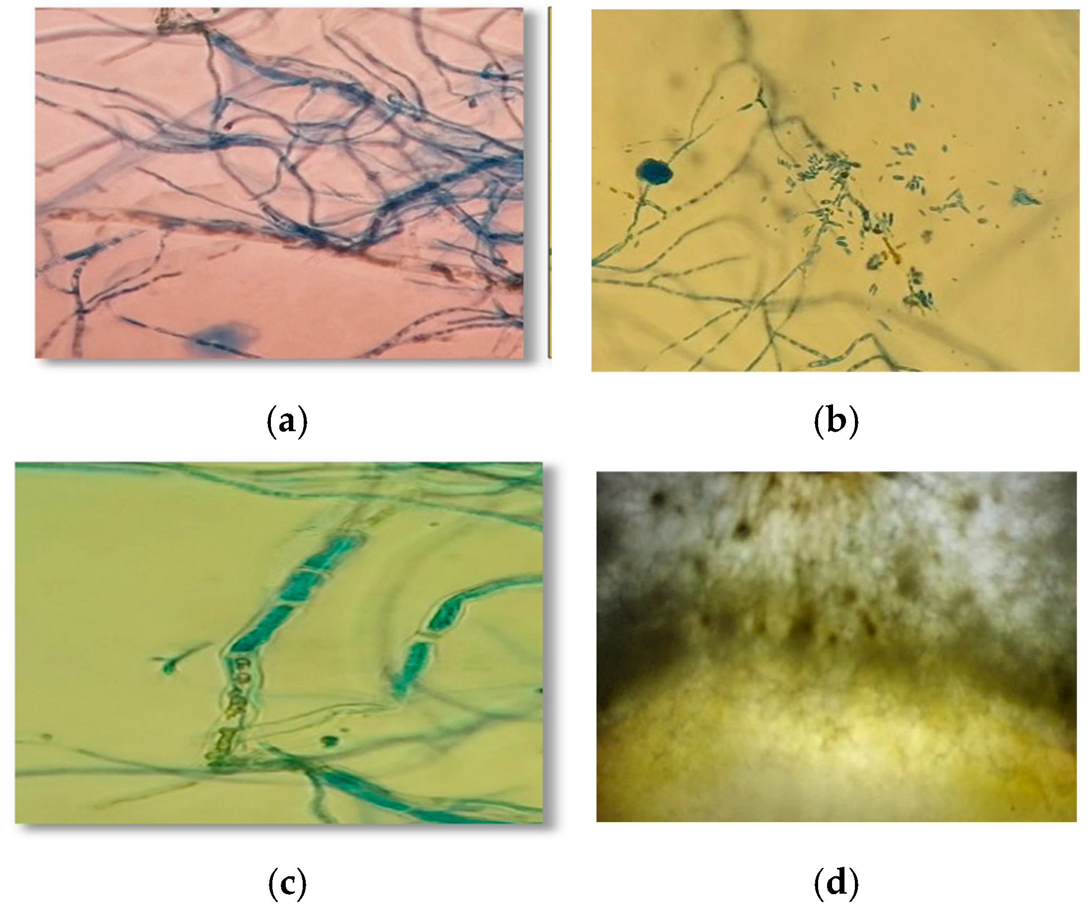
3.4. Mycoparasitism of Trichoderma spp. on S. parahyba Phytopathogens

When Trichoderma sp. CEP-01 and CEP-02 were confronted with Fusarium sp. FE07 and FE08 at incubation temperatures of 24 ± 2 °C and 30 ± 2 °C, distinct hyphal interactions were observed in the confrontation zone. These interactions, which manifested from the moment of contact, included vacuolization, hyphal coiling, sporulation, and spatial invasion, all hallmarks of mycoparasitic behavior (Figure 8).
Furthermore, both Trichoderma sp. isolates CEP-01 and CEP-02 exhibited clear mycoparasitic activity against FE07 and FE08, characterized by hyphal coiling and penetration, which constrained the mycelial growth and physiological activity of the S. parahyba phytopathogens. In contrast, no evidence of hyphal coiling or penetration by Trichoderma spp. was observed in BIOT, suggesting that the limited inhibitory capacity against this pathogen may be attributed to the absence of these parasitic mechanisms.

4. Discussion

Trichoderma spp., recognized for its biological control capacity, demonstrated efficacy in suppressing phytopathogenic fungi under laboratory conditions [35], a result that coincides with that reported by other authors who have demonstrated the mycelial-growth-suppressing activity of phytopathogens of agricultural and forestry importance [24,32,36,37,38].
The Trichoderma sp. CEP-01 and CEP-02 isolates exhibited distinct morphological differences at 24 ± 2 °C and 30 ± 2 °C. CEP-01 fully colonized PDA medium within 72 h at both temperatures, forming white, cottony colonies without concentric rings. In contrast, CEP-02 displayed a light green to yellowish coloration, a powdery texture, and concentric rings, with notably increased conidial production at 30 °C. These observations align with previous findings by [39,40], suggesting differential adaptation of these isolates to specific thermal conditions and potential variations in colonization and reproductive strategies [41,42]. These strategies enable Trichoderma spp. fungi, including T. viride, T. polysporum, and T. harzianum, to biologically control cacao plant pathogens through competition, antibiosis, and mycoparasitism, proving effective against diseases such as black pod rot, witches’ broom, and moniliasis, while also enhancing plant growth and resistance to pathogens [27,30,43].
Fusarium sp. FE07 and FE08 exhibited vigorous growth at 30 °C, achieving colony diameters of 5.7 cm and 5.8 cm, respectively. Morphologically, colonies appeared cottony white with concentric ring formations. However, differences in conidial coloration and the reverse side of the Petri dishes suggest potential adaptive responses to thermal variations. The production of wine-red and orange conidia may indicate a correlation between temperature and conidial pigmentation, as previously noted by [44,45].
Botryodiplodia sp. demonstrated rapid growth, reaching average colony diameters of 8.4 cm at 24 °C and 8.5 cm at 30 °C within 72 h on PDA medium. Colonies were characterized by dark pigmentation, dense cottony mycelium, abundant dark pycnidia, and hyaline conidia that darken as they mature—morphological features typical of this pathogen in woody species [46,47]. The adaptive capacity of Botryodiplodia sp. to thrive under varying temperature conditions enhances its role as a phytopathogen, posing a significant threat to agriculture and forestry. Several studies have shown that this fungus exhibits optimal growth at moderate temperatures, around 25 °C [48], while lower temperatures tend to inhibit both its development and the severity of the diseases it causes [49]. Moreover, its virulence and phytotoxic effects increase within specific temperature ranges, indicating a strong relationship between temperature and pathogenicity [50]. In the context of global climate change, rising temperatures may favor the incidence of diseases caused by this pathogen, increasing the risk to various crops [17]. The observed morphological and growth differences in Trichoderma sp. CEP-01 and CEP-02, Fusarium sp., and Botryodiplodia sp. under distinct temperature conditions have key implications for microbial ecology and the management of agricultural and forestry systems. The differential thermal adaptation of these microorganisms may influence their distribution, resource competition, and overall impact on plant health [16,51].
Understanding these responses is essential for developing integrated disease management strategies in forestry plantations, particularly in the context of climate variability and optimizing the use of biocontrol agents like Trichoderma spp. against pathogens such as Botryodiplodia sp. Previous research has demonstrated the implication of the inhibition of mycelial growth of Botryodiplodia sp., generated by fungi not recognized as biological control agents and biological controllers of the genus Trichoderma, which directly affects the incidence of this phytopathogen [33]. Temperature played a critical role in the interaction between Trichoderma spp. isolates and the phytopathogens, a phenomenon previously reported by [52]. This is particularly relevant to the development of management strategies for these types of diseases, which vary in terms of their parasitic capacity depending on the prevailing environmental conditions and the characteristics of the agroecosystem [16].
In our study, microscopic observations revealed characteristic hyphal coiling behavior in the interaction zone, suggesting an active mycoparasitic strategy by the antagonistic organism. This response has been widely documented, where hyphal coiling induces lysis of the host hyphae in species like Trichoderma atroviride; this behavior is associated with MAPK signaling pathways, which play a key role in regulating hyphal penetration and mycoparasitic effectiveness [36]. Additionally, cytoplasmic alterations were observed in the confrontation zone, including vacuolization and loss of cell wall integrity, which are consistent with parasitic activity. Sporulation was also detected in localized areas of contact, which may indicate a competitive response for space or resources.
Changes in environmental conditions, primarily rising global temperatures and imbalances in rainfall events, can lead to crop relocation and plant diseases caused by a high dependence on pathogen–crop relationships. Trichoderma sp CEP-01 did not exhibit inhibitory activity against Botryodiplodia sp., whereas Trichoderma sp CEP-02 demonstrated notable inhibitory effects, reaching a maximum of 20% RGIP. Both Trichoderma isolates inhibited the growth of FE07 and FE08, with RGIP values ranging from 40% to 53%. When Trichoderma sp. CEP-01 and Trichoderma sp. CEP-02 were co-inoculated, inhibition was enhanced, achieving RGIP values of 61% against FE07 and FE08, and approximately 40% against BIOT.
At 30 ± 2 °C, confrontation assays revealed a significant increase in inhibitory capacity. Trichoderma spp. achieved RGIP values ranging from 62% to 70% against FE07 and FE08 after 168 h of incubation, indicating increased efficacy at elevated temperatures. This is similar to that reported by Daryaei et al. [52] who demonstrated in in vitro studies that Trichoderma species achieve a greater inhibitory effect on the growth of phytopathogens at a temperature of 25 °C and pH 5.5, a response that may be more related to the strain level than to the species level. However, this temperature did not favor inhibition of Botryodiplodia sp., with a maximum RGIP of approximately 17% observed at 24 h when both Trichoderma spp. isolates were combined.

5. Conclusions

Trichoderma sp. CEP-01 and CEP-02 exhibited significant in vitro antagonistic activity against phytopathogenic fungi of S. parahyba, highlighting Trichoderma sp.’s potential as a biological control agent for protecting this forest species. Both CEP-01 and CEP-02 isolates significantly inhibited the growth of Fusarium sp. FE07 and Fusarium sp. FE08 and, to a lesser extent, Botryodiplodia sp., as reflected in the varying degrees of mycelial growth inhibition. These results underscore the critical role of temperature in determining the efficacy of Trichoderma spp. as a biocontrol agent. Overall, these findings emphasize the potential of Trichoderma spp. as a sustainable and effective alternative to conventional chemical disease control methods in S. parahyba plantations.

Author Contributions

Conceptualization, C.B.-P., B.I.-P., B.B.-P., and E.S.-A.; methodology, C.B.-P., B.I.-P., B.B.-P., and E.S.-A.; validation, C.B.-P., B.I.-P., B.B.-P., E.S.-A., P.D.-N., and R.A.V.-P.; formal analysis, C.B.-P., B.I.-P., and B.B.-P.; investigation, C.B.-P., B.I.-P., B.B.-P., and E.S.-A.; resources, C.B.-P., B.I.-P., B.B.-P., and E.S.-A.; writing—original draft preparation, C.B.-P., P.D.-N., and R.A.V.-P.; writing—review and editing, P.D.-N., R.A.V.-P., and C.B.-P.; visualization, P.D.-N., R.A.V.-P., and C.B.-P.; supervision C.B.-P. All authors have read and agreed to the published version of the manuscript.

Funding

This research was supported by the FOCICYT-UTEQ N° PFOC7-42-2020 project “Etiology of the decline and dieback disease in Schizolobium parahybum Vell. S.F. Blake pachaco plantations in the Ecuadorian Humid Tropics.”, and the Fondecyt Initiation Grant No. 11240313, funded by ANID, Chile.

Data Availability Statement

The original contributions presented in this study are included in the article. Further inquiries can be directed to the corresponding author.

Acknowledgments

The authors extend their gratitude to PLANTABAL S.A. for granting access to their Pachaco plantations, which made this study possible.

Conflicts of Interest

The authors declare no conflicts of interest.

References

  1. Ferreira, N.C.d.F.; Gatto, A.; Ramos, M.L.G. Co-Inoculation of Trichoderma harzianum and Bradyrhizobium species Augment the Growth of Schizolobium parahyba var. parahyba (Vell.) Blake Seedlings. Microorganisms 2025, 13, 630. [Google Scholar] [CrossRef]
  2. Belezaca-Pinargote, C.; Morales-Escobar, C.K.; Solano-Apuntes, E.; López-Tobar, R.; Díaz-Navarrete, P. “Decay and Descending Death” New disease of Schizolobium parahybum (Vell.) S.F. Blake (pachaco) in Ecuador. Int. J. Health Sci. 2022, 6, 14269–14281. [Google Scholar] [CrossRef]
  3. Camacho, M.E.; Tatis, H.A.; Ayala, C.C. Morphological characteristics and seed viability of Schizolobium parahyba (Vell.) S.F. Blake. Rev. U.D.C.A Actual. Divulg. Cient. 2020, 23, e1530. [Google Scholar] [CrossRef]
  4. Cely, M.V.T.; Siviero, M.A.; Emiliano, J.; Spago, F.R.; Freitas, V.F.; Barazetti, A.R.; Goya, E.T.; Lamberti, G.D.S.; Dos Santos, I.M.O.; De Oliveira, A.G.; et al. Inoculation of Schizolobium parahyba with mycorrhizal fungi and plant growth-promoting rhizobacteria increases wood yield under field conditions. Front. Plant Sci. 2016, 7, 1708. [Google Scholar] [CrossRef]
  5. Belezaca Pinargote, C.; Suárez Capello, C.; Vera Coello, D. Hongos fitopatógenos asociados a la enfermedad de muerte regresiva y pudrición del fuste de pachaco (Schizolobium parahybum) en el trópico húmedo ecuatoriano (Fungi pathogens associated with dieback and stem rot disease of pachaco [Schizolobium parahybum] in the Ecuadorian humid tropics). Boletín Micológico 2011, 26, 15–22. [Google Scholar]
  6. Shennan-Farpón, Y.; Mills, M.; Souza, A.; Homewood, K. The role of agroforestry in restoring brazil’s atlantic forest: Opportunities and challenges for smallholder farmers. People Nat. 2022, 4, 462–480. [Google Scholar] [CrossRef]
  7. Angelotti, F.; Hamada, E.; Bettiol, W.A. Comprehensive review of climate change and plant diseases in Brazil. Plants 2024, 13, 2447. [Google Scholar] [CrossRef]
  8. Spies, C.F.J.; Mostert, L.; Carlucci, A.; Moyo, P.; van Jaarsveld, W.J.; du Plessis, I.L.; van Dyk, M.; Halleen, F. Dieback and decline pathogens of olive trees in south Africa. Persoonia Mol. Phylogeny Evol. Fungi 2020, 45, 196–220. [Google Scholar] [CrossRef] [PubMed]
  9. Pirttilä, A.M.; Tabas, H.M.P.; Baruah, N.; Koskimäki, J.J. Biofertilizers and biocontrol agents for agriculture: How to identify and develop new potent microbial strains and traits. Microorganisms 2021, 9, 817. [Google Scholar] [CrossRef]
  10. Khazaal, F.A.K.; Ameen, M.K.M.; Ali, A.H. Pathogenic fungi a companied with sudden decline syndrome (wilting disease) of date palm tree (Phoenix dactylifera L.). Basrah J. Sci. 2019, 397, 376. [Google Scholar]
  11. Pimentel, M.F.; Arnão, E.; Warner, A.J.; Subedi, A.; Rocha, L.F.; Srour, A.; Bond, J.P.; Fakhoury, A.M. Trichoderma isolates inhibit Fusarium virguliforme growth, reduce root rot, and induce defense-related genes on soybean seedlings. Plant Dis. 2020, 104, 1949–1959. [Google Scholar] [CrossRef]
  12. Gachango, E.; Hanson, L.E.; Rojas, A.; Hao, J.J.; Kirk, W.W. Fusarium spp. causing dry rot of seed potato tubers in michigan and their sensitivity to fungicides. Plant Dis. 2012, 96, 1767–1774. [Google Scholar] [CrossRef]
  13. Correa-Delgado, R.; Brito-López, P.; Cardoza, R.E.; Jaizme Vega, M.C.; Laich, F.; Gutiérrez, S. Biocontrol potential of a native Trichoderma collection against Fusarium oxysporum f. sp. cubense subtropical race 4. Agriculture 2024, 14, 2016. [Google Scholar] [CrossRef]
  14. Lee, J.H.; Kim, N.K.; Shin, K.; Lee, J.K.; Lee, D.-H. Preliminary report of three entomopathogenic fungi as potential biocontrol agents against the oak wilt vector, Platypus koryoensis. Forests 2025, 16, 1009. [Google Scholar] [CrossRef]
  15. Langa-Lomba, N.; Martín-Ramos, P.; Casanova-Gascón, J.; Julián-Lagunas, C.; González-García, V. Potential of native Trichoderma strains as antagonists for the control of fungal wood pathologies in young grapevine plants. Agronomy 2022, 12, 336. [Google Scholar] [CrossRef]
  16. Vega, D.; Ibarra, S.; Varela Pardo, R.A.; Poggio, S.L. Agroecological management of crop diseases: A review. Agroecol. Sustain. Food Syst. 2023, 47, 919–949. [Google Scholar] [CrossRef]
  17. Broberg, M.; Dubey, M.; Iqbal, M.; Gudmundssson, M.; Ihrmark, K.; Schroers, H.J.; Funck Jensen, D.; Brandström Durling, M.; Karlsson, M. Comparative genomics highlights the importance of drug efflux transporters during evolution of mycoparasitism in clonostachys subgenus bionectria (fungi, ascomycota, hypocreales). Evol. Appl. 2021, 14, 476–497. [Google Scholar] [CrossRef]
  18. Varela-Pardo, R.A.; López-Lastra, C.C.; Manfrino, R.G.; Balcazar, D.; Mónaco, C.; Wright, E.R. Selection of fungal isolates from Buenos Aires, Argentina, as biological control agents of Botrytis cinerea and Sclerotinia sclerotiorum. Rev. La Fac. Cienc. Agrar. UNCuyo 2024, 56, 72–86. [Google Scholar] [CrossRef]
  19. Mukhopadhyay, R.; Kumar, D. Trichoderma: A beneficial antifungal agent and insights into its mechanism of biocontrol potential. Egypt. J. Biol. Pest Control 2020, 30, 133. [Google Scholar] [CrossRef]
  20. Prismantoro, D.; Chua, K.O.; Teo, K.W.E.; Chan, R.; Jefferson, T.A.; Suhaimi, N.S.M.; Mispan, M.S.; Miranti, M.; Doni, F. Whole genome sequence data of Trichoderma yunnanense strain tm10, a plant growth-promoting fungus and biocontrol agent. Data Brief 2025, 58, 111283. [Google Scholar] [CrossRef]
  21. Khan, R.A.A.; Najeeb, S.; Hussain, S.; Xie, B.; Li, Y. Bioactive secondary metabolites from Trichoderma spp. against phytopathogenic fungi. Microorganisms 2020, 8, 817. [Google Scholar] [CrossRef]
  22. Andrés, P.A.; Alejandra, P.M.; Benedicto, M.C.; Nahuel, R.I.; Clara, B.M. A Comparative study of different strains of Trichoderma under different conditions of temperature and pH for the control of Rhizoctonia solani. Agric. Sci. 2022, 13, 702–714. [Google Scholar] [CrossRef]
  23. Abbas, A.; Mubeen, M.; Zheng, H.; Sohail, M.A.; Shakeel, Q.; Solanki, M.K.; Iftikhar, Y.; Sharma, S.; Kashyap, B.K.; Hussain, S.; et al. Trichoderma spp. genes involved in the biocontrol activity against Rhizoctonia solani. Front. Microbiol. 2022, 13, 884469. [Google Scholar] [CrossRef] [PubMed]
  24. Kalimutu, P.K.; Komang Mahardika, I.B.; Anak Agung Sagung, P.R.A. Antagonism test of Trichoderma atroviride and Gliocladium sp. bali local isolates as a disease control of blendok disease (Botryodiplodia theobromae) in Grapefruit (Citrus grandis L. Osbeck). SEAS (Sustain. Environ. Agric. Sci.) 2020, 4, 102–110. [Google Scholar] [CrossRef]
  25. Mesa Vanegas, A.M.; Marín Pavas, D.A.; Calle Osorno, J. Metabolitos secundarios en Trichoderma spp. y sus aplicaciones biotecnológicas agrícolas. Actual. Biológicas 2020, 41, 1–13. [Google Scholar] [CrossRef]
  26. Rubayet, M.T.; Hossain, M.M. Bio-exploration of plant growth-promoting fungus Trichoderma as a potent candidate for plant disease management: An overview. Online J. Biol. Sci. 2025, 25, 22–52. [Google Scholar] [CrossRef]
  27. Rees, H.J.; Drakulic, J.; Cromey, M.G.; Bailey, A.M.; Foster, G.D. Endophytic Trichoderma spp. can protect strawberry and privet plants from infection by the fungus armillaria mellea. PLoS ONE 2022, 17, e0271622. [Google Scholar] [CrossRef]
  28. Kubicek, C.P.; Herrera-Estrella, A.; Seidl-Seiboth, V.; Martinez, D.A.; Druzhinina, I.S.; Thon, M.; Zeilinger, S.; Casas-Flores, S.; Horwitz, B.A.; Mukherjee, P.K.; et al. Comparative genome sequence analysis underscores mycoparasitism as the ancestral life style of Trichoderma. Genome Biol. 2011, 12, R40. [Google Scholar] [CrossRef]
  29. Pratap Singh, S.; Keswani, C.; Pratap Singh, S.; Sansinenea, E.; Xuan Hoat, T. Trichoderma spp. mediated induction of systemic defense response in brinjal against Sclerotinia sclerotiorum. Curr. Res. Microb. Sci. 2021, 2, 100051. [Google Scholar] [CrossRef]
  30. Bai, B.; Liu, C.; Zhang, C.; He, X.; Wang, H.; Peng, W.; Zheng, C. Trichoderma species from plant and soil: An excellent resource for biosynthesis of terpenoids with versatile bioactivities. J. Adv. Res. 2023, 49, 81–102. [Google Scholar] [CrossRef]
  31. Valenzuela, R.V.; Gálvez, G.T.; Villa, R.; Eber, D.; Parra, C.; Fannie, I.; Santoyo, G.; Santos-Villalobos, S. Lipopeptides produced by biological control agents of the genus Bacillus: A review of analytical tools used for their study. Rev. Mex. Cienc. Agrícolas 2020, 11, 419–432. [Google Scholar] [CrossRef]
  32. Joo, J.H.; Hussein, K.A. Biological control and plant growth promotion properties of volatile organic compound-producing antagonistic Trichoderma spp. Front. Plant Sci. 2022, 13, 897668. [Google Scholar] [CrossRef]
  33. Sahlan, F.K.; Rusicha, F.A.; Darmawan, A.A. Isolation and antagonistic test of fungi associated with pummelo citrus against Botryodiplodia theobromae in vitro. IOP Conf. Ser. Earth Environ. Sci. 2023, 1230, 012110. [Google Scholar] [CrossRef]
  34. Rondoy, M.G.; Severino, N.V. Antagonistic capacity of Trichoderma harzianum compared to rhizotecnia, Nakataea sigmoidea, Sclerotium rolfsii and its effect in native strains of Trichoderma isolated form rice crops. Sci. Agropecu. 2019, 10, 199–206. [Google Scholar] [CrossRef]
  35. Asghar, W.; Craven, K.; Kataoka, R.; Mahmood, A.; Asghar, N.; Raza, T.; Iftikhar, F. The application of Trichoderma spp., an old but new useful fungus, in sustainable soil health intensification: A comprehensive strategy for addressing challenges. Plant Stress 2024, 12, 100455. [Google Scholar] [CrossRef]
  36. Viterbo, A.; Harel, M.; Horwitz, B.A.; Chet, I.; Mukherjee, P.K. Trichoderma mitogen-activated protein kinase signaling is involved in induction of plant systemic resistance. Appl. Environ. Microbiol. 2005, 71, 6241–6246. [Google Scholar] [CrossRef]
  37. Rubio-Sosa, A.; Martínez-Bolaños, M.; Gómez-Leyva, J.F.; Lozano-Trejo, S.; Castañeda-Hidalgo, E.; Diaz-Zorrilla, G.O. Inhibition of Colletotrichum spp. causing Anthracnose in Coffee (Coffea Arabica) using native isolates of Trichoderma sp. Mex. J. Phytopathol. 2025, 43, 58. [Google Scholar] [CrossRef]
  38. Khuong, N.Q.; Nhien, D.B.; Thu, L.T.M.; Trong, N.D.; Hiep, P.C.; Thuan, V.M.; Quang, L.T.; Thuc, L.V.; Xuan, D.T. Using Trichoderma asperellum to Antagonize Lasiodiplodia theobromae Causing Stem-End Rot Disease on Pomelo (Citrus maxima). J. Fungi. 2023, 9, 981. [Google Scholar] [CrossRef] [PubMed]
  39. Domingues, M.V.; de Moura, K.E.; Salomão, D.; Elias, L.M.; Patricio, F.R.A. Efeito da temperatura sobre o crescimento micelial de Trichoderma, Sclerotinia Minor e S. Sclerotiorum e Sobre o Micoparasitismo. Summa Phytopathol. 2016, 42, 222–227. [Google Scholar] [CrossRef]
  40. Rodríguez-García, D.; Vargas-Rojas, J. Efecto de la inoculación con Trichoderma sobre el crecimiento vegetativo del tomate (Solanum lycopersicum). Agron. Costarric. 2022, 46, 47. [Google Scholar] [CrossRef]
  41. Wesener, F.; Rillig, M.C.; Tietjen, B. Heat stress can change the competitive outcome between fungi: Insights from a modelling approach. Oikos 2023, 2023, e09377. [Google Scholar] [CrossRef]
  42. Alwadai, A.S.; Perveen, K.; Alwahaibi, M. The isolation and characterization of antagonist Trichoderma spp. from the soil of abha, Saudi Arabia. Molecules 2022, 27, 2525. [Google Scholar] [CrossRef]
  43. Del Carmen López-Ferrer, U.; Brito-Vega, H.; López-Morales, D.; Salaya-Domínguez, J.M.; Gómez-Méndez, E. Papel de Trichoderma en los sistemas agroforestales-cacaotal como un agente antagónico. Trop. Subtrop. Agroecosystems 2017, 20, 91–100. [Google Scholar] [CrossRef]
  44. Gerardo Leyva-Mir, S.; Errol Vega-Portillo, H.; Eduardo Villaseñor-Mir, H.; Tlapal-Bolaños, B.; Vargas-Hernández, M.; Camacho-Tapia, M.; Manuel Tovar-Pedraza, J. Characterization of Fusarium species causing root rot of wheat in the bajio, Mexico. Anim. Sci. Agro-Cienc. 2017, 33, 142–151. [Google Scholar]
  45. Khuong, N.Q.; Thuy Trang, C.T.; Xuan, D.T.; Quang, L.T.; Huu, T.N.; Thanh Xuan, L.N.; Sakagami, J.I.; Thuc, L.V. Evaluation of the antagonistic potential of Trichoderma spp. against Fusarium oxysporum F.28.1A. J. Plant Prot. Res. 2023, 63, 13–26. [Google Scholar] [CrossRef]
  46. Ahimera, N.; Gisler, S.; Morgan, D.P.; Michailides, T.J. Effects of Single-Drop impactions and natural and simulated rains on the dispersal of Botryosphaeria dothidea conidia. Phytopathology 2004, 94, 1189–1197. [Google Scholar] [CrossRef]
  47. Jami, F.; Wingfield, M.J.; Gryzenhout, M.; Slippers, B. Diversity of tree-infecting Botryosphaeriales on native and non-native trees in South Africa and Namibia, Australasian. Plant Pathol. 2017, 46, 529–545. [Google Scholar]
  48. Firmansyah, M.A.; Jayanegara, A.; Ria, M.; Solaya, G.; Ikhwan, D.; Syifaudin, S. The effect of essential oils of Citronella and Patchouli oil on growth of Botryodiplodia sp. in vitro. J. Trop. Silvic. 2023, 14, 39–46. [Google Scholar] [CrossRef]
  49. Pawar, V.P. Impact of ecological factors on development of Botryodiplodia rot of guava fruit. Curr. Bot. 2012, 3, 16. [Google Scholar]
  50. Nazar Pour, F.; Ferreira, V.; Félix, C.; Serôdio, J.; Alves, A.; Duarte, A.S.; Esteves, A.C. Effect of temperature on the phytotoxicity and cytotoxicity of Botryosphaeriaceae fungi. Fungal Biol. 2020, 124, 571–578. [Google Scholar] [CrossRef] [PubMed]
  51. Ruiz-Valencia, J.A.; Vázquez-Sánchez, M.; Burgos-Hernández, M.; Gutiérrez, J.; Terrazas, T. Wood anatomy of Forestiera (oleaceae) species in Mexico. Acta Bot. Mex. 2021, 128, 1–17. [Google Scholar] [CrossRef]
  52. Daryaei, A.; Jones, E.E.; Ghazalibiglar, H.; Glare, T.R.; Falloon, R.E. Effects of temperature, light and incubation period on production, germination and bioactivity of Trichoderma atroviride. J. Appl. Microbiol. 2016, 120, 999–1009. [Google Scholar] [CrossRef] [PubMed]
Figure 1. Colony morphology after 72 h of incubation: (a) Trichoderma sp. CEP-01, and (b) Trichoderma sp. CEP-02 on PDA.
Ijpb 16 00085 g001
Figure 2. Colony morphology after 168 h of incubation: (a) Fusarium sp. FE07 and (b) Fusarium sp. FE08 on PDA. (c) Colony morphology of Botryodiplodia sp. after 72 h of incubation on PDA.
Ijpb 16 00085 g002
Figure 3. Radial growth (cm) of Trichoderma sp. CEP-T01 and CEP-T02 isolates after 72 h at 24 ± 2 °C. Data represent means ± SD and SE (n = 5). Different letters indicate significant differences.
Ijpb 16 00085 g003
Figure 4. Radial growth (cm) of Trichoderma sp. CEP-01 and CEP-02 isolates after 72 h at 30 ± 2 °C. Data represent means ± SD and SE (n = 5). Different letters indicate significant differences.
Ijpb 16 00085 g004
Figure 5. Percentage of radial growth inhibition (RIGP) of S. parahyba phytopathogenic fungi confronted with two Trichoderma sp. isolates (CEP-01 and CEP-02) over 24, 48, 72, 96, 120, 144, and 168 h of incubation at 24 ± 2 °C. Data represent mean RGIP values calculated from five replicate Petri dishes containing PDA medium, along with their respective standard deviations. Different letters indicate significant differences.
Ijpb 16 00085 g005
Figure 6. Percentage of radial growth inhibition (RGIP) of S. parahyba phytopathogenic fungi confronted with two Trichoderma sp. isolates (CEP-01 and CEP-02) over 24, 48, 72, 96, 120, 144, and 168 h of incubation at 30 ± 2 °C. Data represent mean RGIP values from five replicate Petri dishes containing PDA medium, with corresponding standard deviations. Different letters indicate significant differences.
Ijpb 16 00085 g006
Figure 7. In vitro antagonistic activity of Trichoderma sp. CEP-01 and CEP-02 against S. parahyba phytopathogens: (a) Trichoderma sp. CEP-01 vs. Fusarium sp. FE07; (b) Trichoderma sp. CEP-01 vs. Fusarium sp. FE08; (c) Trichoderma sp. CEP-02 vs. Fusarium sp. FE07; (d) Trichoderma sp. CEP-02 vs. Fusarium sp. FE08; (e) Trichoderma spp. CEP-01 vs. Botryodiplodia sp.; and (f) Trichoderma sp. CEP-02 vs. Botryodiplodia sp.
Ijpb 16 00085 g007
Figure 8. Mycoparasitic interactions observed between Trichoderma sp. isolates CEP-01 and CEP-02 and Fusarium sp. FE07 and Fusarium sp. FE08. (a) Hyphal coiling as an antagonistic mechanism; (b) spore formation; (c) vacuolization, and (d) confrontation zone.
Ijpb 16 00085 g008
Table 1. Description of the experiments used to evaluate the effect of temperature on the growth of two species within the genus Trichoderma.
TreatmentsDescription
1Trichoderma sp. CEP-01 5 ± 2 °C
2Trichoderma sp. CEP-01 24 ± 2 °C
3Trichoderma sp. CEP-01 30 ± 2 °C
4Trichoderma sp. CEP-02 5 ± 2 °C
5Trichoderma sp. CEP-02 24 ± 2 °C
6Trichoderma sp. CEP-02 30 ± 2 °C
Table 2. Fungal antagonism scale for biocontrol evaluation.
GradoAntagonistic Capacity
1Trichoderma fully colonizes the medium and overgrows the phytopathogen.
2Trichoderma colonizes two-thirds of the medium and restricts pathogen growth.
3Trichoderma and the phytopathogen each colonize half of the medium, with no dominance.
4The phytopathogen colonizes two-thirds of the medium and restricts Trichoderma growth.
5The phytopathogen completely dominates and inhibits Trichoderma growth.
Table 3. Description of treatments for in vitro assessment of antagonistic capacity of CEP-01 and CEP-02 against S. parahyba phytopathogens.
TreatmentsInteractions
1CEP-01 v/s. FE07
2CEP-01 v/s FE08
3CEP-01 v/s BIOT
4CEP-02 v/s FE07
5CEP-02 v/s FE08
6CEP-02 v/s BIOT
7CEP-01 and CEP-02 v/s FE07
8CEP-01 and CEP-02 v/s FE08
9CEP-01 and CEP-02 v/s BIOT
10BIOT FE07 control
11FE08 control
12BIOT control
Disclaimer/Publisher’s Note: The statements, opinions and data contained in all publications are solely those of the individual author(s) and contributor(s) and not of MDPI and/or the editor(s). MDPI and/or the editor(s) disclaim responsibility for any injury to people or property resulting from any ideas, methods, instructions or products referred to in the content.

