Article

Somatic Embryogenesis in Native Peruvian Fine-Flavor Cocoa Genotypes

by
Karol Rubio
1,2,*,
Santos Leiva
1,2,
Manuel Oliva
1,2,
Jorge R. Diaz-Valderrama
1,2 and
Juan Carlos Guerrero-Abad
3,*
1
Instituto de Investigación para el Desarrollo Sustentable de Ceja de Selva (INDES-CES), Universidad Nacional Toribio Rodríguez de Mendoza de Amazonas, Chachapoyas 01001, Peru
2
Facultad de Ingeniería y Ciencias Agrarias, Universidad Nacional Toribio Rodríguez de Mendoza de Amazonas, Chachapoyas 01001, Peru
3
Instituto de Investigación, Innovación y Desarrollo para el Sector Agrario y Agroindustrial (IIDAA), Facultad de Ingeniería y Ciencias Agrarias, Universidad Nacional Toribio Rodríguez de Mendoza de Amazonas, Chachapoyas 01001, Peru
*
Authors to whom correspondence should be addressed.
Int. J. Plant Biol. 2025, 16(3), 84; https://doi.org/10.3390/ijpb16030084 (registering DOI)
Submission received: 5 June 2025 / Revised: 25 July 2025 / Accepted: 28 July 2025 / Published: 1 August 2025
(This article belongs to the Section Plant Reproduction)

Abstract

Cacao genotypes propagation through plant tissue culture represents a strategic approach for establishing a core collection of elite plants to be used as a donor material source, necessary for increasing new planting areas of cacao. This study aimed to evaluate somatic embryo regeneration in ten native fine-aroma cacao genotypes (INDES-06, INDES-11, INDES-14, INDES-32, INDES-52, INDES-53, INDES-63, INDES-64, INDES-66, INDES-70) from the INDES-CES germplasm collection, under in vitro conditions using culture medium supplemented with different concentrations of Thidiazuron (0, 10, and 20 nM). Our results showed an average of 20 and 100% of callogenesis in all genotypes evaluated, but the callus development did not appear after early stages of its induction; however, primary somatic embryos were observed after 42 days after TDZ treatment in the INDES-52, INDES-53, INDES-64, INDES-66, INDES-70 genotypes. The INDES-52 genotype was more responsive to under 20 nM of TDZ, generating an average of 17 embryos per explant. This study contributes to the adaptation and establishment of a protocol for somatic embryo regeneration of fine-flavor cacao genotypes.

1. Introduction

Cacao domestication started about 5450–5300 years into the Mayo-Chinchipe culture, located in the Upper Amazon region of what is now Ecuador and Peru [1]. Currently, the Peruvian cacao is the second most cultivated perennial crop with the largest agricultural area of 183,043 hectares [2]. However, sustaining and strengthening the cocoa value chain also requires the availability of selected cacao materials with particular characteristics such as productivity and resilience to biotic and abiotic stress, especially genotypes with fine-flavor notes, which are important in the differentiated chocolate industry [3]. In this context, research initiatives across the world are prioritizing the development of cacao cultivars or clones for production, as well as the conservation of important genetic resources to be used in future breeding efforts.
Conventional propagation methods in cacao are based on sexual reproduction, which leads to segregation and, consequently, the loss of desired characteristics with heterogeneous production in plant [4]. Asexual propagation through methods such as grafting or staking is one way to maintain important traits in breeding materials. However, these practices are labor-intensive, costly, and usually have a reduced propagation rate [5]. Because of this, in vitro cacao propagation is becoming a highly valued technique due to its relatively low cost and technical simplicity, which includes somatic embryogenesis or organogenesis [6].
The induction of somatic embryos in cacao from floral explants, such as petals and staminodes, has gained significant attention in recent years. However, the success of this process is highly dependent on the genotype studied and the culture media composition [5,6,7,8,9,10,11]. The culture medium that has shown the greatest success in cacao plant regeneration through somatic embryogenesis is the DKW medium, developed by Driver & Kuniyuki [12] and later modified by Tulecke & McGranahan [13]. This medium is supplemented with different plant growth regulators such as Thidiazuron (TDZ) [5], 2,4- diclorofenoxiacetic acid (2,4-D) [9], and kinetin (KIN) [14]. Nevertheless, the success rates of somatic embryo formation remain low (25%) and are limited to a small number of clones [15]. These outcomes depend on the complex interaction of multiple factors that must be addressed to develop effective regeneration protocols and procedures [9,16].
Based on this, our study aimed to evaluate the embryogenesis somatic response under different concentrations of Thidiazuron (TDZ) in floral staminoids of ten fine-flavor cocoa genotypes from the Amazonas region of Peru. The results show that fine-flavor cocoa genotypes are responsive to TDZ treatment during the somatic embryogenesis and its response is genotype dependent.

2. Materials and Methods

2.1. Plant Material and Explant Preparation

In this study, we included ten fine-flavor cocoa genotypes from the “Instituto de Investigación para el Desarrollo Sustentable de Ceja de Selva” “(INDES-CES)” cacao collection, located in Bagua Grande, Amazonas, Peru (Table 1). As reported by Bustamante et al. [17], these accessions were previously classified as “Peru Unique” based on SNP marker analysis, highlighting their distinct genetic identity within the Amazonas region.
Immature flower buds were collected during the mornings, between 7 and 10 a.m., to prevent bud bursts. The collected buds were immediately immersed in 50 mL conical tubes containing a cold solution of previously sterilized DKW basal salts [12]. Samples were transported at 4 °C to the Plant Physiology and Biotechnology Laboratory from the Toribio Rodríguez de Mendoza National University.
Prior dissection and explant establishment, flower buds were surface sterilized by immersion in a 1% (w/v) calcium hypochlorite solution for 20 min under gentle agitation. Following sterilization, the buds were rinsed three times with sterile distilled water. Subsequently, they were transferred to a sterile Petri dish inside a horizontal laminar flow cabinet, where the staminodes were excised and placed on primary callus growth medium (PCG) [18].

2.2. Culture Medium and Growth Conditions

The primary callus growth (PCG) medium was based on DKW basal medium supplemented with glutamine (250 mg L−1), myo-inositol (100 mg L−1), 2,4-dichlorophenoxyacetic acid (2,4-D) (2 mg L−1), and glucose (20 g L−1). Three concentrations of Thidiazuron (TDZ): 0 nM, 10 nM, and 20 nM, were evaluated by incorporating them into the PCG. TDZ application was only applied during PCG phase. The medium pH was adjusted to 5.8 before adding Phytagel (2.0 g L−1). Staminodes were inoculated on Petri dishes with 30 mL of PCG, followed by incubation in the dark at 25–28 °C for 14 days.
After 14 days of incubation, the staminodes were transferred to a secondary callus growth (SCG) medium and maintained under the same incubation conditions for an additional 14 days. The secondary callus growth (SCG) medium was based on McCown’s basal salts (Sigma M-6774), supplemented with vitamin B5 solution (1 mL L−1), glucose (20 g L−1), 2,4-D (2 mg L−1), 6-benzylaminopurine (6-BA) (0.05 mg L−1), and Phytagel (2.2 g L−1). The pH of the medium was adjusted to 5.7 prior to autoclaving [18].
At the end of the incubation period in SCG medium, the calli were transferred to Petri dishes containing 30 mL of embryo development (ED) medium to induce somatic embryo formation. The ED medium was based on DKW basal salts supplemented with sucrose (30 g L−1), glucose (1 g L−1), and Phytagel (2 g L−1). The cultures were maintained under the same conditions described above and subcultured every 14 days for a total of five times, until a culture period of 98 days was reached.

2.3. Data Analyses

The effect of the three TDZ concentrations on callus induction and somatic embryo formation was assessed using a completely randomized design. The experiment included 30 treatments (3 TDZ levels × 10 accessions). Each treatment consisted of three Petri dishes, with 25 staminodes per dish.
Data on the percentage of callogenic staminodes for each treatment were recorded during the incubation periods on both PCG and SCG media. Callogenesis frequencies were calculated as the percentage of staminodes exhibiting callus formation relative to the total number of staminodes per treatment (75 staminodes). To determine the effect of TDZ on the callogenic response across cacao genotypes, a one-way analysis of variance (ANOVA) was conducted. When significant differences were detected, mean separation was performed using the Scott–Knott clustering test at a significance level of p < 0.05. All statistical analyses were carried out using Infostat v. 2020 software [19].
In the ED culture phase, data on the percentage of embryogenic callus were recorded. Embryogenic callus frequency was calculated as the percentage of calli showing somatic embryo formation relative to the total number of callus-forming staminodes per treatment. In addition, the total number of embryos formed per treatment and per explant was determined. The average number of embryos per explant was calculated by dividing the total number of embryos by the number of embryogenic explants. No statistical analysis was conducted for this stage due to the low frequency of embryogenic responses.

3. Results

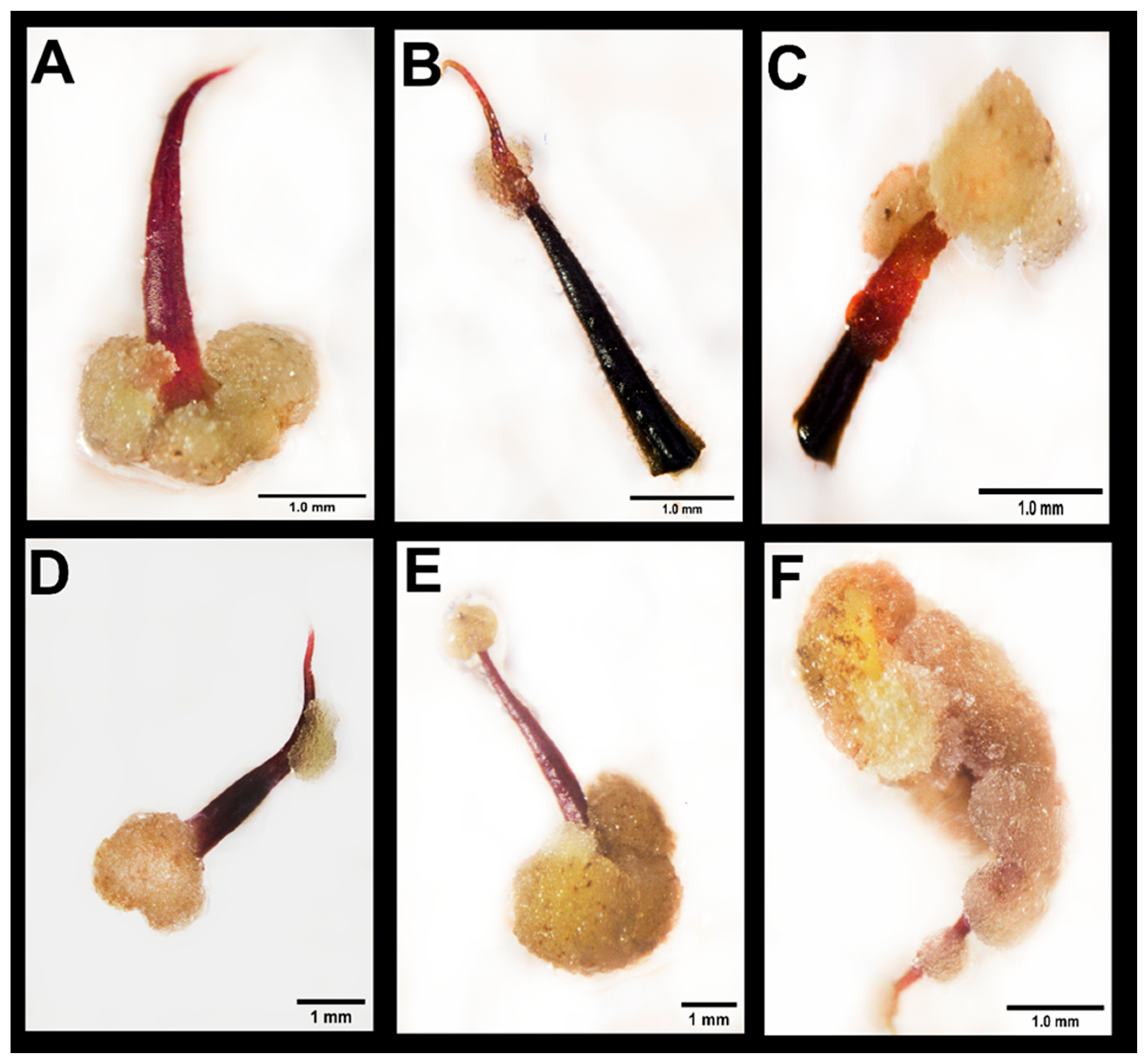
3.1. Effect of Thidiazuron on Callogenesis in Staminodes

After 14 days of culture on the PCG, the explants exhibited varied responses to the applied treatment. Some initiated callus formation, while others became turgid and subsequently gave rise to callus development during the following culture phase (SCG). Different types of calli were observed ranging in color from crystalline to creamy, and textures ranging from fine and coarse granular. Different positions of callus appearance were observed (Figure 1): at the basal (Figure 1A), medial (Figure 1B), and distal parts of staminodes (Figure 1C,E), or covering the whole explant (Figure 1F). Similarly, during the SCG culture phase, various types of calli were observed, exhibiting different coloration (ranging from crystalline to dark) and texture (granular with differing firmness).
The position of callus formation was not exclusive to any particular genotype or treatment. However, certain patterns suggest a potential association between the plant genotype and the specific region of the explant where callus development occurred. In general, callus induction was most commonly observed in the basal zone of the staminode, particularly at the site of excision. Less frequently, calli emerged in the medial region. Notably, in INDES-64 and INDES-66 genotypes, callus formation was more widespread, often occurring along the entire length of the staminode and even covering it completely (Figure 1F).
After 28 days of cultivation on the SCG medium, all genotypes were capable of developing calli, with callus formation rates ranging from 20% to 94.67%. Overall, the lowest average response percentages were observed in treatments without TDZ treatment. However, the INDES-63 and INDES-64 genotypes exhibited reduced callus formation when treated with 10 nM and 20 nM TDZ, respectively (Figure 2).

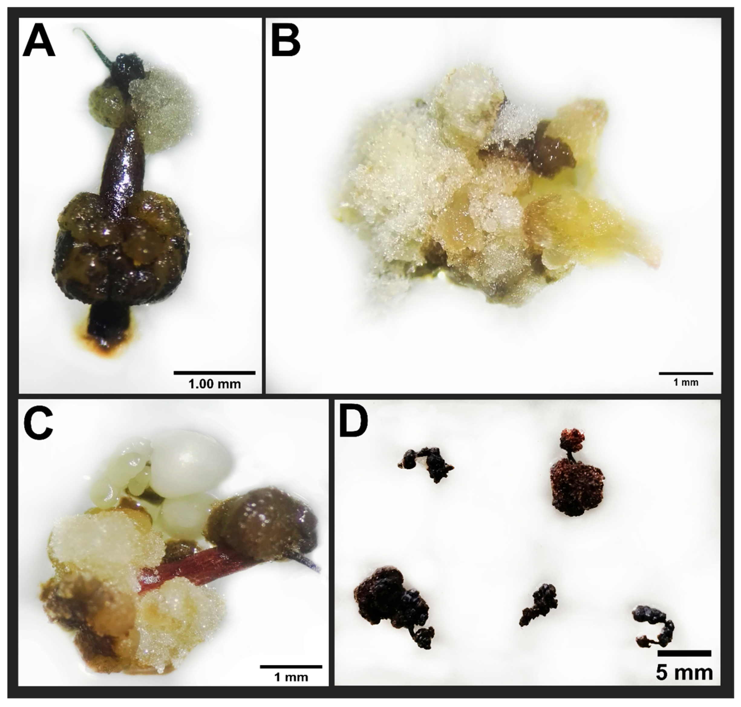
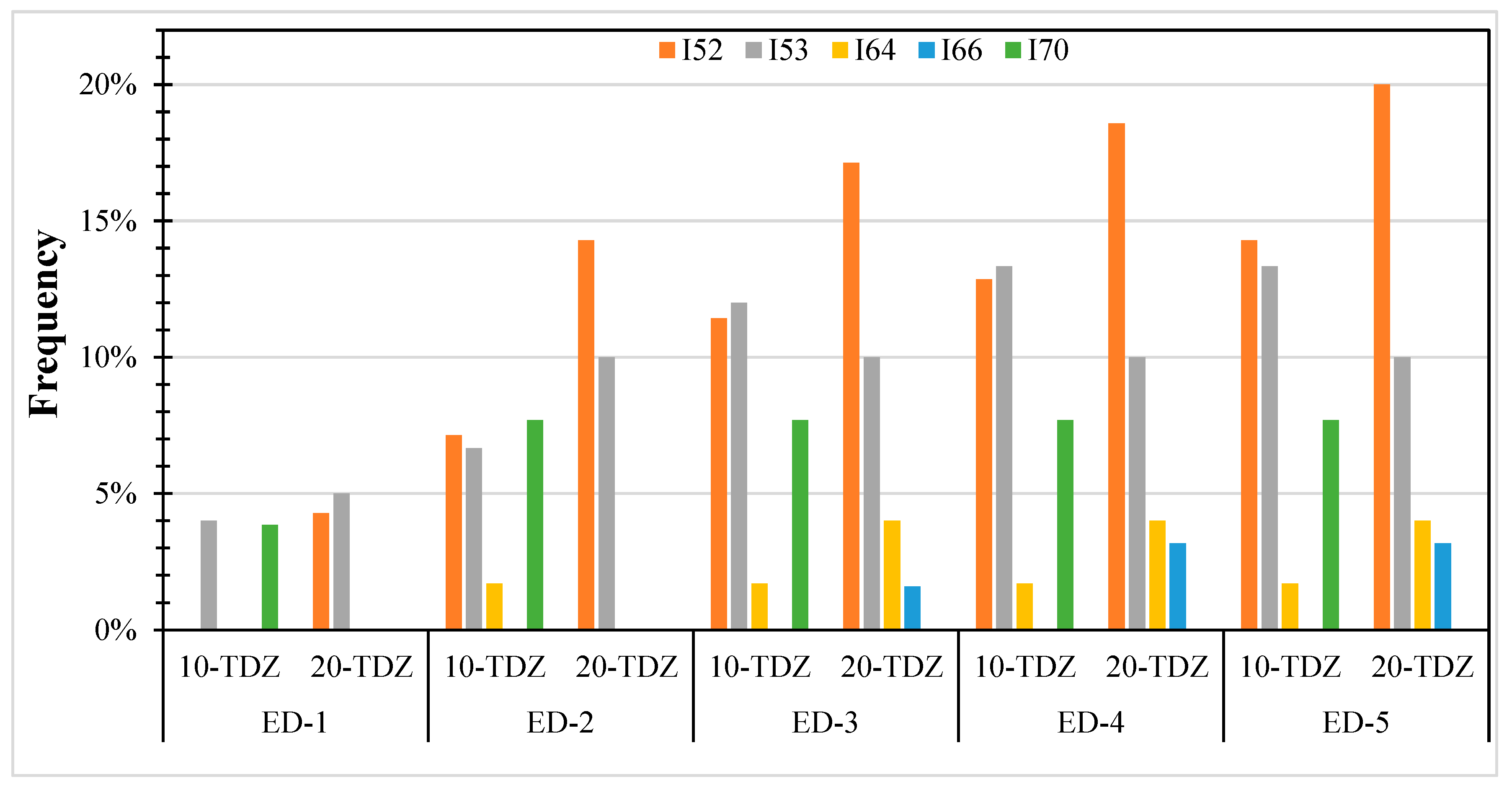
3.2. Effect of Thidiazuron on Somatic Embryogenesis Expression

During the embryogenic development (ED) phase, many calli continued developing under somatic embryogenesis expression without color change, while others became necrotic and ceased growth. During the expression of somatic embryogenesis, some calli, in reactive treatments, coloring changed in a non-synchronous manner, and many calli became dark and necrosed. Throughout the five subcultures performed every 14 days during the ED phase, callus became darker, or showed a combination of dark necrotic calli with the appearance of crystalline or creamy calli as a result of new callus development (Figure 3).
Following the assessment of somatic embryogenesis expression, only eight treatments across five genotypes (INDES-52, INDES-53, INDES-64, INDES-66 and INDES-70) successfully produced somatic embryos (Figure 4). The percentage of embryogenic calli ranged from a low of 2% for INDES-64 genotype cultured on 10 nM L−1 TDZ, to a high 20% inINDES-52 genotype cultured on 20 nM L−1 TDZ. Genotypes of INDES-52, INDES-53, and INDES-70 formed embryos after 14 days of culture on the ED medium. Remarkably, no genotypes formed embryos in the absence of TDZ. For all embryogenic genotypes, callus calli necrosis occurred before embryo development.
The regeneration capacity of somatic embryos varied depends on both genotype and specific treatment applied. For example, INDES-52 genotype, under 20 nM TDZ exhibited a slightly lower average number of embryos per explant compared to 10 nM TDZ treatment; however, it showed a higher overall embryogenic capacity, producing a total of 237 embryos (Table 2). In general, treatments with 20 nM TDZ resulted in a greater total number of embryos across genotypes than those with 10 nM TDZ (Table 2).
During the ED phase, various development stages of somatic embryos were observed (Figure 5). Pro-embryonic structures were first identified by cellular expansion, followed by the formation of globular-stage embryos. These subsequently progressed to heart-shaped, then torpedo-shaped embryos. At the most advanced developmental stages, embryos reached the cotyledonary stage. As embryo formation is not synchronized, different stages can be observed at the same time.

4. Discussion

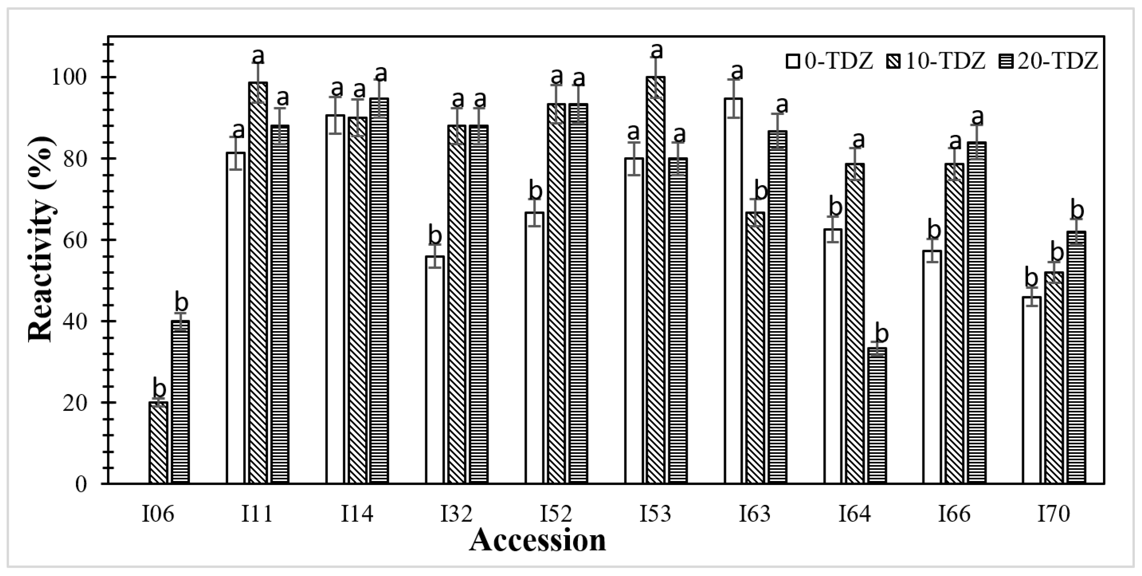
4.1. TDZ Treatment Promotes Explant Reactivity

In previous studies, plant growth regulators play a significant role in the physiological changes at explant level [5,7,9,16,20]. For example, Li et al. [5] found that the addition of TDZ promotes callogenesis in cacao floral explant (staminoids); however, its response is dependent of the genotype [5,7,21,22]. Similar results were found in our experiments, where the callogenesis was variable in all the treatments, depending on the genotypes and TDZ concentration.
To date, only one research on somatic embryogenesis in cacao from Peru has been reported [23]. Nevertheless, studies conducted on cacao genotypes in other countries have demonstrated considerable variation in embryogenic responses. Solano [10] reported primary callus formation on 72–100% in petals and staminodes from five cacao clones from CATIE collection, showing the highest reactivity in staminodes from the ICS-95, CC-137; UF-273; EET-183 and CATIE R7 clones. In Peru, Gárate-Navarro & Arévalo-Gardini [24] evaluated the somatic embryogenesis response of staminodes of ten cacao genotypes from the collection of the Instituto de Cultivos Tropicales (ICT) in San Martín, using DKW medium and different concentrations of 2,4-D (1.50 mg L−1 and 2, 5 mg L−1), and TDZ (5.0 μg L−1 and 7.5 μg L−1). They obtained values of callus formation between 42–100%.
The variability of the response on callus formation may be caused by the genotypic reaction related to the use of TDZ, which may cause different reactions depending on the genotype evaluated [5,7]. Kan Kouassi [16] demonstrated that the interaction between growth regulators such as auxins and cytokinin may affect callus production and subsequent embryogenesis in cacao. In our case, treatments without TDZ affected callus formation. Similar results were observed by Li et al. [5], reporting that the percentage of responsive staminodes was much lower, unless TDZ was added to the culture medium. Nevertheless, higher concentrations of 22 nM of TDZ lowered the percentage of responsive staminodes in most of the cases [5].

4.2. Embryo Formation

After transferring the callus on the ED medium, most explants developed necrosis. Necrosis gradually occurred as subcultures were performed. Some explants were necrosis free and kept growing throughout the entire ED phase. Similar observations were reported by other authors who agree that excessive callus growth is the major bottleneck for plant regeneration via somatic embryogenesis [7,25]. In our study, explants with no necrosis were unable to develop embryos in any case.
The color change during embryogenesis is associated with the effect of phenol accumulation during the culture [25]. The accumulation of phenols negatively affects somatic embryogenesis. This is likely due to their antioxidant properties, which modify the substrate for oxidative enzymes. The activity of these enzymes can lead to the release of auxins that promote embryogenic differentiation [26,27]. Therefore, in cacao, phenols could act as inducers of somatic embryogenesis. However, a balanced distribution and concentration of phenolic compounds are necessary to favor it, as high concentrations have been associated with a non-regenerative callus response. In addition, the histological localization of these compounds could be a tool to distinguish between regenerative and non-regenerative callus [28,29]. Although our study does not aim to measure phenolic compounds in the callus, we consider it an important approach to elucidate the mechanisms underlying embryogenesis.
The appearance of pro-embryonic structures occurred progressively from the first weeks to the end of the evaluation period on the ED medium. Only five genotypes under eight treatments produced somatic embryos, even though essentially all of them formed calli of different types. INDES-52, INDES-53, and INDES-70 genotypes developed embryogenic structures from the first 15 days after ED medium culture. INDES-64 and INDES-66 genotypes initiated embryo formation after 56 days on the ED medium culture. These results are much shorter than the reports from other authors, who observed embryo formation not before 56 days of culture on the ED medium [5,6,7].
The fact that some genotypes did not respond to the induction of somatic embryogenesis and embryo formation supports the results reported by other authors, who found a strong influence of genetic factors and its interaction with other factors during induction on the embryogenic response. Depending on the genotype evaluated, embryos can be easily regenerated on a specific culture medium, while other genotypes did not show the same response [6,7,9,10,11,30]. These differences have also been reported in other species such as Azadirachta indica, Cydonia oblonga, Manguifera sp. and Coffea sp. [31,32,33].
The type of growth regulator, its concentration in the culture medium, and the duration of the incubation period, are key factors influencing the induction of somatic embryogenesis [20]. In addition, the induction of somatic embryogenesis is influenced by other factors such as medium composition, explant type, and genotype [6,10,11,16,24,34,35]. In our study, embryogenic frequencies were higher in INDES-52 and INDES-53 genotypes when cultured on 10 nM TDZ. INDES-64 genotype reached higher embryogenic frequencies on culture medium with 20 nM TDZ, while INDES-70 and INDES-66 genotypes only responded positively to the medium with 10 nM and 20 nM of TDZ, respectively.
To measure the somatic embryogenic response for a specific genotype, it is necessary to evaluate the frequency of somatic embryogenesis and the average number of embryos per explant [7]. In this case, INDES-52 exhibited the highest response to the applied treatment, with an average of 338.6 embryos produced per 100 staminodes when induced with 20 nM TDZ.

5. Conclusions

All the fine-favor cacao genotypes evaluated in our study responded to callus induction in staminoid explants. However, the responses were dependent on the genotype and TDZ concentration. Likewise, only five genotypes of the ten evaluated were suitable for generating somatic embryos, among them INDES-52 was the most responsive. Finally, our results introduce the possibility of increasing the knowledge of somatic embryogenesis in fine-flavor cacao genotypes from another regions from Peru, as well as from other countries.

Author Contributions

Conceptualization, J.C.G.-A. and K.R.; methodology, K.R. and J.C.G.-A.; formal analysis, K.R.; investigation, K.R. and S.L.; data curation, K.R.; writing—original draft preparation, S.L. and K.R.; writing—review and editing, K.R. and J.R.D.-V.; supervision, M.O.; funding acquisition, M.O. and S.L. All authors have read and agreed to the published version of the manuscript.

Funding

The authors thank the Fondo Nacional de Desarrollo Científico, Tecnológico y de Innovación Tecnológica (FONDECYT) for funding this research through the Contract No. 026-2016 “(CINCACAO)”. Also thanks the project CUI No. 2315092 “Construcción del centro de investigación en forestería y agrosilvopastura del Instituto de Investigación para el Desarrollo Sustentable de Ceja de Selva (INDES-CES)”—CEINFOR; project CUI No. 2252878 “Creación del servicio de un laboratorio de fisiología y biotecnología vegetal de la Universidad Nacional Toribio Rodríguez de Mendoza región Amazonas”—FISIOBVEG, project CUI No. 2315081 “Creación e Implementación del Centro de Investigación e Innovación Tecnológica en Cacao de la Universidad Nacional Toribio Rodríguez de Mendoza de Amazonas”—CEINCACAO and Vicerrectorado de Investigación of the Universidad Nacional Toribio Rodríguez de Mendoza de Amazonas (UNTRM).

Data Availability Statement

The data that support the findings of this study are available from the corresponding author upon reasonable request.

Conflicts of Interest

The authors declare no conflicts of interest.

References

  1. Zarrillo, S.; Gaikwad, N.; Lanaud, C.; Powis, T.; Viot, C.; Lesur, I.; Fouet, O.; Argout, X.; Guichoux, E.; Salin, F.; et al. The Use and Domestication of Theobroma cacao during the Mid-Holocene in the Upper Amazon. Nat. Ecol. Evol. 2018, 2, 1879–1888. [Google Scholar] [CrossRef]
  2. SEIA. Estadística Agropecuaria. Available online: https://siea.midagri.gob.pe/portal/siea_bi/index.html (accessed on 24 September 2022).
  3. Centro de Comercio Internacional. Cacao: Guía de Prácticas Comerciales; UNCTAD/OMC: Geneva, Switzerland, 2001. [Google Scholar]
  4. Paredes, J.; Canals, M.; González, A.; Ventura, M. Evaluación de Sustratos en el Enraizamiento de Estacas de Cacao (Theobroma cacao L.). In Proceedings of the 14th International Cocoa Research Conference, Accra, Ghana, 13–18 October 2003; pp. 400–497. [Google Scholar]
  5. Li, Z.; Traore, A.; Maximova, S.; Guiltinan, M.J. Somatic Embriogenesis and Plant Rengeneration from Floral Explants of Cacao (Theobroma cacao L.) Using Thidiazuron. Vitr. Cell. Dev. Biol.-Plant 1998, 34, 293–299. [Google Scholar] [CrossRef]
  6. Henao Ramírez, A.M.; de la Hoz Vasquez, T.; Ospina Osorio, T.M.; Atehortúa Garcés, L.; Urrea Trujillo, A.I. Evaluation of the Potential of Regeneration of Different Colombian and Commercial Genotypes of Cocoa (Theobroma cacao L.) via Somatic Embryogenesis. Sci. Hortic. 2018, 229, 148–156. [Google Scholar] [CrossRef]
  7. Chanatásig, C.I. Inducción de la Embriogénesis Somática en Clones Superiores de Cacao (Theobroma cacao L.), Con Resistencia a Enfermedades Fungosas. Master’s Thesis, Centro Agronómico Tropical de Investigación y Enseñanza, Turrialba, Costa Rica, 2004. [Google Scholar]
  8. Maximova, S.N.; Young, A.; Pishak, S.; Guiltinan, M.J. Field Performance of Theobroma cacao L. Plants Propagated via Somatic Embryogenesis. Vitr. Cell. Dev. Biol.-Plant 2008, 44, 487–493. [Google Scholar] [CrossRef]
  9. Maximova, S.N.; Alemanno, L.L.; Young, A.; Ferriere, N.; Traore, A.; Guiltinan, M.J. Efficiency, Genotypic Variability, and Cellular Origin of Primary and Secondary Somatic Embryogenesis of Theobroma cacao L. Vitr. Cell. Dev. Biol.-Plant 2002, 38, 252–259. [Google Scholar] [CrossRef]
  10. Solano, W. Embriogénesis Somática en Clones Superiores de Cacao (Theobroma cacao L.) Obtenidos en el Programa de Mejoramiento Genético Del CATIE. Master’s Thesis, Centro Agronómico Tropical de Investigación y Enseñanza, Turrialba, Costa Rica, 2008. [Google Scholar]
  11. Urrea, A.I.; Atehortúa, L.; Gallego, A.M. Regeneración vía Embriogénesis Somática de Una Variedad Colombiana Élite de Theobroma cacao L. Rev. Colomb. Biotecnol. 2011, 13, 39–50. [Google Scholar]
  12. Driver, J.; Kuniyuki, A. In Vitro Propagation of Paradox Walnut Rootstock. HortScience 1984, 19, 507–509. [Google Scholar] [CrossRef]
  13. Tulecke, W.; McGranahan, G. Somatic Embryogenesis and Plant Regeneration from Cotyledons of Walnut, Juglans regia L. Plant Sci. 1985, 40, 57–63. [Google Scholar] [CrossRef]
  14. Fontanel, A.; Gire-Bobin, S.; Labbé, G.; Favereau, P.; Álvarez, M.; Rutte, S.; Pétiard, V. In Vitro Multiplication and Plant Regeneration of Theobroma cacao L. via Stable Embryogenic Calli. 10 IAPTC Congress. Plant Biotechnol. 2002, 1, 23–28. [Google Scholar]
  15. Garcia, C.; Marelli, J.P.; Motamayor, J.C.; Villela, C. Somatic Embryogenesis in Theobroma cacao L. In Methods in Molecular Biology; Humana Press Inc.: Totowa, NJ, USA, 2018; Volume 1815, pp. 227–245. [Google Scholar]
  16. Kan Kouassi, M.; Kahia, J.; Kouame, C.N.; Tahi, M.G.; Kouablan, K. Comparing the Effect of Plant Growth Regulators on Callus and Somatic Embryogenesis Induction in Four Elite. HortScience 2017, 52, 142–145. [Google Scholar] [CrossRef]
  17. Bustamante, D.E.; Motilal, L.A.; Calderon, M.S.; Mahabir, A.; Oliva, M. Genetic Diversity and Population Structure of Fine Aroma Cacao (Theobroma cacao L.) from North Peru Revealed by Single Nucleotide Polymorphism (SNP) Markers. Front. Ecol. Evol. 2022, 10, 895056. [Google Scholar] [CrossRef]
  18. Penn State University. Integrated System for Vegetative Propagation of Cacao: Protocol Book; Guiltinan, M.J., Maximova, S.N., Eds.; Penn State University: University Park, PA, USA, 2010. [Google Scholar]
  19. Di Rienzo, J.A.; Casanoves, F.; Balzarini, M.G.; Gonzalez, L.; Tablada, M.; Robledo, C.W. InfoStat, version 2020; Universidad Nacional de Córdoba: Córdoba, Argentina, 2020.
  20. Lopez-Báez, O.; Moreno, J.; Pacheco, S. Avanzes en Propagación de Cacao Theobroma cacao Por Embriogénesis Somática en México. In Proceedings of the International Workshop on New Technologies and Cocoa Breeding, Kota Kinabalu, Malaysia, 16–17 October 2000; Ingenic: Montpellier, France, 2001; pp. 163–176. [Google Scholar]
  21. Ajijah, N.; Rubiyo; Sudarsono, D. Callogenesis and Somatic Embryogenesis of Cacao Using Thidiazuron through One Step of Callus Induction. J. Penelit. Tanam. Ind. 2014, 20, 179–186. [Google Scholar] [CrossRef]
  22. Ajijah, N.; Hartati, R.S.; Rubiyo; Sukma, D.; Sudarsono, S. Effective Cacao Somatic Embryo Regeneration on Kinetin Supplemented DKW Medium and Somaclonal Variation Assessment Using SSRs Markers. Agrivita 2016, 38, 80–92. [Google Scholar] [CrossRef]
  23. Paisic-Ramirez, R.; Hernández-Amasifuen, A.D.; Sánchez-Aguilar, W.D.; Corazon-Guivin, M.A.; Bobadilla, L.G.; Mansilla-Córdova, P.J.; Caetano, A.C.; Silva-Zuta, Z.M.; Guerrero-Abad, J.C. Effect of Osmoregulatory on the Secondary Somatic Embryogenesis of Cocoa (Theobroma cacao L.). J. Appl. Biol. Biotechnol. 2024, 12, 177–183. [Google Scholar] [CrossRef]
  24. Gárate-navarro, M.A.; Arévalo-gardini, E. Induction of Somatic Embryogenesis from Cocoa Farmer Field Collection of ICT-Peru. Int. Ann. Sci. 2017, 2, 6–11. [Google Scholar] [CrossRef]
  25. Alemanno, L.; Berthouly, M.; Michaux-Ferrière, N. Histology of Somatic Embryogenesis from Floral Tissues Cocoa. Plant Cell Tissue Organ Cult. 1996, 46, 187–194. [Google Scholar] [CrossRef]
  26. Ndoumou, D.O.; Ndzomo, G.T.; Niemenak, N. Phenol Content, Acidic Peroxidase and IAA-Oxidase during Somatic Embryogenesis in Theobroma cacao L. Biol. Plant 1997, 39, 337–347. [Google Scholar] [CrossRef]
  27. Quiroz-Figueroa, F.; Mendez-Zeel, M.; Larque-Saavedra, A.; Layola-Vargas, V. Picomolar Concentrations of Salicycates Induce Cellular Growth and Enhance Somatic Embriogénesis in Coffea arabica Tissue Culture. Plant Cell Rep. 2001, 20, 679–684. [Google Scholar] [CrossRef]
  28. Alemanno, L. Localization and Identification of Phenolic Compounds in Theobroma cacao L. Somatic Embryogenesis. Ann. Bot. 2003, 92, 613–623. [Google Scholar] [CrossRef] [PubMed]
  29. Gallego, A.; Henao, A.M.; Urrea, A.; Atehortúa, L. Análisis de Distribución de Polifenoles y Sustancias de Reserva en Embriogénesis Somática de Cacao. Rev. Acta Biol. Colomb. 2016, 21, 335–345. [Google Scholar]
  30. Tan, C.L.; Furtek, D.B. Development of an in Vitro Regeneration System for Theobroma cacao from Mature Tissues. Plant Sci. 2003, 164, 407–412. [Google Scholar] [CrossRef]
  31. Fernández, R.; Villarroel, A.; Cuamo, L.; Storaci, V. Evaluation of a Somatic Embryogenesis Regenetation System for Neem (Azadirachta indica). Acta Biol. Colomb. 2016, 21, 581–592. [Google Scholar] [CrossRef]
  32. Fisichella, M.; Silvi, E.; Morini, S. Regeneration of Somatic Embryos and Roots from Quince Leaves Cultured on Media with Different Macroelement Composition. Plant Cell Tissue Organ Cult. 2000, 63, 101–107. [Google Scholar] [CrossRef]
  33. Litz, R. In Vitro Somatic Embriogénesis from Nucellar Callus of Monoembryonic Mango. HortScience 1984, 19, 715–717. [Google Scholar] [CrossRef]
  34. Issali, A.E.; Traoré, A.; Konan, J.L.; Mpika, J.; Andi, J.; Ngoran, K.; Sangaré, A. Relationship between Five Climatic Parameters and Somatic Embryogenesis from Sporophytic Floral Explants of Theobroma cacao L. Afr. J. Biotechnol. 2010, 9, 6614–6625. [Google Scholar]
  35. Minyaka, E.; Nimeenak, N.; Fotso; Sangara, A.; Ndoumuo, D. Effect of MgSO4 and K2SO4 on Somatic Embryo Differentiation in Theobroma cacao L. Plant Cell Tissue Organ Cult. 2008, 94, 149–160. [Google Scholar] [CrossRef]
Figure 1. Cacao callus development after 15 days in PCG. (A): Callus at the base of the staminode. (B): Callus in the middle part of the staminode. (C): Callus in the distal part of the staminode. (D): Callus on the basal and middle part of the staminode. (E): Callus on the basal and distal part of the staminode. (F): Callus formation around the entire staminode. (C,D): INDES-63; (A,B,E,F): INDES-14.
Ijpb 16 00084 g001
Figure 2. Percent reactivity of staminodes in ten cacao genotypes after 28 days of culture on the SCG medium (n = 75). Different letters indicate differences between treatment means according to the Scott–Knott test (p < 0.05); ANOVA results: F = 2.32, p: 0.0019).
Ijpb 16 00084 g002
Figure 3. Types of calli observed at different stages of development. (A): Callus developing on necrotic tissue (INDES-14 after 45 days of culture). (B): Callus exhibiting both necrotic and actively growing regions (INDES-14 after 45 days of culture). (C): Callus with mixed pigmentation with the presence of somatic embryos (INDES-53 after 60 days of culture). (D): Fully necrotic callus with no embryo development (INDES-64 after 60 days of culture).
Ijpb 16 00084 g003
Figure 4. Embryogenic frequency of five native genotypes of cacao. From left to right: daily embryogenic frequencies recorded during culture in the embryo development (ED) medium. (n = I52 × 10 nM TDZ (70); I52 × 20 nM TDZ (70); I53 × 10 nM TDZ (75); I53 × 20 nM TDZ (40); I64 × 10nM TDZ (59); I64 × 20nM TDZ (25); I66 × 20nM TDZ (63); I70 × 10nM TDZ (29)).
Ijpb 16 00084 g004
Figure 5. Cacao somatic embryogenic stages observed from INDES-53 during the ED cultures. (A): Globular (g). (B,C): Heart (h). (D): Torpedo (t). (E,F): Cotyledonary (ct).
Ijpb 16 00084 g005
Table 1. Genotypes used in this study and their geographical location.
Accession CodeAcronymUTMSectorAltitude (m.a.s.l.)
INDES-06I069369168 17M 787894El Chalán754
INDES-11I119369112 17M 787792El Chalán736
INDES-14I149366961 17M 793728El Limoncito817
INDES-32I329367833 17M 779564Quebrada Seca421
INDES-52I529366649 17M 794441Diamante Bajo725
INDES-53I539366665 17M 794453Diamante Bajo757
INDES-63I639365734 17M 793806Naranjos Alto727
INDES-64I649364133 17M 792251Naranjos Alto665
INDES-66I669364181 17M 792346Naranjos Alto667
INDES-70I709371938 17M 787756Lluhuana970
Table 2. Embryogenic frequency and number of embryos over 70 days of culture in embryo development (ED) medium (E: Total of embryogenic explants per treatment; EE: Average number of embryos per explant; ME: Maximum number of embryos per explant; TE: Total embryos).
Genotypes[TDZ]TreatmentsED5 (70 Days)
EEEMETE
INDES-5210-TDZ141017.644176
INDES-5220-TDZ151416.9339237
INDES-5310-TDZ17105.11051
INDES-5320-TDZ18415.53862
INDES-6410-TDZ231111
INDES-6420-TDZ241222222
INDES-6620-TDZ2723.547
INDES-7010-TDZ292283256
Disclaimer/Publisher’s Note: The statements, opinions and data contained in all publications are solely those of the individual author(s) and contributor(s) and not of MDPI and/or the editor(s). MDPI and/or the editor(s) disclaim responsibility for any injury to people or property resulting from any ideas, methods, instructions or products referred to in the content.

