Article

Deciphering Arabidopsis Aquaporin Networks: Comparative Analysis of the STRING and BioGRID Interactomes

by
Alvaro Lopez-Zaplana
R&D Department, 3A Biotech, 30565 Las Torres de Cotillas, Murcia, Spain
Int. J. Plant Biol. 2025, 16(1), 28; https://doi.org/10.3390/ijpb16010028
Submission received: 25 December 2024 / Revised: 13 February 2025 / Accepted: 20 February 2025 / Published: 26 February 2025
(This article belongs to the Section Plant Communication)

Abstract

:
Aquaporins are transmembrane proteins that mediate the transport of water, as well as various ions and molecules. In plants, they play a critical role in numerous processes, including stress adaptation, nutrition, cellular communication, and transpiration. Therefore, understanding the function and interactions of these proteins with others—known as interactomes—is of significant agronomic and biological interest. This study aims to analyse the interactome of all aquaporins in Arabidopsis thaliana L. using two distinct databases, STRING and BioGRID. After analysing both interactomes, a wide range of interactions were identified between each aquaporin and a diverse array of proteins, including nutrient transporters for ammonium, potassium, phosphorus, sulphur, copper, and sugars; proteins related to responses to abiotic stresses; proteins mediating vesicle membrane fusion, such as synaptobrevins and syntaxins; ubiquitinases; kinases; and other transmembrane proteins. These extensive connections further underscore the critical importance of aquaporins in numerous biological processes, positioning them as central modulators and integration points for cellular and systemic responses in plants.

1. Introduction

Aquaporins, or major intrinsic proteins (MIPs), are transmembrane proteins that mediate the passive transport of water or small polar molecules across membranes. In plants, there are five subfamilies based on their sequence similarity and function: plasma membrane intrinsic proteins (PIPs), tonoplast intrinsic proteins (TIPs), NOD26-like intrinsic proteins, also called NOD26-like MIPs (NIPs), small and basic intrinsic proteins (SIPs), and X-intrinsic proteins (XIPs) [1]. Despite their colloquial name, aquaporins’ transport function extends well beyond the movement of water. Aquaporins have been shown to facilitate the transport of other molecules, such as glycerol [2], urea [3], ammonium [4], CO2 [5], H2O2 [6], or metalloids such as boron (B) [7], silicon (Si) [8], selenium (Se) [9], arsenic (As) [10], or antimony (Sb) [11]. In the last 10 years, in addition to the already mentioned elements or compounds, new substances have emerged that are also proposed to pass through aquaporins. Some of these are sodium (Na), potassium (K) [12], lithium (Li), caesium (Cs), rubidium (Rb), nickel (Ni), or copper (Cu) [13,14].
Aquaporins present a signature motif of Asn-Pro-Ala, known as the NPA motif, along with certain variants that are essential for the specific functioning of the particular aquaporin, such as heterodimerization, acting as a constriction point to serve as a size barrier and functioning as a proton exclusion filter [15,16]. Their ternary structure consists of six alpha helices connected by five loops that form a monomer. Each monomer can interact with three additional monomers to generate a functional tetramer with four pores, one from each monomer and one central pore [4,17,18]. In this way, aquaporins are not only capable of interacting with each other or with other aquaporins to form different types of functional homo- and heterotetramers, but they are able to interact with other proteins or membrane components, such as in the cases of ZmPIP2;5 and syntaxin SYP121, a SNARE protein [19], or AtPIP2;7 with SYP61 and SYP121 [20]. Furthermore, aquaporins show regulatory mechanisms akin to those of other nutrient transporters and form gene co-expression networks responding to abiotic stresses [21,22,23]. In Citrus macrophylla L., a reduction in the application of specific nutrients, such as nitrogen (N), led to an increase in the expression of certain nitrate transporters, including CmNRT2, and also induced changes in the expression of aquaporins, such as CmTIP1.1, facilitating the mobilization of internal nitrogen [24]. In Arachis hypogaea L., a recent study showed a hub of genes, including ion-transport genes like HAK8, NHX, or NCL1, two aquaporins, LEA5 (late embryogenesis abundant protein), CIPK11 (CBL-interacting serine/threonine-protein kinase 11), POD3 (peroxidase 3), and MAPK pathway genes in response to saline stress [23].
All of these advances in the understanding of aquaporins have expanded their functionality beyond their original role as simple pore-forming proteins [4,25]. In fact, novel regulatory mechanisms have been revealed thanks to interactomics research, where it has been seen that PIPs can act like a platform for the recruitment of a wide range of transport activities [26] or even interactions with the lipid bilayer [27]. However, not many significant interactions are known between aquaporins and other nutrient transporters, referred to as protein–protein interactions (PPI) and classified as PPI networks [28]. These PPIs are generated through bioinformatics and computational techniques based on experimental results and are stored in databases such as STRING, one of the most popular databases due to its extensive coverage, quality, and abundance, including data from both experimental and computational sources [29]. To construct a more comprehensive interactome, the BioGRID database was also used, which is another repository for interaction data that contains over 1.5 million manually curated interactions from nearly 56,000 publications [30,31].
Given the importance of the topic and the need to summarize the latest advances in the interaction between proteins and aquaporins, this study will delve into the interactome of all the aquaporins of Arabidopsis thaliana L., one of the most used plant models, which contains 35 aquaporin genes (13 PIPs, 10 TIPs, 9 NIPs, and 3 SIPs) [32] and had a study of its general interactome published recently [33]. To achieve this objective, the STRING and the BioGRID databases will be used to provide an overview of the main interacting groups of Arabidopsis aquaporins. Additionally, AlphaFold will be employed with a relevant nutrient transporter identified in the selected aquaporin.

2. Materials and Methods

2.1. A. thaliana L. Aquaporins Analysis

The 35 aquaporin sequences of A. thaliana L. were obtained from the Arabidopsis Information Resource database (TAIR) and compared to data from NCBI. For the alignment and phylogenetic tree construction of A. thaliana L. aquaporins, Mega11 was employed following previous methods [34]. Shortly, all the sequences were aligned with the MUSCLE method, and we then used a Neighbour Joining (NJ) algorithm, employing 1000 bootstrap replicates, a Poisson model, and pairwise deletion.
Subcellular localization for each aquaporin was predicted in Plant-mPLoc 2.0 [35] and WoLF PSORT, an extension of PSORT II, a protein localization prediction software [36,37]. The highest punctuation of WOLF PSORT was selected to determinate subcellular position prediction.

2.2. Interactome Analysis with STRING

Protein interaction networks were determined by using STRING: functional protein association networks [37]. The functional interactome of each aquaporin was calculated at a 0.7 confidence factor with a full STRING network. The active interaction sources were text mining, experiments, databases, co-expression analyses, neighbourhood, gene fusion, and co-occurrence. For general STRING overview, PIP1;4 aquaporin was selected as starting point, with a high confidence interaction score of 0.7. Three times more nodes were added to the current network, and k-means clustering with four clusters was employed to classify the resulting interactions.

2.3. Interactome Analysis with BioGRID

The same analysis was also performed in the BioGRID database. In this case, filtering by confidence was not possible since most interactions had only one experimental evidence. In the case of BioGRID, there are no interactions based on text mining, which sometimes affected the results obtained with STRING. In BioGRID, only interactors with physical high-throughput evidence or low-throughput evidence are included, allowing filtering by other criteria, such as the type of interactome we intend to explore.
For the construction of the interaction table with BioGRID, only known proteins were considered, excluding those without known structures or hypothetical proteins. For cases involving more than 20 proteins, the following prioritization was applied: strongest evidence of interaction > aquaporins > nutrient transporters > repetitive genes analysed with STRING > repetitive genes with similar aquaporins > large gene families (SNARE and MAPKs, among others).

2.4. AlphaFold and ChimeraX for Structure Prediction and Protein Interaction

ColabFold, a combination of MMseqs2 with AlphaFold2 coupled with Google Colaboratory, was used to build predictions of protein structures [38]. The reliability of the generated models was assessed based on the predicted local distance difference test (pLDDT) score and the confidence in interaction interface predictions. For structural analysis and visualization, ChimeraX 1.9 was used [39]. Distance measurements and surface contact analyses were performed to evaluate potential interactions between the studied proteins.

3. Results

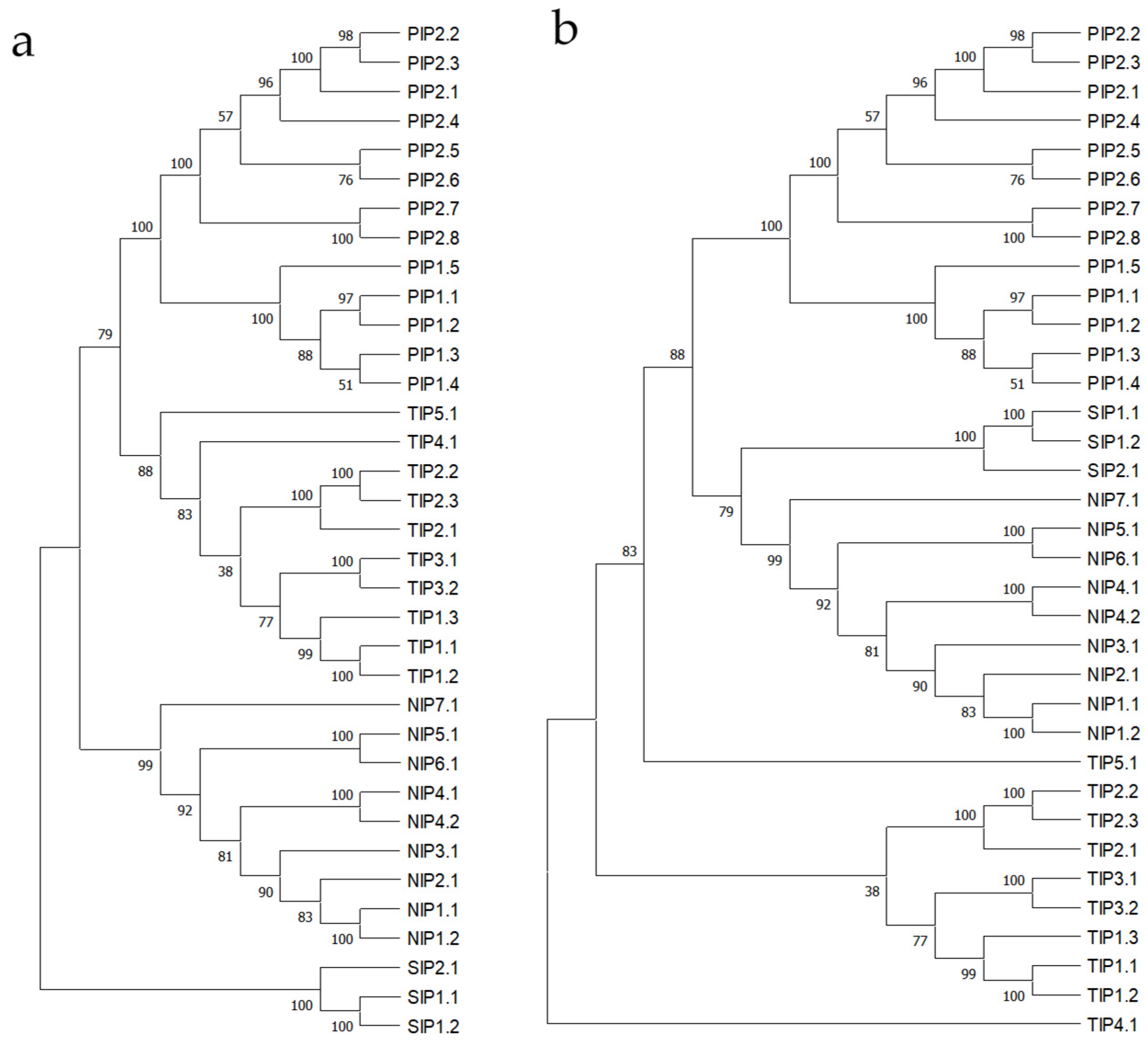
3.1. A. thaliana L. Aquaporins, Multiple Sequence Alignment, and Phylogenetic Tree

The 35 sequences corresponding to the aquaporin family in A. thaliana L. were aligned. In the alignment, the two characteristic NPA motifs of aquaporins were detected in all PIP1, PIP2, and TIP proteins. NIP1;1 and NIP1;2 presented one NPA motif and one NPG motif, NIP5;1 showed NPS and NPV, NIP6;1 had an NPA and an NPV, and NIP7;1 displayed NPS and NPA. The remaining NIPs presented both intact NPA motifs. In contrast, the SIP proteins all showed motif variants: SIP1;1 had NPT and NPA, SIP1;2 had NPC and NPA, and SIP2;1 displayed NPL and NPA (Table 1).
Regarding subcellular localization, most PIPs were predicted by both tools to be located in the plasma membrane, except for PIP2;4, which WOLF predicted to localize in the thylakoid membrane within the chloroplast. Most TIPs were predicted to be located in the vacuolar membrane or tonoplast, except for TIP1;3, which also appears in the plasma membrane, and TIP5;1, which is found in both the plasma membrane and chloroplast. NIPs were primarily located in the plasma membrane, except for NIP3;1, which was also indicated to be in the vacuolar membrane. Lastly, SIPs showed variable localization, appearing in the plasma membrane, vacuole, and endoplasmic reticulum (Table 1).
In Figure 1, we can see how each aquaporin subfamily remains grouped within the same general branches of the tree, demonstrating their similarity to one another. Both TIP4;1 and TIP5;1 appear to be somewhat different. Additionally, TIP5;1 is the only aquaporin that did not fall under the vacuolar membrane localization category (Table 1), suggesting that this aquaporin might have a function beyond residing in the tonoplast membrane.

3.2. A. thaliana L. Aquaporins Interactome by STRING

A general interactome of aquaporins in A. thaliana L. was constructed (Figure 2), resulting in four differentiated nodes: one corresponding to PIPs, another to TIPs and NIPs, a third to the SNARE complex and vesicular transport, and a fourth to the Casparian bands and nitrogen transporters.
To examine each aquaporin in greater detail, an individualized analysis was performed by entering the TAIR Locus identifier from Table 1 into the STRING database (Table 2).
Using STRING, PIPs generally exhibited interactions with each other and with other members of the same subfamily. Notably, there were numerous interactions with SIPs across nearly all PIPs. Several proteins associated with vesicular membrane fusion, such as NPSN12, SYP61, and SYP121, were also highlighted. Additionally, Tryptophan-rich Sensory Protein-related (TSPO) emerged as a significant protein, which, as will be discussed later, plays a role in the regulation of several aquaporins. This protein is sensitive to environmental changes and is associated with abiotic stresses, particularly water stress. Other stress-inducible proteins, specifically responsive to cold and dehydration, are Rare Cold-Inducible 2 (RCI2A and RCI2B), which can also interact with cellular membranes.
The TIPs showed interactions with SIPs, as well as with stress-related signalling proteins such as the transcription factor NAC091, which is linked to stress and cellular defence, and the calcium sensor CBL6. Additionally, TIPs interacted with several vacuolar proteins, including VHA-a3 and phosphatidylinositol-4,5-bisphosphate-dependent protein kinase 24 (PAT24), which is involved in the regulation of membrane lipids.
The NIPs exhibited interactions with SIPs, primarily with several nutrient transporters such as BOR1, BOR2, BOR3, BOR4, BOR5, BOR7, DUR7, and ACR3. They also interacted with proteins related to development and growth, such as SPD6, and NAC091, similar to the TIPs.
Regarding SIPs, connections were found solely with other aquaporins, primarily through text mining interactions. This underscores how underexplored these proteins are, particularly outside the broader context of aquaporin research in an organism.

3.3. Aquaporins Interactome by BioGRID

The total number of interactions recorded in BioGRID was extensive (Table 3), with the majority supported by only a single line of evidence. Notably, PIPs (plasma membrane intrinsic proteins) interacting with other PIPs stood out, as these were the only cases with two or three experimental evidences. The rest of interactions presented 1 evidence although 5-6 concrete cases related with specific aquaporin PIP1;2 and PIP2;1 interactome analysis. Beyond interacting amongst themselves, PIPs also displayed significant interaction with a wide range of proteins from the SNARE complex (synaptobrevin or syntaxin), nutrient transporters (for sugars, ammonium, potassium, phosphorus, sulphur, copper, among others), hormone and signalling molecule transporters, stress-associated proteins like RING/U-box superfamily proteins, which enable rapid adaptations through peptide ubiquitination, and up to 29 transmembrane proteins. Interestingly, this analysis did not reveal interactions with other aquaporins such as SIPs, although in STRING, SIPs were frequently associated with almost all aquaporins, as were TIPs and NIPs, which appeared less frequently but were still noted in some aquaporins.
The TIPs showed interactions with other aquaporins, including TIP1;1 and TIP3;1, TIP1;3 and TIP2;2, and other aquaporins such as TIP2;1 with NIP1;1, in addition to nitrate transporters, phosphatases, tryptophan-rich sensory protein-like proteins, and other proteins that frequently interact with various PIPs, such as polyubiquitin 3 (UBQ3), some vesicle-associated proteins like VAP, and TSPO. TIP3;2 interacted with two Na/H and Ca exchangers, while TIP4;1 was associated with certain vacuolar proteins, such as vacuolar protein sorting protein 60.1 (VPS60.1). Overall, TIPs appear to be either less studied or to interact with fewer proteins.
NIPs also interacted with other aquaporins. Unlike PIPs, which mainly interacted among themselves, NIPs showed interactions with PIPs, TIPs, other NIPs, and SIPs. NIP1;1 displayed a high number of interactions, notably with many vesicle fusion-related proteins and SNARE complex proteins. Far fewer interactors were identified for subsequent aquaporins, with none observed for NIP3;1, NIP4;2, and NIP5;1. Numerous proteins previously shown to interact with PIPs stood out, particularly ubiquitination-related proteins such as UBC32 and UBC34, auxin efflux carrier component (PIN4), and sugar transporters (SUT2).
Regarding the SIPs, SIP1;1 showed no data, SIP1;2 interacted with NIP1;1, and SIP2;1 exhibited up to 11 interactions. Among these, VPS60.1, a vacuolar protein-sorting protein previously associated with TIP4;1, several transmembrane proteins, and three cytochrome isoforms (CB5-D, CB5-E, and CYP81D8) were notable.

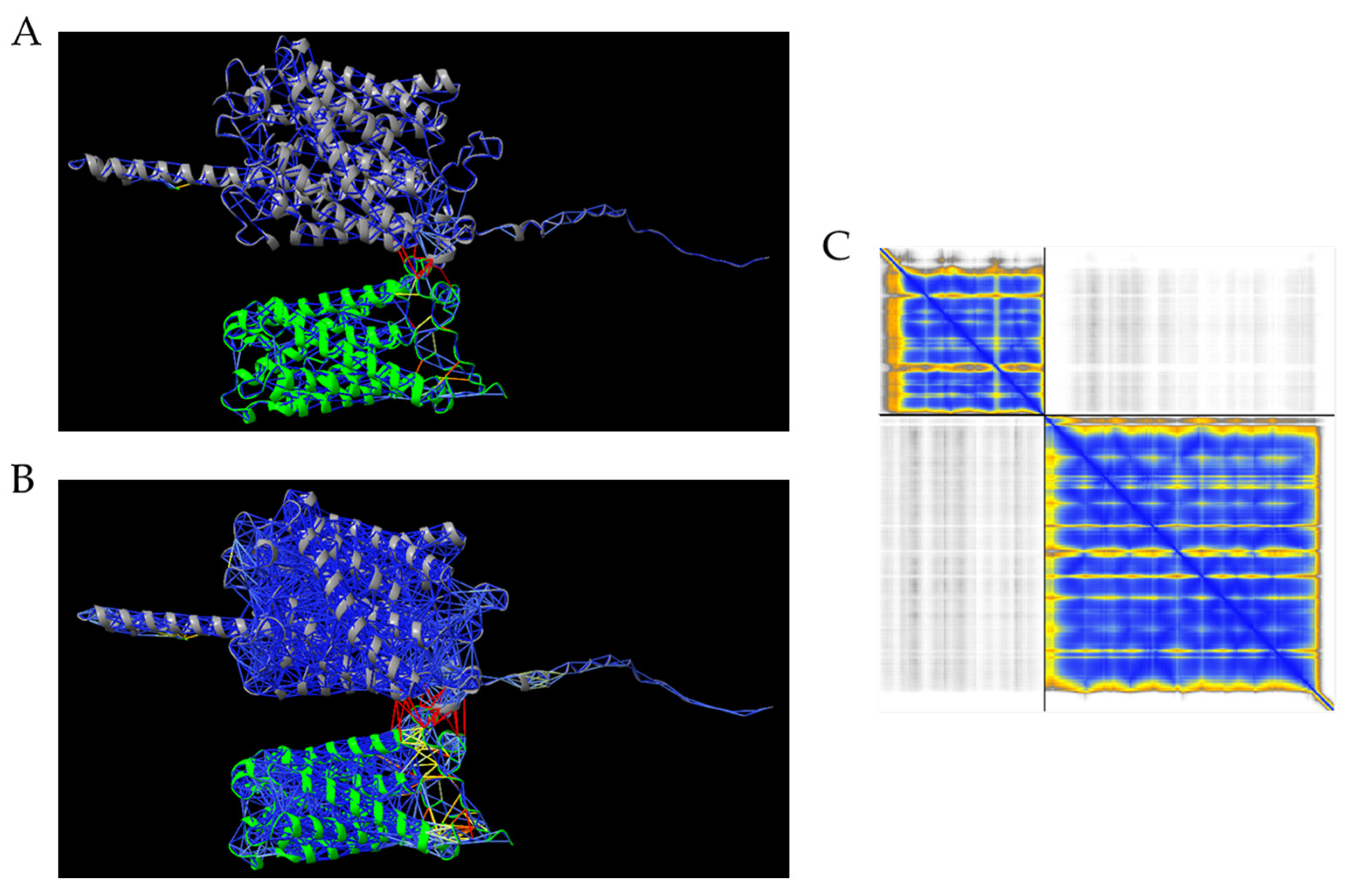
3.4. Aquaporin-Nutrient Transporter Interaction by AlphaFold

To determine how a nutrient transporter interacts with an aquaporin, a structural and interaction prediction assay was conducted. Since no transporter was found to interact with the same aquaporins in both STRING and BioGRID, it was decided to select one that interacted with multiple aquaporins. This was the case for N transporter AMT1;3 in BioGRIND, which was found to interact with PIP1;1, PIP1;2, and PIP1;3 (Figure 3). Additionally, other transporters from the same family, AMT1;1 and AMT1;2, also interacted with other PIPs. The generation of protein structures and interactions between the ammonium transporter and the aquaporin using AlphaFold shows low confidence regarding their potential interaction under these conditions.

4. Discussion

More than 30 years have passed since the initial studies conducted by Preston and Agre [40,41] and the subsequent coining of the term aquaporin in 1993 [42], leading to the first studies identifying the different types of aquaporins. From that point forward, research on aquaporins has advanced, uncovering the modulation of their activity at both the transcriptional and post-transcriptional levels, interaction with other proteins, and relation with plant physiology and environmental changes [43,44,45,46].

4.1. Aquaporins Interact with Other Aquaporins

Across the two methods employed for analysis, all aquaporins were found to interact with at least one other aquaporin with high confidence in STRING. In BioGRID, most of them showed some interactions with other aquaporins. This is not surprising and it is well-known, as each pore is formed by four monomers, which are four aquaporins that can be identical or different, thus influencing the type of activity carried out [47]. Since aquaporins can form functional homo- and heterotetramers with other aquaporins, it was expected that many interactions would be found among them [48]. Some particularly strong and significant interactions observed in STRING included the interaction between PIP1;2, PIP2;1, and PIP2;2, and between PIP2;5, PIP2;7, and PIP2;8 [26]. These interactions were also validated by BioGRID, but most of them only possessed one experimental evidence. In the individualized analyses of each aquaporin, SIP group aquaporins appeared consistently, primarily due to interactions identified through text mining in STRING. This can be explained by the fact that heterodimerization studies of aquaporins have been conducted mainly on PIPs, while SIPs are generally examined superficially and are often only mentioned in genome-wide studies of aquaporins.

4.2. Aquaporins’ Redistribution and Activation in Response to Stress

Other well-known kinds of interactions were found in some proteins that regulate the distribution of aquaporins or their response against abiotic stresses. That was the case of RCI2 genes and some aquaporins, mainly PIPs2. RCI2, which also have transmembrane domains, have been shown in species like Camelina sativa L. to interact with aquaporins, redistributing aquaporins under saline and osmotic stress and inhibiting their water transport activity [49]. Another interaction with a stress regulator was found through fluorescent resonance energy transfer assays between PIP2;7 and the multi-stress regulator TSPO, which is induced by abiotic stresses. TSPO is induced by abscisic acid and regulates the physiology of the cell by physical interaction of PIP2;7, forming a complex that is degraded through the autophagy pathway [50]. This was one of the most frequently appearing proteins in the interactomes of Arabidopsis aquaporins in both STRING and BioGRID, being present in PIP2;6, PIP2;7, TIP1;1, TIP1;2, TIP2;1, TIP2;2, and TIP2;3. Related to abiotic stresses, early-responsive to dehydration stress protein (ERD4), which is rapidly activated in response to dehydration stresses such as drought, stabilizing cell membranes, interacting with other proteins, and regulating gene expression, showed interaction with PIP1;2 [51].
Aquaporins can also modify their activity through phosphorylation and dephosphorylation and can interact with these phosphorylation-type proteins. For example, in Zea maize L. it is known that the activation of endogenous protein kinase A increases the osmotic water permeability coefficient of ZmPIP2;1, pointing out that phosphorylation can activate its channel activity [52]. In this regard, the receptor-like kinase (RLK) family has been identified multiple times through physical interactions with various aquaporins, such as PIP1;2, PIP2;1, RLK902, and RKL1. The expression of these proteins enhances cell proliferation and growth by regulating apoplastic pH and reactive oxygen species (ROS) [53,54,55], possibly modulating the transport activity of H2O2 and H2O in these aquaporins, which are capable of transporting these molecules [56,57]. Furthermore, other protein kinases like K22F20.5 have been suggested to interact with TIP2;3, an aquaporin involved in vacuolar compartmentalization and ammonium detoxification, both implicated in hypocotyl development [58], pointing to the activation of different kinds of signalling pathways related not only to water stress, but also detoxification processes. Calcium-dependent protein kinase 34 (CPK34), which may play a role in signal transduction pathways involving Ca as a secondary messenger, showed association in curated databases and was co-mentioned in a PubMed abstract with NIP4;1, pointing to a relation between them. NIP4;1 and NIP4;2 are regulated by phosphorylation at Ser-267 by CPK34, acting as a principal actor for pollen germination and pollen tube elongation [59,60].
Combining the phosphorylation and change in activity due to stress, it has been observed that the probable inactive receptor kinase At2g26730 (F18A8.10) may modulate the activity of certain PIP2s through dephosphorylation, inhibiting their function of decreasing water permeability in response to cold or salt stresses [58,61,62]. As can be observed, the interaction of aquaporins with stress-response proteins that internalize them or with activation cascades of various kinases can inhibit the function of these aquaporins, primarily in the plasma membrane, thereby reducing the transport of water as well as other components and signalling molecules.

4.3. Aquaporins Interact with Vesicle-Trafficking Proteins

A prominent family that aquaporins often interact with is the vesicle-trafficking proteins, such as syntaxin. For instance, PIP2;7 shows strong interaction with syntaxin-61 and syntaxin-121 (AtSYP61 and AtSYP121), detected through affinity chromatography assays [20,63]. In BioGRID, it was found that ATBET12, or Bet-1-like SNARE 1-2, and NPSN13, also known as novel plant SNARE 13, interact with PIP1;2 and PIP1;3 [26,64]. With NIP1;1 aquaporin, more interactions related with membrane vesicles were found with ATBET12, like AT1G27700, AT4G09580, NPSN12, NPSN13, SYP31, SYP31, SYP51, SYP132, SYP121, or SAR1, most of them syntaxin or vesicle-associated membrane proteins [65], pointing to the implication of this aquaporin in vesicle trafficking. By lowering the STRING confidence level, other aquaporins, such as PIP2;8, are also seen to interact with PVA11, which encodes for Vesicle-Associated Protein 27, detected by two-hybrid array assays [65]. This same interaction was found in BioGRID, adding the interaction between PIP2;3 and VAMP/Synaptobrevin-associated protein 27-1 (VAP27) [65]. These well-studied interactions reveal the intense trafficking of aquaporins, specifically PIPs, from vesicles to the plasma membrane and back, which introduces an additional layer of regulation by preventing them from performing their function at the plasma membrane. This level of regulation and interactions is not exclusive to aquaporins, as the subnetwork of the SNARE complex encompasses approximately 1830 interactions, which corresponds to about 2% of the total known interactome of A. thaliana [33].

4.4. Aquaporins Interact with Nutrients Transporters

Another type of relationship that stood out is the one between aquaporins and nutrient transporters, as these interactions are among the least studied and understood [66]. Aquaporins are capable of transporting numerous nutrients [13], with some of them also known for their ability to transport metalloids, such as NIPs [67]. However, although there is some knowledge regarding functional cooperation and shared regulation [22], the physical interactions between nutrient transporters and aquaporins is not yet well understood.
PIPs showed interactions with very variable transporters, mainly in BioGRID: ammonium transporters, such as AMT1;1, AMT1;2, or AMT1;3; sulphate transporters, such as SULTR1;2; phosphate transporters, such as PHT1;1; potassium transporters, such as AKT1 or KUP7; copper transporters, such as COPT2; or sugar transporters such as UTR2, UTR3, ATVEX1, STP1, STP4, or STP7, among others [26,64]. Previous studies have shown a correlation between low nitrogen levels and a decrease in the expression and activity of PIPs [68,69]. However, the interaction between ammonium transporters such as AMT and aquaporins remains somewhat unclear.
For the TIPs, many of them did not show interactions with nutrient transporters, likely due to their low number of interactions. However, some interesting interactions were found in STRING, such as TIP5;1. TIP5;1, which showed interaction with DUR3 and BOR4, like several NIPs, a urea-proton symporter and an efflux-type boron transporter both localized in roots. TIP5;1 and DUR3 are related to N-transport and urea remobilization [70]. On the other hand, the second pair is related with excess-B tolerance [71]. Furthermore, DUR3 also enhances the sensibility to B toxicity when it is expressed in yeast but did not affect the cellular B concentration [72]. This finding could point to a potential transport and storage mechanism in the tonoplast to prevent high levels of cytoplasmic boron, thereby avoiding toxicity from this excess.
Regarding the NIPs, we found significant differences between the results obtained with STRING and those obtained with BioGRID. In the former, interactions were identified with a large number of nutrient transporters, such as B (BOR1, BOR2, BOR3, BOR4, BOR7), N (DUR7), or As (ACR3). In contrast, in the latter, most NIPs did not show curated interactions (NIP3;1, NIP4;2, NIP5;1) and exhibited only a single interaction (NIP1;2, NIP6;1), and none of the transporters identified in STRING overlapped. Instead, several sugar transporters appeared, such as SUT2 (sucrose) and GONST1 (mannose) associated with NIP7;1. The most widespread synergy regarding nutrient transport by NIPs occurs with boron (B). Typically, aquaporins such as NIP5;1 are capable of transporting B and act in combination with certain borate transporter genes, such as BOR1 [73]. In that line, other common interactions identified involve certain aquaporins and proteins that interact with the metabolites transported by these aquaporins, operating in a coordinated manner. For instance, this is the case of NIP1;2, an aluminium-permeable aquaporin, and ALMT1, a protein that facilitates Al exclusion through exudation, thereby enhancing plant tolerance to Al [74,75]. SIPs did not exhibit interactions with any nutrient transporters, possibly due to their less-studied role compared to PIPs, TIPs, and NIPs, as well as their more limited presence in terms of number and expression levels across different species [76].

4.5. Protein–Protein Interaction Between AtPIP1;1 and AtAMT1;3

After performing the interaction analysis with AlphaFold, no physical interaction between PIP1;1 and AMT1;3 could be detected. However, it is important to consider that the interaction of these proteins with transmembrane regions may also be mediated or facilitated by the presence of the cell membrane itself, slightly modifying its structure and enhancing the interaction [77].
Due to the structure of most nutrient transporters, a potential interaction between the transmembrane domains of aquaporins and those of the transporters themselves has been hypothesized [78,79]. This interaction between nutrient transporters and aquaporins could polarize certain cells or regions of cells into large conduits, facilitating the exchange and integration of nutrients into the cell and throughout the plant. This idea is not new and aligns with the existing knowledge that aquaporins are strategically located in the membrane across a wide range of epithelia, with well-defined physiological functions related to fluid absorption and secretion [80].
This hypothesis requires further in-depth investigation and could open new lines of research, positioning aquaporins as key drivers of intercellular trafficking at the plasma membrane level, not only of water but also of nutrients, and intracellular trafficking between the tonoplast and the cytoplasm.

4.6. Comparison Between STRING and BioGRID

It is important to consider that these types of databases currently do not account for different cell types, tissues, or even plant developmental stages, resulting in the loss of cellular context. Instead, they focus solely on the molecular level, which may lead to the identification of false interactions. Another important point to note and handle carefully when conducting these types of analyses is that databases can sometimes establish connections between proteins that may not be relevant or of real biological interest. This issue was observed with several aquaporins, where STRING reported connections with confidence scores as high as 0.7 in early iterations (without increasing the number of nodes in the tree). In most cases, these relationships are derived from both the aquaporin’s name and the newly detected gene co-occurring within the same abstract or paragraph, where sometimes the relationship was simply due to the gene appearing with a selected housekeeping gene or in a gene list for RNA-seq validation. These genes are listed in Table 1 and are marked with an asterisk.
Regarding these system-induced errors, an especially striking example was found: a gene that STRING labels as PIP1, which, rather than being an aquaporin, is actually described as the “PAMP-induced secreted peptide”, an endogenous peptide that acts as an elicitor of immune responses and a positive regulator of defence responses. This frequent occurrence arises from the text relating an aquaporin, such as PIP2;1, with this PAMP-induced peptide instead of an actual PIP1 aquaporin. The issue extends further, as STRING links PIP2;1 to the PAMP-induced peptide rather than any genuine PIP1 aquaporin. This misassociation leads STRING to also connect PIP2;1 with other proteins involved in plant defence mechanisms against bacterial or viral infections due to the erroneous linkage with this misidentified PIP1. A similar phenomenon occurs with SDP and certain aquaporins, such as NIP1;2, where text mining identified them due to frequent co-occurrences, as co-mentioned in PubMed abstracts. In these cases, the term SDP appears as ‘specificity-determining position’ in an aquaporin, rather than referring to SDP6, the glycerol-3-phosphate dehydrogenase, in the specific example of NIP1;2.
BioGRID, in turn, presents many more interactions for each of the aquaporins, as it displays all available interactions; however, the reliability of these interactions is low, since most have only been analysed once. Nevertheless, this issue also serves as an advantage, as it allows for the identification of certain interactions that may be overlooked by STRING. Additionally, BioGRID offers advanced filters to locate specific interactors, aliases, organisms, or evidence types, which can enhance the search process. A drawback to note about BioGRID is that up to three aquaporins showed no interactome results, whereas they did show interactions in STRING. This discrepancy may be due to BioGRID’s use of a semi-manual curation and review process, meaning that the protein of interest might not appear even if some interactors are already known. It should be noted that the quantity of interactomes varies depending on the number of studies conducted or the scope of these studies, which differs for each aquaporin. For example, Bellati et al. (2016) studied PIP1;2 and PIP2;1, finding that 436 proteins interact with PIP1;2 and 388 with PIP2;1, with 343 interactants in common, while similar aquaporins such as PIP1;1 had only 12 interactors identified in BioGRID [26].

5. Conclusions

All of these findings highlight the involvement of aquaporins in plant metabolism, nutrition, and signalling processes, enhancing their functional complexity and establishing aquaporins as multifunctional channels with roles that extend beyond water transport. Indeed, novel regulatory mechanisms have been uncovered through interactomics research, revealing that PIPs can function as platforms for the recruitment of diverse transport activities. Considering this cross-talk and the fact that water serves as the medium through which nutrients are transported, both aquaporins and typical nutrient transporters should be studied in tandem to better understand plant nutrition as well as the physiological changes and adaptations they undergo. Regarding which platform offers more reliable results, BioGRID provides outcomes based on evidence and curated interactions from scientific experiments such as two hybrid assays, protein-fragment complementation assays, or affinity capture–mass spectrometry, whereas STRING may present results derived from scientific and other criteria, such as term proximity or simultaneous usage, including those based on text mining. While this approach can lead to certain inaccuracies, STRING also offers greater filtering capabilities, enabling more comprehensive statistical analyses and facilitating data interpretation. Therefore, the choice of platform should depend on the type of analysis being conducted. In the case of this study, which aims to provide an overview of all aquaporins interactomes in A. thaliana L. based on experimental evidence, I consider BioGRID to be the more suitable platform.

Funding

This research received no external funding.

Data Availability Statement

No new data were created or analyzed in this study.

Acknowledgments

Thanks to Valentina Origüela for her support and help with the references.

Conflicts of Interest

Author Alvaro Lopez-Zaplana was employed by the company 3A Biotech. The remaining authors declare that the research was conducted in the absence of any commercial or financial relationships that could be construed as a potential conflict of interest.

References

  1. Maurel, C.; Boursiac, Y.; Luu, D.T.; Santoni, V.; Shahzad, Z.; Verdoucq, L. Aquaporins in Plants. Physiol. Rev. 2015, 95, 1321–1358. [Google Scholar] [CrossRef] [PubMed]
  2. Dean, R.M.; Rivers, R.L.; Zeidel, M.L.; Roberts, D.M. Purification and Functional Reconstitution of Soybean Nodulin 26. An Aquaporin with Water and Glycerol Transport Properties†. Biochemistry 1998, 38, 347–353. [Google Scholar] [CrossRef] [PubMed]
  3. Gerbeau, P.; Güçlü, J.; Ripoche, P.; Maurel, C. Aquaporin Nt-TIPa Can Account for the High Permeability of Tobacco Cell Vacuolar Membrane to Small Neutral Solutes. Plant J. 1999, 18, 577–587. [Google Scholar] [CrossRef]
  4. Bertl, A.; Kaldenhoff, R. Function of a Separate NH3-Pore in Aquaporin TIP2;2 from Wheat. FEBS Lett. 2007, 581, 5413–5417. [Google Scholar] [CrossRef]
  5. Kaldenhoff, R. Mechanisms Underlying CO2 Diffusion in Leaves. Curr. Opin. Plant Biol. 2012, 15, 276–281. [Google Scholar] [CrossRef] [PubMed]
  6. Bienert, G.P.; Chaumont, F. Aquaporin-Facilitated Transmembrane Diffusion of Hydrogen Peroxide. Biochim. Biophys. Acta 2014, 1840, 1596–1604. [Google Scholar] [CrossRef]
  7. Takano, J.; Wada, M.; Ludewig, U.; Schaaf, G.; Von Wirén, N.; Fujiwara, T. The Arabidopsis Major Intrinsic Protein NIP5;1 Is Essential for Efficient Boron Uptake and Plant Development under Boron Limitation. Plant Cell 2006, 18, 1498–1509. [Google Scholar] [CrossRef] [PubMed]
  8. Chiba, Y.; Mitani, N.; Yamaji, N.; Ma, J.F. HvLsi1 Is a Silicon Influx Transporter in Barley. Plant J. 2009, 57, 810–818. [Google Scholar] [CrossRef] [PubMed]
  9. Zhao, X.Q.; Mitani, N.; Yamaji, N.; Shen, R.F.; Ma, J.F. Involvement of Silicon Influx Transporter OsNIP2;1 in Selenite Uptake in Rice. Plant Physiol. 2010, 153, 1871–1877. [Google Scholar] [CrossRef]
  10. Bienert, G.P.; Jahn, T.P. Major Intrinsic Proteins and Arsenic Transport in Plants: New Players and Their Potential Role. Adv. Exp. Med. Biol. 2010, 679, 111–125. [Google Scholar] [CrossRef] [PubMed]
  11. Bienert, G.P.; Thorsen, M.; Schüssler, M.D.; Nilsson, H.R.; Wagner, A.; Tamás, M.J.; Jahn, T.P. A Subgroup of Plant Aquaporins Facilitate the Bi-Directional Diffusion of As(OH)3 and Sb(OH)3 across Membranes. BMC Biol. 2008, 6, 1–15. [Google Scholar] [CrossRef] [PubMed]
  12. Tran, S.T.H.; Imran, S.; Horie, T.; Qiu, J.; McGaughey, S.; Byrt, C.S.; Tyerman, S.D.; Katsuhara, M. A Survey of Barley PIP Aquaporin Ionic Conductance Reveals Ca2+-Sensitive HvPIP2;8 Na+ and K+ Conductance. Int. J. Mol. Sci. 2020, 21, 7135. [Google Scholar] [CrossRef] [PubMed]
  13. Tyerman, S.D.; McGaughey, S.A.; Qiu, J.; Yool, A.J.; Byrt, C.S. Adaptable and Multifunctional Ion-Conducting Aquaporins. Annu. Rev. Plant Biol. 2021, 72, 703–736. [Google Scholar] [CrossRef]
  14. Noronha, H.; Araújo, D.; Conde, C.; Martins, A.P.; Soveral, G.; Chaumont, F.; Delrot, S.; Gerós, H. The Grapevine Uncharacterized Intrinsic Protein 1 (VvXIP1) Is Regulated by Drought Stress and Transports Glycerol, Hydrogen Peroxide, Heavy Metals but Not Water. PLoS ONE 2016, 11, e0160976. [Google Scholar] [CrossRef] [PubMed]
  15. Murata, K.; Mitsuoka, K.; Hiral, T.; Walz, T.; Agre, P.; Heymann, J.B.; Engel, A.; Fujiyoshi, Y. Structural Determinants of Water Permeation through Aquaporin-1. Nature 2000, 407, 599–605. [Google Scholar] [CrossRef] [PubMed]
  16. Ikeda, M.; Andoo, A.; Shimono, M.; Takamatsu, N.; Taki, A.; Muta, K.; Matsushita, W.; Uechi, T.; Matsuzaki, T.; Kenmochi, N.; et al. The NPC Motif of Aquaporin-11, Unlike the NPA Motif of Known Aquaporins, Is Essential for Full Expression of Molecular Function. J. Biol. Chem. 2011, 286, 3342–3350. [Google Scholar] [CrossRef] [PubMed]
  17. Ozu, M.; Galizia, L.; Acuña, C.; Amodeo, G. Aquaporins: More Than Functional Monomers in a Tetrameric Arrangement. Cells 2018, 7, 209. [Google Scholar] [CrossRef]
  18. Gonen, T.; Walz, T. The Structure of Aquaporins. Q. Rev. Biophys. 2006, 39, 361–396. [Google Scholar] [CrossRef] [PubMed]
  19. Besserer, A.; Burnotte, E.; Bienert, G.P.; Chevalier, A.S.; Errachid, A.; Grefen, C.; Blatt, M.R.; Chaumont, F. Selective Regulation of Maize Plasma Membrane Aquaporin Trafficking and Activity by the SNARE SYP121. Plant Cell 2012, 24, 3463–3481. [Google Scholar] [CrossRef] [PubMed]
  20. Hachez, C.; Laloux, T.; Reinhardt, H.; Cavez, D.; Degand, H.; Grefen, C.; De Rycke, R.; Inzé, D.; Blatt, M.R.; Russinova, E.; et al. Arabidopsis SNAREs SYP61 and SYP121 Coordinate the Trafficking of Plasma Membrane Aquaporin PIP2;7 to Modulate the Cell Membrane Water Permeability. Plant Cell 2014, 26, 3132–3147. [Google Scholar] [CrossRef] [PubMed]
  21. Wang, M.; Ding, L.; Gao, L.; Li, Y.; Shen, Q.; Guo, S. The Interactions of Aquaporins and Mineral Nutrients in Higher Plants. Int. J. Mol. Sci. 2016, 17, 1229. [Google Scholar] [CrossRef]
  22. Barzana, G.; Rios, J.J.; Lopez-Zaplana, A.; Nicolas-Espinosa, J.; Yepes-Molina, L.; Garcia-Ibañez, P.; Carvajal, M. Interrelations of Nutrient and Water Transporters in Plants under Abiotic Stress. Physiol. Plant. 2021, 171, 595–619. [Google Scholar] [CrossRef]
  23. Wang, F.; Miao, H.; Zhang, S.; Hu, X.; Chu, Y.; Yang, W.; Wang, H.; Wang, J.; Shan, S.; Chen, J. Weighted Gene Co-Expression Network Analysis Reveals Hub Genes Regulating Response to Salt Stress in Peanut. BMC Plant Biol. 2024, 24, 1–19. [Google Scholar] [CrossRef]
  24. Garcia-Gomez, P.; Olmos-Ruiz, R.; Nicolas-Espinosa, J.; Carvajal, M. Effects of Low Nitrogen Supply on Biochemical and Physiological Parameters Related to Nitrate and Water, Involving Nitrate Transporters and Aquaporins in Citrus Macrophylla. Plant Biol. 2023, 25, 944–955. [Google Scholar] [CrossRef] [PubMed]
  25. Uehleln, N.; Lovisolo, C.; Siefritz, F.; Kaldenhoff, R. The Tobacco Aquaporin NtAQP1 Is a Membrane CO2 Pore with Physiological Functions. Nature 2003, 425, 734–737. [Google Scholar] [CrossRef]
  26. Bellati, J.; Champeyroux, C.; Hem, S.; Rofidal, V.; Krouk, G.; Maurel, C.; Santoni, V. Novel Aquaporin Regulatory Mechanisms Revealed by Interactomics. Mol. Cell. Proteomics 2016, 15, 3473–3487. [Google Scholar] [CrossRef] [PubMed]
  27. Martínez-Ballesta, M.d.C.; Carvajal, M. Mutual Interactions between Aquaporins and Membrane Components. Front. Plant Sci. 2016, 7, 206371. [Google Scholar] [CrossRef] [PubMed]
  28. Shoemaker, B.A.; Panchenko, A.R. Deciphering Protein–Protein Interactions. Part I. Experimental Techniques and Databases. PLoS Comput. Biol. 2007, 3, e42. [Google Scholar] [CrossRef]
  29. Szklarczyk, D.; Morris, J.H.; Cook, H.; Kuhn, M.; Wyder, S.; Simonovic, M.; Santos, A.; Doncheva, N.T.; Roth, A.; Bork, P.; et al. The STRING Database in 2017: Quality-Controlled Protein-Protein Association Networks, Made Broadly Accessible. Nucleic Acids Res. 2017, 45, D362–D368. [Google Scholar] [CrossRef] [PubMed]
  30. Oughtred, R.; Stark, C.; Breitkreutz, B.J.; Rust, J.; Boucher, L.; Chang, C.; Kolas, N.; O’Donnell, L.; Leung, G.; McAdam, R.; et al. The BioGRID Interaction Database: 2019 Update. Nucleic Acids Res. 2019, 47, D529–D541. [Google Scholar] [CrossRef]
  31. Stark, C.; Breitkreutz, B.J.; Reguly, T.; Boucher, L.; Breitkreutz, A.; Tyers, M. BioGRID: A General Repository for Interaction Datasets. Nucleic Acids Res. 2006, 34, D535–D539. [Google Scholar] [CrossRef]
  32. Johanson, U.; Karlsson, M.; Johansson, I.; Gustavsson, S.; Sjövall, S.; Fraysse, L.; Weig, A.R.; Kjellbom, P. The Complete Set of Genes Encoding Major Intrinsic Proteins in Arabidopsis Provides a Framework for a New Nomenclature for Major Intrinsic Proteins in Plants. Plant Physiol. 2001, 126, 1358–1369. [Google Scholar] [CrossRef] [PubMed]
  33. Yilmaz, M.; Paulic, M.; Seidel, T. Interactome of Arabidopsis thaliana. Plants 2022, 11, 350. [Google Scholar] [CrossRef]
  34. Lopez-Zaplana, A.; Nicolas-Espinosa, J.; Carvajal, M.; Bárzana, G. Genome-Wide Analysis of the Aquaporin Genes in Melon (Cucumis Melo L.). Sci. Rep. 2020, 10, 22240. [Google Scholar] [CrossRef]
  35. Chou, K.C.; Shen, H. Bin Plant-MPLoc: A Top-Down Strategy to Augment the Power for Predicting Plant Protein Subcellular Localization. PLoS ONE 2010, 5, e11335. [Google Scholar] [CrossRef] [PubMed]
  36. Horton, P.; Park, K.J.; Obayashi, T.; Fujita, N.; Harada, H.; Adams-Collier, C.J.; Nakai, K. WoLF PSORT: Protein Localization Predictor. Nucleic Acids Res. 2007, 35, W585–W587. [Google Scholar] [CrossRef]
  37. Franceschini, A.; Szklarczyk, D.; Frankild, S.; Kuhn, M.; Simonovic, M.; Roth, A.; Lin, J.; Minguez, P.; Bork, P.; Von Mering, C.; et al. STRING v9.1: Protein-Protein Interaction Networks, with Increased Coverage and Integration. Nucleic Acids Res. 2013, 41, D808–D815. [Google Scholar] [CrossRef] [PubMed]
  38. Mirdita, M.; Schütze, K.; Moriwaki, Y.; Heo, L.; Ovchinnikov, S.; Steinegger, M. ColabFold: Making Protein Folding Accessible to All. Nat. Methods 2022, 19, 679–682. [Google Scholar] [CrossRef]
  39. Meng, E.C.; Goddard, T.D.; Pettersen, E.F.; Couch, G.S.; Pearson, Z.J.; Morris, J.H.; Ferrin, T.E. UCSF ChimeraX: Tools for Structure Building and Analysis. Protein Sci. 2023, 32, e4792. [Google Scholar] [CrossRef] [PubMed]
  40. Preston, G.M.; Agre, P. Isolation of the CDNA for Erythrocyte Integral Membrane Protein of 28 Kilodaltons: Member of an Ancient Channel Family. Proc. Natl. Acad. Sci. USA 1991, 88, 11110–11114. [Google Scholar] [CrossRef]
  41. Agre, P.; Saboori, A.M.; Asimos, A.; Smith, B.L. Purification and Partial Characterization of the Mr 30,000 Integral Membrane Protein Associated with the Erythrocyte Rh(D) Antigen. J. Biol. Chem. 1987, 262, 17497–17503. [Google Scholar] [CrossRef]
  42. Agre, P.; Sasaki, S.; Chrispeels, M.J. Aquaporins: A Family of Water Channel Proteins. Am. J. Physiol. 1993, 265, F461. [Google Scholar] [CrossRef] [PubMed]
  43. Yepes-Molina, L.; Bárzana, G.; Carvajal, M. Controversial Regulation of Gene Expression and Protein Transduction of Aquaporins under Drought and Salinity Stress. Plants 2020, 9, 1662. [Google Scholar] [CrossRef] [PubMed]
  44. Ariani, A.; Barozzi, F.; Sebastiani, L.; di Toppi, L.S.; di Sansebastiano, G.P.; Andreucci, A. AQUA1 Is a Mercury Sensitive Poplar Aquaporin Regulated at Transcriptional and Post-Translational Levels by Zn Stress. Plant Physiol. Biochem. 2019, 135, 588–600. [Google Scholar] [CrossRef] [PubMed]
  45. Christou, A.; Filippou, P.; Manganaris, G.A.; Fotopoulos, V. Sodium Hydrosulfide Induces Systemic Thermotolerance to Strawberry Plants through Transcriptional Regulation of Heat Shock Proteins and Aquaporin. BMC Plant Biol. 2014, 14, 42. [Google Scholar] [CrossRef]
  46. Lopez-Zaplana, A.; Martinez-Garcia, N.; Carvajal, M.; Bárzana, G. Relationships between Aquaporins Gene Expression and Nutrient Concentrations in Melon Plants (Cucumis melo L.) during Typical Abiotic Stresses. Environ. Exp. Bot. 2022, 195, 104759. [Google Scholar] [CrossRef]
  47. Jozefkowicz, C.; Berny, M.C.; Chaumont, F.; Alleva, K. Heteromerization of Plant Aquaporins. In Plant Aquaporins. Signaling and Communication in Plants; Chaumont, F., Tyerman, S., Eds.; Springer: Cham, Switzerland, 2017; Volume 29, pp. 29–46. [Google Scholar] [CrossRef]
  48. Berny, M.C.; Gilis, D.; Rooman, M.; Chaumont, F. Single Mutations in the Transmembrane Domains of Maize Plasma Membrane Aquaporins Affect the Activity of Monomers within a Heterotetramer. Mol. Plant 2016, 9, 986–1003. [Google Scholar] [CrossRef] [PubMed]
  49. Shin, J.-H.; Kim, H.-S.; Kim, S.; Ahn, S.-J. Osmotic Stress-Induced CsRCI2E Endosomal Trafficking Promotes the Redistribution of Aquaporin CsPIP2 at the Plasma Membrane of Camelina sativa L. 2023; preprint. [Google Scholar] [CrossRef]
  50. Hachez, C.; Veljanovski, V.; Reinhardt, H.; Guillaumot, D.; Vanhee, C.; Chaumont, F.; Batoko, H. The Arabidopsis Abiotic Stress-Induced TSPO-Related Protein Reduces Cell-Surface Expression of the Aquaporin PIP2;7 through Protein-Protein Interactions and Autophagic Degradation. Plant Cell 2015, 26, 4974–4990. [Google Scholar] [CrossRef] [PubMed]
  51. Wu, G.; Tian, N.; She, F.; Cao, A.; Wu, W.; Zheng, S.; Yang, N. Characteristics Analysis of Early Responsive to Dehydration Genes in Arabidopsis thaliana (AtERD). Plant Signal Behav. 2023, 18, 2105021. [Google Scholar] [CrossRef] [PubMed]
  52. Van Wilder, V.; Miecielica, U.; Degand, H.; Derua, R.; Waelkens, E.; Chaumont, F. Maize Plasma Membrane Aquaporins Belonging to the PIP1 and PIP2 Subgroups Are in Vivo Phosphorylated. Plant Cell Physiol. 2008, 49, 1364–1377. [Google Scholar] [CrossRef] [PubMed]
  53. Tarutani, Y.; Sasaki, A.; Yasuda, M.; Nakashita, H.; Yoshida, S.; Yamaguchi, I.; Suzuki, Y. Identification of Three Clones Which Commonly Interact with the Kinase Domains of Highly Homologous Two Receptor-like Kinases, RLK902 and RKL1. Biosci. Biotechnol. Biochem. 2004, 68, 2581–2587. [Google Scholar] [CrossRef] [PubMed]
  54. Marshall, A.; Aalen, R.B.; Audenaert, D.; Beeckman, T.; Broadley, M.R.; Butenko, M.A.; Caño-Delgado, A.I.; de Vries, S.; Dresselhaus, T.; Felix, G.; et al. Tackling Drought SECEPTOR-LIKE KINASES Present New Approaches. Plant Cell 2012, 24, 2262. [Google Scholar] [CrossRef]
  55. Haruta, M.; Sabat, G.; Stecker, K.; Minkoff, B.B.; Sussman, M.R. A Peptide Hormone and Its Receptor Protein Kinase Regulate Plant Cell Expansion. Science 2014, 343, 408–411. [Google Scholar] [CrossRef]
  56. Israel, D.; Lee, S.H.; Robson, T.M.; Zwiazek, J.J. Plasma Membrane Aquaporins of the PIP1 and PIP2 Subfamilies Facilitate Hydrogen Peroxide Diffusion into Plant Roots. BMC Plant Biol. 2022, 22, 566. [Google Scholar] [CrossRef] [PubMed]
  57. Dynowski, M.; Schaaf, G.; Loque, D.; Moran, O.; Ludewig, U. Plant Plasma Membrane Water Channels Conduct the Signalling Molecule H2O2. Biochem. J. 2008, 414, 53–61. [Google Scholar] [CrossRef] [PubMed]
  58. Xu, Z.; Wang, M.; Shi, D.; Zhou, G.; Niu, T.; Hahn, M.G.; O’Neill, M.A.; Kong, Y. DGE-Seq Analysis of MUR3-Related Arabidopsis Mutants Provides Insight into How Dysfunctional Xyloglucan Affects Cell Elongation. Plant Sci. 2017, 258, 156–169. [Google Scholar] [CrossRef] [PubMed]
  59. Lee, H.; Ganguly, A.; Baik, S.; Cho, H.T. Calcium-Dependent Protein Kinase 29 Modulates PIN-FORMED Polarity and Arabidopsis Development via Its Own Phosphorylation Code. Plant Cell 2021, 33, 3513–3531. [Google Scholar] [CrossRef]
  60. Wang, Y.; Zhao, Z.; Liu, F.; Sun, L.; Hao, F. Versatile Roles of Aquaporins in Plant Growth and Development. Int. J. Mol. Sci. 2020, 21, 9485. [Google Scholar] [CrossRef]
  61. He, W.D.; Gao, J.; Dou, T.X.; Shao, X.H.; Bi, F.C.; Sheng, O.; Deng, G.M.; Li, C.Y.; Hu, C.H.; Liu, J.H.; et al. Early Cold-Induced Peroxidases and Aquaporins Are Associated with High Cold Tolerance in Dajiao (Musa Spp. ‘Dajiao’). Front. Plant Sci. 2018, 9, 323137. [Google Scholar] [CrossRef]
  62. Hsu, J.L.; Wang, L.Y.; Wang, S.Y.; Lin, C.H.; Ho, K.C.; Shi, F.K.; Chang, I.F. Functional Phosphoproteomic Profiling of Phosphorylation Sites in Membrane Fractions of Salt-Stressed Arabidopsis thaliana. Proteome Sci. 2009, 7, 42. [Google Scholar] [CrossRef]
  63. Laloux, T.; Matyjaszczyk, I.; Beaudelot, S.; Hachez, C.; Chaumont, F. Interaction Between the SNARE SYP121 and the Plasma Membrane Aquaporin PIP2;7 Involves Different Protein Domains. Front. Plant Sci. 2021, 11, 631643. [Google Scholar] [CrossRef] [PubMed]
  64. Jones, A.M.; Xuan, Y.; Xu, M.; Wang, R.S.; Ho, C.H.; Lalonde, S.; You, C.H.; Sardi, M.I.; Parsa, S.A.; Smith-Valle, E.; et al. Border Control—A Membrane-Linked Interactome of Arabidopsis. Science 2014, 344, 711–716. [Google Scholar] [CrossRef] [PubMed]
  65. Dreze, M.; Carvunis, A.-R.; Charloteaux, B.; Galli, M.; Pevzner, S.J.; Tasan, M.; Ahn, Y.-Y.; Balumuri, P.; Barabási, A.-L.; Bautista, V.; et al. Evidence for Network Evolution in an Arabidopsis Interactome Map. Science 2011, 333, 601–607. [Google Scholar] [CrossRef]
  66. Roche, J.V.; Törnroth-Horsefield, S. Aquaporin Protein-Protein Interactions. Int. J. Mol. Sci. 2017, 18, 2255. [Google Scholar] [CrossRef]
  67. Pommerrenig, B.; Diehn, T.A.; Bienert, G.P. Metalloido-Porins: Essentiality of Nodulin 26-Like Intrinsic Proteins in Metalloid Transport. Plant Sci. 2015, 238, 212–227. [Google Scholar] [CrossRef] [PubMed]
  68. Ding, L.; Li, Y.; Gao, L.; Lu, Z.; Wang, M.; Ling, N.; Shen, Q.; Guo, S. Aquaporin Expression and Water Transport Pathways inside Leaves Are Affected by Nitrogen Supply through Transpiration in Rice Plants. Int. J. Mol. Sci. 2018, 19, 256. [Google Scholar] [CrossRef]
  69. Di Pietro, M.; Vialaret, J.; Hem, S.; Prado, K.; Rossignol, M.; Maurel, C.; Santoni, V. Coordinated Post-Translational Responses of Aquaporins to Abiotic and Nutritional Stimuli in Arabidopsis Roots. Mol. Cell. Proteom. 2013, 12, 3886–3897. [Google Scholar] [CrossRef] [PubMed]
  70. Bohner, A.; Kojima, S.; Hajirezaei, M.; Melzer, M.; Von Wirén, N. Urea Retranslocation from Senescing Arabidopsis Leaves Is Promoted by DUR3-Mediated Urea Retrieval from Leaf Apoplast. Plant J. 2015, 81, 377–387. [Google Scholar] [CrossRef]
  71. Wakuta, S.; Fujikawa, T.; Naito, S.; Takano, J. Tolerance to Excess-Boron Conditions Acquired by Stabilization of a BOR1 Variant with Weak Polarity in Arabidopsis. Front. Cell Dev. Biol. 2016, 4, 176044. [Google Scholar] [CrossRef]
  72. Huai, Z.; Peng, L.; Wang, S.; Zhao, H.; Shi, L.; Xu, F. Identification and Characterization of an Arabidopsis thaliana Mutant Lbt with High Tolerance to Boron Deficiency. Front. Plant Sci. 2018, 9, 362361. [Google Scholar] [CrossRef]
  73. Jothi, M.; Takano, J. Understanding the Regulatory Mechanisms of B Transport to Develop Crop Plants with B Efficiency and Excess B Tolerance. Plant Soil 2023, 487, 1–20. [Google Scholar] [CrossRef]
  74. Wang, Y.; Yu, W.; Cao, Y.; Cai, Y.; Lyi, S.M.; Wu, W.; Kang, Y.; Liang, C.; Liu, J. An Exclusion Mechanism Is Epistatic to an Internal Detoxification Mechanism in Aluminum Resistance in Arabidopsis. BMC Plant Biol. 2020, 20, 122. [Google Scholar] [CrossRef] [PubMed]
  75. Wang, Y.; Li, R.; Li, D.; Jia, X.; Zhou, D.; Li, J.; Lyi, S.M.; Hou, S.; Huang, Y.; Kochian, L.V.; et al. NIP1;2 Is a Plasma Membrane-Localized Transporter Mediating Aluminum Uptake, Translocation, and Tolerance in Arabidopsis. Proc. Natl. Acad. Sci. USA 2017, 114, 5047–5052. [Google Scholar] [CrossRef] [PubMed]
  76. Johanson, U.; Gustavsson, S. A New Subfamily of Major Intrinsic Proteins in Plants. Mol. Biol. Evol. 2002, 19, 456–461. [Google Scholar] [CrossRef] [PubMed]
  77. Muller, M.P.; Jiang, T.; Sun, C.; Lihan, M.; Pant, S.; Mahinthichaichan, P.; Trifan, A.; Tajkhorshid, E. Characterization of Lipid-Protein Interactions and Lipid-Mediated Modulation of Membrane Protein Function through Molecular Simulation. Chem. Rev. 2019, 119, 6086–6161. [Google Scholar] [CrossRef]
  78. Duart, G.; Grau, B.; Mingarro, I.; Martinez-Gil, L. Methodological Approaches for the Analysis of Transmembrane Domain Interactions: A Systematic Review. Biochim. Biophys. Acta Biomembr. 2021, 1863, 183712. [Google Scholar] [CrossRef] [PubMed]
  79. Teese, M.G.; Langosch, D. Role of GxxxG Motifs in Transmembrane Domain Interactions. Biochemistry 2015, 54, 5125–5135. [Google Scholar] [CrossRef]
  80. Brown, D.; Katsura, T.; Kawashima, M.; Verkman, A.S.; Sabolic, I. Cellular Distribution of the Aquaporins: A Family of Water Channel Proteins. Histochem. Cell Biol. 1995, 104, 1–9. [Google Scholar] [CrossRef]
Figure 1. (a) Phylogenetic tree of aquaporin family proteins of A. thaliana L. (b) Bootstrap consensus tree. Generated using MEGA11. Neighbour Joining (NJ) algorithm, a Poisson model, and the pairwise deletion method was employed.
Ijpb 16 00028 g001
Figure 2. STRING analysis protein–protein interactome of A. thaliana L. starting from PIP1;4. High confidence (0.700) was selected. Yellow cluster is for PIPs aquaporins, red cluster is related to passive transport and aquaporins TIPs and NIPs, light blue cluster is membrane fusion, SNAP receptor activity, and SNARE interactions in vesicular transport, and green cluster is related to nitrate import and Casparian strip integrity. The lines between nodes represent predicted interactions: green is gene neighbourhood, red is gene fusions, blue is gene co-occurrence, yellow is text mining, black is co-expression, and soft blue is protein homology. Dotted lines represent interactions between different clusters with the same code pattern.
Ijpb 16 00028 g002
Figure 3. (A,B) AtPIP1;1 (green) and AtAMT1;3 (grey) proteins’ structures. As the distance decreases from 10 Å (A) to 3 Å (B), several low-confidence interactions appear (red lines) between PIP1;1 and AMT1;3. (C) AlphaFold error plot. Colours for each pair of residues (horizontal and vertical axis), with blue indicating a good relative position, yellow indicating moderate confidence, and red indicating low confidence in the relative position.
Ijpb 16 00028 g003
Table 1. Arabidopsis MIPs. Aquaporins names a proposed by Johanson et al., 2001 [32]. Gene identifier in TAIR b. Protein accession number c is the protein sequence for a translation of the corresponding gene in NCBI. NPA Motifs d variations were found. Location prediction e according to Plant-mPLoc and WOLF PSORT. PM means plasma membrane, C means chloroplast thylakoid membrane, V means vacuole membrane, ER means endoplasmic reticulum.
Name aTAIR Locus bAccession No cNPA Motifs dLocation e (mPlot/WOLF)
PIP1;1AT3G61430CAB71073NPA/NPAPM/PM
PIP1;2AT2G45960AAC28529NPA/NPAPM/PM
PIP1;3AT1G01620AAF81320NPA/NPAPM/PM
PIP1;4AT4G00430AAF02782NPA/NPAPM/PM
PIP1;5AT4G23400CAA20461NPA/NPAPM/PM
PIP2;1AT3G53420CAB67649NPA/NPAPM/PM
PIP2;2AT2G37170AAD18142NPA/NPAPM/C
PIP2;3AT2G37180AAD18141NPA/NPAPM/PM
PIP2;4AT5G60660BAB09839NPA/NPAPM/PM
PIP2;5AT3G54820CAB41102NPA/NPAPM/PM
PIP2;6AT2G39010AAC79629NPA/NPAPM/PM
PIP2;7AT4G35100CAA17774NPA/NPAPM/PM
PIP2;8AT2G16850AAC64216NPA/NPAPM/PM
TIP1;1AT2G36830AAD31569NPA/NPAV/V
TIP1;2AT3G26520BAB01832NPA/NPAV/V
TIP1;3AT4G01470AAC62778NPA/NPAV/PM
TIP2;1AT3G16240BAB01264NPA/NPAV/V
TIP2;2AT4G17340CAB10515NPA/NPAV/V
TIP2;3AT5G47450BAB09071NPA/NPAV/V
TIP3;1AT1G73190AAG52132NPA/NPAV/V
TIP3;2AT1G17810AAF97261NPA/NPAV/V
TIP4;1AT2G25810AAC42249NPA/NPAV/V
TIP5;1AT3G47440CAB51216NPA/NPAPM/C
NIP1;1AT4G19030CAA16760NPA/NPGPM/PM
NIP1;2AT4G18910CAA16748NPA/NPGPM/PM
NIP2;1AT2G34390AAC26712NPA/NPAPM/PM
NIP3;1AT1G31885AAG50717NPA/NPAPM/V
NIP4;1AT5G37810BAB10360NPA/NPAPM/PM
NIP4;2AT5G37820BAB10361NPA/NPAPM/PM
NIP5;1AT4G10380CAB39791NPS/NPVPM/PM
NIP6;1AT1G80760AAF14664NPA/NPVPM/PM
NIP7;1AT3G06100AAF30303NPS/NPAPM/PM
SIP1;1AT3G04090AAF26804NPT/NPAPM/V
SIP1;2AT5G18290BAB09487NPC/NPAPM and V/V
SIP2;1AT3G56950CAB72165NPL/NPAPM/ER
Table 2. Proteins interacting with all aquaporins of A. thaliana according to STRING. A confidence score of 0.7 was used. Potential errors, mainly introduced due to STRING’s text mining, are marked with an asterisk (*) and discussed in the Section 4.
NameSTRING Interactors
PIP1;1PIP2;3, PIP1 *
PIP1;2PIP2;1, SIP2;1, PIP1 *
PIP1;3SIP2;1, PIP1 *
PIP1;4SIP2;1, PIP1 *, NPSN12
PIP1;5PIP1 *
PIP2;1PIP1;2, PIP2;2, SIP1;1, SIP2;1, SIP2;2, PIP1 *, RCI2A, RCI2B
PIP2;2PIP2;1, SIP1;1, SIP1;2, SIP2;1, PIP1 *, T20K18.110 *, F11A12.3 *, dl3910c *, MTG13.3 *
PIP2;3PIP1;1, PIP2;8; SIP1;1
PIP2;4NIP2, SIP1;1, SIP1;2, SIP2;1, PIP1 *
PIP2;5PIP2;7, PIP2;8, SIP1;1, SIP1;2, SIP2;1, SYP121, PIP1 *
PIP2;6SIP2;1, TEL1 *, F18A8.10 *
PIP2;7PIP2;5, PIP2;8, SIP1;2, SIP2;1, SYP121, TSPO, MTG13.3, SYP61, PIP1 *
PIP2;8PIP2;3, PIP2;5, PIP2;7, SIP1;2, SIP2;1, PIP1 *
TIP1;1SIP1;1, SIP1;2, SIP2;1, PAT24, NAC091, PIP1 *
TIP1;2SIP1;1, PAT24, NAC091, PIP1 *,
TIP1;3SIP1;1, SIP1;2, SIP2;1, PAT24, T21P5.16, PIP1 *
TIP2;1SIP2;1, PAT24, NAC091, F4J3I0_ARATH
TIP2;2SIP1;1, SIP1;2, SIP2;1, PAT24, NAC091
TIP2;3SIP1;1, SIP1;2, SIP2;1, PAT24, NAC091, K22F20.5
TIP3;1SIP1;1, SIP2;1, PAT24, NAC091, VHA-a3, T13K14.180, CBL6
TIP3;2PAT24, NAC091
TIP4;1ACT2, GAPC1, GAPC2, MON1, SKIP16, YLS8, AP2M, F17M5.140, PP2AA3, TIP41L
TIP5;1SIP1;1, SIP1;2, SIP2;1, NIP2, BOR4, DUR3, AA1, AGD13, PAT24, NAC091
NIP1;1SIP1;1, SIP1;2, SIP2;1, F7K2.10, CPK31, MLP36, F7F1.4
NIP1;2SIP1;1, SIP1;2, SIP2;1, ACR3, SDP6, ALMT1, T9J23.9, T7I23.21
NIP2;1NIP2, SIP1;1, SIP1;2, SIP2;1
NIP3;1SIP1;1, SIP1;2, SIP2;1, ACR3
NIP4;1NIP2, SIP1;1, SIP1;2, SIP2;1, CPK34, NAC091, PPAN, F26K9_220, Q6NLB7_ARATH *
NIP4;2SIP1;1, SIP1;2, SIP2;1, CPK34, NAC091
NIP5;1SIP1;1, SIP1;2, SIP2;1, NIP2, BOR1, BOR2, BOR3, BOR4, BOR7, AAA1 *
NIP6;1SIP1;1, SIP1;2, SIP2;1, NIP2, BOR1, BOR3, BOR4, BOR7, DUR7, ACR3
NIP7;1SIP1;1, SIP1;2, SIP2;1, NIP2, ACR3, SDP6, BOR4, F21O3.16 *
SIP1;1PIP2;2, PIP2;5, TIP1;1, TIP2;2, TIP3;1, NIP1;2, NIP2;1, NIP4;1, NIP4;2, NIP5;1
SIP1;2PIP2;4, PIP2;8, TIP2;2, TIP5;1, NIP1;2, NIP4;1, NIP4;2, NIP5;1, NIP6;1. NIP7;1
SIP2;1PIP2;4, PIP2;8, TIP3;1, TIP5;1, NIP1;2, NIP4;1, NIP4;2, NIP5;1, NIP6;1. NIP7;1
Table 3. Proteins selection that interacts with each aquaporin of A. thaliana according to BioGRID. Total number of interactors is in parentheses. Potential errors are marked with an asterisk (*) and discussed in the Section 4.
NameBioGRID Interactors
PIP1;1PIP1;2, PIP2;1, PIP2;3, PIP2;5, PIP2;7, SYP132, UBQ3, RLK7 *, ABCB4, ABCB19, AMT1;3 (12)
PIP1;2PIP1;1, PIP1;2, PIP1;3, PIP1;5, PIP2s, ABCB4, ABCB19, PEN3, PDR6, PDR7, PIN5, AMT1;1/1;2/1;3, COPT2, AKT1, SULTR1;2, PHT1;1, ERD4, STP1/4/7, RKL1 (376)
PIP1;3PIP1;2, PIP2;5, PIP1;4, PIP1;5, PIP2;1, PIP2;3, PIP2;7, PIP2;8, ABC4, ABC19, AMT1;3, ATBET12, PIN5, UTR2, VP2 (66)
PIP1;4PIP1;2, PIP1;3, PIP2;1, PIP2;2, PIP2;3, PIP2;5, PIP2;6, PIP2;7, ABC4, ABC19, AMT1;3, T1F15.6, FACE2, FMA, UBQ3 (17)
PIP1;5PIP1;2, PIP1;3, PIP1;5, PIP2;1, PIP2;2, PIP2;3, PIP2;5, PIP2;6, PIP2;7, PIP2;8, NIP1;1, NIP7;1, ABC4/19, AMT1;3, ATVEX1, UTR2/3, UBC34, SEC61β, HHP4, CEV1, BCB (99)
PIP2;1PIP1s, PIP2s except PIP2;8, FER, NHL3, PLDδ, PLDγ, RKL1, RKL902, ABC4/19, ADK1, ACA8, ACA10, PDR6/7/9, ALA1, ACA8, AMT1;1/1;2/1;3, SULTR1;2 (306)
PIP2;2PIP1;2, PIP1;3PIP1;4, PIP1;5, PIP2;1, PIP2;7, ABC4, ABC19, AMT1;3, CHAL, CHX9SYP132 (14)
PIP2;3PIP1s, PIP2;5, PIP2;1, PIP2;5,PIP2;8, NIP1;1, VAP, NTL9, LPAT4, ABCB4, AMT1;3 (17)
PIP2;4PIP1;2, PIP2;1, ABCB4, ABCB19, AMT1;3, SYP132, UBQ3 (7)
PIP2;5PIPs1, PIP2;3, PIP2;7, PIP2;8, CHX9, NTL9 (13)
PIP2;6PIP1;2, PIP1;3, PIP1;5, PIP2;1, PIP2;7, AMT1;3, TSPO (7)
PIP2;7PIP1s, PIP2s except PIP2;4, NIP1;1, NIP2;1, SYP61, SYP121, TSPO, ABCB19, ABCB4, AMT1;3, ACBP6, AT2G28315, CERK1, ELIP2, RD28, SYP132, UTR2, VAP (49)
PIP2;8PIP1;2, PIP1;3, PIP1;4, PIP1;5, PIP2;3, PIP2;5, PIP2;7, NIP1;1, KUP7, UBQ3, VAP (18)
TIP1;1TIP3;1, HHP2, IQD6, NHL3, PAP3, TSPO, AT1G22570, AT1G34640, AT5G49540 (12)
TIP1;2UBQ3, TSPO (2)
TIP1;3TIP2;2 (1)
TIP2;1NIP1;1, NAC089, NTL9, TSPO, UBQ3 (6)
TIP2;2TIP1;3, NIP1;1, NAC089, TSPO, UPS1, VAP, AT1G34640, AT3G16310 (12)
TIP2;3AT1G63110, TSPO (2)
TIP3;1TIP1;1, NIP1;1, NAC089, AT3G16310 (5)
TIP3;2CAX7, CNGC18, GCL1, NHX8 (4)
TIP4;1AGP27, AT1G29060, HHP2, NAC089, NHL3, NTL9, UBC34, VPS60.1, WAK3 (16)
TIP5;1AT3G49190 (O-acyltransferase family protein) (1)
NIP1;1PIP1;5, PIP2;3, PIP2;7, PIP2;8, TIP2;2, TIP3;1, SIP1;2, ANTR2, VIT1,UTR1, UTR3, NPSN12, NPSN13, ATBET12, PYR6, SAR1, SEP1, SYP31, SY51, SYP121, SYP132, (184)
NIP1;2GRF3 (1)
NIP2;1PIP2;7, AT1G29060, FACE2, HHP2, HHP4, IQD6, PAP3, SK42, TBL18, UBC34 (24)
NIP3;1No curated interaction data for this protein (0)
NIP4;1ATBET12, HHP2, HHP4, IQD6, MAPR3, NHL3, TBL18, UBC32, UBC34 (17)
NIP4;2No curated interaction data for this protein (0)
NIP5;1No curated interaction data for this protein (0)
NIP6;1AT3G17620 (putative F-box protein) (1)
NIP7;1PIP1;5, CNGC10, CNGC18, GDU4, GONST1, MSBP1, PIN4, SUT2 (12)
SIP1;1No curated interaction data for this protein (0)
SIP1;2NIP1;1, AT3G26110 (anther-specific agp1-like protein) (2)
SIP2;1AT4G29450, CB5-D, CB5-E, CYP81D8, HHP2, HHP4, NAC089, NHL3, VPS60.1 (11)
Disclaimer/Publisher’s Note: The statements, opinions and data contained in all publications are solely those of the individual author(s) and contributor(s) and not of MDPI and/or the editor(s). MDPI and/or the editor(s) disclaim responsibility for any injury to people or property resulting from any ideas, methods, instructions or products referred to in the content.

