Article

Rapid Amperometric Assessment of Substrate Oxidation and Acute Toxicant Responses in Acidithiobacillus Strain Thio1

G.K. Skryabin Institute of Biochemistry and Physiology of Microorganisms, Federal Research Center “Pushchino Scientific Center for Biological Research of the Russian Academy of Sciences”, Pushchino 142290, Russia
*
Author to whom correspondence should be addressed.
Microbiol. Res. 2026, 17(8), 162; https://doi.org/10.3390/microbiolres17080162
Submission received: 31 May 2026 / Revised: 15 August 2026 / Accepted: 17 August 2026 / Published: 19 August 2026

Abstract

Obligate acidophilic bacteria of the genus Acidithiobacillus are widely used and well known in the biomining industry. Over the past decade, it has been shown that bacterial activity can be determined in short-term experiments via amperometric measurements of microbial oxygen consumption, as changes in the Clark electrode current are proportional to changes in the concentration of dissolved oxygen. This article presents a study of Acidithiobacillus sp. strain Thio1, which was isolated from pyrite–chalcopyrite copper ore and is closely related to A. ferrooxidans. Oxygen consumption by strain Thio1 was measured during the bacterial oxidation of substrates as changes in the electrode current. To evaluate the acute respiratory response of the bacteria to Cu, Zn, and As, respiration suppression was measured at increasing concentrations of the toxicants. The proposed amperometric method is not a substitute for the long-term biogeotechnological evaluation of strain activity using specific ore or pulp samples. At the same time, it can be used as a complementary microbiological method. The proposed approach offers distinct advantages: testing takes mere minutes (rapid analysis), and comparisons of substrates or toxicants can be performed using a single biomass sample (standardization). The amperometric method also demonstrated that oxygen was consumed during the corrosion of solid specimens (steel and chalcopyrite) by Acidithiobacillus sp. strain Thio1. However, applying this method to biocorrosion requires further study because the proportion of oxygen consumption directly related to microbial corrosion remains unknown.

1. Introduction

Bioleaching of valuable metals is rooted in industrial microbiology and “green biotechnology”. This biological approach to metal extraction is more environmentally friendly than pyrometallurgy or chemical acid hydrometallurgy [1]. Common leaching microbial consortia include Acidithiobacillus ferrooxidans or Leptospirillum ferrooxidans bacteria, since representatives of these species: (1) are autotrophic, do not require any organic additives; (2) are extremely acidophilic (pH 1–2), i.e., active together with acid chemical leaching; (3) oxidize mineral sulfides, forming sulfuric acid; and (4) oxidize iron to Fe3+, which is a chemical oxidizer of chalcopyrite, etc. [2,3,4].
As a rule, strict acidophilic autotrophic iron-oxidizing strains are isolated in the classical standard Silverman–Lundgren medium 9K [5]. The leaching activity of the strains or their consortia is usually tested in this medium or a modification thereof; the latter replaces the main energy substrate Fe2+ for ore, so the target bacterial activity is assessed by the yield of dissolved metals [6,7]. Commonly, the resistance of strains to toxic components of ore is evaluated in the same manner. Conventional tests of the physiological characteristics of the strains and consortia, carried out using various growth dynamics parameters, are lengthy and labor-intensive. Meanwhile, it has already been shown that the activity of the acidophilic autotrophic iron-oxidizing bacterium A. ferrooxidans can be assessed using a rapid method of measuring oxygen consumption by the biomass [8].
The method is based on the amperometric measurement of the current (nanoampere, nA) produced in a test chamber by bacterial oxygen consumption. This process is initiated by injection of the substrate. Two controls are measured and subtracted: (i) endogenous bacterial respiration without a substrate, and (ii) sterile chemical oxidation of the substrate. While bacterial substrate oxidation leads to an increase in current, toxicant-induced suppression of the bacteria results in a current decrease (indicating suppressed endogenous respiration). The method facilitates real-time recording and quantification of bacterial responses within minutes [9,10,11].
In recent years, researchers have used biosensors to measure electrode current during the microbial oxidation of dissolved substrates. These studies were used to determine the presence and concentration of specific substances in solution [12,13,14]. In contrast, the present study tested the hypothesis that this method could be applied not only to chemical analysis, but also to the study of the properties of bacterial strains using known substrates.
The main aim of this study was to develop a rapid method for assessing the comparative activity of Acidithiobacillus strain based on the amperometric measurement of electrode current associated with bacteria-induced changes in dissolved oxygen. The obligately acidophilic, autotrophic, iron-oxidizing Acidithiobacillus sp. strain Thio1, isolated from a pyrite–chalcopyrite deposit and presumably adapted to copper, was used as a model organism. To fulfill the stated objectives, experiments were conducted to evaluate: (i) the biooxidation of energy substrates (Fe2+ or formate), and (ii) the effects of chemical inhibitors (zinc, copper, arsenate). The formate supplement was chosen based on the ability of A. ferrooxidans to utilize its oxidation as an energy source [15]. The amperometric method was also used to check the response during the potential involvement of the bacteria in the microbiologically induced corrosion (MIC) of solid materials (steel or chalcopyrite).

2. Materials and Methods

Microorganisms and Culturing. A culture of autotrophic acidophilic iron-oxidizing bacteria was isolated from acidic drainage of a copper pyrite–chalcopyrite deposit (Orenburg region, Russia); the ore and drainage samples were provided by Uralpromengineering LLC, Ekaterinburg, Russia. Isolation, cultivation and biomass harvesting were carried out in the modified 9K medium [5], g/L: (NH4)2SO4—3.0, KCl—0.1, K2HPO4—0.5, MgSO4 × 7H2O—0.5, Ca(NO3)2—0.01, FeSO4 × 7H2O—18.0, 10 N H2SO4—4.5 mL/L (Sigma-Aldrich/Merck, Germany; Reakhim, Russia), pH 2.2. The cultivation to obtain biomass for subsequent experiments was carried out in a stationary mode at 28 °C for 7 days.
Strain Identification by Molecular Methods. The isolated strain Thio1 was identified using 16S rDNA gene sequencing. Genomic DNA was isolated as described in [16], and the amount of isolated DNA was determined using a NanoDrop 2000c (Thermoscientific, Waltham, MA, USA). Universal prokaryotic 16S rRNA primers 27f (5′-AGAGTTTGATCCTGGCTCAG-3′) and 1492r (5′-TACGGYTACCTTGTTACGACTT-3′) were used for 16S rDNA gene amplification.
Experiments on the Ore Leaching and Bioleaching. The study on acid leaching of pyrite–chalcopyrite ore was conducted in the 9K medium [5] without iron, pH 2.2. The added substrate was the ore. All experiments were carried out in 750 mL conical flasks containing 200 mL of the basal medium. The ore specimens weighed 20 g each one. Increasing concentrations of iron, sulfur, and copper in the solution served as the leaching indicators. The experiments were conducted in three variants: (1) chemical leaching in the medium without bacteria, (2) chemical–biological leaching in the inoculated medium, and (3) chemical–biological leaching in the inoculated medium supplemented with formate, 65.11 mM (0.3%). Initial cell density in the inoculated flasks was 2 × 10³/mL. After inoculation, the flasks with cotton stoppers were incubated in a steady-state mode at 24 °C for 12 days.
Analyses of Leached and Dissolved Chemical Elements (Fe, S, Cu) were carried out with Bruker S6 JAGUAR spectrometer (Bruker AXS GmbH, Karlsruhe, Germany) using the method of the wavelength-dispersive X-ray fluorescence (WDXRF). X-ray intensity was measured with a counter-detector. The equipment, standard solutions, etc., were provided by the Uralpromengineering LLC (Russia). A range of specified concentrations was measured in a single calibration using a linearity range (with a linear count rate) of up to 2 million counts/s. At low element concentrations, linearity was well maintained, significantly facilitating spectrometer calibration; relative errors were below 5%. The liquid samples were analyzed directly. The intensity of the emitted sulfur signal depends purely on the number of sulfur atoms present, regardless of whether they are bound in organic compounds or inorganic compounds [17] so the sulfur content data are presented as “total sulfur”. Potassium sulfate K2SO4 was used for the calibration solution.
Experiments on Substrate Oxidation with Evaluation of Cell Number. The experiments were also carried out in the medium [5] without iron and under the same conditions. In contrast, the added substrates were not ore but ferrous iron Fe2+ or formate, or their mixture (1:1), 3.6 mM. Three replicates (flasks) were used for each experiment. Bacterial growth was determined by the change in the bacterial number, cells per 1 mL. The bacterial count was evaluated by the direct cell counting which is commonly accepted for super-acidophilic bacteria in microscopic fields of view [6,7,18,19].
Experiments on Substrate Oxidation with Amperometric Measurement of Oxygen Consumption.
Electrode current associated with bacteria-induced changes in dissolved oxygen (oxygen consumption) was measured amperometrically using a Clark electrode chamber (Kronas, Moscow, Russia). Previous studies utilizing the Kronas biosensors provide descriptions of this chamber and the biomass preparation procedures [14,20,21]. Details regarding the preparation and maintenance of the biomass in the present study are given below. To measure bacterial activity, the strain biomass was immobilized on glass paper Whatman GF/A, UK (Whatman International Ltd, Cytiva, Maidstone, UK) and placed in the cell. The procedures for biomass preparation and immobilization were described in detail [8]. The wet biomass was 5 mg in each case; it was immobilized on a glass paper disk of 7 mm2. The contact area of the immobilized biomass and the electrode was constant in all cases and was determined by the electrode’s working area of 4 mm2. When stored in the buffer solution at 6 ± 1 °C, the immobilized biomass maintained constant respiratory activity for 10–12 days. The latter was established in a separate preliminary study and is very significant, as it allows experiments to be conducted with the same washed biomass but with different substrates over the course of a week. Electrode current (nA) associated with bacteria-induced changes in dissolved oxygen (oxygen consumption) was measured. The current was also measured during endogenous respiration of immobilized biomass washed from the medium. Then, a solution of the oxidizable substrate was introduced into the electrode cell, and bacterial oxidation of the substrates was measured. All experiments were carried out in a buffer solution (basal 9K medium without iron), pH 2.2, at 22–24 °C; magnetic stirring was used. The volume of the buffer solution in the electrode cell was 1900 µL, the added substrate/blank solution was 100 µL. The figures in the article present the experimental results with the subtraction of endogenous bacterial respiration and background chemical oxidation. Endogenous respiration was measured as the electrode current in the chamber with immobilized biomass before the addition of the substrate solution. Background chemical oxygen demand was measured as the electrode current in a similar experiment without bacteria, but with the test solution added to the chamber. Concentrations of the test substrates (ferrous iron Fe2+ or formate, or their mixture (1:1)) were 180 μM in the same 9K basal medium; microorganisms responded immediately to their addition to the electrode cell.
The routine use of biosensors to determine the concentration of an analyte requires calibration of the electrical signal versus the concentration of the oxidized substrate. Calibration should also be performed when changing the electrode material [14]. In the present study, the same electrodes and the same portions of washed immobilized biomass were used. This experimental design allowed data to be presented without additional calibrations and recalculations of the relationship between electrode current (nA) and dissolved oxygen. The results are presented as a comparison of electrode current (nA) values in experiments with different substrates.
Biocorrosion of solid substrates (steel and chalcopyrite) was evaluated with the same method and equipment. Specimens of steel were the steel paper clips (Staff Profi, Ningbo/Yiwu, Zhejiang, China). Specimens of chalcopyrite were sampled from the pyrite–chalcopyrite ore (Uralpromengineering LLC, Ekaterinburg, Russia). The working area of the solid specimens was 4 mm2. Experiments were carried out in the buffer solution (basal medium 9K without iron), pH 2.2, at 22–24 °C. In contrast to the experiments with dissolved substrates, the produced electrode current, nA, was measured once per day during 7 days. As in the experiments with dissolved substrates, the subtracted controls were the chemical oxidation of the specimens and endogenous bacterial respiration.
Experiments on the Acute Respiratory Response to Toxicants with Amperometric Measurement of Oxygen Consumption. The bacterial response to toxicants was also assessed by electrode current (nA) associated with bacteria-induced changes in dissolved oxygen (oxygen uptake). The same amperometric equipment (Kronas, Moscow, Russia) was used in the experiments to measure the electrode current as an indicator of oxygen uptake. In this case, the suppression of endogenous bacterial respiration was measured upon the addition of toxicants to the electrode chamber. The basal medium 9K without iron was used. Testing was conducted at various toxicant concentrations, which were selected in preliminary experiments, mM/L: copper sulfate hydrate—4.0, 20.0, and 60.0; zinc sulfate hydrate—3.48, 17.4, and 52.2 mM; arsenate (arsenic acid)—0.026, 2.6, and 26.4 mM. In experiments with copper, and zinc, microorganisms responded immediately to these supplementations; however, with arsenate, the bacterial reaction started in 100 s.
Microscopy of Solid Substrates. Amperometric testing of oxygen consumption during corrosion of solid materials was accompanied by microscopic scanning of the objects in 5 days. An AXIO Imager A1 light microscope (Carl Zeiss AG, Oberkochen, Germany), an AxioCam digital camera (Carl Zeiss AG, Oberkochen, Germany), and AxioVision AC data processing software were used for these observations.
The surface of the samples was examined using scanning electron microscopy (SEM). The samples were fixed at 4 °C for 24 h in glutaraldehyde vapor and then post-fixed at 20 °C for 3 h in OsO4 vapor. After dehydration in propylene oxide vapor, the samples were sputter-coated with gold (Fine Coat Ion Sputter JFC-1100, Tokyo, Japan) and examined using a JSM-6510LV scanning microscope (JEOL, Tokyo, Japan).
Statistical Analysis of Results. All biological experiments and chemical measurements were performed in at least three replicates. The number of independent flasks and analytical measurements corresponding to every value was 3 (n = 3). The standard deviation (SD) was calculated using the STDEV.S function in Excel and is indicated by error bars in the figures to show how widely values are spread around the mean (error bars).
Electrochemical measurements were repeated at three electrodes (n = 3) to obtain results for the statistical analysis of reproducibility. In all cases, the recorded data are presented as electrode current (nA) and its change versus time of the experiment (s).

3. Results and Discussion

3.1. Strain Identification

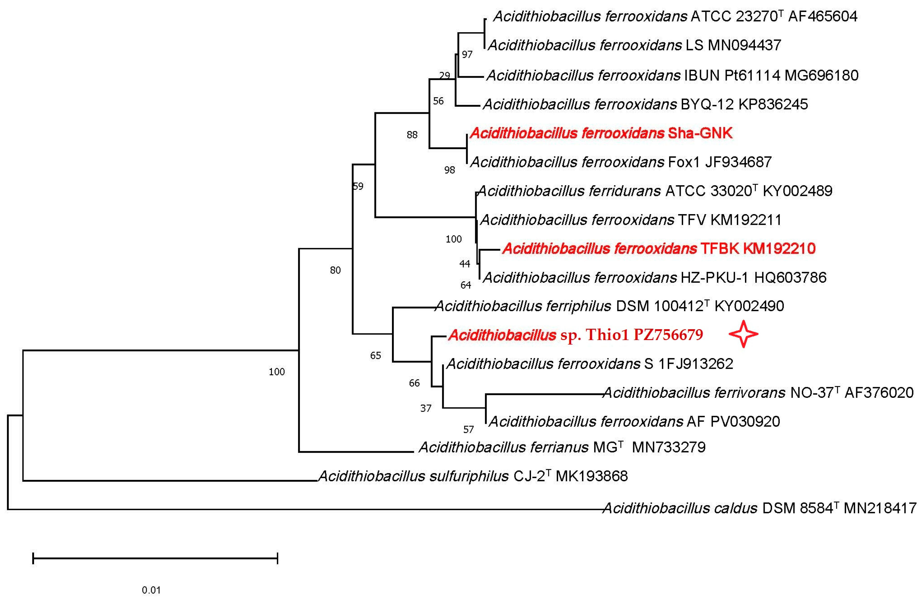
The strain was isolated from the acidic drainage of a pyrite–chalcopyrite deposit, i.e., from mineral ecosystem that contained inorganic sulfides and iron. Since the isolation was carried out in the 9K medium supplemented with iron at pH 2, the culture was represented by obligate acidophilic iron-oxidizing autotrophic bacteria, strain Thio1. According to the bacterial physiology and morphology, it was suggested that the strain belongs to the genus Acidithiobacillus, most likely to A. ferrooxidans. Molecular identification based on the 16S rRNA gene sequences confirmed this suggestion. Figure 1 shows the position of strain Acidithiobacillus sp. Thio1 among other representatives of the species. The genetic data of strain Thio1 have been submitted to GenBank under the accession number PZ756679.
Based on its phylogenetic position, Acidithiobacillus sp. Thio1 is closely related to the strains A. ferrooxidans A 1FJ913262 and AF PV030920. Meanwhile, in the phylogenetic tree, the studied strain Thio1 is also located in the same group as the type strains of the species A. ferrivorans and A. ferriphilus. These data suggest the usefulness of a future taxonomic revision for the Acidithiobacillus genus as a whole.
We also compared the position of Acidithiobacillus sp. Thio1 isolated from sulfide ore (pyrite–chalcopyrite) in the Orenburg region with two other strains. The compared A. ferrooxidans strains were studied in our previous research investigations and were isolated as follows: (1) ShA-GNK in the same Orenburg region from silicate ore [8] and (2) TFBk from sulfide (arsenopyrite) ore in the Republic of Kazakhstan [19]. Figure 1 shows that the two strains isolated from sulfide ores of different countries were more closely related than the two strains originating from different types of ores in the same region.

3.2. Leaching and Bioleaching of the Pyrite–Chalcopyrite Ore

The ability of Acidithiobacillus sp. Thio 1 to leach Fe, S, and Cu from the pyrite–chalcopyrite ore was studied with the WDXRF via the comparison of their concentrations in the solution (Table 1). Concentrations of the dissolved Fe, S, and Cu elements increased over 12 days. Chemical (abiotic) leaching was relatively low and not significant for any potential production process. In the presence of A. ferrooxidans Thio1, the concentration of dissolved copper increased twofold while the concentrations of dissolved iron and sulfur increased by more than an order of magnitude (Table 1). These experiments demonstrated that Acidithiobacillus sp. Thio1 is an active agent of metal bioleaching from solid ore.
The presented data also showed that formate supplementation stimulated copper dissolution throughout the experiment. A comparison of variants with and without formate revealed different effects on the release of iron and sulfur. However, on the 12th day of leaching, the iron concentration in the solution without formate was higher than that with formate. This result suggests that formate is not only utilized as a substrate by A. ferroxidans [15] but can also act as an alternative substrate for iron oxidation.

3.3. Bacterial Iron Oxidation with and Without Formate

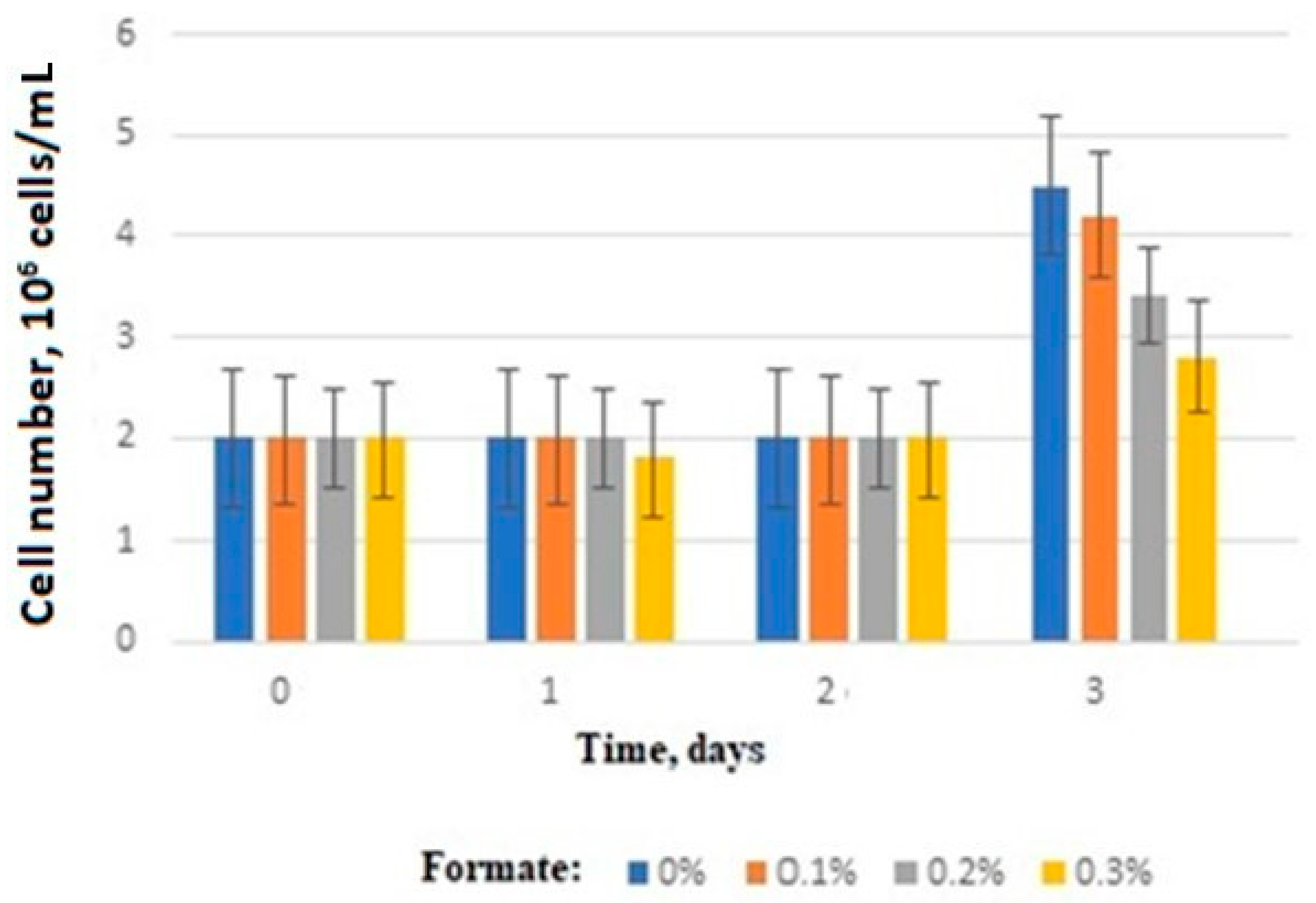
The presented data (Table 1) also led to the hypothesis that A. ferrooxidans Thio1 bacteria can switch between the oxidation of two alternative substrates. To address this question, Acidithiobacillus sp. Thio1 was grown in the 9K medium containing iron and various concentrations of formate. The results demonstrate that the presence of formate from day 3 onwards onward suppressed bacterial growth, as indicated by direct cell counts (Figure 2).

3.4. Amperometric Evaluation of the Bacterial Substrate Oxidation

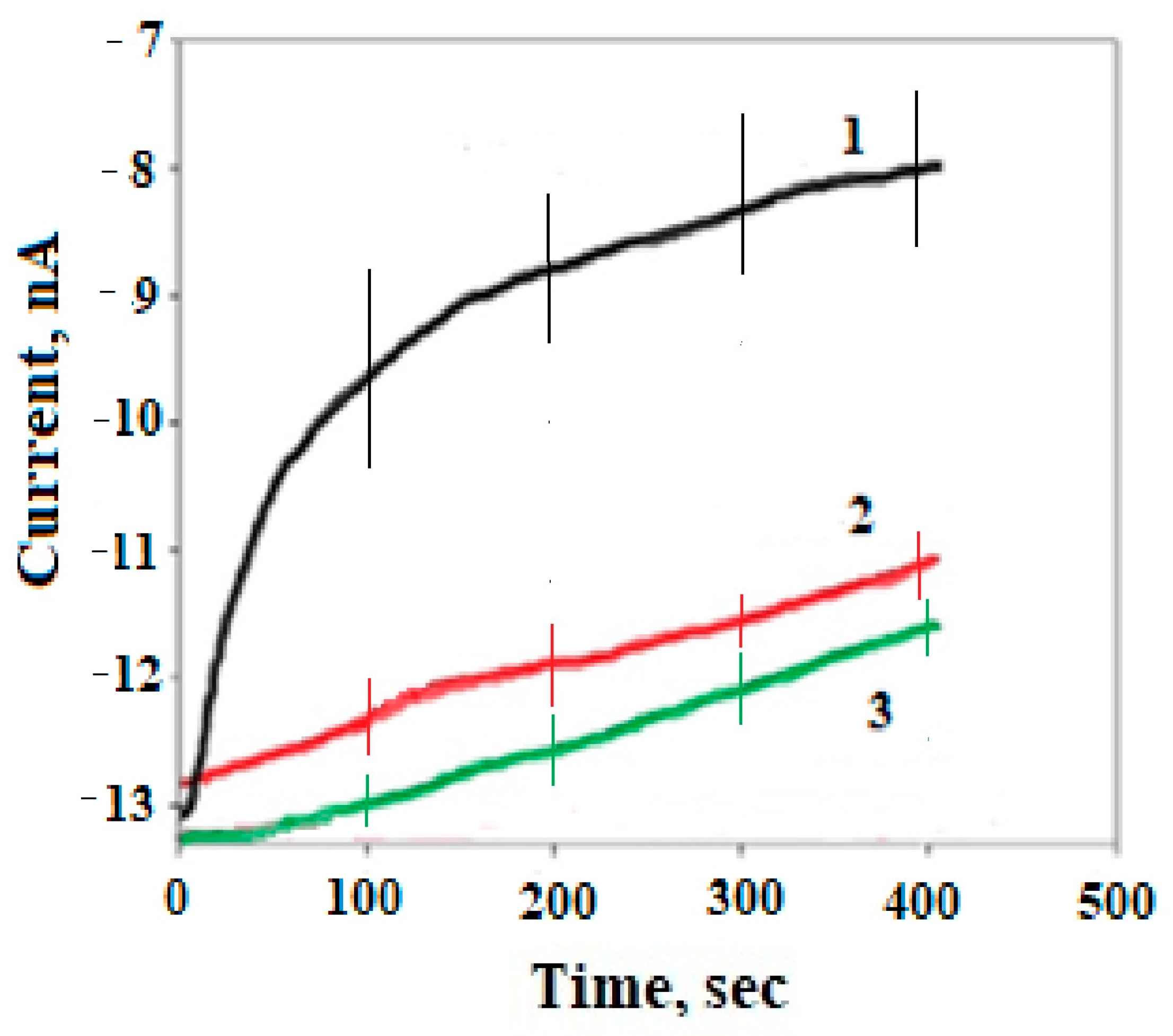
The hypothesis regarding the competition of two alternative substrates in A. ferrooxidans Thio1 was tested in additional experiments. Substrate oxidation activity was assessed using an amperometric method based on oxygen consumption. The tested substrates were iron, formate, and their mixture (1:1), added to the solution at an equimolar concentration of 180 μM. Figure 3 shows the measurement results after subtracting the endogenous respiration of the bacteria. The results indicated that the strain Acidithiobacillus sp. Thio1 exhibited the highest activity in iron oxidation. This is consistent with the ability to oxidize iron as a characteristic feature of strain Thio1: the strain was isolated from an iron-containing deposit and was initially maintained in the 9K medium with iron. The strain Acidithiobacillus sp. Thio1 was also capable of oxidizing formate (Figure 3). Consistent with these data, the oxidation activity for the formate–iron mixture showed an intermediate level. However, this intermediate level was much lower than the average oxygen consumption curves for ferrous iron and formate taken separately, which may indicate some suppression of activity when using alternative substrates.

3.5. Amperometric Evaluation of the Bacterial Acute Respiratory Response to Toxicants

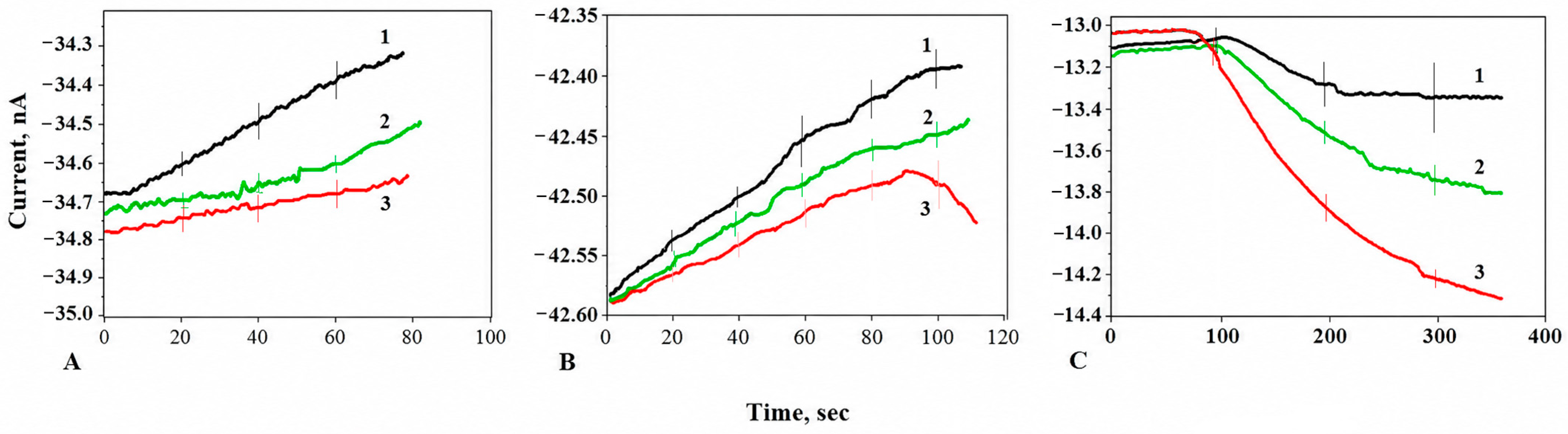
The Acidithiobacillus sp. Thio1 respiratory response to the tested toxicants was assessed based on the maintained respiratory activity of the biomass in the 9K medium. As described above, these parameters were measured amperometrically as changes in the electrode current. Oxygen consumption data, adjusted by subtracting endogenous respiration, served as indicators of the respiratory response to toxicants. The results, excluding endogenous biomass respiration, are presented in Figure 4.
The data showed that both zinc and copper ions suppressed the respiratory/oxidative activity of the bacteria (Figure 4A,B). Increasing concentrations of toxic metal ions led to a decrease in bacterial activity. Nevertheless, respiratory activity was maintained despite the absence of oxidizable substrates in the medium. This phenomenon indicates that bacterial oxygen consumption continued due to defense mechanisms and cellular reserves, showing some short-term tolerance. These experiments only show the acute effects of toxicants on respiratory activity, so they do not establish long-term resistance. The results also show the potential for comparing various strains by their acute respiratory response to toxicants, indicating that the Acidithiobacillus sp. Thio1 strain may be promising for the industrial leaching of pyrite–chalcopyrite ore.
Bacterial inhibition by arsenate increased with its concentration (Figure 4C). In contrast to the results of the experiments with toxic metal ions, all obtained values were below the initial level of endogenous respiration (Figure 4C). Differences in bacterial activity at various arsenate concentrations suggested that the bacteria may not be completely killed. Nevertheless, using the amperometric method does not allow the determination of residual bacterial activity, but it does record varying decreases in the measured electrode current. In these cases, it is possible to interpret this decrease as respiration inhibition, which was different at various arsenate concentrations.
Thus, the presented data do not provide a predictive model for the long-term survival of bacteria in the presence of toxicants. However, they demonstrate the feasibility of using the amperometric method to obtain the short-term acute respiratory response of strains to various toxicants and their concentrations, which expands present microbiological knowledge.

3.6. Amperometric Evaluation of Solid Substrates Oxidation

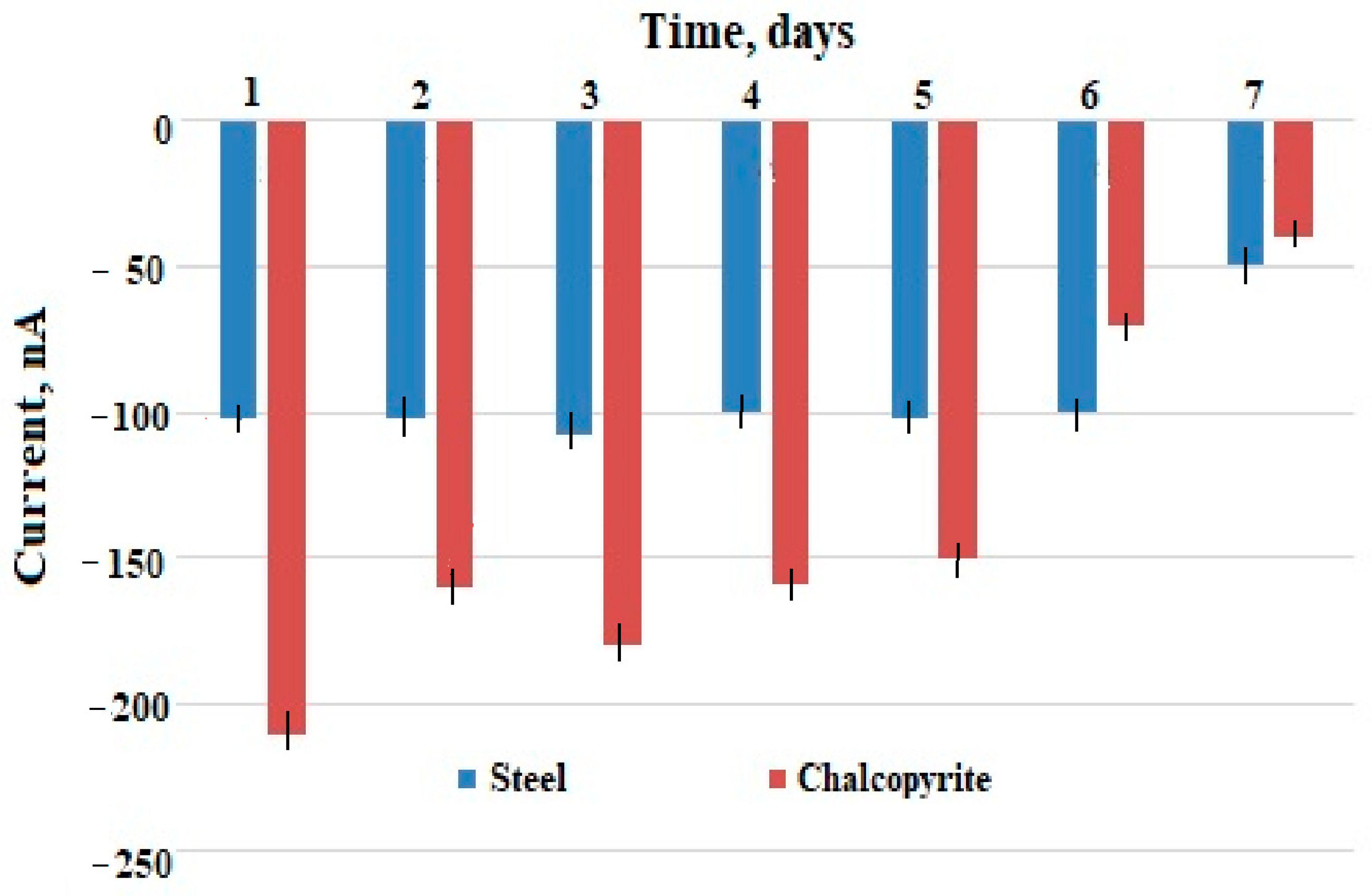
The biocorrosion of solid substrates cannot be assessed with the same accuracy as the bacterial reaction of Acidithiobacillus sp. Thio1 with dissolved substrates, since the surface of the metal or ore specimens in contact with the biomass is not uniform at the microscopic level. In addition, the interaction of bacteria with a solid substrate requires initial cell adhesion [22,23]; therefore, measurements were carried out once a day over a 7-day period. As shown in Figure 5, the steel began to undergo noticeable corrosion only on the 7th day. The less uniform ore contained not only chalcopyrite but also pyrite, which is more accessible to iron-oxidizing acidithiobacilli. These specimens demonstrated an increase in electrode current generation starting from the first days of incubation (Figure 5). Corrosion or leaching of the pyrite–chalcopyrite ore by Acidithiobacillus sp. Thio1 is also shown above in Table 1. It can be assumed that current generation during corrosion by iron-oxidizing bacteria, especially A. ferrooxidans [24], provides a basis for studying various materials under biocorrosion conditions. Meanwhile, the direct quantitative evaluation of biocorrosion via oxygen consumption is not yet applicable because the chemical composition of the produced oxides remains unknown.

3.7. Microscopic Evaluation of Solid Substrates Oxidation

The previous paragraph noted that the resulting electric current was generated in the electrode cell with solid samples and was therefore attributed to corrosion processes. These processes were linked to the activity of the iron-oxidizing bacterium Acidithiobacillus sp. Thio1. However, this area of research is new and poorly understood. Therefore, we conducted a microscopic examination of the samples to confirm the presence of microbially induced corrosion and verify its absence in the sterile controls.
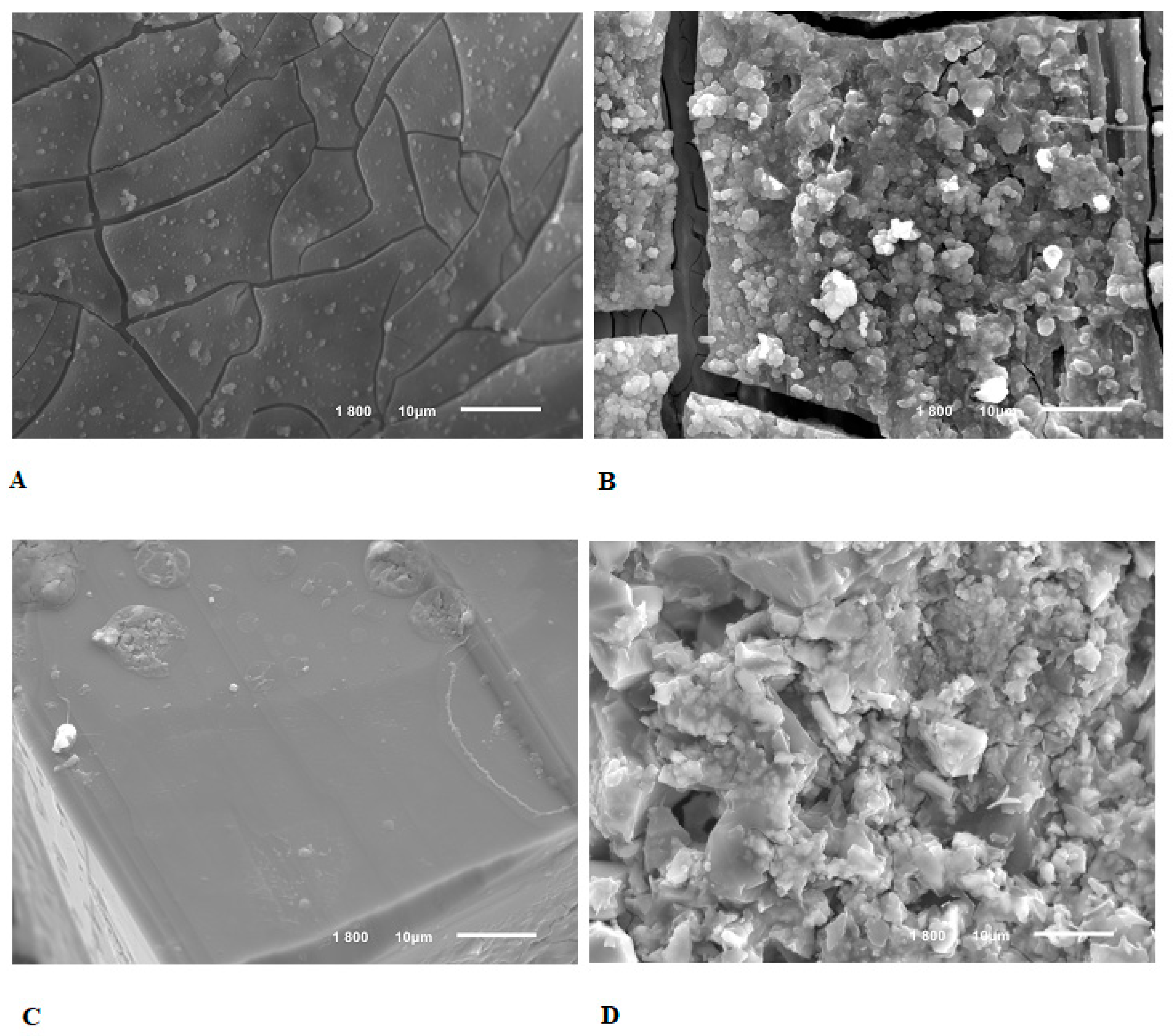
Scanning electron micrographs (Figure 6) demonstrate that uninoculated steel and pyrite–chalcopyrite ore samples remained relatively undamaged after 7 days in an acidic environment (Figure 6A,C). In contrast, the inoculated samples became covered with noticeable biofouling and inorganic microdeposits, likely representing corrosion products, over the same period. Overall, the microscopic analysis supports the corrosive role of Acidithiobacillus sp. Thio1 on steel and ore, aligning with the bacterial origin of the increased electrode current observed in the amperometric measurements (Figure 5) and the results of the ore leaching tests (Table 1).
Overall, studying the bacterial corrosion of solid inorganic substrates, especially metals, requires more detailed analyses. These include elemental and structural characterization, the investigation of galvanic coupling and impedance currents, and the tracking of the behavior of carbon in steel, silumin, and other alloys. Such experiments can serve as a basis for future research into the resistance of metallic alloys to biocorrosion. The presented data demonstrate that the amperometric method can be effectively incorporated into the toolbox for assessing metal resistance to biocorrosion.

4. Conclusions

Iron-oxidizing bacteria, Acidithiobacillus are of practical importance due to their use in the biomining of metals, including from deposits containing toxic copper and/or arsenic. Typically, the activity of these bacteria is studied in long-term laboratory experiments that simulate industrial conditions as closely as possible. Over the past decade, it has been shown that bacterial activity can also be measured in short-term experiments by assessing the electrode current generated during substrate oxidation.
The object of this study was the obligate acidophilic autotrophic iron-oxidizing bacterium Acidithiobacillus sp. strain Thio1. The amperometric assay detected changes in electrode current—and thus in bacterial respiratory activity—after the addition of dissolved energy substrates or toxicants. The study involved the comparative oxidation of various compounds using the same biomass specimen. This approach appears useful for comparative research in short-term experiments.
Additionally, the biocorrosion of pyrite–chalcopyrite ore and steel was examined. Scanning electron microscopy revealed biofouling only in the inoculated specimens. Oxygen consumption, as evaluated via the amperometric method, was in good agreement with these results. Furthermore, the amperometric data agreed with the results of conventional pyrite–chalcopyrite bioleaching experiments (Table 1).
Experiments with solid substrates are not yet sufficient for the practical implementation of the amperometric method, but they represent a possible avenue for future applications. Overall, the study demonstrated that the amperometric method can be integrated into the microbiological toolbox as a complementary technique.

Author Contributions

Conceptualization, A.Y. and M.V.; methodology, A.Y., T.K., A.Z., T.A. and A.R.; validation, T.A.; investigation, A.Y., T.K., A.Z. and T.A.; writing—original draft preparation, review and editing, M.V.; supervision, M.V.; funding acquisition, M.V. and A.R. All authors have read and agreed to the published version of the manuscript.

Funding

The work was carried out with the financial support of the Ministry of Science and Higher Education of the Russian Federation, state assignment FMRM-2026-0020.

Institutional Review Board Statement

Not applicable.

Informed Consent Statement

Not applicable.

Data Availability Statement

The original contributions presented in this study are included in the article. Further inquiries can be directed to the corresponding author.

Conflicts of Interest

The authors declare no conflicts of interest. The funders had no role in the design of the study; in the collection, analyses, or interpretation of data; in the writing of the manuscript; or in the decision to publish the results.

References

  1. Kumar, A.; Lima, A.T.; Kirkelund, G.M.; Jensen, P.E.; Ottosen, L.M.; Funari, V.; Shemi, A.; Ndlovu, S.; Gomes, H.I. Bioleaching: From natural ores to urban mines for sustainability, circularity, and carbon neutrality. Resour. Conserv. Recycl. 2026, 227, 108746. [Google Scholar] [CrossRef] [Scilit]
  2. Xia, L.; Yin, C.; Dai, S.; Qiu, G.; Chen, X.; Liu, J. Bioleaching of chalcopyrite concentrate using Leptospirillum ferriphilum, Acidithiobacillus ferrooxidans and Acidithiobacillus thiooxidans in a continuous bubble column reactor. J. Ind. Microbiol. Biotechnol. 2010, 37, 289–295. [Google Scholar] [CrossRef] [Scilit] [PubMed]
  3. Zhang, R.; Sun, C.; Kou, J.; Zhao, H.; Wei, D.; Xing, Y. Enhancing the leaching of chalcopyrite using Acidithiobacillus ferrooxidans under the induction of surfactant Triton X-100. Minerals 2019, 9, 11. [Google Scholar] [CrossRef] [Scilit]
  4. Liu, X.; Li, Y.; Cao, J.; Zeng, Z.; Liu, X.; Zhang, R.; Li, Q.; Sand, W. Bioleaching of chalcopyrite waste rock in the presence of the copper solvent extractant LIX984N. Front. Microbiol. 2022, 13, 820052. [Google Scholar] [CrossRef] [Scilit] [PubMed]
  5. Silverman, M.P.; Lundgren, D.G. Studies on the chemoautotrophic iron bacterium Ferrobacillus ferrooxidans. I. An improved medium and a harvesting procedure for securing high cell yields. J. Bacteriol. 1959, 77, 642–647. [Google Scholar] [CrossRef] [Scilit] [PubMed]
  6. Fomchenko, N.V.; Novikov, G.V.; Melamud, V.S.; Muravyov, M.I. Bioleaching of nickel-containing metallurgical slag by chemolithotrophic microorganisms at different temperatures. Appl. Biochem. Microbiol. 2025, 61, 1133–1140. [Google Scholar] [CrossRef] [Scilit]
  7. Bulaev, A.G.; Artykova, A.V.; Elkina, Y.u.A.; Kolosov, A.V.; Nechaeva, A.V.; Beletsky, A.V.; Kadnikov, V.V.; Melamud, V.S.; Mardanov, A.V. Bioleaching of copper-zinc concentrate at different temperature conditions. Microbiology 2024, 93, 773–784. [Google Scholar] [CrossRef] [Scilit]
  8. Yachkula, A.; Kuvichkina, T.; Zvonarev, A.; Abashina, T.; Reshetilov, A.; Vainshtein, M. Extremophilic bacteria Acidithiobacillus ferrooxidans: Substrate preference and response to As-compounds evaluated by oxygen consumption. Microbiology 2026, 95, 243–250. [Google Scholar] [CrossRef] [Scilit]
  9. Chiang, A.J.; Hasty, J. Design of synthetic bacterial biosensors. COMICR 2023, 76, 102380. [Google Scholar] [CrossRef] [Scilit] [PubMed]
  10. Zlatev, R.; Magnin, J.P.; Ozil, P.; Stoytcheva, M. Bacterial sensors based on Acidithiobacillus ferrooxidans: Part I. Fe2+ and S2O32− determination. Biosens. Bioelectron. 2006, 21, 1493–1500. [Google Scholar] [CrossRef] [Scilit] [PubMed]
  11. Zlatev, R.; Magnin, J.P.; Ozil, P.; Stoytcheva, M. Bacterial sensors based on Acidithiobacillus ferrooxidans: Part, I.I. Cr(VI) determination. Biosens. Bioelectron. 2006, 21, 1501–1506. [Google Scholar] [CrossRef] [Scilit] [PubMed]
  12. Banik, R.M.; Mayank; Prakash, R.; Upadhyay, S.N. Microbial biosensor based on whole cell of Pseudomonas sp. for online measurement of p-Nitrophenol. Sens. Actuators B Chem. 2008, 131, 295–300. [Google Scholar] [CrossRef] [Scilit]
  13. Arlyapov, V.; Kamanin, S.; Ponamoreva, O.; Reshetilov, A. Biosensor analyzer for BOD index express control on the basis of the yeast microorganisms Candida maltosa, Candida blankii, and Debaryomyces hansenii. Enzym. Microb. Technol. 2012, 50, 215–220. [Google Scholar] [CrossRef] [Scilit] [PubMed]
  14. Tarasov, S.E.; Plekhanova, Y.V.; Bykov, A.G.; Kadison, K.V.; Medvedeva, A.S.; Reshetilov, A.N.; Arlyapov, V.A. Novel conductive polymer composite PEDOT:PSS/bovine serum albumin for microbial bioelectrochemical devices. Sensors 2024, 24, 905. [Google Scholar] [CrossRef] [Scilit] [PubMed]
  15. Pronk, J.T.; Meijer, W.M.; Hazeu, W.; van Dijken, J.P.; Bos, P.; Kuenen, J.G. Growth of Thiobacillus ferrooxidans on formic acid. Appl. Environ. Microbiol. 1991, 57, 2057–2062. [Google Scholar] [CrossRef] [Scilit] [PubMed]
  16. Van de Peer, Y.; De Wachter, R. TREECON for Windows: A software package for the construction and drawing of evolutionary trees for the Microsoft Windows environment. Bioinformatics 1994, 10, 569–570. [Google Scholar] [CrossRef] [Scilit] [PubMed]
  17. Sverchkov, I.P.; Povarov, V.G. Quantitative determination of sulfur forms in bottom sediments for rapid assessment of the industrial facilities impact on aquatic ecosystems. J. Min. Inst. 2024, 267, 372–380. [Google Scholar]
  18. Franklin, R.B.; Campbell, A.H.; Higgins, C.B.; Barker, M.K.; Brown, B.L. Enumerating bacterial cells on bioadhesive coated slides. J. Microbiol. Methods 2011, 87, 154–160. [Google Scholar] [CrossRef] [Scilit] [PubMed]
  19. Yachkula, A.; Rozova, O.; Abashina, T.; Vainshtein, M.; Grouzdev, D.; Bulaev, A. Attempts to stimulate leaching activity of Acidithiobacillus ferrooxidans strain TFBk. Minerals 2022, 12, 1051. [Google Scholar] [CrossRef] [Scilit]
  20. Kuvichkina, T.N.; Nosulich, V.E.; Doronina, N.V.; Makarenko, A.A.; Reshetilov, A.N. Phenol oxidation by immobilized Pseudomonas monteiliizima cells. Bull. Tula State Univ. Nat. Sci. 2020, 2, 44–55. [Google Scholar]
  21. Kuvichkina, T.N.; Kaparullina, E.N.; Doronina, N.V.; Reshetiloiv, A.N. Rhodococcus qingshengii GlMm1 as the basis for a biosensor for determination of the fungicide carbendazim. Microbiology 2024, 93, 160–162. [Google Scholar] [CrossRef] [Scilit]
  22. San Martín, F.; Aguilar, C. Study of the Adhesion Mechanism of Acidithiobacillus ferrooxidans to Pyrite in Fresh and Saline Water. Minerals 2019, 9, 306. [Google Scholar] [CrossRef] [Scilit]
  23. Tonietti, L.; Esposito, M.; Cascone, M.; Barosa, B.; Fiscale, S.; Muscari Tomajoli, M.T.; Sbaffi, T.; Santomartino, R.; Covone, G.; Cordone, A.; et al. Unveiling the bioleaching versatility of Acidithiobacillus ferrooxidans. Microorganisms 2024, 12, 2407. [Google Scholar] [CrossRef] [Scilit] [PubMed]
  24. Lin, L.; Tsou, C.-H.; Dou, B.; Yan, S.; Zeng, Y.; Gong, M. Electrochemical corrosion behavior and mechanism of iron-oxidizing bacteria Thiobacillus ferrooxidans from acid mine drainage on Q235 carbon steel. New J. Chem. 2022, 46, 20279–20291. [Google Scholar] [CrossRef] [Scilit]
Figure 1. A phylogenetic tree based on 16S rRNA gene sequences shows positions of three Acidithiobacillus strains in the author’s collection, including Thio1, among representatives of the genus and species. Strain Thio1 is marked with an asterisk. The scale bar corresponds to 0.01 substitutions per nucleotide position (evolutionary distance).
Microbiolres 17 00162 g001
Figure 2. Growth dynamics of Acidithiobacillus sp. Thio1 in the 9K medium with various formate concentrations. Abscissa axis—incubation time, days; ordinate axis—cell number, 106 cells/mL. Error bars represent the standard deviation values.
Microbiolres 17 00162 g002
Figure 3. Generated electric current as an indicator of oxygen consumption by Acidithiobacillus sp. Thio1 in presence of different substrates, 180 μM. The substrate designations are shown in figure: 1—ferrous iron; 2—mixture of ferrous iron and formate, 1:1; 3—formate. Abscissa axis—time, sec; ordinate axis—electric current, nA. Error bars represent the standard deviation values.
Microbiolres 17 00162 g003
Figure 4. Generated electric current as an indicator of the oxygen consumption by Acidithiobacillus sp. Thio1 in presence of added toxic compounds, mM. (A) Copper sulfate: 1—4.0, 2—20.0, and 3—60.0; (B) zinc sulfate: 1—3.48, 2—17.4, and 3—52.2; (C) arsenate: 1—0.026, 2—2.60, 3—26.4. Abscissa axis—incubation time, sec; ordinate axis—electric current, nA. Error bars represent the standard deviation values.
Microbiolres 17 00162 g004
Figure 5. Generated electric current as an indicator of the oxygen consumption by Acidithiobacillus sp. Thio1 in presence of steel (blue) or pyrite–chalcopyrite (red). Abscissa axis—time, days; ordinate axis—current, nA. Error bars represent the standard deviation values.
Microbiolres 17 00162 g005
Figure 6. Scanning microscopy of the solid specimens on the 7th day of incubation. (A) steel, not inoculated; (B) steel, inoculated with Acidithiobacillus sp. Thio1; (C) pyrite–chalcopyrite ore, not inoculated; (D) pyrite–chalcopyrite ore, inoculated with Acidithiobacillus sp. Thio1. All images were captured at 1800× magnification. The scale bar is shown in Figure, 10 µm.
Microbiolres 17 00162 g006
Table 1. Leaching of pyrite–chalcopyrite ore with Acidithiobacillus sp. Thio1 at 24 °C. The best final results are highlighted in bold. Initial pH values were 1.9–2.2, and the final ones were 1.7–1.9. Analytical data are presented as the mean value ± SD. The coefficient of variation (CV) values did not exceed 5%.
Dissolved
Chemicals
Concentrations of the Chemicals (Mean Value ± SD), mg/L, on Different Days of Incubation
Days: 151012
Chemical leaching
Fe0.13 ± 0.0050.16 ± 0.0050.21 ± 0.0100.32 ± 0.012
S0.10 ± 0.0030.12 ± 0.0040.17 ± 0.0050.33 ± 0.011
Cu0.01 ± <0.0010.01 ± <0.0010.04 ± <0.0010.05 ± <0.001
Chemical and bacterial leaching
Fe0.17 ± 0.0060.97 ± 0.0403.50 ± 0.1704.70 ± 0.230
S0.10 ± 0.0030.70 ± 0.0252.00 ± 0.1002.18 ± 0.108
Cu0.00 ± <0.0010.01 ± <0.0010.08 ± 0.0020.10 ± 0.002
Chemical and bacterial leaching with supplemented formate
Fe0.04 ± 0.0020.22 ± 0.0103.75 ± 0.1754.20 ± 0.200
S0.04 ± 0.0010.86 ± 0.0331.50 ± 0.0602.75 ± 0.110
Cu0.01 ± <0.0010.14 ± 0.0030.15 ± 0.0030.16 ± 0.003
Disclaimer/Publisher’s Note: The statements, opinions and data contained in all publications are solely those of the individual author(s) and contributor(s) and not of MDPI and/or the editor(s). MDPI and/or the editor(s) disclaim responsibility for any injury to people or property resulting from any ideas, methods, instructions or products referred to in the content.