Article

Preliminary Experimental Study on the Removal of Staphylococcus epidermidis and Pseudomonas aeruginosa from Surgical Instrument Surfaces Under Controlled Conditions


by
Edmar Gonçalves Pereira Filho
1,
Stéfanne Rodrigues Rezende Ferreira
2,
Amanda Veiga Paiva Simões
1,
Eli Júnior Pereira Rodrigues
1,
Iorrana Morais de Oliveira
1,
Marillia Lima Costa
1,
Adeliane Castro da Costa
1,
Berendina Elsina Bouwman
1 and
Hanstter Hallison Alves Rezende
1,*
1
Laboratory of Bacteriology and Micology, Institute of Health Sciences, Federal University of Jataí, Jataí 75801-615, Goiás, Brazil
2
Institute of Tropical Pathology and Public Health, Federal University of Goiás, Goiânia 74605-050, Goiás, Brazil
*
Author to whom correspondence should be addressed.
Microbiol. Res. 2026, 17(4), 77; https://doi.org/10.3390/microbiolres17040077

Submission received: 22 February 2026 / Revised: 1 April 2026 / Accepted: 3 April 2026 / Published: 8 April 2026

Abstract

The objective of this study is to evaluate the efficiency of surgical instruments’ manual cleaning versus automated cleaning in an ultrasonic cleaner for the removal of biofilms on surgical forceps contaminated with Staphylococcus epidermidis and Pseudomonas aeruginosa. Subsequently, the residual microbial load was quantified through microbiological culture, aiming to evaluate the effectiveness of biofilm removal under different reprocessing conditions. Cleaning is an essential step in the processing of surgical instruments to ensure the effective removal of dirt and microorganisms. Through adhesion, microorganisms can attach to surfaces and form biofilms, organized structures surrounded by an extracellular matrix consisting of various components, which favor metabolic exchanges, adaptation, resistance, and bacterial dispersion. These biofilms increase the pathogenic potential of microorganisms, contributing to the occurrence of Healthcare-Associated Infections, and to avoid these, it is essential that preventive measures aimed at microbial reduction are adopted. Automated cleaning proved more effective than manual cleaning, and the combined approach achieved the greatest microbial reduction, though persistent contamination was still observed. The ability of adhesion and biofilm formation on the surfaces of surgical instruments is regarded as a challenge for complete microbial removal. These findings enhance the need for more rigorous reprocessing protocols and complementary strategies to ensure greater safety in the use of reusable instruments in clinical practice.

1. Introduction

Cleaning is an essential step in the processing of surgical instruments, as its purpose is to remove dirt and microorganisms that can compromise subsequent steps, such as disinfection and sterilization [1].
Instrument cleaning can be performed in two ways: manually and automatically. The former is carried out mechanically, using brushes, enzymatic detergents, and running water. It requires extra care to ensure that all surfaces, joints, and lumens are properly scrubbed and rinsed. The latter is carried out using equipment such as ultrasonic cleaners and thermodisinfectors, which promote greater process standardization, reduction of occupational exposure, and better efficiency in removing dirt [2].Cleaning failures can favor the occurrence of healthcare-associated infections (HAIs). These infections are defined as clinical manifestations acquired after healthcare procedures or hospitalization, generally from the third day of hospitalization, putting patients and healthcare professionals’ safety at risk [3,4]. According to estimates from the World Health Organization (WHO), HAIs affect millions of people annually, with a prevalence of about 7% in developed countries and up to ~15% in developing countries. They represent one of the main factors contributing to antimicrobial resistance, being an adverse event with predictability and potential for prevention [5].
Among HAIs, those associated with invasive procedures, such as surgical site Infections (SSIs), stand out, and, when acquired, lead to varied impacts on the patient, ranging from mild discomfort to serious complications. This problem becomes even more serious given the spread of multidrug-resistant microorganisms and their ability to form biofilms [3,6].
Biofilms are microbial communities surrounded by an extracellular matrix. This matrix establishes a protective microenvironment that shields bacterial cells from chemical, biological, and mechanical agents, hindering complete removal by conventional cleaning and sterilization [7,8]. The development process is described in five stages: attachment, multiplication, exodus, maturation, and dispersion, which ensure both the persistence and dissemination of microorganisms [9,10,11]. This structure provides greater resistance to antimicrobial agents and hinders the cleaning process of contaminated materials. The extracellular matrix creates a microenvironment that protects bacterial cells against chemical, biological, and mechanical agents, making complete removal by conventional cleaning and subsequent disinfection and/or sterilization processes difficult [12].
In this context, opportunistic microorganisms gain clinical prominence. Recent studies demonstrate the biofilm-forming capacity of Klebsiella spp. isolated in ICUs, as well as E. coli in urinary tract infections (UTIs) and Enterococcus spp. colonizing healthcare products [13,14,15,16]. In this scenario, two microorganisms stand out: Staphylococcus epidermidis and Pseudomonas aeruginosa. Staphylococcus epidermidis, a common inhabitant of human skin, is capable of colonizing the surfaces of medical devices, showing high competence in the formation of biofilms and increasing resistance to antimicrobials [17,18]. When colonizing asymptomatically as part of the microbiota, it is exposed to medical devices and is therefore frequently identified in infections, with potential progression to systemic infections, representing a high risk factor in neonatal, elderly, and immunocompromised patients [19].
In addition to its clinical relevance, due to its well-known ability to form biofilms, it has become one of the main model bacteria in research on this structure [20]. Adhesion molecules of Staphylococcus epidermidis facilitate its attachment to skin and/or surfaces through accumulation-associated proteins (Aap), extracellular matrix-binding proteins (Embp), and MSCRAMMs, as well as the polysaccharide intercellular adhesin (PIA), which contributes to biofilm stability and reduces dehydration. Aap plays a dual role, promoting initial binding and cellular accumulation, while Embp and MSCRAMMs reinforce adhesion as an essential factor for colonization, consolidating S. epidermidis as a strong biofilm former [21,22]
Likewise, species of the genus Pseudomonas, in particular P. aeruginosa, present rapid environmental adaptation, multiple virulence factors, and high tolerance to disinfectants, which favors persistent colonization and the progression to chronic infections [23,24,25]. This microorganism stands out due to its significant clinical impact in hospital settings. Its ability to easily adhere to medical devices and tissues has made it widely studied in biofilms [26]. This structure plays a central role in virulence by directly interfering with other factors, such as toxin production and antimicrobial resistance. In addition, it remodels survival strategies, reinforcing its notoriety as an agent of persistent infections [19,27].
Biofilm formation in P. aeruginosa is initially facilitated by adhesion through flagella and type IV pili. Another important factor is the quorum-sensing (QS) communication system, which modulates gene expression and other factors, together with coordinated integration with nutrient availability and stress conditions, ensuring adaptability and persistence in clinical environments [28,29].
In conjunction with direct colonization of devices, environmental contamination is also a determining factor in HAIs. Hospital environments, such as surgical suites, can act as reservoirs for microorganisms, spread by patients, professionals, air, or equipment. This enhances the need for strict handling and storage protocols for reprocessed products [25,30]. Biofilm formation, both on biotic (mucosal) and abiotic surfaces (implants, instruments, and health products), contributes to microbial persistence and hinders infection treatment [6].
Given this scenario, adequate processing of instruments becomes essential. According to the Spaulding classification and WHO recommendations [31,32], critical and semi-critical devices must be cleaned before disinfection or sterilization, preferably through automated methods. Although these methods provide better results, manual cleaning still plays a complementary role in hard-to-reach areas.
However, research shows that even the combination of techniques does not guarantee the complete removal of biofilms. This finding highlights the need for additional strategies, continuous monitoring, and process improvement [30,33,34,35]. This study aimed to comparatively evaluate manual, ultrasonic, and combined cleaning methods in reducing bacterial biofilms on critical areas of surgical forceps. The objective was to assess not only statistical reductions but also the ability of these methods to achieve the absence of detectable CFU, a clinical parameter essential for critical instruments. This comparative approach is justified because biofilm persistence on reusable surgical instruments remains a recognized challenge in infection control and patient safety. By quantifying microbial reduction and highlighting residual contamination under controlled experimental conditions, the study provides evidence that supports discussions on the adequacy of current reprocessing protocols and reinforces the need for complementary strategies to strengthen biosafety.

2. Materials and Methods

2.1. Experimental Design

The experiment was performed in duplicate using 24 plain anatomical forceps with 14 cm straight serration (6B Invent Germany®, Água Branca, São Paulo, Brazil), made of AISI 420 surgical stainless steel according to ABNT NBR ISO 7153-1 [36] a material predominantly used in the manufacture of critical surgical instruments, particularly forceps and scissors. Fragments (~1 cm) were obtained with a manual guillotine (Ferramenta Vitalicia®, Olhão, Portugal). Fragments were obtained for contamination by immersion, and the shaft and serrated tip regions were selected due to their irregular surfaces, which pose greater challenges to the cleaning process as they represent critical areas for the retention of debris and biofilms. This experimental design allowed greater control and comparability among the tested methods, simulating irregular surfaces with difficult access commonly encountered in healthcare settings. However, the use of fragments represents a limitation, as it does not fully replicate the complete surgical instrument.
The fragments were distributed between the microorganisms S. epidermidis and P. aeruginosa, so that each microorganism was tested at three different time points with four fragments at each time, distributed into four groups with one fragment per group. The experiment was conducted in duplicate, totaling 24 fragments for each microorganism. The choice of these microorganisms was based on their clinical relevance and on the fact that they are classical models of biofilm formation.

2.2. Sterilization and Controlled Contamination

The fragments were initially subjected to cleaning in an ultrasonic bath (Solidsteel®, Piracicaba, São Paulo, Brazil) for 15 min, rinsed in sterile distilled water, and dried. They were then packaged in surgical-grade paper and sterilized in an autoclave (Prismatec®, Itu, São Paulo, Brazil) with saturated steam under pressure, using the standard cycle of 121 °C for 15 min.
After sterility was confirmed, the fragments were contaminated by immersion in 50 mL of Tryptic Soy Broth (HiMedia Labs®, Kennett Square, PA, USA) containing 106 CFU/mL of Staphylococcus epidermidis (ATCC 12228) or Pseudomonas aeruginosa (ATCC 0027). The concentration of 106 CFU/mL was standardized to ensure uniform and controlled contamination, avoiding saturation of the fragment surfaces. We acknowledge, however, that this model does not reproduce the presence of organic load, such as blood or tissue. This approach allowed the isolation of the effect of cleaning methods on bacterial biofilms, ensuring comparability of results among the experimental groups.
The fragments were kept in a BOD incubator (Lucadema®, São Jose do Rio Preto, São Paulo, Brazil) at 35 ± 2 °C, in aerobic conditions, for 1, 6, or 12 h, with periodic manual agitation to promote contact between the cells and the metallic surfaces. This time-course evaluation enabled the assessment of cleaning under progressive conditions, aiming to represent the different stages of biofilm development.

2.3. Experimental Groups

After contamination, the fragments were aseptically removed and distributed into four groups.
Group 1—Positive control (contamination): fragments examined without cleaning, to confirm adhesion and biofilm.
Group 2—Manual cleaning: the fragments were subjected to chemical action by immersion in 10 mL of enzymatic detergent (Dinâmica®, São Paulo, Brazil; 100 mL/L in sterile distilled water) for 5 min, followed by mechanical action through friction with a soft-bristled brush (Bettanin SA®, Esteio, Brazil), five unidirectional movements, on a sterile Petri dish, followed by rinsing in distilled water.
Group 3—Automated cleaning: fragments immersed in the same detergent solution and subjected to an ultrasonic cleaner (Solidsteel®, Piracicaba, São Paulo, Brazil) operated at 40 kHz and 40 °C for 15 min, with subsequent rinsing in distilled water, thereby promoting mechanical, chemical (detergent), and thermal (water heating) action.
Group 4—Manual + automated cleaning: sequential application of the procedures described in Groups 2 and 3.

2.4. Bacterial Recovery

After each procedure, the fragments in each group were transferred to sterile microtubes containing 1.0 mL of sterile distilled water and were subsequently vortexed for two 1-min cycles (30-s interval) to detach the adhered cells. Aliquots of 10 µL were seeded in three replicates on Nutrient Agar (MBioLog®, Contagem, Minas Gerais, Brazil), using a Drigalski loop, for quantification in CFU. For the control group, serial dilutions up to 1:100 were performed, plating in three replicates and incubation at 37 °C in aerobiosis for 24 h, followed by CFU counting.

2.5. Statistical Analysis

The results were recorded in Microsoft Excel®, transformed into log10 values, and analyzed in GraphPad Prism 9.0® (GraphPad Software, San Diego, CA, USA). The Shapiro–Wilk test was applied, showing a distribution close to normality, which justified the use of one-way ANOVA and the Mann–Whitney test for pairwise comparisons between groups.

3. Results

After the contamination periods (1, 6, and 12 h), the fragments were processed according to the respective experimental groups, and the recovered bacterial load was quantified (Table 1). In general, a gradual increase in bacterial load was observed as a function of exposure time for both microorganisms tested. The progressive increase over time reinforced the need for immediate cleaning, since even small exposures favor the initial formation of biofilms. Upon further analysis, it was possible to observe that the data from S. epidermidis showed lower variability, displaying a more uniform behavior. For P. aeruginosa, the results were more heterogeneous, with higher standard deviations and differences between duplicates.
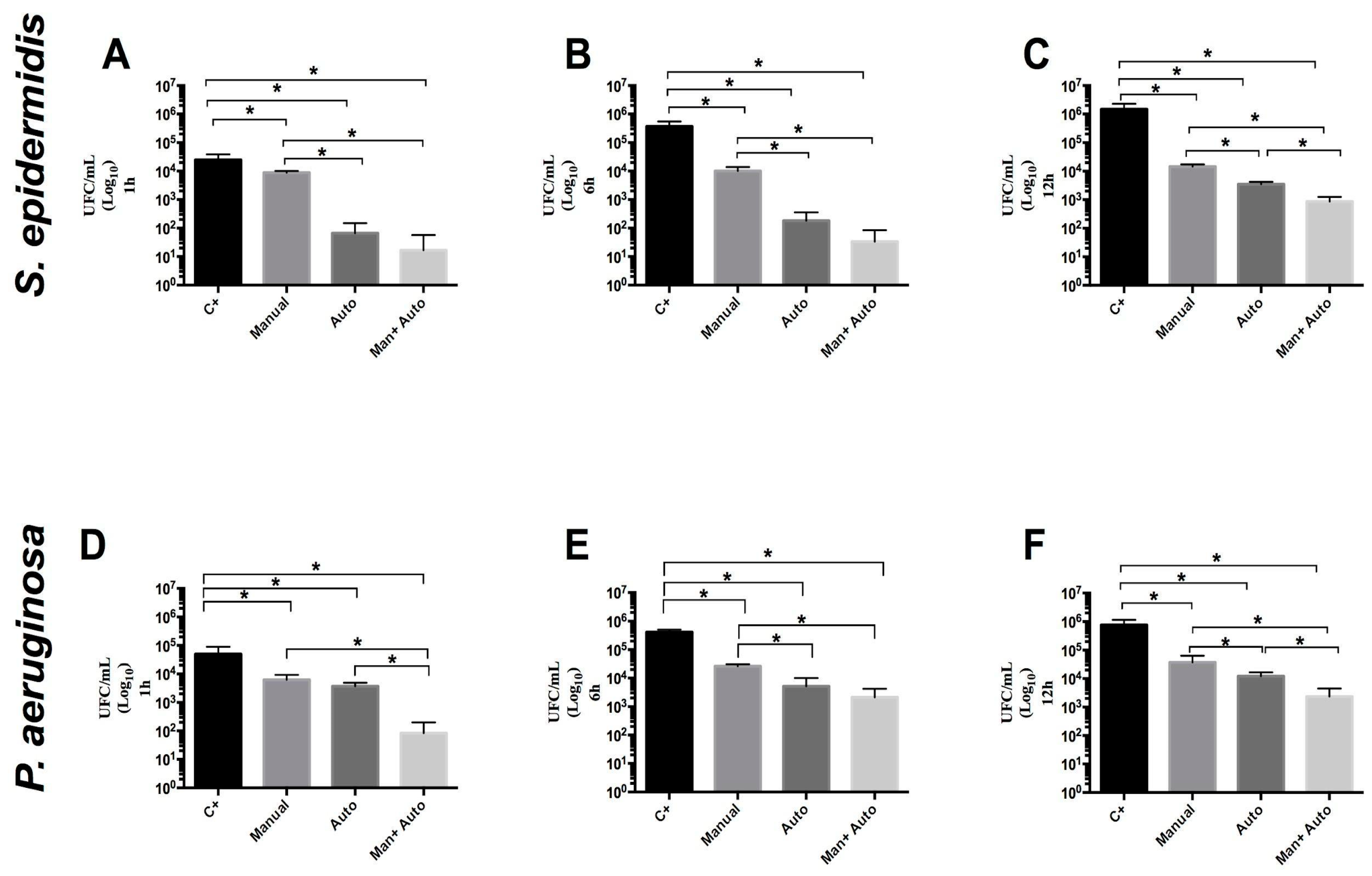
As demonstrated in Figure 1A (S. epidermidis) and 1D (P. aeruginosa), after one hour of exposure, the fragments subjected to the different cleaning methods showed a significant reduction (* p < 0.05) in relation to the control group. There was also a significant difference between the manual cleaning group and the groups subjected to automated and manual + automated modalities. In the case of S. epidermidis, automated cleaning alone presented a higher bacterial load compared to manual + automated cleaning, but this difference was not statistically significant (p > 0.05). The more consistent results suggested that S. epidermidis biofilms were more resistant to mechanical removal. To P. aeruginosa, however, the difference between the two methods was significant (* p < 0.05), which could have been related to structural heterogeneity.
After six hours of exposure (Figure 1B,E), an increase in bacterial load was observed compared to the one-hour period. Even so, the fragments undergoing manual cleaning showed a significant reduction (* p < 0.05) in relation to the control group. The automated and manual + automated cleaning groups showed greater microbial reduction compared to manual cleaning alone. However, direct comparison between automated cleaning and the manual + automated combination revealed no significant difference (p > 0.05) for both microorganisms.
After twelve hours, the highest bacterial load was recorded between the analyzed times (1 and 6 h). As illustrated in Figure 1C,F, all groups subjected to some type of cleaning showed a significant reduction in bacterial load compared to the control (* p < 0.05). In this condition, ultrasonic cleaning was more effective than manual cleaning, and the manual + automated combination promoted a statistically significant additional reduction when compared to automated cleaning alone (* p < 0.05). This finding confirmed the superiority of the combined strategy, showing that the association of methods improved bacterial removal.
Despite the significant reductions, the persistence of contamination highlighted the difficulty of eliminating biofilms under real clinical conditions. The presence of resistant biofilms may favor the dissemination of opportunistic microorganisms and increase the risk of healthcare-associated infections. The potential impact on patient safety and on the reliability of sterilization processes reinforces the need for complementary strategies in reprocessing protocols.

4. Discussion

Recommendations for immediate processing of instruments after use are based on the premise that early cleaning favors the complete removal of microorganisms. Studies indicate that cleaning stainless steel surgical instruments is effective when done within six hours of use [37]. However, when analyzing the exposure period of just one hour, considerable bacterial growth was observed even in this short interval. A study demonstrated significant recovery of Escherichia coli in fragments of surgical instruments after one hour of exposure [37]. Similarly, other work with polypropylene meshes revealed the immediate adhesion of Staphylococcus aureus and Enterococcus faecalis, structured microbial accumulation formation up to 50 µm thick and 100 µm long in just two hours [38].
In the present study, it was found that manual cleaning, regardless of the incubation time, was ineffective, resulting in a high bacterial load, with an average reduction of only 1.30 log10 in relation to the control group. Conversely, automated cleaning showed an average reduction of 3.30 log10 CFU compared to the control, demonstrating superior performance and corroborating previous results described in the literature [39].
Despite this, residual bacterial growth was observed even when automated cleaning was associated with manual cleaning (group 4), indicating that this procedure does not guarantee complete removal of contamination. This difficulty in eliminating adherent Staphylococcus epidermidis from surfaces had already been reported in previous studies. For example, titanium and steel discs contaminated with S. aureus, S. epidermidis, E. faecalis and Propionibacterium acnes, obtained from patients with prosthetic joint infection, were evaluated. These studies concluded that sonication in an ultrasonic bath can help remove adherent microbial structures, although it is ineffective in sterilizing instruments contaminated with S. epidermidis, showing that this microorganism, when surface-associated, proved to be highly resistant [40].
This fact may be related to the composition of surface-associated communities of S. epidermidis, in which the matrix formed by the production of polysaccharide intercellular adhesin (PIA/PNAG) via the ica operon generated organized microbial structures with a homogeneous structure resistant to mechanical and chemical actions [41]. The data obtained in the analyses with P. aeruginosa showed a more heterogeneous behavior. Despite the limitations of the experimental environment, this variability should not be interpreted as experimental inconsistency. A possible explanation for this behavior is the biofilm structure of this bacterial species. While S. epidermidis surface-associated structures may present a homogeneous structure mainly due to PIA, the biofilm of P. aeruginosa exhibited a complex structure regulated by exopolysaccharides, enzymes, virulence factors, and internal gradients of oxygen and nutrients. These factors were primarily regulated by the quorum-sensing system, which played a central role in this regulation and was fundamental for the biofilm to be dynamic and adaptive. Thus, within the biofilm, distinct regions could occur, being more resistant or more susceptible due to this adaptability [27,42].
Another important difference between these two biofilms lies in the cell-wall architecture and desiccation tolerance of each microorganism. The Gram-positive cell wall of Staphylococcus epidermidis is thicker and largely composed of peptidoglycan, conferring greater mechanical resistance and protection against external stressors, and it exhibited higher tolerance to desiccation, forming a stable biofilm that favors its persistence in the environment [10,18,43]. In contrast, the Gram-negative cell envelope of Pseudomonas aeruginosa includes an outer membrane, a periplasmic space, and high metabolic versatility that confer adaptive advantages; its biofilm producing heterogeneous regions—some more susceptible, others less—within the biofilm, which ensured the persistence of this microorganism in such environments [44,45]. Moreover, it should be considered that under clinical conditions, biofilms frequently exhibit a multispecies composition. This fact increases the complexity, since distinct microorganisms may cooperate. These aspects highlight the importance of interpreting microbial reduction data with caution, particularly when translating experimental findings to clinical contexts. These findings indicate that manual cleaning alone is limited, intensifying the need for automated methods under the tested conditions.
Our results, with mean reductions of approximately 3.3 log10 CFU, were within the variability reported in the literature, such as in other studies with S. epidermidis (reductions of approximately 1–2 log10 for manual cleaning, reductions of 1–3 log10 for ultrasonic washer automation) [39], or in other studies comparing the efficiency of removal according to the tested instrument, among other factors [46,47,48]. In addition, international guidelines indicate that cleaning processes can reduce microbial load by approximately 2–6 log10 [49]. These findings reinforce the superiority of automated cleaning, but do not guarantee the absence of detectable CFU, requiring complementary strategies.
Although some previous studies have evaluated cleaning and quantified microbial reduction, current international guidelines do not establish a well-defined quantitative threshold to determine when cleaning can be considered effective [50]. Cleaning is interpreted as the ability to reduce microbial load prior to sterilization, which aims to achieve a Sterility Assurance Level (SAL) of 10−6 [50,51]. This aspect is particularly relevant when considering more complex instrument geometries, which can facilitate microbial retention and microbial persistence, thereby impairing sterilization efficacy by protecting microorganisms and increasing tolerance. The microorganisms evaluated are known for their ability to persist on surfaces under certain conditions [52]. Therefore, assessing the efficiency of different cleaning strategies is essential to understand how effectively microbial contamination is removed prior to sterilization, since cleaning can influence the effectiveness of the entire reprocessing chain and is among the most common causes of sterilization failure [53,54]. In this study, the results suggest greater efficiency of the automated system, especially in the group subjected to manual cleaning followed by automated cleaning. However, the detection of microbial load indicates that contamination cannot be completely removed. These findings contribute to understanding the behavior of cleaning processes in instruments with challenging areas, such as serrated surfaces. Moreover, the results highlight the importance of continuously evaluating the cleaning process to ensure adequate decontamination prior to sterilization.
The persistence of adherent microorganisms observed in this study suggests the need for further investigation into cleaning strategies. Cleaning protocols should consider not only mechanical and chemical efficacy but also the structural diversity of microbial communities in clinical settings. These aspects should be addressed in future studies designed under clinically relevant conditions.
In this context, it is important to emphasize that disinfection and validation protocols must comply with international standards. Among them, the ASTM E2197—Standard Quantitative Disk Carrier Test, which evaluates bactericidal, fungicidal, and virucidal activity of chemical agents on metallic surfaces, and ISO 15883—Washer-disinfectors, which defines requirements and tests for washer-disinfectors used in cleaning and disinfecting medical devices, are particularly relevant [55,56]. These standards provide important reference frameworks for future validation studies.
As limitations, the present study was limited to laboratory conditions, which may not fully reflect the variability of the clinical environment, and only specific bacterial species were evaluated, in contrast to the diversity of microorganisms in these settings. Moreover, the use of fragments of approximately 1 cm does not fully reproduce the complexity of surgical instruments, although it allowed the simulation of critical areas with difficult access. The experimental contamination was carried out by immersion in a bacterial suspension without an organic load. This approach enabled standardization and comparability among the groups but did not reproduce the clinical complexity, in which organic residues can protect microorganisms, reduce the effectiveness of cleaning agents, and promote microbial persistence.

5. Conclusions

Under the controlled experimental conditions employed in this study, all evaluated cleaning methods reduced the levels of adherent bacterial contamination on surgical instrument surfaces. The combination of manual and ultrasonic cleaning resulted in greater microbial reduction compared to either method alone.
However, residual contamination was still detected after all cleaning procedures, indicating that complete removal of adherent bacteria may not be achieved under the tested conditions. These findings highlight the limitations of cleaning alone when evaluated in isolation.
Given the simplified experimental design, including the absence of organic load and the use of standardized surface fragments, the results should be interpreted with caution and should not be directly extrapolated to clinical practice. Instead, this study provides preliminary quantitative data under controlled conditions, which may serve as a basis for future studies incorporating more complex and clinically relevant models.

Author Contributions

Conceptualization: E.G.P.F., A.V.P.S. and H.H.A.R.; methodology E.G.P.F., A.V.P.S., S.R.R.F., E.J.P.R. and I.M.d.O.; formal analysis: E.G.P.F., S.R.R.F. and H.H.A.R.; investigation: E.G.P.F. and A.V.P.S.; data curation: E.G.P.F., S.R.R.F., A.C.d.C., B.E.B. and H.H.A.R.; writing—original draft preparation: E.G.P.F., S.R.R.F., E.J.P.R., I.M.d.O., M.L.C. and H.H.A.R.; writing—review and editing: E.G.P.F., A.C.d.C., M.L.C., B.E.B. and H.H.A.R.; supervision: A.C.d.C. and H.H.A.R.; project administration: H.H.A.R. All authors have read and agreed to the published version of the manuscript.

Funding

Fundação de Amparo à Pesquisa do Estado de Goiás (FAPEG) and the Coordenação de Aperfeiçoamento de Pessoal de Nível Superior (CAPES).

Data Availability Statement

Data are contained within the article.

Acknowledgments

The authors acknowledge the Fundação de Amparo à Pesquisa do Estado de Goiás (FAPEG) and the Coordenação de Aperfeiçoamento de Pessoal de Nível Superior (CAPES) for their essential support of the research activities. The resources provided by these institutions enabled the acquisition of equipment and materials crucial for conducting the laboratory experiments. The continuous support of these agencies has been decisive for the scientific and technological advancement of this project.

Conflicts of Interest

The authors declare no conflict of interest.

References

  1. Vista do Incidentes Relacionados à Limpeza de Artigos Para a Saúde que Comprometem a Segurança do Paciente | Global Academic Nursing Journal [Internet]. Available online: https://www.globalacademicnursing.com/index.php/globacadnurs/article/view/335/481 (accessed on 20 September 2025).
  2. SOBECC—Associação Brasileira de Enfermeiros de Centro Cirúrgico, Recuperação Anestésica e Centro de Material e Esterilização. Diretrizes de Práticas em Enfermagem Perioperatória e Processamento de Produtos Para Saúde, 8th ed.; SOBECC: São Paulo, Brazil, 2021; 972p. [Google Scholar]
  3. Oliveira, A.C.; Paula, A.O.; Iquiapaza, R.A.; de Lacerda, A.C.S. Infecções relacionadas à assistência em saúde e gravidade clínica em uma unidade de terapia intensiva. Rev. Gaúcha Enferm. 2012, 33, 89–96. Available online: https://seer.ufrgs.br/index.php/rgenf/article/view/25068 (accessed on 17 August 2025). [CrossRef]
  4. Cavalcante, E.F.O.; Pereira, I.R.B.O.; Leite, M.J.V.F.; Santos, A.M.D.; Cavalcante, C.A.A. Implementação dos núcleos de segurança do paciente e as infecções relacionadas à assistência à saúde. Rev. Gaúcha Enferm. 2019, 40, e20180306. [Google Scholar] [CrossRef]
  5. WHO. WHO Launches First Ever Global Report on Infection Prevention and Control; World Health Organization: Geneva, Switzerland, 2024; p. 94. [Google Scholar]
  6. Liu, J.Y.; Dickter, J.K. Nosocomial Infections: A History of Hospital-Acquired Infections. Gastrointest. Endosc. Clin. N. Am. 2020, 30, 637–652. [Google Scholar] [CrossRef] [PubMed]
  7. Schulze, A.; Mitterer, F.; Pombo, J.P.; Schild, S. Biofilms by bacterial human pathogens: Clinical relevance-development, composition and regulation-therapeutical strategies. Microb. Cell 2021, 8, 28. [Google Scholar] [CrossRef] [PubMed]
  8. Zhao, A.; Sun, J.; Liu, Y. Understanding bacterial biofilms: From definition to treatment strategies. Front. Cell. Infect. Microbiol. 2023, 13, 1137947. [Google Scholar] [CrossRef] [PubMed]
  9. Flemming, H.C.; van Hullebusch, E.D.; Neu, T.R.; Nielsen, P.H.; Seviour, T.; Stoodley, P.; Wingender, J.; Wuertz, S. The biofilm matrix: Multitasking in a shared space. Nat. Rev. Microbiol. 2023, 21, 70–86. [Google Scholar] [CrossRef] [PubMed]
  10. Schilcher, K.; Horswill, A.R. Staphylococcal Biofilm Development: Structure, Regulation, and Treatment Strategies. Microbiol. Mol. Biol. Rev. 2020, 84, e00026-19. [Google Scholar] [CrossRef] [PubMed]
  11. Moormeier, D.E.; Bayles, K.W. Staphylococcus aureus biofilm: A complex developmental organism. Mol. Microbiol. 2017, 104, 365–376. [Google Scholar] [CrossRef] [PubMed]
  12. Dadi, N.C.T.; Radochová, B.; Vargová, J.; Bujdáková, H. Impact of Healthcare-Associated Infections Connected to Medical Devices—An Update. Microorganisms 2021, 9, 2332. [Google Scholar] [CrossRef]
  13. Sadanandan, B.; Yogendraiah, K.M. Enterococcus faecalis Biofilm: A Clinical and Environmental Hazard. Med. Sci. Forum 2025, 35, 5. [Google Scholar] [CrossRef]
  14. Tamrat, E.; Asmare, Z.; Geteneh, A.; Sisay, A.; Getachew, E.; Kassanew, B.; Dessale, M.; Gashaw, Y.; Jemal, A.; Gashaw, M.; et al. The global prevalence of biofilm-forming Enterococcus faecalis in clinical isolates: A systematic review and meta-analysis. BMC Infect. Dis. 2025, 25, 981. [Google Scholar] [CrossRef] [PubMed]
  15. Abo-alella, D.; Abdelmoniem, W.; Tantawy, E.; Asaad, A. Biofilm-producing and carbapenems-resistant Escherichia coli nosocomial uropathogens: A cross-sectional study. Int. Microbiol. 2024, 27, 1633–1640. [Google Scholar] [CrossRef] [PubMed]
  16. Li, Y.; Ni, M. Regulation of biofilm formation in Klebsiella pneumoniae. Front. Microbiol. 2023, 14, 1238482. [Google Scholar] [CrossRef]
  17. Otto, M. Staphylococcal Biofilms. Microbiol. Spectr. 2018, 6, 4. [Google Scholar] [CrossRef] [PubMed]
  18. Skovdal, S.M.; Jørgensen, N.P.; Meyer, R.L. JMM Profile: Staphylococcus epidermidis. J. Med. Microbiol. 2022, 71, 001597. [Google Scholar] [CrossRef] [PubMed]
  19. Fernández-Calderón, M.C.; Fernández-Babiano, I.; Navarro-Pérez, M.L.; Pazos-Pacheco, C.; Calvo-Cano, A. Biofilm formation and role of other pathogenic factors in the virulence of Staphylococcus epidermidis clinical isolates. Front. Cell. Infect. Microbiol. 2025, 15, 1630341. [Google Scholar] [CrossRef] [PubMed]
  20. Cheung, G.Y.C.; Otto, M. Staphylococcus epidermidis—Key to understanding biofilms, commensalism, and more. J. Bacteriol. 2025, 207, e00165-25. [Google Scholar] [CrossRef] [PubMed]
  21. Severn, M.M.; Horswill, A.R. Staphylococcus epidermidis and its dual lifestyle in skin health and infection. Nat. Rev. Microbiol. 2023, 21, 97–111. [Google Scholar] [CrossRef] [PubMed]
  22. Schaeffer, C.R.; Hoang, T.M.N.; Sudbeck, C.M.; Alawi, M.; Tolo, I.E.; Robinson, D.A.; Horswill, A.R.; Rohde, H.; Fey, P.D. Versatility of Biofilm Matrix Molecules in Staphylococcus epidermidis Clinical Isolates and Importance of Polysaccharide Intercellular Adhesin Expression during High Shear Stress. mSphere 2016, 1, e00165-16. [Google Scholar] [CrossRef]
  23. Pires, E.J.V.C.; da Silva Júnior, V.V.; de Lopes, A.C.S.; Veras, D.L.; Leite, L.E.; Maciel, M.A.V. Análise epidemiológica de isolados clínicos de Pseudomonas aeruginosa provenientes de hospital universitário. Rev. Bras. Ter. Intensiv. 2009, 21, 384–390. [Google Scholar] [CrossRef]
  24. Revista de Epidemiologia e Controle de Infecção [Internet]. Available online: https://seer.unisc.br/index.php/epidemiologia/index (accessed on 19 August 2025).
  25. Tuon, F.F.; Dantas, L.R.; Suss, P.H.; Tasca Ribeiro, V.S. Pathogenesis of the Pseudomonas aeruginosa Biofilm: A Review. Pathogens 2022, 11, 300. [Google Scholar] [CrossRef] [PubMed]
  26. Rawat, N.; Sheoran, S.; Khan, J.; Khan, R.; Alzobaidi, N.; Alhalmi, A. Different aspects of Pseudomonas aeruginosa biofilm: An in-depth analysis from formation to detection. Naunyn-Schmiedeberg’s Arch. Pharmacol. 2025, 398, 9529–9549. [Google Scholar] [CrossRef] [PubMed]
  27. Zhang, X.; Zhang, D.; Zhou, D.; Zheng, S.; Li, S.; Hou, Q.; Li, G.; Han, H. A comprehensive review of the pathogenic mechanisms of Pseudomonas aeruginosa: Synergistic effects of virulence factors, quorum sensing, and biofilm formation. Front. Microbiol. 2025, 16, 1619626. [Google Scholar] [CrossRef]
  28. Chen, G.; Fanouraki, G.; Anandhi Rangarajan, A.; Winkelman, B.T.; Winkelman, J.T.; Waters, C.M.; Mukherjee, S. Combinatorial control of Pseudomonas aeruginosa biofilm development by quorum-sensing and nutrient-sensing regulators. mSystems 2024, 9, e00372-24. [Google Scholar] [CrossRef] [PubMed]
  29. Visão da Formação do Biofilme de Pseudomonas aeruginosa: Estratégias de Antibiofilme e Métodos Convencionais de Avaliação. [Internet]. Available online: https://jbarbiomed.com/home/article/view/234/215 (accessed on 19 December 2025).
  30. Rutala, W.A.; Weber, D.J. Risk of disease transmission to patients from “contaminated” surgical instruments and immediate use steam sterilization. Am. J. Infect. Control. 2023, 51, A72–A81. [Google Scholar] [CrossRef] [PubMed]
  31. Report on the Burden of Endemic Health Care-Associated Infection Worldwide Clean Care Is Safer Care [Internet]. 2011. Available online: www.who.int (accessed on 19 August 2025).
  32. Rowinski, A.; Von Schreeb, J. Decontamination of surgical instruments for safe wound care surgeries in disasters: What are the options? A scoping review. Prehosp. Disaster. Med. 2021, 36, 645–650. [Google Scholar] [CrossRef] [PubMed]
  33. Agência Nacional de Vigilância Sanitária (ANVISA). Segurança do Paciente em Serviços de Saúde: Higienização das Mãos; ANVISA: Brasília, Brazil, 2009; 105p.
  34. Rutala, W.A.; Gergen, M.F.; Sickbert-Bennett, E.E.; Weber, D.J. Comparative evaluation of the microbicidal activity of low-temperature sterilization technologies to steam sterilization. Infect. Control Hosp. Epidemiol. 2020, 41, 391–395. [Google Scholar] [CrossRef] [PubMed]
  35. de Melo Costa, D.; Castillo, R.; Vickery, K.; Ferreira Veiga Tipple, A.; de Oliveira Lopes, L.K.; Hu, H. Hinged surgical instruments: Efficacy of double manual cleaning versus automated cleaning on biofilm removal. J. Hosp. Infect. 2022, 124, 67–71. [Google Scholar] [CrossRef] [PubMed]
  36. NBR ISO 7153-1; Instrumentos Cirúrgicos—Materiais—Parte 1: Metais. Associação Brasileira de Normas Técnicas (ABNT): Rio de Janeiro, Brazil, 2023. Available online: https://www.abntcatalogo.com.br (accessed on 29 March 2026).
  37. Li, X.L.; Ji, G.Y. Evaluation of the Direct relationship Between Bacterial Load on Contaminated Stainless Steel Surgical Instruments and the Holding Time Prior to Disinfection and Also to Analyse the Efficacy of Different Disinfecting Solutions [Internet]. Available online: www.biomedres.info (accessed on 19 August 2025).
  38. Stoodley, P.; Sidhu, S.; Nistico, L.; Mather, M.; Boucek, A.; Hall-Stoodley, L.; Kathju, S. Kinetics and morphology of polymicrobial biofilm formation on polypropylene mesh. FEMS Immunol. Med. Microbiol. 2012, 65, 283–290. [Google Scholar] [CrossRef] [PubMed]
  39. de Evangelista, S.S.; Guimaraes, N.R.; Garcia, N.B.; dos Santos, S.G.; de Oliveira, A.C. Effectiveness of manual versus automated cleaning on Staphylococcus epidermidis biofilm removal from the surface of surgical instruments. Am. J. Infect. Control 2020, 48, 267–274. [Google Scholar] [CrossRef] [PubMed]
  40. Bjerkan, G.; Witso, E.; Bergh, K. Sonication is superior to scraping for retrieval of bacteria in biofilm on titanium and steel surfaces in vitro. Acta Orthop. 2009, 80, 245–250. [Google Scholar] [CrossRef] [PubMed]
  41. Schaeffer, C.R.; Woods, K.M.; Longo, G.M.; Kiedrowski, M.R.; Paharik, A.E.; Büttner, H.; Christner, M.; Boissy, R.J.; Horswill, A.R.; Rohde, H.; et al. Accumulation-Associated Protein Enhances Staphylococcus epidermidis Biofilm Formation under Dynamic Conditions and Is Required for Infection in a Rat Catheter Model. Infect. Immun. 2015, 83, 214–226. [Google Scholar] [CrossRef] [PubMed]
  42. Moradali, M.F.; Ghods, S.; Rehm, B.H.A. Pseudomonas aeruginosa lifestyle: A paradigm for adaptation, survival, and persistence. Front. Cell. Infect. Microbiol. 2017, 7, 39. [Google Scholar] [CrossRef] [PubMed]
  43. Amaeze, N.J.; Akinbobola, A.B.; Kean, R.; Ramage, G.; Williams, C.; Mackay, W. Transfer of micro-organisms from dry surface biofilms and the influence of long survival under conditions of poor nutrition and moisture on the virulence of Staphylococcus aureus. J. Hosp. Infect. 2024, 150, 34–39. [Google Scholar] [CrossRef] [PubMed]
  44. Dayton, H.; Kiss, J.; Wei, M.; Chauhan, S.; LaMarre, E.; Cornell, W.C.; Morgan, C.J.; Janakiraman, A.; Min, W.; Tomer, R.; et al. Cellular arrangement impacts metabolic activity and antibiotic tolerance in Pseudomonas aeruginosa biofilms. PLoS Biol. 2024, 22, e3002205. [Google Scholar] [CrossRef]
  45. Yin, R.; Cheng, J.; Wang, J.; Li, P.; Lin, J. Treatment of Pseudomonas aeruginosa infectious biofilms: Challenges and strategies. Front. Microbiol. 2022, 13, 955286. [Google Scholar] [CrossRef]
  46. Evangelista, S.D.S.; Dos Santos, S.G.; De Resende Stoianoff, M.A.; De Oliveira, A.C. Analysis of microbial load on surgical instruments after clinical use and following manual and automated cleaning. Am. J. Infect. Control 2015, 43, 522–527. [Google Scholar] [CrossRef] [PubMed]
  47. Alfa, M.J. Monitoring and improving the effectiveness of cleaning medical and surgical devices. Am. J. Infect. Control 2013, 41, S56–S59. [Google Scholar] [CrossRef] [PubMed]
  48. Vassey, M.; Budge, C.; Poolman, T.; Jones, P.; Perrett, D.; Nayuni, N.; Bennett, P.; Groves, P.; Smith, A.; Fulford, M.; et al. A quantitative assessment of residual protein levels on dental instruments reprocessed by manual, ultrasonic and automated cleaning methods. Br. Dent. J. 2011, 210, E14. [Google Scholar] [CrossRef] [PubMed]
  49. Limpeza | Controle de Infecção | CDC [Internet]. Available online: https://www.cdc.gov/infection-control/hcp/disinfection-sterilization/cleaning.html?utm_source=chatgpt.com (accessed on 29 March 2026).
  50. Authors, Disclosure, Acknowledgements | Infection Control | CDC [Internet]. Available online: https://www.cdc.gov/infection-control/hcp/disinfection-sterilization/authors.html?utm_source=chatgpt.com (accessed on 29 March 2026).
  51. World Health Organization. Decontamination and Reprocessing of Medical Devices for Health-Care Facilities. Available online: https://iris.who.int/handle/10665/250232 (accessed on 29 March 2026).
  52. Flemming, H.C.; Wingender, J. The biofilm matrix. Nat. Rev. Microbiol. 2010, 8, 623–633. [Google Scholar] [CrossRef] [PubMed]
  53. Huang, J.; Fu, Q.; Shao, X.; Li, Y. Ultrasonic strategies for mitigating microbial adhesion and biofilm formation on medical surfaces: A mini review. Front. Microbiol. 2025, 16, 1558035. [Google Scholar] [CrossRef] [PubMed]
  54. Weber, D.J.; Rutala, W.A.; Anderson, D.J.; Sickbert-Bennett, E.E. Biofilms on medical instruments and surfaces: Do they interfere with instrument reprocessing and surface disinfection. Am. J. Infect. Control 2023, 51, A114–A119. [Google Scholar] [CrossRef] [PubMed]
  55. ISO 15883-1:2024; Lavadoras-Desinfetadoras—Parte 1: Requisitos Gerais, Termos e Definições e Testes. ISO: Geneva, Switzerland, 2024. Available online: https://www.iso.org/standard/81249.html?utm_source=copilot.com (accessed on 9 January 2026).
  56. ASTM E2197-17e1; Standard Quantitative Disk Carrier Test Method for Determining Bactericidal, Virucidal, Fungicidal, Mycobactericidal, and Sporicidal Activities of Chemicals. ASTM International: West Conshohocken, PA, USA, 2017. [CrossRef]
Figure 1. Bacterial load in colony-forming units (CFU) per mL, recovered from instrument fragments (serrated tip and shaft, surgical forceps) at one, six, and twelve hours, in the groups subjected to manual cleaning, automated cleaning, and manual plus automated cleaning in relation to the control group. (A) S. epidermidis, serrated tip, 1 h; (B) S. epidermidis, serrated tip, 6 h; (C) S. epidermidis, serrated tip, 12 h; (D) P. aeruginosa, serrated shaft, 1 h; (E) P. aeruginosa, serrated shaft, 6 h; (F) P. aeruginosa, serrated shaft, 12 h. * Statistically significant difference (p < 0.05).
Microbiolres 17 00077 g001
Table 1. Bacterial load recovered in tests with microorganisms S. epidermidis and P. aeruginosa in each experimental group, expressed as mean ± standard deviation (Jataí, Goiás, 2024).
MicroorganismTime (h)Experiment *Positive ControlManualAutomatedManual + Automated
S. epidermidis1A4.49 ± 0.203.90 ± 0.061.43 ± 1.250.67 ± 1.15
B4.20 ± 0.173.99 ± 0.030.67 ± 1.150.00 ± 0.00
6A5.31 ± 0.084.13 ± 0.060.67 ± 1.150.00 ± 0.00
B5.73 ± 0.043.82 ± 0.072.52 ± 0.070.67 ± 1.15
12A6.35 ± 0.024.21 ± 0.083.52 ± 0.043.08 ± 0.08
B5.87 ± 0.084.11 ± 0.053.56 ± 0.122.73 ± 0.05
P. aeruginosa1A4.04 ± 0.514.46 ± 0.491.57 ± 1.503.11 ± 0.58
B4.75 ± 0.044.49 ± 0.381.57 ± 1.504.33 ± 0.08
6A4.93 ± 0.354.79 ± 0.394.31 ± 0.263.90 ± 0.04
B4.48 ± 0.584.79 ± 0.393.44 ± 0.532.42 ± 2.09
12A4.78 ± 0.133.98 ± 1.463.50 ± 0.254.15 ± 0.45
B4.88 ± 0.464.53 ± 0.324.68 ± 0.373.82 ± 0.15
* Experiment: A corresponds to the mean (±SD) of three replicates, expressed in log10 CFU; B corresponds to the mean (±SD) of three replicates from the independent duplicate of the experiment.
Disclaimer/Publisher’s Note: The statements, opinions and data contained in all publications are solely those of the individual author(s) and contributor(s) and not of MDPI and/or the editor(s). MDPI and/or the editor(s) disclaim responsibility for any injury to people or property resulting from any ideas, methods, instructions or products referred to in the content.