Case Report

Preliminary Assessment of Cow-Derived Fermented Product (CDFP) Effects on the Human Gut Microbiome: A Single-Subject Case Study

1
Department of Biosciences, Veer Narmad South Gujarat University, Surat 395007, India
2
Department of Biological Science and Technology, National Pingtung University of Science and Technology, Neipu, Pingtung 912301, Taiwan
3
Department of Microbiology, Shree Ramkrishna Institute of Computer Education and Applied Sciences, Surat 395001, India
4
School of Applied Science & Technology, Gujarat Technological University, Ahmedabad 382424, India
*
Authors to whom correspondence should be addressed.
These authors contributed equally to this work.
Microbiol. Res. 2026, 17(1), 25; https://doi.org/10.3390/microbiolres17010025
Submission received: 9 December 2025 / Revised: 9 January 2026 / Accepted: 16 January 2026 / Published: 18 January 2026

Abstract

Cow’s milk, urine, dung, ghee, and curd possess significant medicinal value in Ayurveda and have been integral to traditional Indian clinical practices for centuries. The cow-derived fermented product (CDFP), a formulation rooted in Ayurvedic tradition, combines these five components as a panchgavya and is believed to offer multifaceted health benefits. In this preliminary single-subject case study, we evaluated the microbial composition of CDFP itself and assessed its effects on the human gut microbiome before and after 7 and 15 days of administration. A single healthy male subject consumed CDFP daily for seven consecutive days. Using 16S rRNA metagenomic sequencing, we observed a prominent increase in gut microbial diversity and a rise in beneficial bacterial genera such as Bifidobacterium, Faecalibacterium, and Akkermansia during and after treatment. Functional profiling revealed significant enhancements in pathways associated with amino acid metabolism, vitamin biosynthesis (e.g., folate, riboflavin), and energy metabolism, along with transient boosts in secondary metabolite synthesis. Metabolomic analysis identified 171 bioactive compounds within CDFP, with 33 exhibiting interactions with human proteins involved in immune modulation, oxidative stress response, and gut barrier integrity. Although conducted on a single participant, this study is the first to elucidate the distinct changes observed in gut microbial composition and function following the seven-day CDFP regimen and provides initial insights that warrant further investigation in larger, controlled studies. These findings highlight the potential of CDFP as a microbiota-targeted intervention with health-supportive properties.

1. Introduction

The cow-derived fermented product (CDFP) is a traditional Ayurvedic formulation that represents a convergence of ancient medicinal practices and emerging microbiome science. This preparation, composed of five ingredients obtained from a healthy cow, namely milk, curd, ghee, urine, and dung, has been integral to Indian traditional medicine for centuries. Used in Ayurvedic practices as Panchgavya, CDFP has traditionally been associated with benefits related to human health, agriculture, environmental purification, and religious rituals [1,2]. Despite its rich historical use, modern science has only recently begun to explore the underlying biological and therapeutic mechanisms of this complex formulation.
In Ayurveda, CDFP has traditionally been described for use in the management of various health conditions, including respiratory disorders like asthma and flu, as well as immune-related disorders such as allergies and chronic illnesses, and cardiovascular diseases, renal disorders, and rheumatoid arthritis [3]. It has also been used in leucoderma, wound healing, leucorrhoea, dietary and gastrointestinal tract disorders, obesity, ulcer, chemical intoxication, and other bacterial, fungal, and viral infections. Classical and ethnomedical sources have also mentioned its use in the context of severe diseases; however, such claims remain largely anecdotal and lack validation through controlled clinical studies [4,5].
CDFP has also been applied in organic farming as a bio-input and is often proposed as an alternative to chemical fertilizers, pesticides, and antibiotics. It also plays a significant part in synthetic fertilizers, pesticides, insecticides, and antibiotics, making it a cornerstone of eco-friendly farming practices. Unlike many commercial manures, CDFP offers a cost-effective solution that not only enriches soil fertility but also enhances biological activity in the soil, particularly by supporting the growth and health of beneficial earthworms. As a natural bio-formulation, it contributes to overall crop vigour and resilience. Moreover, by-products like cow dung and cow urine—key components of CDFP—are also valuable renewable resources, commonly used in the production of biogas and electricity, further extending its benefits from agricultural applications to the realm of sustainable energy generation [3,6,7,8].
Recent advances in microbiome research have revealed the critical role of gut microbes in supporting human health. The gut microbiota regulates a wide range of physiological functions, including digestion and nutrient metabolism, immunological regulation, and even neural function [9,10]. However, scientific understanding of how traditional formulations such as CDFP interact with or alter the gut flora remains limited. This knowledge gap presents an opportunity to investigate potential mechanisms through which traditional preparations might influence health outcomes.
The integration of 16S rRNA metagenomic techniques provides a powerful tool to unravel how traditional formulations like CDFP influence the human gut microbiome. By enabling detailed insights into changes in microbial diversity and functional profiles, this approach offers promising pathways to decode how these ancient formulations interact with human gut ecosystems, potentially bridging traditional knowledge with evidence-based therapeutic development.
In this context, our study aims to conduct a preliminary investigation of CDFP’s effects on the human gut microbiome using 16S rRNA metagenomic analysis. We hypothesize that CDFP administration may be associated with alterations in gut microbial composition and function. While acknowledging the preliminary nature of this single-participant case study, we aim to provide initial insights that could guide more comprehensive future investigations.

2. Materials and Methods

2.1. Preparation of CDFP

Fresh cow milk, curd, ghee, urine, and dung were collected from a healthy indigenous cow maintained at a certified organic dairy farm in Surat, Gujarat, India. To ensure the health and safety of the biological materials, the cow was reared under sanitary conditions with regular veterinary monitoring. All samples were collected in sterile containers during the early morning hours and used for CDFP production.
The collected cow milk, curd, ghee, urine, and dung were combined in equal proportions (1:1:1:1:1) and prepared according to traditional methods [11]. For dung preparation, fresh cow dung was mixed with distilled water in an equal weight ratio and then filtered through sterile muslin cloth to obtain a filtrate. All five ingredients were thoroughly mixed and allowed to ferment at room temperature (25 ± 2 °C) for three hours, followed by filtration through a sterile muslin cloth. To ensure safety and consistency, all materials were collected under veterinary supervision from healthy cows maintained under standardized conditions.

2.2. Metabolite Extraction from CDFP

The extraction of metabolites from CDFP was performed using multi-solvent methods. The extraction solvent consisted of 0.1% (v/v) formic acid in Milli-Q water and acetonitrile, corresponding to mobile phases A and B, respectively. Approximately 10 mL of the CDFP sample was combined with 50 mL of the extraction solvent, incubated for 1 min, and subsequently sonicated for 10 min. The sample was subsequently centrifuged at 12,000× g for 15 min at 4 °C. The resultant supernatant was collected and stored at −20 °C until liquid chromatography–mass spectrometry (LC-MS) analysis.

2.3. Metabolite Profiling by LC-MS/MS

LC-MS/MS analysis was conducted using a Q-Exactive Plus Biopharma mass spectrometer (Thermo Scientific, Waltham, MA, USA) equipped with Thermo Scientific Xcalibur software (Version 4.2.28.14) for data acquisition and Compound Discoverer 3.2 SP1 for data processing. Chromatographic separation of extracted metabolites was performed on a Syncronis C18 column (100 × 2.1 mm, 1.7 µm particle size, Thermo Scientific) with mobile phase A consisting of 0.1% (v/v) formic acid in Milli-Q water and mobile phase B consisting of acetonitrile.
The LC gradient profile was as follows: 0–1 min, 5% B; 1–12 min, 5–95% B; 12–14 min, 95% B; 14–14.1 min, 95–5% B; 14.1–17 min, 5% B, where mobile phase B corresponds to acetonitrile. The flow rate was maintained at 0.3 mL/min, and the column temperature was set at 40 °C. The injection volume was 5 μL. Mass spectra were acquired in both positive and negative ionization modes using the following parameters: spray voltage, 3.5 kV (positive) and 2.5 kV (negative); capillary temperature, 320 °C; sheath gas flow rate, 40 arbitrary units; auxiliary gas flow rate, 10 arbitrary units; S-lens RF level, 55%; scan range, m/z 100–1500; and resolution, 70,000 for MS and 17,500 for MS/MS [12].

2.4. Network Pharmacology Analysis

The metabolites identified through LC-MS/MS analysis were used to construct the protein–protein interaction network in human models using the STITCH Database [13]. The metabolites of CDFP annotated with PubChem ID and corresponding SMILE IDs were used to screen for their potential interaction with human proteins through the STITCH Database. The STITCH protein–protein interaction and the protein–chemical interaction thus obtained were integrated into a comprehensive network illustrating the interactions between CDFP constituents and human proteins. The comprehensive network was further analysed to identify the top 50 human protein–protein and protein–metabolite interactions with the highest cumulative scores involving CDFP constituents and human proteins. Additionally, specific CDFP metabolites interacting with these top-ranked proteins were identified.

2.5. Administration of CDFP and Faecal Sample Collection

To decode the effects of CDFP in the human gut, the author (Mr. Arpan Tapaniya) participated as a volunteer to take the CDFP orally under the observation of Gastroenterologist Dr. Naresh Gabani, Gujarat Hospital, Surat (https://www.gujarathospital.in/, accessed on 8 December 2025). A single oral dose of 10 mL of CDFP was given daily at 06:00 AM on an empty stomach for seven consecutive days, in accordance with guidelines from the Ayurvedic Formulary of India [14].
Stool samples were collected daily during the seven-day treatment phase. These samples were pooled and used for metagenomic DNA extraction to represent the microbial profile during the intervention. To assess temporal shifts in the gut microbiota, stool samples were also collected at three distinct stages:
  • Pre-treatment phase: Daily stool samples were collected for seven consecutive days prior to the initiation of CDFP administration;
  • Treatment phase: On the 7th day, after completing the CDFP treatment;
  • Follow-up phase: Following seven days of CDFP administration and a subsequent seven-day washout period without CDFP intake, a final stool sample was collected on day 15 to assess post-treatment effects on the gut microbiota.
We ensured the volunteer had not taken any medications for at least three months before the initiation of the study, and he was under the observation of a gastroenterologist. During the study tenure, the volunteer adhered to a standardized control diet, as detailed in Table S1.

2.6. DNA Extraction and PCR Amplification

Genomic DNA was extracted from four samples: CDFP after 3 h of preparation, pooled stool samples from the pre-treatment phase (BT), stool samples from the treatment phase (AT7), and stool samples from the follow-up phase (AT15) using the HiPurA™ stool DNA purification kit (Himedia Pvt. Ltd., Mumbai, India) following the manufacturer’s protocol. DNA quality was verified using 0.8% agarose gel electrophoresis, and concentration was quantified with the Qubit dsDNA HS Assay Kit (Life Tech, Shenzhen, China). Amplification of DNA was performed using the Ion 16S Metagenomics Kit (Thermo Fisher Scientific, Waltham, MA, USA).

2.7. Library Preparation and Ion Torrent Sequencing

16S rRNA sequencing libraries were prepared using the Ion 16S Metagenomics Kit in conjunction with the Ion Plus Fragment Library Kit (Thermo Fisher Scientific). The workflow included sequencing, which was carried out using the Ion Torrent S5 Plus system (Thermo Fisher Scientific) according to the manufacturer’s instructions. The workflow was designed for microbiome profiling using the Ion Torrent 510/520/530/540 Kit-chef template preparation system (Thermo Fisher Scientific, Waltham, MA, USA) and included two primer sets that selectively amplified seven hypervariable regions (V2, V3, V4, V6, V7, V8, and V9) of the 16S rRNA gene. A minimum of 11.6 ng total DNA was used for library preparation. Library quantification and quality assessment were conducted using the Agilent 2100 Bioanalyzer with the DNA 1000 chip (Agilent Technologies, Santa Clara, CA, USA).
Sequencing was carried out using the Ion Torrent S5 Plus system (Thermo Fisher Scientific) following the manufacturer’s instructions. Template preparation was performed using the Ion 510/520/530/540 Kit-chef template preparation system (Thermo Fisher Scientific, Waltham, MA, USA).

2.8. Data Analysis

Sequencing data quality was assessed using FASTQC [15]. Alpha diversity was calculated using the Shannon and Simpson indices. Comparative taxonomic analysis of the metagenomic data was performed, and predictive functional metagenomic analysis was conducted using the iVikodak software [16].

3. Results

3.1. CDFP Preparation

CDFP was prepared using the five basic ingredients (milk, curd, ghee, urine, and dung) obtained from a healthy cow. All five ingredients were mixed in equal ratios (1:1:1:1:1), with dung being pre-mixed with distilled water in equal ratio on a weight basis and filtered through muslin cloth. The fermentation process was standardized to a 3 h duration at 25 ± 2 °C, as preliminary analyses showed optimal microbial diversity at this time point.
Metagenomic analysis of CDFP revealed a diverse microbial community comprising 52 phyla, 582 families, 1204 genera, and 2336 species (Table 1). The preparation showed a high abundance of beneficial lactic acid bacteria, including Lactobacillus, Weissella, and Ligilactobacillus, along with Faecalitalea. This diverse microbial composition reflects the contribution from all five cow-derived components and the fermentation process.

3.2. Metabolite Profiling and Network Pharmacology

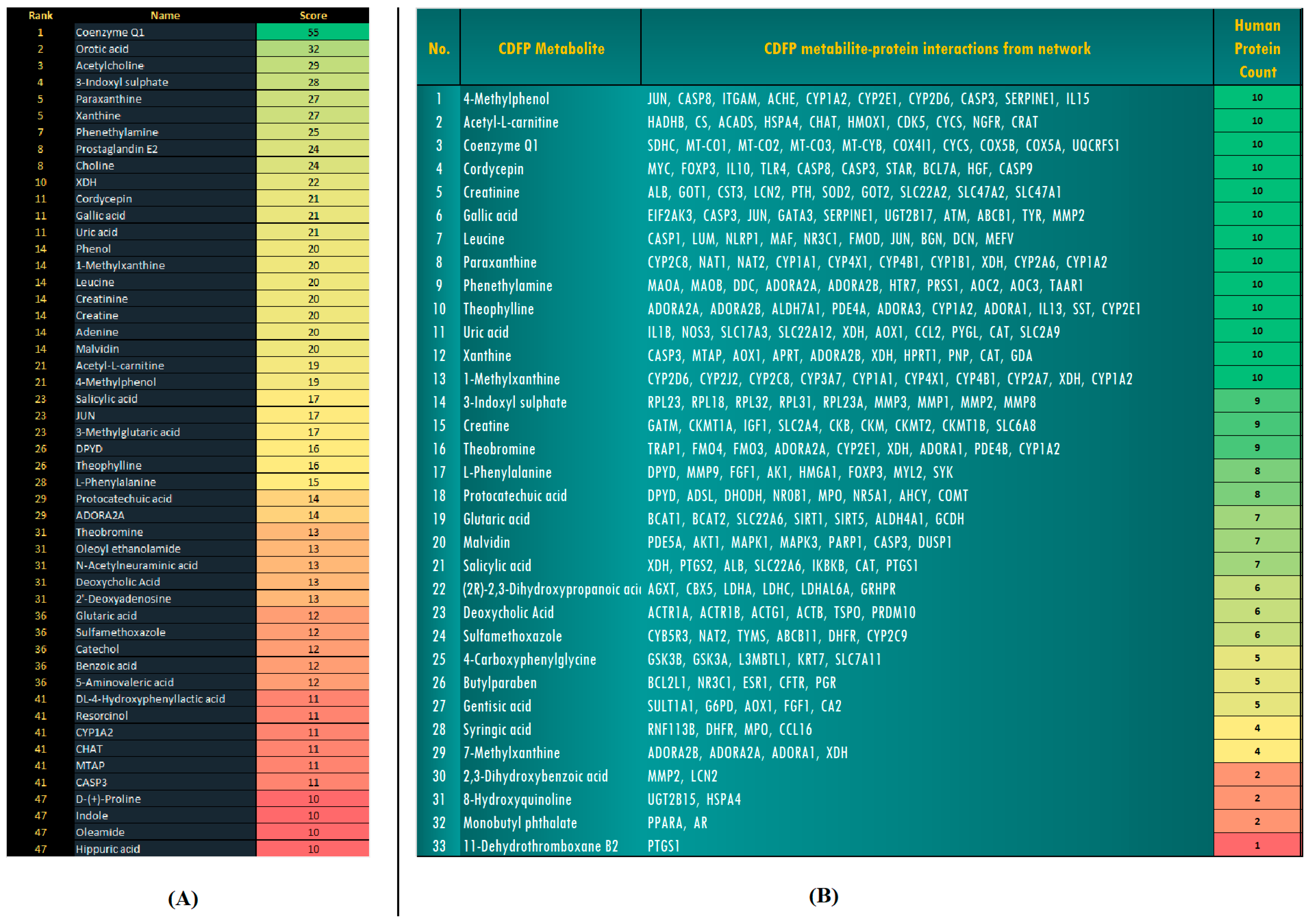
A total of 171 metabolites were identified from CDFP, each assigned a PubChem compound ID and corresponding SMILES notation. These metabolites were screened for interactions with human proteins using the STITCH database. The resulting interaction network (Figure 1), constructed and analysed in Cytoscape, revealed that 33 out of the 171 metabolites (19.3%) exhibited direct interactions with the top 50 Interactions identified based on network centrality and interaction strength (Figure 2) (Table S2).
Among the 33 active CDFP metabolites, six compounds, namely acetyl-L-carnitine, coenzyme Q1, cordycepin, creatine, leucine, and xanthine, showed the highest degree of interaction, each associated with 10 distinct human proteins. 3-Indoxyl sulphate interacted with 9 proteins, while L-phenylalanine and salicylic acid each interacted with 8 proteins. Protocatechuic acid and 7-methylxanthine showed 7 interactions each (Figure 2B). Phytochemicals such as cordycepin, gallic acid, acetyl-L-carnitine, theobromine, theophylline, L-phenylalanine, and malvidin were found to interact significantly with human proteins. Cordycepin and gallic acid showed, respectively, a significant interaction with CASP3 (Caspase-3) and JUN-CYP1A2. Methylxanthine, theobromine, theophylline, and xanthine were found to interact with ADORA2A, whereas XDH levels were also affected by seven CDFP metabolites interacting with xanthine, paraxanthine, and uric acid from CDFP.
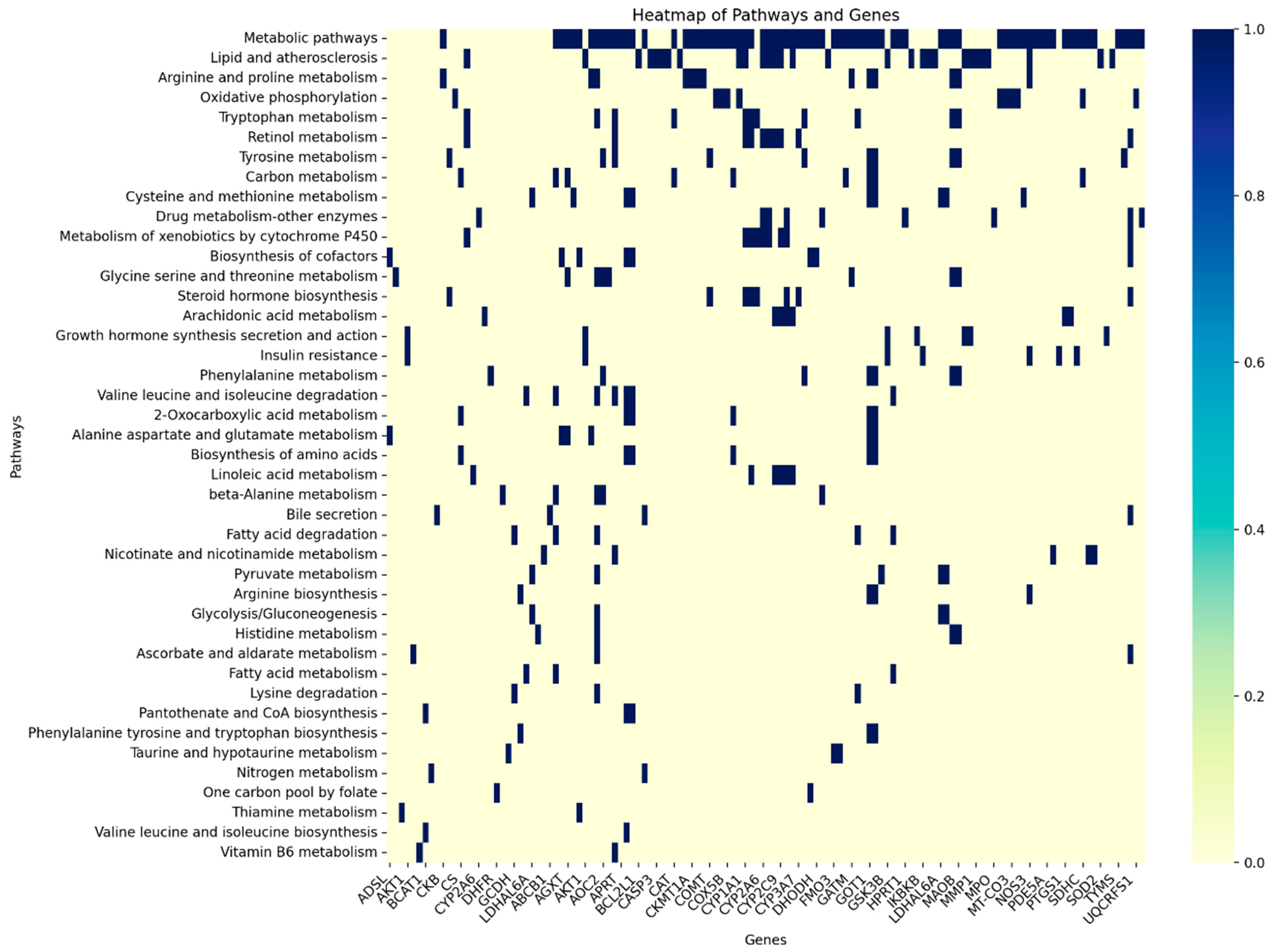
The heatmap visualization reveals intricate relationships between biological pathways and their associated genes, where dark blue cells indicate gene presence within specific pathways (Figure 3). Metabolic and lipid/atherosclerosis pathways emerge as the most gene-rich processes, containing numerous genes across a broad range of functions. Key regulatory genes, including MAPK1, MAPK3, and AKT1, appear in multiple pathways, as evidenced by vertical blue stripes across the visualization. The CYP gene family is predominantly involved in drug metabolism and xenobiotic pathways, while oxidative phosphorylation exhibits a distinct gene signature. Some pathways show highly specialized gene requirements with minimal overlap, while others share substantial genetic components with related processes. The visualization reveals both pathway-specific gene clusters and hub genes that serve as connection points between different biological processes.

3.3. Gut Microbiome Diversity and Composition

Ion Torrent sequencing generated single-end reads for each sample. All sequences are publicly available in the NCBI SRA database under the BioProject PRJNA1150211. Quality assessment confirmed that >95% of reads passed quality filtering across all samples.
The taxonomic analysis revealed varying levels of microbial diversity across samples (Table 1). The total number of reads was highest in the BT group (2,363,299) and comparatively lower in CDFP (2,038,835), AT15 (1,600,110), and AT7 (1,473,598). However, microbial diversity showed a different pattern, with diversity at the phylum level being highest in AT7 (55) compared to BT (42). Similarly, at the genus level, diversity was highest in the CDFP sample (1204), followed by AT7 (991), AT15 (919), and lowest in BT (612). Species-level analysis showed that CDFP contained the most species (2336), while BT had the least (1004).
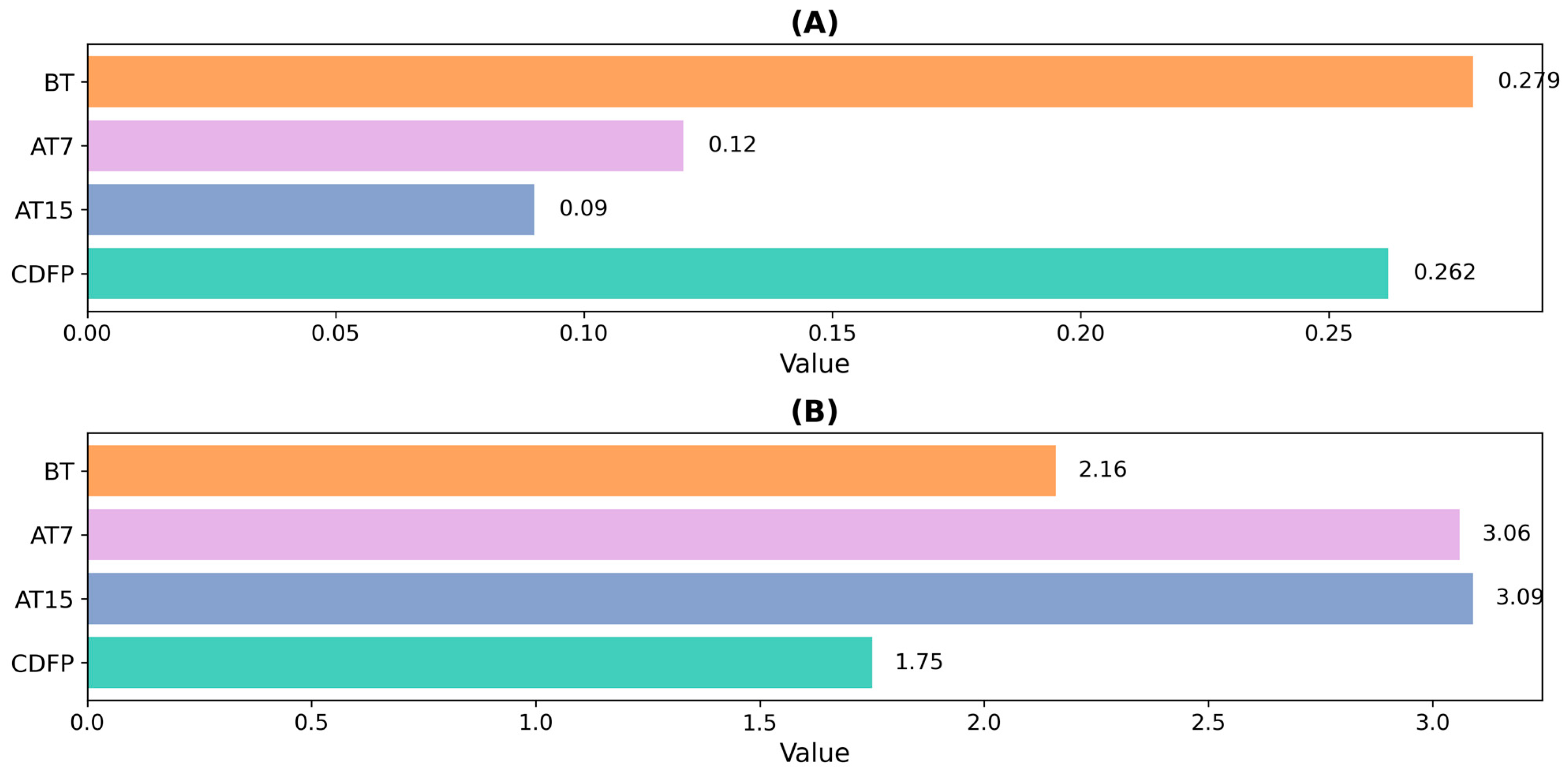
The Shannon and Simpson diversity indices provide insight into bacterial diversity (Figure 4). The Shannon index, which reflects both species abundance and evenness, indicated that AT15 (3.09) and AT7 (3.06) exhibited the highest diversity, followed by BT (2.16) and CDFP (1.75). In contrast, the Simpson index showed that BT had the highest value (0.279), indicating lower diversity, whereas AT15 exhibited the lowest value (0.090), suggesting higher diversity. Together, these indices demonstrated that CDFP and BT exhibited lower microbial diversity, whereas AT15 and AT7 showed higher diversity. Overall, compared to the pre-treatment phase, the gut microbiota exhibited increased alpha diversity following treatment. Furthermore, effect-size analysis showed a 1.42–1.43-fold increase in Shannon diversity at AT7 and AT15 relative to BT-baseline, accompanied by a marked reduction in Simpson index values, indicating increased evenness and diversity following CDFP administration (Table 2).
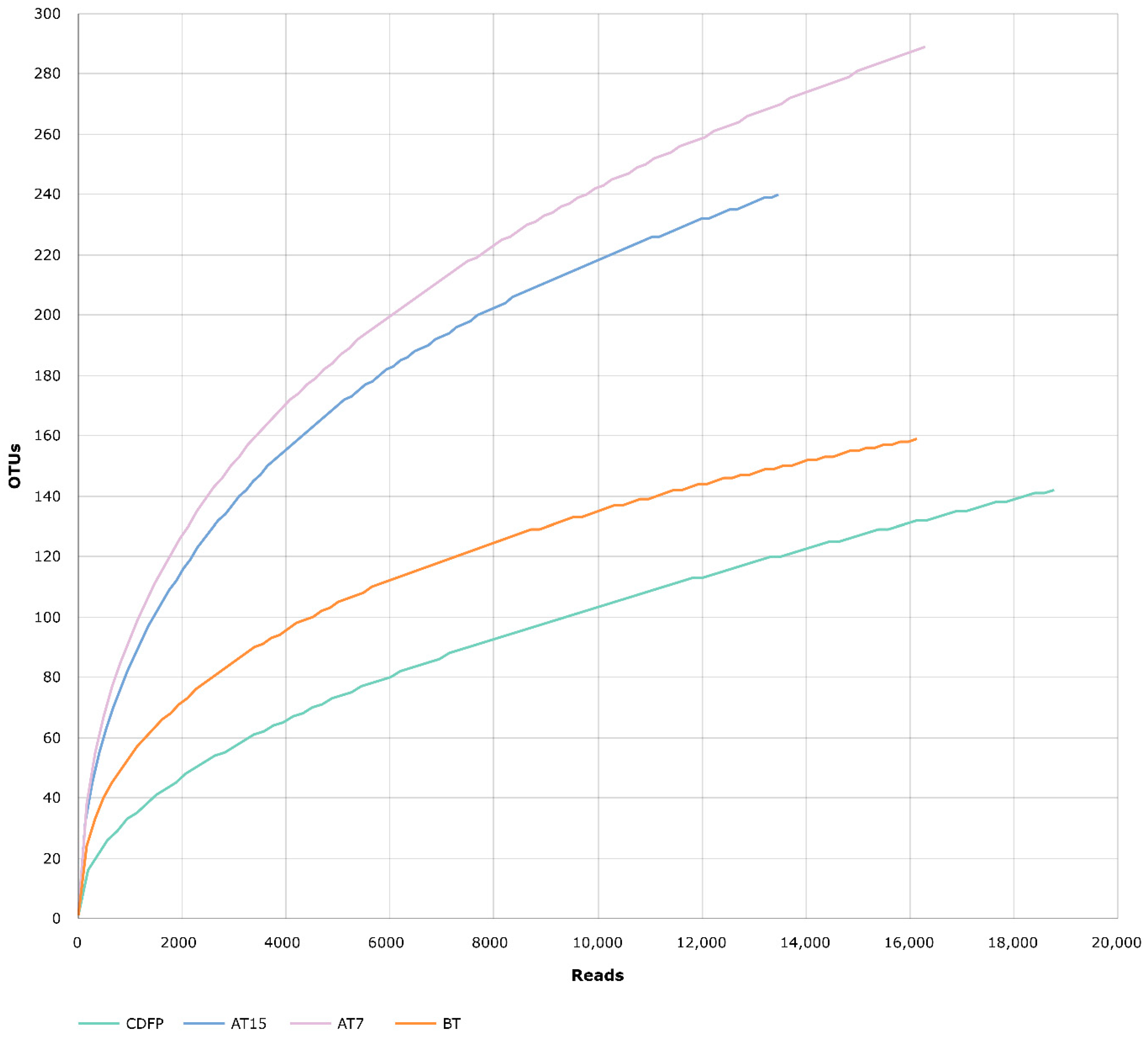
The rarefaction curve illustrates the correlation between the number of reads and the observed operational taxonomic units (OTUs) for all samples (Figure 5). The curve revealed that The rarefaction curves showed that OTU richness increased with increasing sequencing depth, approaching saturation at higher read counts. AT15 and AT7 showed similar patterns, whereas CDFP and BT had slightly different trends.
Taxonomic analysis of the gut microbiome indicated notable changes in bacterial genera, offering insights into the potential effects of cow-derived fermented products on gut microbiota and their possible health implications. The taxonomic analysis revealed distinctive changes in bacterial community composition following CDFP administration (Figure 6). CDFP contains various beneficial genera, including Lactobacillus, Weissella, Faecalitalea, and Akkermansia, along with the substantial presence of Ligilactobacillus. The BT microbial profile shows the volunteer’s native gut community, which may reflect a relatively stable and diverse set of genera. Prevotella and Megamonas were dominant genera, along with a considerable presence of Bacteroides, Barnesiella, Parabacteroides, and Akkermansia. Following 7 days of CDFP treatment (AT7), the gut microbiota exhibited enrichment in several beneficial genera, including Parabacteroides, Alistipes, Ligilactobacillus, Bacteroides, Eubacterium, Anaerostipes, and Barnesiella. While Akkermansia was detected throughout all samples, including in CDFP itself, its abundance showed variable patterns. The follow-up phase (AT15) demonstrated sustained presence of commensal genera including Bifidobacterium, Ruminococcus, Blautia, Faecalibacterium, Faecalitalea, Prevotella, and Christensenella. These observations suggest potential microbial resilience and enrichment of commensal communities following CDFP intervention.

3.4. Functional Composition of the Microbiome

The functional profiling of the gut microbiome revealed several beneficial modulations in core metabolic and cellular pathways following CDFP treatment. Analysis based on KEGG Level-1 functional categories showed that metabolism-related pathways dominated across all samples, accounting for 48.3% (BT), 51.7% (AT7), and 52.3% (AT15) of all functional categories, followed by genetic information processing and environmental information processing.
At KEGG Level-2, notable changes were observed in several metabolic pathways across treatment groups (Table S3). Lipid metabolism showed significant changes, with relative abundance decreasing from 27.5% in BT to 22.1% in AT7, followed by a slight increase to 23.6% in AT15. Amino acid metabolism increased from 19.7% in BT to 24.3% in AT7 and 25.1% in AT15. Energy metabolism also increased, rising from 21.2% in BT to 25.7% in AT7 and 26.3% in AT15. The metabolism of cofactors and vitamins displayed similar trends, increasing from 18.6% in BT to 22.9% in AT7 and 24.3% in AT15.
The heatmap analysis of KEGG Level-3 pathways (Figure 7) revealed enhanced microbial metabolic activity across various pathways following CDFP treatment. Amino acid metabolism pathways, including D-alanine metabolism, taurine metabolism, and D-arginine metabolism, showed low levels in BT but increased progressively in AT7 and AT15. Similarly, vitamin B biosynthesis pathways (riboflavin, folate, vitamin B6, and biotin) showed significant enhancement following treatment. Riboflavin metabolism notably increased from 19.03% in BT to 24.84% in AT15, while folate metabolism increased from 17.82% to 23.56%.
A transient increase in secondary metabolite pathways, such as isoflavonoid, sesquiterpenoid, and triterpenoid biosynthesis, was observed at AT7, followed by a decline at AT15, which may reflect an early adaptive response to CDFP components. Lipopolysaccharide biosynthesis showed a significant increase from 17.51% in BT to 30.85% in AT15. Additionally, lysosomal and sphingolipid metabolism pathways showed consistent activity throughout treatment, peaking at 29.51% in AT7.

4. Discussion

This preliminary case study aimed to investigate the effects of a traditional Ayurvedic preparation (CDFP) on the human gut microbiome. Our findings suggest that short-term CDFP administration may influence both the composition and functional potential of gut microbial communities. While the single-participant design limits generalizability, these initial observations provide valuable insights that merit further investigation in larger, controlled studies.
Traditional fermented preparations have been used across cultures for gastrointestinal health. More than 3000 years ago, ancient Indian Ayurvedic texts described the use of cow dung in the management of gastrointestinal ailments [17]. Similarly, historical accounts indicate that around 400 BC, Chinese physician Li Shizhen employed faecal-based preparations for treating certain illnesses [18,19]. Such historical records highlight a long-standing relationship with microbiome-based approaches, although modern preparation methods for CDFP emphasize safety and standardization.
One of the most notable effects observed in this case study was the increased microbial diversity following CDFP administration. Before treatment, microbial diversity was comparatively lower, which is typically associated with suboptimal gut health. After seven days of therapy and during the follow-up period, the gut microbiota showed enrichment in both diversity and beneficial bacterial populations. This observation aligns with studies suggesting that higher microbial diversity is generally associated with improved gut health and resilience against dysbiosis.
Cow’s urine and distillate have antimicrobial activity comparable to numerous antibiotics, including ofloxacin, cefpodoxime, and gentamicin, against various pathogenic bacteria [20,21]. Similarly, cow dung and its extract have been reported to possess antibacterial activity [22,23]. Cow milk and curd-derived antimicrobial peptides and other antibacterial substances have recently been described [24,25,26]. When these components are combined as CDFP and allowed to ferment, a complex formulation enriched with microbially derived metabolites is generated, which may contribute to potential health-related effects.
The documented antimicrobial properties of cow’s urine, dung, and milk components [20,21,22,23,24,25,26] may contribute to the observed changes in gut microbial composition following CDFP administration. The reduction in certain potentially pathogenic taxa combined with the enrichment of beneficial genera such as Bifidobacterium and Faecalibacterium suggests a selective antimicrobial effect rather than broad-spectrum suppression. This selective modulation may result from the synergistic action of various antimicrobial compounds present in CDFP, including antimicrobial peptides, organic acids, and other bioactive metabolites identified in our LC-MS/MS analysis. The parallel increase in beneficial bacteria suggests that CDFP may create a selective pressure favouring commensal and probiotic species while potentially inhibiting opportunistic pathogens, though direct antimicrobial testing would be needed to confirm this mechanism.
Gene–pathway interaction analysis following CDFP administration suggests its potential involvement in multi-target biological interactions. Several key genes, including AKT1, MAPK1, IGF1, CYP1A2, and COMT, were associated with essential signalling pathways such as PI3K-Akt, MAPK, insulin, and FoxO—pathways crucial for cellular metabolism, survival, and immune regulation [27,28,29]. Increased representation of amino acid metabolism (e.g., tryptophan, D-alanine, and taurine), vitamin biosynthesis (riboflavin, folate, and vitamin B6), and oxidative phosphorylation pathways was observed, suggesting potential modulation of energy production and gut metabolic functions [30,31]. Additionally, detoxification-related genes (CYPs, UGT2B17) were upregulated, suggesting improved hepatic xenobiotic clearance [32]. Bioactive constituents such as cordycepin and gallic acid showed interactions with immune and apoptotic proteins like CASP3 and FOXP3, supporting immunomodulatory and anti-inflammatory effects [33,34]. Neuroactive genes, including CHAT and MAOA, were also associated with the interaction network, suggesting a possible link to gut–brain axis–related pathways [35].
Moreover, genes involved in arginine and proline metabolism (CKB, GATM) and cytochrome P450 pathways (CYP2C9, CYP1A2) point toward an enhanced capacity for nitrogen balance and xenobiotic detoxification, which are vital for gut microbial homeostasis [36]. The increased representation of these pathways may be associated with a gut environment favorable to short-chain fatty acid (SCFA)-producing bacteria, which are known to support mucosal health and immunomodulatory functions [37].
The study observed the presence of probiotic-associated genera Lactobacillus and Weissella across all samples, suggesting a gut microbiota containing beneficial taxa [38,39]. Prevotella abundance indicates a potential shift in carbohydrate metabolism, possibly influenced by dietary or therapeutic interventions [40]. AT7 and AT15 samples showed increased representation of health-associated bacteria such as Bifidobacterium and Faecalibacterium, which are recognized for roles in digestion, immune regulation, and the production of anti-inflammatory metabolites such as butyrate [41,42].
Taxonomic analysis revealed several notable changes in bacterial communities. The increased abundance of Parabacteroides, Alistipes, and Bacteroides following CDFP administration suggests a potential beneficial modulation of the Bacteroidetes phylum, which is often associated with improved metabolic health. Similarly, the enrichment of Bifidobacterium and Faecalibacterium during the follow-up phase is particularly promising, as these genera are recognized for their roles in improving digestion, regulating immune function, and producing anti-inflammatory compounds such as butyrate [43,44].
The presence of Akkermansia, a mucin-degrading bacterium associated with intestinal barrier function and metabolic benefits [45], suggests potentially favorable effects on gut health. Previous studies have linked Akkermansia abundance with reduced inflammation and improved metabolic parameters [46], though these associations would need to be directly measured in future studies.
Following CDFP treatment, microbial pathways related to amino acid metabolism showed increased representation. D-alanine metabolism increased in AT7 and AT15, suggesting enrichment of bacterial populations involved in cell wall biosynthesis, which may contribute to structural stability and microbial resilience within the gut ecosystem [47]. Increased representation of pathways related to SCFA production coincided with higher abundances of SCFA-producing bacteria such as Bifidobacterium and Faecalibacterium [48]. Additionally, taurine and hypotaurine metabolism also showed progressive enhancement. Taurine is known to support bile acid conjugation, oxidative stress regulation, and immune modulation [49]. Increased representation of these pathways may be associated with improved gut homeostasis and host–microbiota interactions. Furthermore, elevated levels of D-arginine and D-ornithine metabolism post-treatment point toward a decrease in proteolytic or dysbiosis-associated microbial populations, supporting a shift to a more balanced gut environment [50].
CDFP administration significantly enhanced the biosynthesis of essential B vitamins, including folate, riboflavin, vitamin B6, and biotin. These vitamins play critical roles in cellular metabolism, immune function, and neurological health [51]. Riboflavin metabolism notably increased from 19.03% in BT to 24.84% in AT15, with similar trends observed for folate and vitamin B6 pathways. This pattern may reflect a selective enrichment of bacterial taxa capable of synthesizing these micronutrients. The increased microbial capacity for B vitamin production is especially important as these nutrients not only support host health directly but also influence gut microbial diversity and functionality [52].
Increased representation of microbial functions related to carbohydrate metabolism was also observed, particularly in fructose and mannose metabolism and oxidative phosphorylation. These pathways contribute to energy production and microbial growth, supporting a more active and efficient gut microbial ecosystem [53]. Alterations in these metabolic routes may influence energy extraction from dietary components and the overall metabolic capacity of the gut microbiota.
We also documented enhanced microbial functions in carbohydrate metabolism, particularly in fructose and mannose metabolism and oxidative phosphorylation, which contribute to energy production and microbial growth [54]. The improvement in these metabolic routes likely supports a more active and efficient gut microbial ecosystem, promoting better energy extraction from the diet. The data also highlight upregulated pathways related to host digestion and hormonal regulation post-treatment. Specifically, protein digestion and absorption, along with insulin secretion, showed marked increases, suggesting improved nutrient assimilation and metabolic control [55]. These functional enhancements may result from a favourable shift in gut microbial composition toward metabolically beneficial taxa that support host physiological functions.
A transient spike in secondary metabolite pathways such as isoflavonoid, sesquiterpenoid, and triterpenoid biosynthesis was noted at AT7, followed by a decline at AT15. This suggests an early microbial adaptive response to CDFP components, potentially contributing to mucosal defence and detoxification, with subsequent regulatory feedback or microbial succession leading to a stabilized environment over time [56].
A significant rise in lipopolysaccharide (LPS) biosynthesis, from 17.51% in BT to 30.85% in AT15, was observed. While excessive LPS is typically associated with inflammation, controlled production can play a role in immune system priming without pathogenic consequences. Additionally, consistent activity in lysosomal and sphingolipid metabolism pathways throughout treatment, peaking at 29.51% in AT7, suggests enhanced microbial mediation of cellular recycling, gut barrier integrity, and mucosal immunity [57].
Our research focuses on identifying beneficial bacteria and metabolites and exploring their potential interactions with human protein targets to examine multi-target biological associations related to metabolism, oxidative stress pathways, and immune function. Although CDFP was evaluated in a single volunteer, the findings provide preliminary baseline data that may inform future, larger-scale investigations. However, validation will require well-designed controlled trials involving larger cohorts and appropriate control groups.

5. Conclusions

Our study represents an initial step toward the scientific evaluation of this ancient Ayurvedic preparation and practice. While this study was conducted with a single participant, the observed temporal changes in gut microbial composition and function following the seven-day CDFP regimen provide preliminary but informative insights. Our analysis revealed a marked increase in beneficial bacterial genera such as Bifidobacterium, Faecalibacterium, and Akkermansia, accompanied by a decline in potentially harmful taxa. Functional profiling further showed significant shifts in key metabolic pathways, including those related to lipid metabolism, amino acid metabolism, energy production, and the metabolism of cofactors and vitamins. After CDFP consumption, the participant’s gut exhibited enhanced microbial diversity, an indicator commonly associated with improved gut health. These results highlight the potential of CDFP as a natural dietary supplement capable of influencing microbial balance. However, these findings must be interpreted cautiously given the single-subject design, and validation through larger, controlled clinical trials is essential before drawing definitive conclusions about therapeutic efficacy. By applying modern scientific tools to a time-honoured practice, this research underscores the value of integrating traditional medicine with evidence-based approaches and opens the door to innovative microbiome-targeted research directions rooted in ancient healing systems. Future research should include randomized controlled trials with appropriate sample sizes, control groups, and clinical outcome measures to fully evaluate CDFP’s therapeutic potential.

Supplementary Materials

The following supporting information can be downloaded at: https://www.mdpi.com/article/10.3390/microbiolres17010025/s1, Table S1: Fixed diet plan of volunteer (Author) 15 days before, during, and after treatment periods; Table S2: Heatmap of interactions between CDFP-derived metabolites and human proteins; Table S3: Functional analysis of microbial metabolic pathways at KEGG Level-2 across different treatment groups.

Author Contributions

Conceptualization, N.D. and N.V.; Methodology, K.A., A.V. and A.T.; Software, A.G., R.C. and R.P.; Validation, A.G., R.C. and R.P.; Formal Analysis, N.D. and N.V.; Investigation, K.A., A.V. and A.T.; Resources, P.D. and D.J.H.S.; Data Curation, A.G., R.C. and R.P.; Writing—Original Draft Preparation, N.D. and N.V.; Writing—Review and Editing, P.D. and D.J.H.S.; Visualization, K.A., A.V. and A.T.; Supervision, P.D. and D.J.H.S.; Project Administration, P.D. and D.J.H.S.; Funding Acquisition, P.D. and D.J.H.S. All authors have read and agreed to the published version of the manuscript.

Funding

This research was financially supported by the Veer Narmad South Gujarat University Research Grant 2021 (Letter No. IPR/UGC/18464/2021). N.V. and K.A. were recipients of an international fellowship through the Taiwan Experience Education Program (TEEP), which supported their research internship at the Functional Genomics Laboratory, Department of Biological Science and Technology, National Pingtung University of Science and Technology (NPUST), Taiwan.

Institutional Review Board Statement

This study involved self-experimentation by one of the authors (Mr. Arpan Tapaniya), who voluntarily participated in assessing the effects of the CDFP formulation on his gut microbiome. The formulation was administered under the clinical observation of Dr. Naresh Gabani (Gastroenterologist, Gujarat Hospital, Surat) and followed the guidelines outlined in the Ayurvedic Formulary of India. As the procedure involved minimal risk, was non-invasive, and conducted with full informed consent by the author himself, formal review and approval from an institutional ethics committee was not required. This approach complies with ethical norms for self-experimentation in minimal-risk studies and is consistent with international research standards such as the Declaration of Helsinki.

Informed Consent Statement

Informed consent was obtained from the participant prior to the study. Written consent has also been obtained for publication of the findings.

Data Availability Statement

The original data presented in the study are openly available in the National Center for Biotechnology Information (NCBI) at https://www.ncbi.nlm.nih.gov/bioproject/PRJNA1150211, accessed on 8 December 2025.

Acknowledgments

The authors are thankful for utilizing the equipment of UGC-SAP-II, DST-FIST-Level-1, and the supercomputer facility of the state government available at the Department of Biosciences, Veer Narmad South Gujarat University, Surat. We sincerely thank the TEEP program and NPUST for their generous support and for providing valuable research opportunities.

Conflicts of Interest

The authors declare no conflicts of interest.

Abbreviations

The following abbreviations are used in this manuscript:
CDFPCow-derived fermented product
BTStool sample before the CDFP treatment
AT7Stool sample after 7 continuous days of CDFP treatment
AT15Stool sample after a 7-day gap following CDFP treatment

References

  1. Bajaj, K.K.; Chavhan, V.; Raut, N.A.; Gurav, S. Panchgavya: A precious gift to humankind. J. Ayurveda Integr. Med. 2022, 13, 100525. [Google Scholar] [CrossRef]
  2. Sayi, D.S.; Mohan, S.; Kumar, K.V. Molecular characterization of a proteolytic bacterium in Panchagavya: An organic fertilizer mixture. J. Ayurveda Integr. Med. 2018, 9, 123–125. [Google Scholar] [CrossRef] [PubMed]
  3. Dhama, K.; Rathore, R.; Chauhan, R.S.; Tomar, S. Panchgavya (Cowpathy): An overview. Int. J. Cow Sci. 2005, 1, 1–15. [Google Scholar]
  4. Schnürer, J.; Magnusson, J. Antifungal lactic acid bacteria as biopreservatives. Trends Food Sci. Technol. 2005, 16, 70–78. [Google Scholar] [CrossRef]
  5. Chauhan, R.S. Panchagavya Therapy (Cow pathy)-Current status and future directions. Indian Cow 2004, 1, 3–7. [Google Scholar]
  6. Tharmaraj, K.; Ganesh, P.; Kumar, R.S.; Anandan, A.; Kolanjinathan, K. A critical review on Panchagavya-a boon plant growth. Int. J. Pharm. Biol. Arch. 2011, 2, 1611–1614. [Google Scholar]
  7. Pathak, R.K.; Ram, R.A. Bio-enhancers: A potential tool to improve soil fertility, plant health in organic production of horticultural crops. Progress. Hortic. 2013, 45, 237–254. [Google Scholar]
  8. Raju, G.S.K.; Nawabpet, P.; Kumar, A. Panchagavya as Soil Conditioner: Ancient traditional knowledge for sustainable agriculture. J. Exp. Agric. Int. 2022, 44, 181–186. [Google Scholar] [CrossRef]
  9. Hou, K.; Wu, Z.X.; Chen, X.Y.; Wang, J.Q.; Zhang, D.; Xiao, C.; Zhu, D.; Koya, J.B.; Wei, L.; Li, J.; et al. Microbiota in health and diseases. Signal Transduct. Target. Ther. 2022, 7, 135. [Google Scholar] [CrossRef]
  10. Sarma, H.H.; Talukdar, N. Dasagavya and Panchagavya: Elixirs of organic farming. Indian Farmer 2024, 11, 198–202. [Google Scholar]
  11. Vaghamshi, N.; Gandhi, H.; Beladiya, U.; Mangrola, A.; Dudhagara, P.; Patel, D.R.; Chaudhari, R. 16s rRNA Amplicon Sequencing Approach for Community and Predictive Functional Diversity of Therapeutically Valuable Formulation of Cow-derivatives. Biosc. Biotech. Res. Comm. 2022, 15, 547–551. [Google Scholar] [CrossRef]
  12. Krishnareddy, P.M.; Basavarajegowda, M.H.; Buela, P.P.; Devanna, P.; Eregowda, P.M.; Sarangi, A.N.; Govindaraju, M.K.; Middha, S.K.; Banakar, S.N. Decoding the microbiome and metabolome of the Panchagavya—An indigenous fermented bio-formulation. Imeta 2022, 1, e63. [Google Scholar] [CrossRef] [PubMed]
  13. Szklarczyk, D.; Santos, A.; Von Mering, C.; Jensen, L.J.; Bork, P.; Kuhn, M. STITCH 5: Augmenting protein–chemical interaction networks with tissue and affinity data. Nucleic Acids Res. 2016, 44, D380–D384. [Google Scholar] [CrossRef] [PubMed]
  14. Ministry of Health and Family Welfare. The Ayurvedic Formulary of India Part 1, 2nd ed.; Govt. of India, Ministry of Health and Family Welfare: New Delhi, India, 2003. [Google Scholar]
  15. Brown, J.; Pirrung, M.; McCue, L.A. FQC Dashboard: Integrates FastQC results into a web-based, interactive, and extensible FASTQ quality control tool. Bioinformatics 2017, 33, 3137–3139. [Google Scholar] [CrossRef]
  16. Nagpal, S.; Haque, M.M.; Mande, S.S. Vikodak—A modular framework for inferring functional potential of microbial communities from 16S metagenomic datasets. PLoS ONE 2016, 11, e0148347. [Google Scholar] [CrossRef]
  17. Meng, H.J.; Dhanashree, B. Antifungal effect of cow’s urine distillate on Candida species. J. Ayurveda Integr. Med. 2017, 8, 233–237. [Google Scholar]
  18. Oprita, R.; Bratu, M.; Oprita, B.; Diaconescu, B. Fecal transplantation–the new, inexpensive, safe, and rapidly effective approach in the treatment of gastrointestinal tract diseases. J. Med. Life 2016, 9, 160. [Google Scholar]
  19. Lewin Ralph, A.; Ralph, A.L. More on merde. Perspect. Biol. Med. 2001, 44, 594–607. [Google Scholar] [CrossRef]
  20. Randhawa, G.K.; Sharma, R. Chemotherapeutic potential of cow urine: A review. J. Intercult. Ethnopharmacol. 2015, 4, 180–186. [Google Scholar] [CrossRef] [PubMed] [PubMed Central]
  21. Pant, L.; Thapa, S.; Dahal, B.; Khadka, R.; Biradar, M.S. In silico and in vitro studies of antibacterial activity of cow urine distillate (CUD). Evid.-Based Complement. Altern. Med. 2024, 2024, 1904763. [Google Scholar] [CrossRef]
  22. Thenmozhi, S.; Mageswari, M.; Chinnamani, S.; Sivasuriyan, S. Antimicrobial activity of animal waste (Cow dung). World J. Sci. Res. 2018, 3, 37–41. [Google Scholar]
  23. Mishra, O.P.; Mehtab, R.P.; Parmar, S.; Doneria, R. Cow dung an undeciphered boon: An overview. Pharma Innov. J. 2020, 9, 84–89. [Google Scholar] [CrossRef]
  24. Singh, A.; Duche, R.T.; Wandhare, A.G.; Sian, J.K.; Singh, B.P.; Sihag, M.K.; Singh, K.S.; Sangwan, V.; Talan, S.; Panwar, H. Milk-derived antimicrobial peptides: Overview, applications, and future perspectives. Probiotics Antimicrob. Proteins 2023, 15, 44–62. [Google Scholar] [CrossRef] [PubMed]
  25. Bielecka, M.; Cichosz, G.; Czeczot, H. Antioxidant, antimicrobial and anticarcinogenic activities of bovine milk proteins and their hydrolysates-a review. Int. Dairy J. 2022, 127, 105208. [Google Scholar] [CrossRef]
  26. Sadewi, S.M.; Nurhasanah, N.; Sudibyo, S.; Windayani, N.; Kiswandono, A.A.; Satria, H. Antioxidant and Antibacterial Activities of Curd and Whey Kefir Produced from Etawa Goat Milk. J. Multidiscip. Appl. Nat. Sci. 2024, 4, 139–145. [Google Scholar] [CrossRef]
  27. Franks, S.E.; Briah, R.; Jones, R.A.; Moorehead, R.A. Unique roles of Akt1 and Akt2 in IGF-IR mediated lung tumorigenesis. Oncotarget 2015, 7, 3297. [Google Scholar] [CrossRef]
  28. Zhang, W.; Thompson, B.J.; Hietakangas, V.; Cohen, S.M. MAPK/ERK signaling regulates insulin sensitivity to control glucose metabolism in Drosophila. PLoS Genet. 2011, 7, e1002429. [Google Scholar] [CrossRef]
  29. Schultze, S.M.; Hemmings, B.A.; Niessen, M.; Tschopp, O. PI3K/AKT, MAPK and AMPK signalling: Protein kinases in glucose homeostasis. Expert Rev. Mol. Med. 2012, 14, e1. [Google Scholar] [CrossRef]
  30. Magnúsdóttir, S.; Heinken, A.; Kutt, L.; Ravcheev, D.A.; Bauer, E.; Noronha, A.; Greenhalgh, K.; Jäger, C.; Baginska, J.; Wilmes, P.; et al. Generation of genome-scale metabolic reconstructions for 773 members of the human gut microbiota. Nat. Biotechnol. 2017, 35, 81–89. [Google Scholar] [CrossRef]
  31. Biesalski, H.K. Nutrition meets the microbiome: Micronutrients and the microbiota. Ann. N. Y. Acad. Sci. 2016, 1372, 53–64. [Google Scholar] [CrossRef]
  32. Pascussi, J.M.; Vilarem, M.J. Inflammation and drug metabolism: NF-kappB and the CAR and PXR xeno-receptors. Med. Sci. 2008, 24, 301–305. [Google Scholar]
  33. Xiong, S.; Jiang, J.; Wan, F.; Tan, D.; Zheng, H.; Xue, H.; Hang, Y.; Lu, Y.; Su, Y. Cordyceps militaris extract and cordycepin alleviate oxidative stress, modulate gut microbiota and ameliorate intestinal damage in LPS-induced piglets. Antioxidants 2024, 13, 441. [Google Scholar] [CrossRef] [PubMed]
  34. Mohamed, E.K.; Hafez, D.M. Gallic acid and metformin co-administration reduce oxidative stress, apoptosis and inflammation via Fas/caspase-3 and NF-κB signaling pathways in thioacetamide-induced acute hepatic encephalopathy in rats. BMC Complement. Med. Ther. 2023, 23, 265. [Google Scholar] [CrossRef] [PubMed]
  35. Chen, Y.; Xu, J.; Chen, Y. Regulation of neurotransmitters by the gut microbiota and effects on cognition in neurological disorders. Nutrients 2021, 13, 2099. [Google Scholar] [CrossRef]
  36. Zhao, L.; Zhang, F.; Ding, X.; Wu, G.; Lam, Y.Y.; Wang, X.; Fu, H.; Xue, X.; Lu, C.; Ma, J.; et al. Gut bacteria selectively promoted by dietary fibers alleviate type 2 diabetes. Science 2018, 359, 1151–1156. [Google Scholar] [CrossRef] [PubMed]
  37. Fusco, W.; Lorenzo, M.B.; Cintoni, M.; Porcari, S.; Rinninella, E.; Kaitsas, F.; Lener, E.; Mele, M.C.; Gasbarrini, A.; Collado, M.C.; et al. Short-chain fatty-acid-producing bacteria: Key components of the human gut microbiota. Nutrients 2023, 15, 2211. [Google Scholar] [CrossRef]
  38. Ahmed, S.; Singh, S.; Singh, V.; Roberts, K.D.; Zaidi, A.; Rodriguez-Palacios, A. The Weissella genus: Clinically treatable bacteria with antimicrobial/probiotic effects on inflammation and cancer. Microorganisms 2022, 10, 2427. [Google Scholar] [CrossRef]
  39. Al-Yami, A.M.; Al-Mousa, A.T.; Al-Otaibi, S.A.; Khalifa, A.Y. Lactobacillus species as probiotics: Isolation sources and health benefits. J. Pure Appl. Microbiol. 2022, 16, 1761–1770. [Google Scholar] [CrossRef]
  40. Yeoh, Y.K.; Sun, Y.; Ip, L.Y.T.; Wang, L.; Chan, F.K.; Miao, Y.; Ng, S.C. Prevotella species in the human gut is primarily comprised of Prevotella copri, Prevotella stercorea and related lineages. Sci. Rep. 2022, 12, 9055. [Google Scholar] [CrossRef]
  41. Kumar, S.; Mukherjee, R.; Gaur, P.; Leal, É.; Lyu, X.; Ahmad, S.; Puri, P.; Chang, C.M.; Raj, V.S.; Pandey, R.P. Unveiling roles of beneficial gut bacteria and optimal diets for health. Front. Microbiol. 2025, 16, 1527755. [Google Scholar] [CrossRef]
  42. Zhao, M.A.; Chu, J.; Feng, S.; Guo, C.; Xue, B.; He, K.; Li, L. Immunological mechanisms of inflammatory diseases caused by gut microbiota dysbiosis: A review. Biomed. Pharmacotreatment 2023, 164, 114985. [Google Scholar] [CrossRef]
  43. Aja, E.; Zeng, A.; Gray, W.; Connelley, K.; Chaganti, A.; Jacobs, J.P. Health Effects and Therapeutic Potential of the Gut Microbe Akkermansia muciniphila. Nutrients 2025, 17, 562. [Google Scholar] [CrossRef] [PubMed]
  44. Olsson, L.M.; Boulund, F.; Nilsson, S.; Khan, M.T.; Gummesson, A.; Fagerberg, L.; Engstrand, L.; Perkins, R.; Uhlen, M.; Bergström, G.; et al. Dynamics of the normal gut microbiota: A longitudinal one-year population study in Sweden. Cell Host Microbe 2022, 30, 726–739. [Google Scholar] [CrossRef]
  45. Wang, S.; Huang, M.; You, X.; Zhao, J.; Chen, L.; Wang, L.; Luo, Y.; Chen, Y. Gut microbiota mediates the anti-obesity effect of calorie restriction in mice. Sci. Rep. 2018, 8, 13037. [Google Scholar] [CrossRef] [PubMed]
  46. Louis, P.; Flint, H.J. Formation of propionate and butyrate by the human colonic microbiota. Environ. Microbiol. 2017, 19, 29–41. [Google Scholar] [CrossRef] [PubMed]
  47. Ridlon, J.M.; Harris, S.C.; Bhowmik, S.; Kang, D.J.; Hylemon, P.B. Consequences of bile salt biotransformations by intestinal bacteria. Gut Microbes 2016, 7, 22–39. [Google Scholar] [CrossRef]
  48. Morrison, D.J.; Preston, T. Formation of short chain fatty acids by the gut microbiota and their impact on human metabolism. Gut Microbes 2016, 7, 189–200. [Google Scholar] [CrossRef]
  49. Magnúsdóttir, S.; Ravcheev, D.; de Crécy-Lagard, V.; Thiele, I. Systematic genome assessment of B-vitamin biosynthesis suggests co-operation among gut microbes. Front. Genet. 2015, 6, 148. [Google Scholar] [CrossRef]
  50. Scott, K.P.; Jean-Michel, A.; Midtvedt, T.; van Hemert, S. Manipulating the gut microbiota to maintain health and treat disease. Microb. Ecol. Health Dis. 2015, 26, 25877. [Google Scholar] [CrossRef]
  51. Holt, R.A.; Cochrane, K. Tumor potentiating mechanisms of Fusobacterium nucleatum, a multifaceted microbe. Gastroenterology 2017, 152, 694–696. [Google Scholar] [CrossRef]
  52. Mao, B.; Li, D.; Zhao, J.; Liu, X.; Gu, Z.; Chen, Y.Q.; Zhang, H.; Chen, W. Metagenomic insights into the effects of fructo-oligosaccharides (FOS) on the composition of fecal microbiota in mice. J. Agric. Food Chem. 2015, 63, 856–863. [Google Scholar] [CrossRef]
  53. Duda-Chodak, A.; Tarko, T.; Satora, P.; Sroka, P. Interaction of dietary compounds, especially polyphenols, with the intestinal microbiota: A review. Eur. J. Nutr. 2015, 54, 325–341. [Google Scholar] [CrossRef]
  54. Vatanen, T.; Kostic, A.D.; d’Hennezel, E.; Siljander, H.; Franzosa, E.A.; Yassour, M.; Kolde, R.; Vlamakis, H.; Arthur, T.D.; Hämäläinen, A.M.; et al. Variation in microbiome LPS immunogenicity contributes to autoimmunity in humans. Cell 2016, 165, 842–853. [Google Scholar] [CrossRef]
  55. Bryan, P.F.; Karla, C.; Edgar Alejandro, M.T.; Sara Elva, E.P.; Gemma, F.; Luz, C. Sphingolipids as mediators in the crosstalk between microbiota and intestinal cells: Implications for inflammatory bowel disease. Mediat. Inflamm. 2016, 2016, 9890141. [Google Scholar] [CrossRef] [PubMed]
  56. Johnson, E.L.; Heaver, S.L.; Walters, W.A.; Ley, R.E. Microbiome and metabolic disease: Revisiting the bacterial phylum Bacteroidetes. J. Mol. Med. 2017, 95, 1–8. [Google Scholar] [CrossRef] [PubMed]
  57. Di Sabatino, A.; Santacroce, G.; Rossi, C.M.; Broglio, G.; Lenti, M.V. Role of mucosal immunity and epithelial–vascular barrier in modulating gut homeostasis. Intern. Emerg. Med. 2023, 18, 1635–1646. [Google Scholar] [CrossRef] [PubMed]
Figure 1. Network pharmacology analysis of CDFP constituents and human protein targets. Blue nodes represent LC-MS/MS-identified CDFP metabolites; yellow to red nodes denote the top 50 ranked human proteins based on interaction scores (yellow: lower score; red: highest score); green nodes indicate other associated human proteins. Interactions were mapped using the STITCH database and visualized in Cytoscape version 3.10.4.
Microbiolres 17 00025 g001
Figure 2. Network pharmacology analysis of CDFP constituents and their interactions with human proteins: (A) Network diagram representing the top 50 interactions among human proteins and bioactive phytochemical constituents derived from CDFP. Each edge represents a significant interaction as determined by network scoring and ranking in Cytoscape. Nodes represent either human proteins or phytochemicals, with edges labelled by interaction rank and confidence score. (B) The highlighted subnetwork focuses on the top 33 key phytochemical constituents involved in these top interactions. For each phytochemical, associated human protein targets are visualized along with their total interaction counts, providing insight into potential multi-target activity. Visualization and ranking were performed using Cytoscape’s Network Analyzer.
Microbiolres 17 00025 g002
Figure 3. The heatmap illustrates the relationships between various biological pathways and their associated genes. Each row represents a distinct pathway, while each column corresponds to a specific gene. The presence of a gene in a pathway is indicated by a color gradient, with darker shades representing stronger associations.
Microbiolres 17 00025 g003
Figure 4. Relationships between alpha diversity across CDFP, BT, AT7, and AT15 samples, as indicated by (A) the Shannon diversity index and (B) the Simpson diversity index for bacterial communities.
Microbiolres 17 00025 g004
Figure 5. Rarefaction curve comparing operational taxonomic units (OTUs) across CDFP, AT15, AT7, and BT samples based on bacterial diversity.
Microbiolres 17 00025 g005
Figure 6. Heatmap of genus-level abundance across treatment groups. The colour intensity represents the log10-transformed relative abundance of bacterial genera across four sample groups: CDFP (fermented product composition), BT (before treatment), AT7 (after 7 days of treatment), and AT15 (after 15 days, including a 7-day washout period). Abundance values were normalized and scaled for visualization.
Microbiolres 17 00025 g006
Figure 7. KEGG Level-3 functional pathway heatmap illustrating the abundance of specific metabolic and biosynthetic processes in the gut microbiome across different treatment groups (CDFP, BT, AT7, and AT15).
Microbiolres 17 00025 g007
Table 1. Microbial diversity indices of the human gut microbiome across different study phases. Values represent the total number of unique taxa identified at each taxonomic level based on 16S rRNA sequencing analysis. CDFP = cow-derived fermented product; BT = before treatment (baseline); AT7 = after 7 days of treatment; AT15 = after 15 days (7 days post-treatment cessation).
LevelCDFPBTAT7AT15
Phylum52425552
Family582356518500
Genus1204612991919
Species2336100417601577
Table 2. Effect-size analysis of alpha diversity following CDFP treatment.
SampleShannon IndexFold Change v/s BTSimpson IndexFold Change v/s BT
BT (Baseline)2.161.000.2791.00
AT73.061.42 ↑0.1120.40 ↓
AT153.091.43 ↑0.0900.32 ↓
↑ indicates increase; ↓ indicates decrease compared to baseline BT.
Disclaimer/Publisher’s Note: The statements, opinions and data contained in all publications are solely those of the individual author(s) and contributor(s) and not of MDPI and/or the editor(s). MDPI and/or the editor(s) disclaim responsibility for any injury to people or property resulting from any ideas, methods, instructions or products referred to in the content.