Article

Microbiological Profile and Resistance Patterns in Periprosthetic Joint Infections: A Regional Multicenter Study in Spain

by
Lucia Henriquez
1,2,*,
Ander Uribarri
1,2,
Iñaki Beguiristain
1,2,
Ignacio Sancho
3,
Carmen Ezpeleta Baquedano
1,2 and
Maria Eugenia Portillo
1,2
1
Department of Clinical Microbiology, University Hospital of Navarra, 31008 Pamplona, Spain
2
Institute of Healthcare Research of Navarra (IdiSNa), 31008 Pamplona, Spain
3
Department of Orthopedics and Trauma Surgery, University Hospital of Navarra, 31008 Pamplona, Spain
*
Author to whom correspondence should be addressed.
Microbiol. Res. 2025, 16(7), 142; https://doi.org/10.3390/microbiolres16070142
Submission received: 5 May 2025 / Revised: 15 June 2025 / Accepted: 17 June 2025 / Published: 1 July 2025

Abstract

Due to the significant number of microbiologically negative periprosthetic joint infections (PJIs), understanding the trend in etiology and resistance patterns is essential for the correct management of these infections. Currently, few studies have been published in Spain. In this study, we analyzed the incidence, clinical characteristics, etiology, and antibiotic resistance in patients with PJIs over the last 5 years in Navarra. In this multicentric and retrospective study, all patients diagnosed with PJIs in Navarra from 2019 to 2023 were included. Of the total 156 PJIs, 23% had negative cultures and 56% of these patients had been treated with antibiotics prior to sampling. Staphylococcus epidermidis with methicillin resistance was the predominant etiological agent, followed by Staphylococcus aureus and Cutibacterium acnes. Forty percent of the Gram-positive cocci (GPC) and 35% of the Gram-negative bacilli (GNB) were multidrug-resistant organisms (MDROs). Quinolone resistance was 46% for staphylococci and 18% for Gram-negatives. In addition, 9% of staphylococci were resistant to rifampicin. Antibiotic therapy administration prior to sampling is one of the main problems for microbiological diagnosis and is present more frequently in culture-negative PJIs (56%). New sequencing techniques could improve this difficulty. The high percentage of resistance in the microorganisms causing PJI leads us to reconsider the empirical treatment for suspected PJI, with the use of different therapeutic approaches depending on the time of infection and the possible use of new non-antibiotic therapies.

1. Introduction

Periprosthetic joint infections (PJIs) represent one of the most devastating complications of arthroplasty due to their high morbidity and mortality and associated healthcare cost. The overall incidence of PJI ranges from 0.5 to 2%. However, the number of PJIs is experiencing an increasing trend due to the upsurge in the implantation of joint replacements in recent years [1]. Due to the aging of the population, this number of infections is expected to continue to increase [2], especially in Spain, which is one of the countries leading the increase in the aging population in Europe.
Although diagnosis has advanced enormously in recent years with improved culture methods [3,4], sonication of explanted materials, and progress in molecular biology and sequencing techniques [5,6], the microbiological diagnosis of implant-associated infections remains a challenge. Despite the fact that some authors claim that culture techniques have reached their maximum yield for the diagnosis of osteoarticular infections [7], recent studies have shown that the percentage of culture-negative PJI exceeds 20%, which is a relatively frequent finding associated with high rates of treatment failure [8]. This high percentage has been related to the use of antibiotics prior to sampling, the use of inappropriate diagnostic techniques, or the presence of difficult-to-grow microorganisms [9].
On the other hand, according to data from the European Centre for Disease Prevention and Control (ECDC), Spain has one of the highest rates of multidrug-resistant organisms (MDROs) [10], many of which are gaining importance in the etiology of osteoarticular infections [11]. Like PJIs, this rate is expected to increase further in the coming years, as well as the number of deaths directly attributable to drug resistance [10].
Due to the large number of microbiologically negative PJIs, it is essential to understand the trends in etiology and resistance patterns for the correct management of these infections. Studies with multicenter national data, adjusted to the epidemiologic and demographic characteristics of each region, would enable us to establish and optimize empirical treatment protocols and reduce inadequate approaches in patients with PJIs.
Although there is considerable data regarding the microbiological trends of PJI at the European level, published data on the incidence and etiology in Spain over the past five years remain limited.
In this study, we analyzed the incidence, clinical, etiological, and antibiotic resistance characteristics of PJI in elderly patients diagnosed in the hospitals of Navarra over the past five years.

2. Materials and Methods

2.1. Study Design and Population

This observational, multicenter, retrospective study included all patients aged ≥18 years diagnosed with a PJI (n = 156) in one of the three tertiary hospitals covering the entire population of Navarra (≈700,000 inhabitants): the Hospital Universitario de Navarra (≈1000 beds), Hospital Reina Sofía de Tudela (≈200 beds), Hospital García Orcoyen de Estella (≈100 beds), and Clínica Ubarmin (≈100 beds) from 2019 to 2023. Patients with aseptic failure (AF) were excluded from the study. The following information was recorded: demographic, clinical, laboratory, and microbiological data, as well as antimicrobial therapy. The distributions of the pathogenic microorganisms and antibiotic resistance patterns in patients diagnosed with PJI over 5 years were analyzed.

2.2. Study Definitions

The diagnosis of PJI was defined according to the European Bone and Joint Infection Society (EBJIS) 2021 criteria. PJI diagnosis was established when at least two periprosthetic tissue cultures yielded the same microorganism or when ≥50 CFU/mL were detected in sonication fluid culture. Aseptic failure (AF) was considered when the prosthesis was removed in the absence of PJI criteria. The type of PJI was categorized using an adaptation of Zimmerli’s classification [12]: early, <3 months; low-grade, 3–24 months; late, >24 months; and, finally, categorized as acute or chronic infection according to clinical symptomatology. A hematogenous origin was considered in infections where the microorganism was isolated from the blood culture and in late infections that presented with acute symptoms or in which a virulent microorganism was isolated.
Antimicrobial pretreatment was defined as the administration of any antibiotic agent for at least 1 day within the 14 days prior to surgery. Preoperative prophylaxis with 2 g of cefazolin was administered to all patients. Vancomycin was used for patients with a documented allergy to beta-lactam antibiotics.

2.3. Diagnostic Methods

Microbiological processing of samples was performed centrally at the Hospital Universitario de Navarra, which is the reference center for the region of Navarre. This ensured consistent methodology across all samples from the three participating hospitals, thereby minimizing potential biases related to microbiological processing.

2.3.1. Periprosthetic Tissue

A total of 3–5 periprosthetic tissues per patient were collected in sterile vials and subjected to automatic homogenization (Avantor VWR, Radnor, PA, USA).

2.3.2. Sonication of Removed Implants

All removed prostheses were sonicated in the microbiology laboratory (Sonicator SM25E-MT, Branson Ultrasonics Corporation, Geneva, Switzerland) for 1 min at a frequency of 40 ± 5 kHz.

2.3.3. Microbiological Culture

Aliquots of the samples (synovial fluid, intraoperative tissue, and implant sonication fluid) were inoculated onto Schaedler agar with 5% sheep blood, PolyViteX agar (BioMérieux, Marcy L’Etoile, France), and thioglycolate broth (BBL Enriched Thioglycolate Medium with Vitamin K and Hemin; Becton Dickinson and Company, Franklin Lakes, NJ, USA). Sonication fluid samples were further inoculated into aerobic and anaerobic blood culture bottles (BACTEC BD, Sparks, MD, USA). Aerobic cultures were incubated at 37 °C (5% CO2) for one week, and anaerobic cultures were incubated for two weeks. All isolates were identified by MALDI-TOF (Bruker Daltonics, Billerica, MA, USA).

2.3.4. Antibiotic Sensitivity Testing

Antibiotic sensitivity testing was performed by broth microdilution (MicroScan Walkaway, Beckman Coulter, Brea, CA, USA) following the EUCAST v.14 breakpoints. Microorganisms classified as multidrug-resistant organisms (MDROs) were those meeting the definition of Magiorakos 2012. The same definition as for methicillin-resistant S. aureus was applied to coagulase-negative staphylococci (CoNS). Suspected AmpC hyperproduction and extended-spectrum beta-lactamases (ESBL) by phenotypic pattern were confirmed genotypically by PCR. Additionally, microorganisms resistant to quinolones and rifampicin, which are used for antibiofilm treatment of PJI, were also considered. Whole genome sequencing (WGS) was performed on 18 methicillin-resistant Staphylococcus spp. isolates using the Illumina DNA Prep kit and the MiSeq System (Illumina, San Diego, CA, USA). Multilocus sequence typing (MLST) and mutations analysis was carried out using the Bactopia pipeline.

2.3.5. Statistical Analysis

Qualitative variables, including demographic trends and microorganism distributions were assessed using chi-square (χ2) tests. Univariable logistic regression was applied to identify independent risk factors, preoperative antibiotics, and culture-negative rates. Statistical significance was considered for p-values < 0.05.

3. Results

3.1. Demographic and Clinical Outcomes

A total of 4827 arthroplasties were performed during the 5-year study period, of which 156 cases were classified as PJIs: 23 in 2019, 31 in 2020, 33 in 2021, 30 in 2022, and 39 in 2023. The overall incidence was 3%. Sixty-five percent of the affected patients were male, with a mean age of 71 years (range 33–94). Regarding the type of prosthesis, 51% were knee prostheses, 42% hip prostheses, and 7% shoulder prostheses.
Fifty-five percent (85/156) of patients with PJI presented with acute clinical symptoms of infection (inflammation, acute pain, surgical wound drainage, or fever). Meanwhile, 45% (71/156) were chronic infections, mainly characterized by signs such as prosthetic loosening, chronic pain, or the presence of a fistula. Only 19% (30) of infections had blood cultures taken, which hindered the determination of a possible hematogenous origin of the PJI. A hematogenous origin was considered in 25% of the infections.
Nearly half of the patients (42%) had received some antibiotic in the 15 days prior to infection. The empirical treatments used were highly heterogeneous, as no protocol had been established. The most commonly used combination was levofloxacin with rifampicin (17%) and ceftazidime with vancomycin (7%).

3.2. Microbiological Results

Regarding the etiology of the PJIs, 23% of infections had negative cultures. Prior antibiotic therapy was significantly associated with a negative microbiological culture. Among patients with negative cultures, 56% had received prior antibiotics, compared to 35% of those with positive cultures. The odds of having a negative culture were 2.32 times higher in patients who had received prior antibiotics (OR = 2.32, 95% CI: 1.08–4.97, p = 0.03).
Ninety percent of PJIs with positive cultures were monomicrobial, and 68% (p < 0.05) were caused by Gram-positive cocci (GPC). The percentages of anaerobes and Gram-negative bacilli (GNB) were 17% and 13%, respectively. CoNS were the main etiological agents, and no significant differences were observed based on the type of infection. The most frequently isolated CoNS species were Staphylococcus epidermidis (76%), Staphylococcus lugdunensis (12%), Staphylococcus haemolyticus (4%), and Staphylococcus capitis (4%).
S. epidermidis was the principal etiological agent accounting for the majority of all periprosthetic joint infections in the cohort, followed by S. aureus (57%) in early infections and Cutibacterium acnes (47%) in late infections.
C. acnes was the causal agent in 60% (6/10) of the shoulder PJIs. However, of the total isolates, 37% (7/19) were in hip implants and 36% (6/19) were in knee implants.
There were six polymicrobial infections; three of which had prior antimicrobial treatment. Of these, five were caused by two microorganisms and one was a coinfection involving three microorganisms: Providencia stuartii, Proteus mirabilis, and Enterococcus faecalis. Table 1 shows the microbiological results for the different types of PJI. Table 2 summarizes the stratification of the most frequent microorganisms by year and implant type.

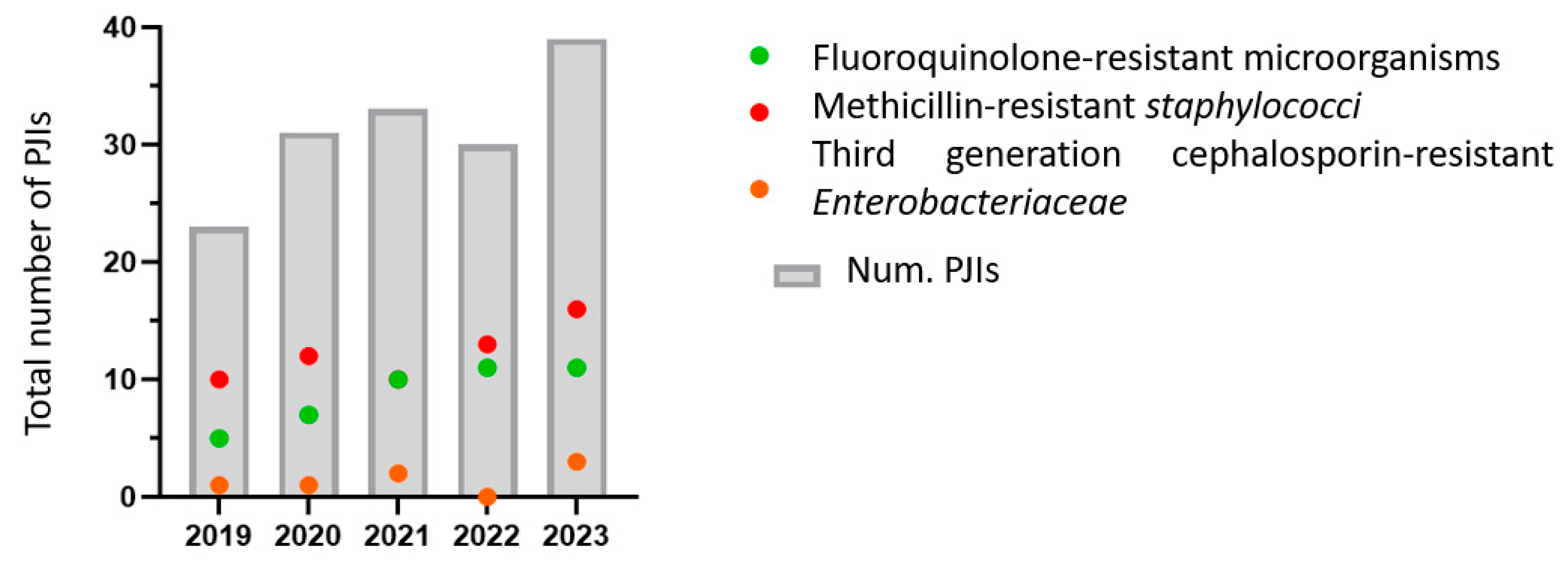
3.3. Antimicrobial Resistance

Thirty-two percent of the isolated microorganisms were MDROs, representing 40% (34/86) of the GPC and 35% (6/17) of the GNB. The resistance to quinolones was 46% for staphylococci and 20% for Enterobacteriaceae.
Forty-five percent of patients with quinolone-resistant isolates had received prior antibiotic therapy. Nine percent of the staphylococci, all CoNS, were resistant to rifampicin. All rifampicin-resistant staphylococci presented the Ile527Met mutation in rpoB, which is associated with this resistance. All of these also exhibited resistance to methicillin and quinolones. Of the 18 methicillin-resistant CoNS that were sequenced and where the mecA gene was detected, 39% belonged to sequence type ST2 and 17% belonged to ST5. Furthermore, all ST2 isolates presented the virulence biofilm-forming factor icaC. All quinolone-resistant sequenced strains (78%) presented quinolone resistance-determining regions mutations: S84Y in gyrA and S80F, as well as D84Y in parC.
Table 3 presents the antibiotic resistance data for first-line treatments used in PJIs due to their antibiofilm activity. One E. coli producing ESBL and five Enterobacteriaceae with hyperproduced chromosomal AmpC were isolated: one Serratia marcescens and four Enterobacter cloacae. Of these Enterobacteriaceae resistant to third-generation cephalosporins, four were isolated in 2023. Figure 1 shows the evolution of resistance to quinolones, rifampicin, and cephalosporins during the study years. Resistance to all three antibiotic groups increased over time.
All C. acnes isolates were susceptible to penicillin and all other beta-lactams. Only 16% (3/19) were resistant to metronidazole.

4. Discussion

Although PJI is one of the most common complications of prosthetic implants, its demographic and microbiological profile, as well as its resistance patterns across different geographic areas in Spain, have been poorly studied in recent years. Our analysis shows that the number of PJIs is increasing, as is occurring globally, making it a public health issue [1,13]. The highest number of cases occurred in the last year, with 39 cases compared to 30 in 2022. We propose that this increase is attributable to the higher number of prosthetic implants.
Approximately a quarter of PJIs (23%) had negative cultures. Although this percentage is comparable to, or even lower than, those reported in other European countries [14,15], it is concerning that more than half of these patients (56%) had received prior antibiotic therapy, which is a risk factor that reduces the diagnostic sensitivity of the infection [16]. We observed a more than twofold increase in the odds of negative cultures among patients exposed to antibiotics prior to sampling (OR = 2.32).
Other possible causes related to culture-negative PJIs, such as the use of inadequate culturing tools or an insufficient culturing period, can be ruled out in our cohort, as sonication of the explanted prostheses was performed and a prolonged incubation time of two weeks was applied. Both strategies have been shown to significantly increase the sensitivity of microbiological diagnosis [17,18]. In recent years, in order to optimize these techniques and minimize their limitations, recent advances such as enzymatic treatment, m6A modification, and new pre-processing methods have been evaluated [4,19,20].
Therefore, it is evident that the initiation of antibiotic treatment prior to sample collection results in a decrease in diagnostic efficacy. This challenge could be addressed with the implementation of new molecular biology techniques and NGS (next-generation sequencing) in sonication fluid as a complementary diagnostic method. Given that culture-based techniques seem to have already reached their maximum performance [7], NGS methods, which detect genetic material from both viable and non-viable microorganisms, would allow for the identification of all such infections where no microbiological agent is isolated, significantly improving diagnostic performance [21,22,23].
Regarding microbial etiology, as is common, GPC, particularly staphylococci, were the most frequent agents in all types of PJIs [24,25,26]. Similar to recent studies conducted at the national level, the main causative agents were methicillin-resistant coagulase-negative staphylococci (MR-CoNS) [27,28]. This differs from other nearby European countries, where the main causative agent of PJI is S. aureus [15,24]. The majority of MR-CoNS belonged to sequence types ST2 and ST5, which are both associated with nosocomial infections, suggesting a possible perioperative origin. They showed virulence factors related to biofilm formation (icaC gene), which has been shown to be associated with PJIs, representing one of the major challenges in PJI management [29].
Given that the majority of PJIs in our cohort were caused by staphylococci, NGS may provide substantial benefits in the detection of difficult-to-culture staphylococci, such as small-colony variants, as well as fastidious microorganisms. This improvement in the detection of rare pathogens by mNGS leads to reduced antibiotic-related complications, shorter hospital stays and antibiotic durations, and better treatment outcomes, particularly in culture-negative cases [22,30].
In our cohort, the low number of S. aureus and MRSA cases stands out, and there was only one isolation of P. aeruginosa over the 5-year period. While in numerous European and American studies, S. aureus and GNB were the main agents involved in early infections, in our case CoNS remained predominant [25,26]. It is likely that many of the early PJIs with negative cultures were caused by these virulent microorganisms (S. aureus and GNB), which were not successfully isolated.
The high number of PJIs caused by anaerobic microorganisms is noteworthy, as well as the fact that of the 18 PJIs caused by C. acnes, 67% occurred in joints other than the shoulder, which is typically the most common site of isolation [31]. The isolation of GNB was infrequent, mostly causing (59%) early acute infections. These findings highlight the importance of antibiotic coverage against anaerobic microorganisms in chronic infections of any joint type in our region. Furthermore, the use of empirical treatment with a Gram-negative spectrum would only be particularly relevant in early acute infections.
While previous studies have shown that polymicrobial infections typically account for around 15–17% of PJIs [32], in this study, the percentage was significantly lower at 4%. This low number of polymicrobial infections may also be attributed to the inappropriate use of antibiotic therapy prior to sample collection. Furthermore, no significant differences were observed between the three types of infections (early, low-grade, and late), nor did they follow any pattern of microorganism combinations.
For the optimization of treatment, it is as important to identify the causative pathogen as it is to understand the patterns of antibiotic sensitivity. In our study, over 30% of the pathogens were MDROs. The primary microbiological agent was S. epidermidis, with the majority (74%) being methicillin-resistant. Given this therapeutic challenge, some authors suggest a benefit in using combination therapies with vancomycin or daptomycin for PJIs in areas with a high prevalence of methicillin-resistant staphylococci, despite the nephrotoxicity and that they are considered to have broad coverage within Gram-positive bacteria, due to the risk/benefit ratio [24]. Of these methicillin-resistant S. epidermidis, 81% were also resistant to quinolones, and six isolates were resistant to rifampicin.
It is concerning that close to half of the staphylococci (46%) were resistant to quinolones and 10% to rifampicin, which are the main antibiotics used for the treatment of PJIs due to their high anti-biofilm activity [33]. Additionally, there has been an upward trend in resistance to these antibiotics in recent years. These findings align with numerous studies demonstrating that resistance in staphylococci is on the rise, representing an emerging threat [28,33]. These organisms are associated with increased treatment failure, prolonged hospital stays, and elevated healthcare costs [34]. Consequently, rapid microbiological diagnosis and early initiation of targeted therapy are not only essential for optimizing patient outcomes but also for reducing the broader healthcare burden and limiting the spread of antimicrobial resistance. Monotherapy or suboptimal dosing with rifampicin, as well as incorrect treatment with quinolones, has been shown to be a triggering factor for rifampicin resistance, leading to mutations in the rpoB gene [35]. In our cohort, of the six cases with rifampicin resistance presenting the Ile527Met mutation in rpoB, three (50%) met one of these criteria: two were incorrectly treated with quinolones and one with rifampicin monotherapy.
In many of our patients, early treatment was initiated with the combination of two antibiotics, fluoroquinolones with rifampicin, whereas it is recommended to do so only after the implantation of the definitive prosthesis, with dry surgical wounds and no drains [33,35]. We propose a link between the inappropriate use of antibiotics and the high resistance rate in our community. Moreover, the high percentage of quinolone resistance (20%), which also increased over the years, is noteworthy.
In the case of Enterobacteriaceae, 40% were resistant to third-generation cephalosporins, a percentage much higher than that described in previous national studies (3%) [25]. This resistance followed an upward trend, reaching 100% of the isolates in the last year (Enterobacteriaceae with hyperproduction of chromosomal AmpC). On the other hand, the combination of ceftazidime with vancomycin is one of the most commonly used empirical treatment options in our hospitals despite the low prevalence of P. aeruginosa in our community. Based on our local etiology and antimicrobial susceptibility data, and because it has proven to be an effective therapeutic option, we suggest combining carbapenems [36] or cefepime [37] instead of ceftazidime as an empirical treatment for acute PJIs, given the higher prevalence of acute PJI caused by Enterobacteriaceae in our setting.
Infected arthroplasties with resistant microorganisms have been shown to have a treatment failure rate ranging from 33 to 82% [28]. New therapeutic strategies not based on antibiotics, such as stem cell-AMPs, CRISPR-Cas 9, probiotics, or nanobiotics, enable progress in addressing the current situation [38]. Phages and phage-derived enzymes developed through engineering, or macrophage activation, could offer significant future benefits against these PJIs caused by antibiotic-resistant microorganisms [39]. The efficacy of phage–liposome nanoconjugates in the eradication of orthopedic biofilms has been demonstrated in a recent study. Given that the components are already approved for clinical application, these nanoconjugates represent a promising strategy with high translational potential [40].

5. Conclusions

In conclusion, these data on the current etiological patterns associated with PJIs would allow the establishment of more targeted empirical and definitive treatment protocols, based on the type of infection, for all cases with negative cultures. New molecular sequencing techniques (NGS) could help reduce the number of infections without etiological identification and could be useful as a screening tool for patients in whom no causative agent can be isolated. However, the lack of standardization and the heterogeneity of NGS methods currently used in PJIs highlight the need for studies that optimize and enable the establishment of unified protocols (appropriate sample type, extraction, pipelines, and cut-off) to standardize a diagnostic technique. On the other hand, the high percentage of resistance in our area to the main antibiofilm agents raises the question of the future benefit of new non-antibiotic treatment strategies, which could be promising in the management of PJIs.

6. Limitations

Our study has certain limitations. First of all, it is a retrospective study with limited sample size and geographic scope. Future multi-regional or national studies over a longer period of time would enable us to improve generalizability and detect broader resistance patterns. On the other hand, detection by molecular biology methods, PCR or NGS, was not performed and this could influence the rate of culture-negative PJIs. The absence of molecular diagnostics potentially impacts pathogen detection and resistance analysis. These findings highlight the need for advanced molecular techniques, such as 16S rRNA sequencing or mNGS, to enhance microbiological yield. Notably, NGS has demonstrated higher sensitivity and diagnostic accuracy compared to conventional culture methods, although with a slightly lower specificity [41]. Studies with a larger number of samples using molecular biology techniques, which are capable of detecting non-viable microorganisms that are difficult to isolate or with a low bacterial load, are necessary to confirm a microbiological diagnosis. Finally, since these are regional data, the results and conclusions cannot be extrapolated to areas with different epidemiologic distributions and microorganism patterns but rather only to regions with similar microbiological distributions.

Author Contributions

Conceptualization, L.H. and M.E.P.; methodology, L.H., M.E.P. and I.S.; software, A.U.; validation, M.E.P. and I.S.; formal analysis, I.B.; investigation, L.H. and A.U.; resources, L.H. and A.U.; data curation, M.E.P. and I.S.; writing—original draft preparation, L.H. and A.U.; writing—review and editing, M.E.P. and I.S.; visualization, I.B.; supervision, M.E.P. and C.E.B.; project administration, M.E.P.; funding acquisition, M.E.P. and L.H. All authors have read and agreed to the published version of the manuscript.

Funding

The APC was funded by Navarra Biomed (NB)—Servicio Navarro de Salud-Osasunbidea (SNS-O). This study was supported by a project funded by the Government of Navarra. Proyecto DIPAN GºNa 56/22. Principal investigator: M.E.Portillo.

Institutional Review Board Statement

This study was conducted according to the guidelines of the Declaration of Helsinki. Approval for the study was obtained from the local institutional review board (Comité de Ética de Investigación Clínica en Navarra (CEIN)) approved on 21 June 2016 (No. PI_2021/92).

Informed Consent Statement

Patient consent was waived due to the anonymous clinical data management.

Data Availability Statement

The original contributions presented in this study are included in the article. Further inquiries can be directed to the corresponding author.

Conflicts of Interest

The authors declare no conflict of interest. The funders had no role in the design of the study; in the collection, analyses, or interpretation of data; in the writing of the manuscript; or in the decision to publish the results.

References

  1. Patel, R. Periprosthetic Joint Infection. N. Engl. J. Med. 2023, 388, 251–262. [Google Scholar] [CrossRef] [PubMed]
  2. Portillo, M.E.; Sancho, I. Advances in the Microbiological Diagnosis of Prosthetic Joint Infections. Diagnostics 2023, 13, 809. [Google Scholar] [CrossRef]
  3. Peel, T.N.; Sedarski, J.A.; Dylla, B.L.; Shannon, S.K.; Amirahmadi, F.; Hughes, J.G.; Cheng, A.C.; Patel, R. Laboratory Workflow Analysis of Culture of Periprosthetic Tissues in Blood Culture Bottles. J. Clin. Microbiol. 2017, 55, 2817–2826. [Google Scholar] [CrossRef] [PubMed]
  4. Henríquez, L.; Martín, C.; Echeverz, M.; Lasa, Í.; Ezpeleta, C.; Portillo, M.E. Evaluation of the Use of Sonication Combined with Enzymatic Treatment for Biofilm Removal in the Microbiological Diagnosis of Prosthetic Joint Infection. Microbiol. Spectr. 2024, 12, e0002024. [Google Scholar] [CrossRef]
  5. Ivy, M.I.; Thoendel, M.J.; Jeraldo, P.R.; Greenwood-Quaintance, K.E.; Hanssen, A.D.; Abdel, M.P.; Chia, N.; Yao, J.Z.; Tande, A.J.; Mandrekar, J.N.; et al. Direct Detection and Identification of Prosthetic Joint Infection Pathogens in Synovial Fluid by Metagenomic Shotgun Sequencing. J. Clin. Microbiol. 2018, 56, e00402-18. [Google Scholar] [CrossRef]
  6. Kullar, R.; Chisari, E.; Snyder, J.; Cooper, C.; Parvizi, J.; Sniffen, J. Next-Generation Sequencing Supports Targeted Antibiotic Treatment for Culture Negative Orthopedic Infections. Clinical. Infect. Dis. 2023, 76, 359–364. [Google Scholar] [CrossRef]
  7. Esteban, J.; Gómez-Barrena, E. An Update about Molecular Biology Techniques to Detect Orthopaedic Implant-Related Infections. EFORT Open. Rev. 2021, 6, 93–100. [Google Scholar] [CrossRef] [PubMed]
  8. Tan, T.L.; Kheir, M.M.; Shohat, N.; Tan, D.D.; Kheir, M.; Chen, C.; Parvizi, J. Culture-Negative Periprosthetic Joint Infection: An Update on What to Expect. JB JS Open Access 2018, 3, e0060. [Google Scholar] [CrossRef]
  9. Hong, H.L.; Flurin, L.; Thoendel, M.J.; Wolf, M.J.; Abdel, M.P.; Greenwood-Quaintance, K.E.; Patel, R. Targeted Versus Shotgun Metagenomic Sequencing-Based Detection of Microorganisms in Sonicate Fluid for Periprosthetic Joint Infection Diagnosis. Clin. Infect. Dis. 2023, 76, e1456–e1462. [Google Scholar] [CrossRef]
  10. Antimicrobial Resistance Surveillance in Europe 2023–2021 Data; European Centre for Disease Prevention and Control and World Health Organization: Stockholm, Sweden, 2023.
  11. da Silva, R.B.; Salles, M.J. Outcomes and Risk Factors in Prosthetic Joint Infections by Multidrug-Resistant Gram-Negative Bacteria: A Retrospective Cohort Study. Antibiotics 2021, 10, 340. [Google Scholar] [CrossRef]
  12. Sendi, P.; Zimmerli, W. Diagnosis of Periprosthetic Joint Infections in Clinical Practice. Int. J. Artif. Organs 2012, 35, 913–922. [Google Scholar] [CrossRef] [PubMed]
  13. Zardi, E.M.; Franceschi, F. Prosthetic joint infection. A relevant public health issue. J. Infect. Public Health 2020, 13, 1888–1891. [Google Scholar] [CrossRef]
  14. Mussa, M.; Manciulli, T.; Corbella, M.; Mariani, B.; Cambieri, P.; Gipsz, N.; Scudeller, L.; Abbott, D.M.; Brunetti, E.; Mosconi, M.; et al. Epidemiology and Microbiology of Prosthetic Joint Infections: A Nine-Year, Single-Center Experience in Pavia, Northern Italy. Musculoskelet. Surg. 2021, 105, 195–200. [Google Scholar] [CrossRef] [PubMed]
  15. Sebastian, S.; Sezgin, E.A.; Stučinskas, J.; Tarasevičius, Š.; Liu, Y.; Raina, D.B.; Tägil, M.; Lidgren, L.; W-Dahl, A. Different Microbial and Resistance Patterns in Primary Total Knee Arthroplasty Infections—A Report on 283 Patients from Lithuania and Sweden. BMC Musculoskelet. Disord. 2021, 22, 800. [Google Scholar] [CrossRef]
  16. Kalbian, I.; Park, J.W.; Goswami, K.; Lee, Y.K.; Parvizi, J.; Koo, K.H. Culture-Negative Periprosthetic Joint Infection: Prevalence, Aetiology, Evaluation, Recommendations, and Treatment. Int. Orthop. 2020, 44, 1255–1261. [Google Scholar] [CrossRef] [PubMed]
  17. Bellova, P.; Knop-Hammad, V.; Königshausen, M.; Mempel, E.; Frieler, S.; Gessmann, J.; Schildhauer, T.A.; Baecker, H. Sonication of Retrieved Implants Improves Sensitivity in the Diagnosis of Periprosthetic Joint Infection. BMC Musculoskelet. Disord. 2019, 20, 623. [Google Scholar] [CrossRef]
  18. Talsma, D.T.; Ploegmakers, J.J.W.; Jutte, P.C.; Kampinga, G.; Wouthuyzen-Bakker, M. Time to Positivity of Acute and Chronic Periprosthetic Joint Infection Cultures. Diagn. Microbiol. Infect. Dis. 2021, 99, 115178. [Google Scholar] [CrossRef]
  19. Cai, Y.; Chen, X.; Huang, C.; Chen, Y.; Zhang, C.; Huang, Z.; Zhang, W.; Tang, Y.; Fang, X. Alteration of M6A-Tagged RNA Profiles in Bone Originated from Periprosthetic Joint Infection. J. Clin. Med. 2023, 12, 2863. [Google Scholar] [CrossRef]
  20. Cai, Y.; Ding, H.; Chen, X.; Chen, Y.; Huang, C.; Zhang, C.; Huang, Z.; Huang, Y.; Li, W.; Zhang, W.; et al. Optimization and Standardization of MNGS-Based Procedures for the Diagnosis of Mycoplasma Periprosthetic Joint Infection: A Novel Diagnostic Strategy for Rare Bacterial Periprosthetic Joint Infection. Front. Cell. Infect. Microbiol. 2023, 13, 1089919. [Google Scholar] [CrossRef]
  21. Kato, H.; Hagihara, M.; Asai, N.; Umemura, T.; Hirai, J.; Yamagishi, Y.; Iwamoto, T.; Mikamo, H. Comparison of Microbial Detection Rates in Microbial Culture Methods versus Next-Generation Sequencing in Patients with Prosthetic Joint Infection: A Systematic Review and Meta-Analysis. J. Orthop. Surg. Res. 2023, 18, 604. [Google Scholar] [CrossRef]
  22. Henríquez, L.; Uribarri, A.; Portillo, M.E. Revisiting Diagnostics: Practical Application of next-Generation Sequencing Technologies for Infectious Diseases. Clin. Microbiol. Infect. 2025; in press. [Google Scholar] [CrossRef]
  23. Olearo, F.; El Zein, S.; Portillo, M.E.; Zapf, A.; Rohde, H.; Berbari, E.F.; Wouthuyzen-Bakker, M. Diagnostic Accuracy of 16S RDNA PCR, Multiplex PCR and Metagenomic next-Generation Sequencing in Periprosthetic Joint Infections: A Systematic Review and Meta-Analysis. Clin. Microbiol. Infect. 2025, 37, 1115–1125. [Google Scholar] [CrossRef] [PubMed]
  24. Casenaz, A.; Piroth, L.; Labattut, L.; Sixt, T.; Magallon, A.; Guilloteau, A.; Neuwirth, C.; Amoureux, L. Epidemiology and Antibiotic Resistance of Prosthetic Joint Infections According to Time of Occurrence, a 10-Year Study. J. Infect. 2022, 85, 492–498. [Google Scholar] [CrossRef]
  25. Benito, N.; Mur, I.; Ribera, A.; Soriano, A.; Rodríguez-Pardo, D.; Sorlí, L.; Cobo, J.; Fernández-Sampedro, M.; Del Toro, M.D.; Guío, L.; et al. The Different Microbial Etiology of Prosthetic Joint Infections According to Route of Acquisition and Time after Prosthesis Implantation, Including the Role of Multidrug-Resistant Organisms. J. Clin. Med. 2019, 8, 673. [Google Scholar] [CrossRef] [PubMed]
  26. Tai, D.B.G.; Patel, R.; Abdel, M.P.; Berbari, E.F.; Tande, A.J. Microbiology of Hip and Knee Periprosthetic Joint Infections: A Database Study. Clin. Microbiol. Infect. 2022, 28, 255–259. [Google Scholar] [CrossRef] [PubMed]
  27. Barbero Allende, J.M.; Gómez-Junyent, J.; Sorlí Redó, L.; Rodríguez-Pardo, D.; Murillo Rubio, Ó.; Fernández Sampedro, M.; Escudero-Sánchez, R.; García Gutiérrez, M.; Portillo, M.E.; Sancho, I.; et al. Description of Reinfection of Joint Prosthesis after 2-Stage Replacement (Infection of the 2nd Stage Prosthesis): A Multicenter Study. Enferm. Infecc. Microbiol. Clin. (Engl. Ed.) 2024, 42, 354–360. [Google Scholar] [CrossRef]
  28. Hays, M.R.; Kildow, B.J.; Hartman, C.W.; Lyden, E.R.; Springer, B.D.; Fehring, T.K.; Garvin, K.L. Increased Incidence of Methicillin-Resistant Staphylococcus Aureus in Knee and Hip Prosthetic Joint Infection. J. Arthroplast. 2023, 38, S326–S330. [Google Scholar] [CrossRef]
  29. Izakovicova, P.; Borens, O.; Trampuz, A. Periprosthetic Joint Infection: Current Concepts and Outlook. EFORT Open Rev. 2019, 4, 482–494. [Google Scholar] [CrossRef]
  30. Hu, H.; Ding, H.; Lyu, J.; Chen, Y.; Huang, C.; Zhang, C.; Li, W.; Fang, X.; Zhang, W. Detection of Rare Microorganisms in Bone and Joint Infections by Metagenomic Next-Generation Sequencing: Discrimination, Decision-Making, and Clinical Benefits. Bone Jt. Res. 2024, 13, 401–410. [Google Scholar] [CrossRef]
  31. Boisrenoult, P. Cutibacterium Acnes Prosthetic Joint Infection: Diagnosis and Treatment. Orthop. Traumatol. Surg. Res. 2018, 104, S19–S24. [Google Scholar] [CrossRef]
  32. Flurin, L.; Greenwood-Quaintance, K.E.; Patel, R. Microbiology of Polymicrobial Prosthetic Joint Infection. Diagn. Microbiol. Infect. Dis. 2019, 94, 255–259. [Google Scholar] [CrossRef] [PubMed]
  33. Wallimann, A.; Achermann, Y.; Ferris, C.; Morgenstern, M.; Clauss, M.; Stadelmann, V.; Rüdiger, H.A.; O’Mahony, L.; Moriarty, T.F. Emergence of Rifampicin-Resistant Staphylococci on the Skin and Nose of Rifampicin-Treated Patients with an Orthopaedic-Device-Related Infection. J. Bone Jt. Infect. 2024, 9, 191–196. [Google Scholar] [CrossRef]
  34. Murray, C.J.; Ikuta, K.S.; Sharara, F.; Swetschinski, L.; Robles Aguilar, G.; Gray, A.; Han, C.; Bisignano, C.; Rao, P.; Wool, E.; et al. Global Burden of Bacterial Antimicrobial Resistance in 2019: A Systematic Analysis. Lancet 2022, 399, 629–655. [Google Scholar] [CrossRef] [PubMed]
  35. Achermann, Y.; Eigenmann, K.; Ledergerber, B.; Derksen, L.; Rafeiner, P.; Clauss, M.; Nüesch, R.; Zellweger, C.; Vogt, M.; Zimmerli, W. Factors Associated with Rifampin Resistance in Staphylococcal Periprosthetic Joint Infections (PJI): A Matched Case-Control Study. Infection 2013, 41, 431–437. [Google Scholar] [CrossRef] [PubMed]
  36. Antony, S.; Khan, I.; Chowdhury, O.; Heydemann, J.; Antony, N.; Heydemann, J.; Isaac, D. Proposed Guidelines for the Management of ESBL in Prosthetic Joint Infections. Infect. Disord. Drug. Targets. 2020, 20, 563–569. [Google Scholar] [CrossRef]
  37. Triffault-Fillit, C.; Mabrut, E.; Corbin, K.; Braun, E.; Becker, A.; Goutelle, S.; Chaudier, P.; Fessy, M.H.; Dupieux, C.; Laurent, F.; et al. Tolerance and Microbiological Efficacy of Cefepime or Piperacillin/Tazobactam in Combination with Vancomycin as Empirical Antimicrobial Therapy of Prosthetic Joint Infection: A Propensity-Matched Cohort Study. J. Antimicrob. Chemother. 2020, 75, 2299–2306. [Google Scholar] [CrossRef]
  38. Semeshchenko, D.; Slullitel, P.A.; Farinati, A.; Albani-Forneris, A.F.; Piuzzi, N.S.; Buttaro, M.A. Unconventional Therapies in Periprosthetic Joint Infections: Prevention and Treatment: A Narrative Review. J. Clin. Med. 2025, 14, 2610. [Google Scholar] [CrossRef]
  39. Ponce Benavente, L.; Wagemans, J.; Hinkel, D.; Aguerri Lajusticia, A.; Lavigne, R.; Trampuz, A.; Gonzalez Moreno, M. Targeted Enhancement of Bacteriophage Activity against Antibiotic-Resistant Staphylococcus Aureus Biofilms through an Evolutionary Assay. Front. Microbiol. 2024, 15, 1372325. [Google Scholar] [CrossRef]
  40. Wang, L.; Tkhilaishvili, T.; Jiang, Z.; Pirlar, R.F.; Ning, Y.; Millán Laleona, A.; Wang, J.; Tang, J.; Wang, Q.; Trampuz, A.; et al. Phage-Liposome Nanoconjugates for Orthopedic Biofilm Eradication. J. Control. Release 2024, 376, 949–960. [Google Scholar] [CrossRef]
  41. Su, S.; Wang, R.; Zhou, R.; Bai, J.; Chen, Z.; Zhou, F. Higher Diagnostic Value of Next-Generation Sequencing versus Culture in Periprosthetic Joint Infection: A Systematic Review and Meta-Analysis. Knee Surg. Sports Traumatol. Arthrosc. 2024, 32, 2277–2289. [Google Scholar] [CrossRef]
Figure 1. Time trend of microorganisms resistant to quinolones, rifampicin, and third-generation cephalosporins. The third-generation cephalosporin-resistant Enterobacteriaceae were resistant to cefotaxime, ceftriaxone, and ceftazidime.
Microbiolres 16 00142 g001
Table 1. Etiology of periprosthetic joint infections (PJIs) in Navarra over the last 5 years (2019–2023).
Early
(<3 Months)
(n = 59)
Delayed
(3–24 Months)
(n = 39)
Late
(>24 Months)
(n = 58)
Total

(n = 156)
Monomicrobial42 (71%)27 (69%)45 (78%)114
Polimicrobial2 (3%)2 (5%)2 (3%)6
Negative culture PJI15 (25%)10 (26%)11 (19%)36 (23%)
With previously antibiotic treatment10 (67%)6 (60%)4 (36%)20 (56%)
No. of isolates47 3148126
MDR16 (34%)12 (39%)12 (25%)40 (32%)
GPC31 (66%)23 (74%)32 (67%)86
CoNS*14161242
S. aureus112619
S. lugdunensis1146
S. pneumoniae1001
Beta-hemolític Streptococci2125
S. agalactiae (Group B)1102
S. dysgalactiae (Group C)1012
S. equi zooepidemicu (Group C)0011
Streptococcus viridans group1258
Enterococcus faecalis.1135
GPB001 (2%)1
Listeria monocytogenes0011
GNB10 (21%)1 (3%)6 (13%)17
Enterobacteriaceae91515
B. melitensis0011
P. aeruginosa1001
Anaerobes6 (13%)7 (23%)9 (19%)22
C. acnes46919
F. magna1102
Gemella spp.1001
Note: PJI: periprosthetic joint infection; MDR: multidrug-resistant; GPC: Gram-positive cocci; CoNS*: coagulase-negative staphylococci (excluding S. lugdunensis); GPB: Gram-positive bacilli; GNB: Gram-negative bacilli.
Table 2. Distribution of the most frequently isolated microorganisms by anatomical location (shoulder, hip, and knee) and year of diagnosis.
20192020202120222023TOTAL
Knee PJI9 (39%)21 (68%)11 (33%)21 (70%)18 (46%)80
S. epidermidis15354
S. aureus03122
C.acnes03111
Enterobacteriacee10321
Hip PJI13 (57%)10 (32%)17 (52%)7 (23%)19 (49%)10
S. epidermidis23535
S. aureus51122
C.acnes01411
Enterobacteriacee1201 *3
Shoulder PJI1 (4%)0 (0%)5 (15%)2 (7%)2 (5%)66
S. epidermidis00000
S. aureus00000
C.acnes00312
Enterobacteriacee00000
TOTAL PJI2331333039156
Note. PJI: periprosthetic joint infection. * PJI caused by two Enterobacteriaceae (P. stuartii and P. mirabilis).
Table 3. Resistance data of staphylococci and Enterobacteriaceae from PJI.
MeticilineFluoroquinolonesRifampicinThird Generation Cephalosporins *

Staphylococcus aureus (n = 19)
3 (16%)
(SARM)
7 (37%)0
-
ConS (n = 42)31 (74%)24 (57%)6 (14%)-
Enterobacteriaceae (n = 15)-3 (20%)-6 (40%)
Note: * All of them showed resistance to cefotaxime, ceftriaxone, and ceftazidime.
Disclaimer/Publisher’s Note: The statements, opinions and data contained in all publications are solely those of the individual author(s) and contributor(s) and not of MDPI and/or the editor(s). MDPI and/or the editor(s) disclaim responsibility for any injury to people or property resulting from any ideas, methods, instructions or products referred to in the content.

