Article

Insights into the Thriving of Bacillus megaterium and Rhodotorula mucilaginosa in Mining Areas: Their Adaptation and Tolerance Under Extreme Levels of Cu and Mn

by
Alfonso Álvarez-Villa
1,
Maribel Plascencia-Jatomea
2,*,
Kadiya Calderón
3,
Katiushka Arévalo-Niño
4,
Guadalupe López-Avilés
1 and
Francisco Javier Almendariz-Tapia
1,*
1
Department of Chemical Engineering and Metallurgy, University of Sonora, Hermosillo 83000, Mexico
2
Department of Research and Postgraduate in Food, University of Sonora, Hermosillo 83000, Mexico
3
Department of Scientific and Technological Research, University of Sonora, Hermosillo 83000, Mexico
4
Institute of Biotechnology, Autonomous University of Nuevo León, San Nicolás de los Garza 66455, Mexico
*
Authors to whom correspondence should be addressed.
Microbiol. Res. 2025, 16(7), 140; https://doi.org/10.3390/microbiolres16070140
Submission received: 14 May 2025 / Revised: 25 June 2025 / Accepted: 26 June 2025 / Published: 1 July 2025

Abstract

Understanding microbial adaptation and tolerance based on the cellular concentration and biosorption capacity provides critical insights for evaluating microbial performance under heavy metal stress, which is essential for selecting efficient strains or consortia for bioremediation applications. In this study, the adaptation and tolerance of Bacillus megaterium and Rhodotorula mucilaginosa to elevated concentrations of copper (Cu) and manganese (Mn) were investigated by introducing the maximum adaptation concentration (MAC) alongside the maximum tolerable concentration (MTC) and the minimum inhibitory concentration (MIC). A Gaussian model was fitted to the relative growth responses to estimate the MACs, MTCs, and MICs. B. megaterium exhibited MACs of 4.6 ppm Cu and 393.9 ppm Mn, while R. mucilaginosa showed MACs of 59.6 ppm Cu and 64.4 ppm Mn, corresponding to concentrations that stimulated their maximum cell density. A biosorption analysis revealed average capacities of 6.3 ± 5.3 mg Cu/g biomass and 28.6 ± 17.2 mg Mn/g biomass, positively correlated with the MTCs, indicating enhanced metal uptake under sublethal stress. The co-culture assays demonstrated dynamic microbial interactions shaped by the type and concentration of metal, including coexistence, competitive substitution, and dominance by tolerance. These findings support the use of MACs as indicators of growth stimulation and MTCs as thresholds for enhanced metal uptake, providing a dual-parameter framework for selecting metallotolerant microorganisms for metal recovery strategies.

1. Introduction

Worldwide, mining pollution has affected more than 400,000 km of river channels, causing toxic effects [1]. Heavy metals persist in an environment due to their non-biodegradability and bioaccumulation, severely polluting the water and soil [2]. Heavy metal pollution arises from natural and anthropogenic sources, with mining and industrial waste being the primary contributors. Heavy metals, such as arsenic, cadmium, chromium, copper, iron, lead, manganese, mercury, and zinc, have densities above 5 g/mL and are associated with environmental toxicity [3,4]. High concentrations of copper and manganese were detected in the sediments of two rivers affected by acid mine spills in Sonora, Mexico [5,6].
The types and concentrations of heavy metals significantly influence the composition and function of microbial microbiomes [7]. An ecological process for the remediation of environments polluted with heavy metals is the use of microorganisms as sorbents [8,9], including metallotolerant species. The metabolic activity and growth stimulation of these microorganisms allow them to uptake heavy metals from soil and water [10,11].
Microorganisms possess both intracellular and extracellular resistance mechanisms that regulate metal bioavailability and mitigate toxicity, including metal sequestration, enzymatic detoxification, and efflux pumps. These mechanisms can be activated by specific metals, influencing microbial adaptation in heavy metal-rich environments [3,12]. Bioremediation strategies leverage microbial sorption (Figure 1) and resistance mechanisms to regulate metal bioavailability and reduce toxicity in contaminated environments [13,14].
In extreme environments, abiotic factors, such as the physicochemical properties of the water and soil, do not allow for the growth of a wide variety of microbial species, allowing for only highly adapted microorganisms to reach high cell densities [15]. These selective pressures lead to the development of specialized microbiomes that tolerate heavy metals. Adaptation is the capacity of a microbial species to adjust morphologically, physiologically, and metabolically in response to continuous environmental variations, including fluctuating concentrations of toxic substances [16,17].
Microorganisms cope with environmental stress through three interrelated processes: tolerance, resistance, and adaptation. Tolerance allows microorganisms to endure metal toxicity under transient exposure conditions [18,19]. In contrast, resistance to heavy metal stress is an evolutionary trait that enables microorganisms to grow and reproduce in the presence of toxic metal concentrations, regardless of the duration of exposure, through specific genetic modifications, such as mutations or the acquisition of resistance genes [20,21]. Adaptation, as a broader concept, includes both tolerance and resistance, depending on whether genetic modifications are involved [22,23,24]. It is essential to differentiate bacterial tolerance from resistance, as they involve distinct mechanisms by which microorganisms respond to toxic environmental conditions, such as heavy metals [18,19].
Tolerance to heavy metals is often measured in millimolar (mM) concentrations or converted to parts per million (ppm, mg/L, and mg/kg) by multiplying by the atomic weight of the metal [25,26]. At concentrations exceeding one mM (63.5 ppm Cu and 54.9 ppm Mn), copper and manganese tend to bioaccumulate [27]. Furthermore, the dual function of Cu-Zn and Mn superoxide dismutases (SODs) as tolerance and adaptation mechanisms has been highlighted in environments with high levels of reactive oxygen species (ROS) generated by Fenton reactions [28].
Metallotolerant and metallophilic microorganisms possess the ability to survive in environments with high concentrations of heavy metals [29,30,31]. Some acidophilic microorganisms have acquired tolerance to heavy metals by thriving in extreme environments [32]. Studies have shown that Bacillus cereus and Pseudomonas putida isolated from mine wastelands in Poland exhibit both antibiotic and metal tolerance [33]. Extremophilic microorganisms, particularly haloarchaea, represent promising candidates for the bioremediation of heavy metals like cadmium due to their unique metabolic capabilities and tolerance to high-salinity conditions [34].
The maximum tolerable concentration (MTC) and the minimum inhibitory concentration (MIC) are commonly used to assess microbial tolerance and resistance to toxic substances. The MTC is the highest concentration at which microbial growth is still observed during exposure [35,36]. It is related to the minimum bactericidal concentration (MBC) and the minimum duration required to kill 99% of the population (MDK99), both of which reflect the partial survival of the microbial population. The MIC, on the other hand, is the lowest concentration that results in complete growth inhibition [37,38]. Mathematical predictive models relate resistance to the MIC and tolerance to the MDK99, since the MIC is not sufficient to study how the concentration of a toxic substance affects growth [39]. However, an analysis of tolerance and resistance using the MTC and MIC does not necessarily indicate that a microorganism can adapt to extreme conditions.
Although the MTC and MIC can help assess microbial tolerance and resistance, they do not account for the stimulatory effects that specific metal concentrations may have on microbial growth during adaptation. This study introduces the maximum adaptation concentration (MAC) as a novel metric to quantify microbial adaptation better by evaluating the adaptation and tolerance of Bacillus megaterium and Rhodotorula mucilaginosa to copper and manganese stress, using metrics such as the MAC, MTC, and MIC. Additionally, the biosorption capacity at these thresholds and the dynamics of microbial interactions in co-culture systems were assessed to provide insights for more effective bioremediation strategies.

2. Materials and Methods

2.1. Metallotolerant Microorganisms

The metallotolerant microorganisms B. megaterium and R. mucilaginosa were isolated from zones near copper mining activities along the San Pedro River (30°59′38.85000″ N, 110°18′47.63520″ W; the river flows into the United States) in Sonora, Mexico, where high concentrations of copper and manganese have been reported [5].

2.2. Copper and Manganese Adaptation and Tolerance Study

The adaptation and tolerance of B. megaterium and R. mucilaginosa to copper (Cu) and manganese (Mn) were assessed using a beef extract, peptone, and glucose (BPG) medium. The BPG medium consisted of nutrient broth (3 g/L beef extract and 5 g/L peptone) supplemented with glucose (6 g/L) and adjusted to pH 5.5. It was sterilized in an autoclave at 121 °C for 15 min. Copper and manganese were added to the BPG medium as CuSO4·5H2O and MnSO4·H2O, respectively, to achieve a concentration range of 0–2000 ppm, with 0 ppm serving as the control. The enriched medium was dispensed into Erlenmeyer flasks at 20% v/v. Each flask was inoculated with 1% v/v of an inoculum obtained from a culture with 24 h of growth and incubated at 30 °C with shaking at 150 rpm. After 48 h, samples were taken to measure the cell concentrations (cells/L) using a Neubauer chamber.
To maintain the solubility and bioavailability of the copper and manganese ions (Cu2+, Mn2+, and SO42− dissociation) in the culture medium, the nutrient and heavy metal solutions were sterilized separately and mixed at ambient temperature. The BPG medium was maintained at a pH of 5.5 for the adaptation study to ensure that soluble copper and manganese remained bioavailable. At pH values above 5.5 in a culture medium enriched with cupric nitrate and malt extract, copper ions were shown to precipitate due to hydroxyl ion interactions [40]. Similarly, manganese ions were shown to precipitate as hydroxides at a pH above 4.5 in acid mine water [41]. Therefore, the composition and pH of the culture medium were carefully controlled to avoid the formation of insoluble compounds and ensure metal bioavailability [42].

2.3. Detection of Oxidative Stress by Reactive Oxygen Species (ROS)

The ROS was determined by cellular analysis using fluorescence microscopy (Zeiss filter set 38 Ex/Em = 470/525 nm). After 48 h of growth at experimental MTCs of Cu and Mn, the cells were stained with the dye 2′,7′-Dichlorofluorescin diacetate (Sigma-Aldrich, Saint Louis, MO, USA) for ROS detection at a concentration of 50 μM, dissolved in ethanol–water (30:70). Samples of 10 μL of growth cells were placed on glass microscope slides with 10 μL of phosphate-buffered saline (PBS) and 20 μL of the dye, then incubated for 30 min in the dark [43,44].

2.4. Inhibitory Model

An inhibitory model of the Gaussian peak 3P was curve-fitted to the experimental data of relative growth %(C) (% of growth compared to control) vs. the concentration of heavy metal C (ppm of Cu and Mn) using JMP 17 statistical software. Equation (1) represents the Gaussian model, where %max is the maximum relative growth (height of the peak) at the maximum adaptation concentration (MAC), and W is related to the control of the peak width.
% ( C ) = % m a x · e C M A C 2 2 · W 2

2.5. Correlation Analysis Among Adaptation, Tolerance, and Biosorption Capacity

From the Cu and Mn adaptation and tolerance study at experimental MACs and MTCs (initial concentration, C0), the biomass yield (X) in dry weight (g/L) and the final concentration (Cf) of Cu and Mn were determined after 48 h of growth in the BPG medium. The biosorption capacity (q) in mg of metal sorbed per gram of biomass (mg/g) was determined using Equation (2), where the initial C0 and final Cf concentrations of Cu and Mn were analyzed by atomic absorption spectroscopy. A correlation analysis of Cu and Mn was performed using JMP 17 statistical software.
q = C 0 C f X

2.6. Co-Culture Study

The interaction between the microorganisms and heavy metals was investigated using a co-culture of B. megaterium and R. mucilaginosa in a culture medium enriched with copper (Cu) and manganese (Mn). The co-culture experiments were performed in triplicate in Erlenmeyer flasks containing 20% v/v of BPG medium, which consisted of nutrient broth (3 g/L beef extract and 5 g/L peptone) supplemented with glucose (6 g/L) and adjusted to pH 5.5. The medium was sterilized in an autoclave at 121 °C for 15 min. Copper (CuSO4·5H2O) and manganese (MnSO4·H2O) were added to the BPG medium to achieve a concentration range of 0–100 ppm for both metals, with 0 ppm serving as the control. Each flask was inoculated with 1% v/v of an inoculum obtained from a culture with 24 h of growth and incubated at 30 °C with shaking at 150 rpm. The abundance of each species in the co-culture was determined and compared to the axenic (pure) cultures. The species abundance was quantified as the cell concentration (cells/L) using a Neubauer chamber for the total counts and green fluorescent protein (GFP) staining combined with fluorescence microscopy to differentiate the microbial populations.

2.7. Statistical Analysis

The results of the cellular concentration were expressed as the mean ± standard deviation (error bars). The data from the inhibitory model and co-culture study were analyzed by using JMP 17 statistical software.

3. Results

3.1. Copper and Manganese Microbial Adaptation and Tolerance

In the presence of Cu and Mn, B. megaterium and R. mucilaginosa exhibited distinct adaptive responses, as evidenced by increased cell concentrations relative to the metal-free controls (Figure 2). The microbial adaptation and tolerance were evaluated using three key parameters: the MAC, defined as the highest metal concentration where the cell concentration was equal to or greater than that of the control (0 ppm); the MTC, representing the highest sublethal concentration with partial growth inhibition; and the MIC, the lowest concentration at which complete growth inhibition occurred.
The results show that B. megaterium had MAC and MTC values of 800 ppm and 1500 ppm Mn, respectively, while R. mucilaginosa achieved an MAC of 125 ppm Cu and an MTC of 150 ppm Cu. These results confirm that both species can persist and grow in environments enriched with heavy metals, exhibiting physiological traits associated with environmental adaptation. Establishing the MAC, MTC, and MIC for Cu and Mn in these two strains provides a foundational step for modeling growth dynamics under heavy metal stress.

3.2. Detection of Oxidative Stress Under MTC Conditions

The oxidative stress was assessed under MTC conditions using fluorescence microscopy to explore the stress responses further (Figure 3). Green fluorescence, indicative of reactive oxygen species (ROS) generation, was detected in B. megaterium at 25 ppm Cu and 1500 ppm Mn, and R. mucilaginosa at 180 ppm Mn. The cell elongation in B. megaterium at 1500 ppm Mn suggests morphological adaptation to mitigate Mn toxicity. Future work will include evaluating co-cultures under combined Cu and Mn exposure, quantifying the oxidative stress markers, and performing detailed morphometric analyses under sublethal stress.

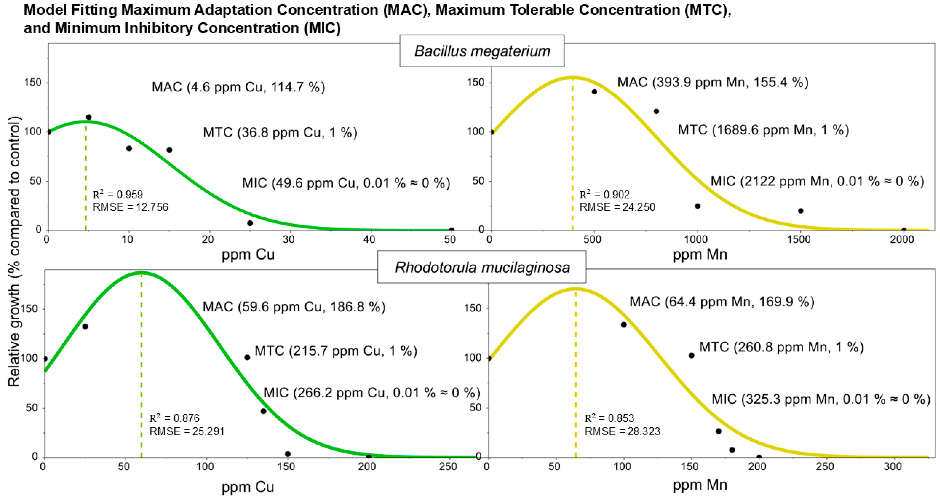
3.3. Model Fitting of MACs, MTCs, and MICs

To mathematically describe the microbial responses to Cu and Mn, a Gaussian peak model was fitted to the experimental data representing the relative cell concentration (% of growth compared to the control) as a function of the metal concentration (Figure 4). This model provides a biologically meaningful framework for identifying three key thresholds: the maximum adaptation concentration (MAC) corresponds to the concentration yielding the highest relative growth (peak height), the maximum tolerable concentration (MTC) is defined as the point where growth is reduced to 1%, and the minimum inhibitory concentration (MIC) is defined as 0.01% relative growth, equivalent to the minimum duration for killing 99.99% of the population (MDK99.99) [37].
The model successfully captured the experimental behavior of both B. megaterium and R. mucilaginosa, yielding accurate curve fits and parameter estimates. B. megaterium exhibited an MAC of 4.6 ppm Cu and a much greater MAC of 393.9 ppm Mn, confirming its preferential adaptation to manganese. In contrast, R. mucilaginosa showed an MAC of 59.6 ppm Cu and an MAC of 64.4 ppm Mn, reflecting a broader tolerance and stimulation capacity for copper and, to a lesser extent, manganese. This asymmetry in the fitted curves reflects species-specific detoxification strategies. The height of the Gaussian peak, representing the maximum relative growth, is a quantitative indicator of how efficiently a microorganism metabolizes and adapts under increasing metal stress. A broader peak suggests a wider adaptation window, while a steep decline past the MAC indicates sharp toxicity effects and limited metabolic compensation.
The results show that B. megaterium demonstrated MAC and MTC values of 800 ppm and 1500 ppm Mn, respectively, while R. mucilaginosa achieved an MAC of 125 ppm Cu and an MTC of 150 ppm Cu. These results confirm that both species can persist and grow in environments enriched with heavy metals, exhibiting physiological traits associated with environmental adaptation. Establishing the MAC, MTC, and MIC for Cu and Mn in these two strains provides a foundational step for modeling growth dynamics under heavy metal stress.

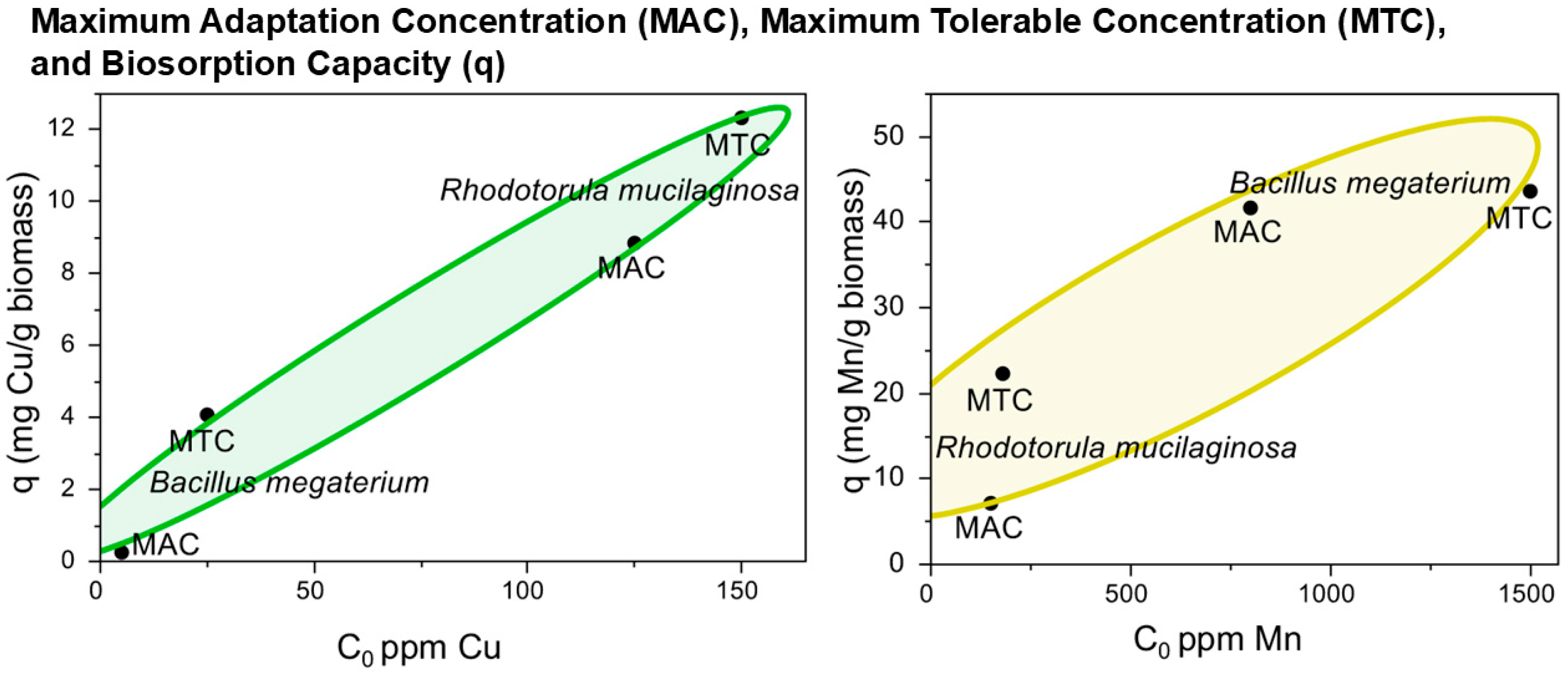
3.4. Correlation Analysis Among Experimental MACs, MTCs, and Biosorption Capacity

A correlation analysis evaluated the relationship among the microbial MACs, MTCs, and biosorption capacity in B. megaterium and R. mucilaginosa (Figure 5). The average biosorption capacities were 6.3 ± 5.3 mg Cu/g biomass and 28.6 ± 17.2 mg Mn/g biomass, indicating a higher affinity for manganese uptake across both strains.
When comparing the MAC and MTC data, the biosorption capacity was consistently higher at the MTCs than at the MACs, suggesting that increased metal stress near sublethal limits stimulates metal binding (Table 1). Furthermore, the species-specific patterns observed, R. mucilaginosa for Cu and B. megaterium for Mn, highlight a strong association between the tolerance phenotype and biosorption capacity. This insight highlights the importance of integrating tolerance and biomass productivity (optimal near the MAC) when selecting strains for biotechnological applications. The MAC defines the concentration windows ideal for large-scale biomass production, while the MTC reveals the biosorption thresholds that microorganisms can withstand.

3.5. Co-Culture with Copper and Manganese

The interaction between B. megaterium and R. mucilaginosa under metal stress demonstrates the ecological advantage of physiological flexibility. The two species coexisted without metals (0 ppm Cu and Mn), each maintaining its cell concentration profile compared to the axenic cultures (Figure 6).
However, when exposed to 10 ppm Cu, R. mucilaginosa outcompeted B. megaterium, becoming the dominant species. In contrast, at 100 ppm Mn, B. megaterium prevailed, indicating a shift in the community structure driven by metal-specific tolerance. Under combined Cu and Mn stress (10 ppm Cu + 100 ppm Mn), B. megaterium emerged as the dominant organism, suggesting its superior adaptability to manganese and combined metal conditions.

4. Discussion

The initial evaluation of microbial tolerance to copper and manganese revealed species-specific biomass production profiles, with distinct concentration thresholds for B. megaterium and R. mucilaginosa. These patterns suggest differential resistance mechanisms and adaptation potential in response to metal exposure. Such variability in metal toxicity and microbial response is consistent with earlier studies showing that tolerance is species-dependent and linked to specific resistance mechanisms [45].
For instance, strains of Pseudomonas are known to produce specific proteins to resist high concentrations of copper [13]. Other studies have demonstrated microbial adaptation at defined concentrations of metals, such as cadmium and zinc, as seen in soils near a lead–zinc smelter in Poland, where the microorganisms adapted to bioavailable concentrations of 6 ppm Cd and 103 ppm Zn at pH 7 [46]. Similarly, tolerance ranges can be broad and species-specific: Paecilomyces marquandii exhibited growth stimulation at 159 ppm Cu, partial inhibition at 318 ppm, and complete inhibition at 477 ppm in Sabouraud medium [47]. In the case of R. mucilaginosa, previous studies have reported an unchanged biomass at 50 ppm Cu in Yeast Mold medium, although extracellular polymeric substance (EPS) production was enhanced under stress conditions [48].
In contrast, at concentrations close to the MIC of bismuth, Brevundimonas diminuta reduced the production of metabolites [49]. Klebsiella pneumoniae also showed metal-specific responses under lead stress, with increased growth at 100 ppm, partial inhibition at 800 ppm, and an MIC of 900 ppm [50]. These examples underscore the importance of experimentally defining the full spectrum of microbial growth responses to metal stress, particularly the zones of stimulation (MAC), partial inhibition (MTC) associated with oxidative stress induced by ROS, and complete inhibition (MIC). The presence of ROS and morphological changes observed under fluorescence microscopy serve to corroborate that the MTC corresponds not just to partial growth inhibition, but also to active cellular responses, such as oxidative stress modulation, which are known to influence metal uptake and tolerance mechanisms [44].
The Gaussian peak model was fitted to characterize the adaptation and tolerance responses under increasing metal concentrations, estimating the MAC, MTC, and MIC values. For the bioremediation of divalent copper, Acinetobacter guillouiae demonstrated tolerance up to 150 ppm Cu, and its growth data were used to evaluate several inhibition models, including the Monod, Powell, Haldane, Luong, and Edwards models, each providing substrate inhibition constants [51]. These models typically assume the growth response starts at the origin (zero growth at zero substrate concentration). However, this assumption is invalid in assays involving relative microbial growth, where the response is normalized to 100% under control conditions. As a result, such models often fail to adequately fit experimental data that begin at a defined maximum rather than at zero. In contrast, the Gaussian peak model is more suitable for these experimental setups, as it does not require the response curve to originate from zero.
A correlation analysis was conducted among the MACs, MTCs, and biosorption capacity to relate these adaptation and tolerance thresholds to the bioremediation potential. The chemical behavior of Cu2+ and Mn2+ further explains the observed patterns. According to the Irving–Williams series (Mn2+ < Fe2+ < Co2+ < Ni2+ < Cu2+ > Zn2+), Cu2+ forms more stable complexes than Mn2+ due to its smaller ionic radius and higher charge density (Cu2+ > Mn2+), leading to preferential binding in metalloproteins [52,53]. In mixed metal environments, Cu2+ can displace Mn2+ from binding sites, potentially disrupting enzymes that require manganese. This competitive binding necessitates precise cellular regulation; bacterial regulators, such as CueR and MntR, maintain a balance between the availability of essential metals and their toxicity. CueR modulates copper efflux to avoid the mismetalation of non-cognate metalloproteins, while MntR promotes manganese uptake to support antioxidant defenses and metabolic enzymes [52].
The role of metal-specific interactions was further confirmed through co-culture experiments. These interaction patterns illustrate three key microbial responses to metal stress—coexistence, competitive substitution, and dominance by tolerance—confirmed through GFP visualization and biomass quantification. The capacity of both species to shift dominance depending on the type and concentration of metal underscores their adaptive plasticity, a crucial trait for surviving fluctuating environments. From an applied perspective, co-culturing offers practical advantages by simulating the complexity of natural microbial consortia in mining-impacted systems. While axenic cultures reveal species-specific responses, co-cultures expose emergent community behaviors, such as metabolic cooperation and resource competition, which are critical for optimizing microbial consortia in bioremediation strategies. Using measurement techniques, such as fluorescence microscopy and spectroscopic analysis, allows for detailed insights into microbial interactions and metabolite dynamics within complex microbial communities [54].
The ecological advantage of physiological flexibility observed in the co-culture under metal stress aligns with findings that intracellular metal availability and competition influence protein metalation, such as MncA, and cellular adaptation mechanisms regulated by genes like mntS and copA [55]. Studies have emphasized that bacterial species rarely cooperate, often competing in shared environments. This competitive dynamic is reflected in our findings, where metal-specific stress led to shifts in dominance between R. mucilaginosa and B. megaterium, highlighting their distinct adaptive strategies [56]. The contrasting roles of Cu2+ as an oxidant (Fenton reactions) and Mn2+ as an antioxidant (MnSOD), along with the increased availability of Mn2+ under oxidative conditions, likely contributed to the observed microbial resilience and dominance under combined Cu and Mn stress [57].
Environmental stressors, such as metal exposure, can disrupt microbial coexistence by altering the interaction dynamics, as previously demonstrated in spatially structured microbial communities [58]. The observed metal-specific tolerance of R. mucilaginosa and B. megaterium supports previous findings that microbial detoxification is highly dependent on the strain and toxin involved [59]. The adaptation of R. mucilaginosa under copper stress highlights the importance of high-affinity copper-buffering systems in microbial competition [60]. Manganese-dependent systems might similarly influence the physiology and competitive advantage of other microbes, such as B. megaterium, under combined metal stress conditions [61].
One limitation of this study is the use of controlled laboratory conditions, which, while essential for reproducibility, may not fully replicate the physicochemical heterogeneity of mining-polluted environments. In situ factors, such as porosity, particle size, soil type, organic matter content, redox potential, and competing microbial populations, can significantly influence microbial metabolism. Additionally, the adaptation and tolerance metrics were based primarily on the biomass yield, which may not capture the complete microbiome response, particularly the secondary metabolite production, EPS secretion, or biofilm formation. Studies have shown that microbial metabolites play key roles in stress modulation, efflux pump activation, and detoxification pathways that remain underexplored [62]. Functional adaptations to cadmium, copper, and nickel pollution often involve specialized sulfur- and iron-cycling microorganisms, as demonstrated in hot springs, hydrothermal vents, subsurface aquifers, and mine tailings, underscoring the importance of integrating metagenomic and transcriptomic analyses to improve our understanding of microbe–metal interactions significantly [63,64].
To further advance the application of metallotolerant microorganisms to environmental remediation, future research should integrate transcriptomics, proteomics, and metabolomics approaches to elucidate the regulatory networks and resistance genes involved in metal detoxification, including copA, mntR, and SODs [52,55]. The profiling of secondary metabolites, particularly carotenoids and EPS, may also reveal key antioxidant and stress-response mechanisms [48,57]. Additionally, pilot-scale or microcosm studies are necessary to validate microbial performance under dynamic environmental conditions, such as variable metal concentrations, pH, and redox gradients. By combining microbial metrics, such as the MAC and MTC, with multi-omics insights and bioprocess innovations, it may be possible to understand the dynamics of consortia and their capability to thrive in mining-impacted environments, offering a sustainable strategy for turning heavy metal pollution into valuable metal recovery solutions.

5. Conclusions

In this study, the adaptation and tolerance of the microorganisms Bacillus megaterium and Rhodotorula mucilaginosa to high concentrations of copper and manganese were investigated. To discern between adaptation and tolerance, the maximum adaptation concentration (MAC) was introduced and evaluated alongside the maximum tolerable concentration (MTC), providing quantitative insights into microbial resistance under metal stress and its relationship to biosorption capacity. In the co-culture systems, both species withstood sublethal concentrations of copper and manganese, exhibiting dynamic biomass production and interaction patterns that shifted between coexistence, competitive substitution, and dominance by tolerance. These findings highlight their ecological plasticity and underscore their biotechnological relevance. As the demand for critical metals increases, microbiome engineering and enhancing metal–microbe interactions will offer promising pathways for sustainable metal recovery strategies.

Author Contributions

Conceptualization, A.Á.-V., M.P.-J. and F.J.A.-T.; methodology, M.P.-J., K.C., K.A.-N. and F.J.A.-T.; software, A.Á.-V. and F.J.A.-T.; validation, M.P.-J., K.C., K.A.-N. and F.J.A.-T.; formal analysis, M.P.-J., K.C., K.A.-N. and F.J.A.-T.; investigation, A.Á.-V.; resources, M.P.-J., G.L.-A. and F.J.A.-T.; data curation, A.Á.-V., M.P.-J. and F.J.A.-T.; writing—original draft preparation, A.Á.-V., M.P.-J. and F.J.A.-T.; writing—review and editing, A.Á.-V. and G.L.-A.; visualization, G.L.-A.; supervision, M.P.-J. and F.J.A.-T.; project administration, F.J.A.-T.; funding acquisition, F.J.A.-T. All authors have read and agreed to the published version of the manuscript.

Funding

This research was funded by the Secretariat of Science, Humanities, Technology and Innovation (SECIHTI), grant number 2015-01-1594.

Institutional Review Board Statement

Not applicable.

Informed Consent Statement

Not applicable.

Data Availability Statement

The original contributions presented in this study are included in the article. Further inquiries can be directed to the corresponding authors.

Acknowledgments

The authors thank the Department of Chemical Engineering and Metallurgy at the University of Sonora for providing the experimental facilities.

Conflicts of Interest

The authors declare no conflicts of interest.

Abbreviations

The following abbreviations are used in this manuscript:
MACMaximum adaptation concentration
MTCMaximum tolerable concentration
MICMinimum inhibitory concentration

References

  1. Macklin, M.G.; Thomas, C.J.; Mudbhatkal, A.; Brewer, P.A.; Hudson-Edwards, K.A.; Lewin, J.; Scussolini, P.; Eilander, D.; Lechner, A.; Owen, J.; et al. Impacts of metal mining on river systems: A global assessment. Science 2023, 381, 1345–1350. [Google Scholar] [CrossRef]
  2. Ayangbenro, A.S.; Babalola, O.O. A New Strategy for Heavy Metal Polluted Environments: A Review of Microbial Biosorbents. Int. J. Environ. Res. Public Health 2017, 14, 94. [Google Scholar] [CrossRef] [PubMed]
  3. Roane, T.M.; Pepper, I.L.; Gentry, T.J. Chapter 18—Microorganisms and Metal Pollutants. In Environmental Microbiology, 3rd ed.; Pepper, I.L., Gerba, C.P., Gentry, T.J., Eds.; Academic Press: San Diego, CA, USA, 2015; pp. 415–439. [Google Scholar]
  4. Jacob, J.M.; Karthik, C.; Saratale, R.G.; Kumar, S.S.; Prabakar, D.; Kadirvelu, K.; Pugazhendhi, A. Biological approaches to tackle heavy metal pollution: A survey of literature. J. Environ. Manag. 2018, 217, 56–70. [Google Scholar] [CrossRef] [PubMed]
  5. Gómez-Álvarez, A.; Valenzuela-García, J.L.; Meza-Figueroa, D.; O-Villanueva, M.d.l.; Ramírez-Hernández, J.; Almendariz-Tapia, J.; Pérez-Segura, E. Impact of mining activities on sediments in a semi-arid environment: San Pedro River, Sonora, Mexico. Appl. Geochem. 2011, 26, 2101–2112. [Google Scholar] [CrossRef]
  6. León-García, G.J.; Gómez-Álvarez, A.; Meza-Figueroa, D.M.; Valenzuela-García, J.L.; Encinas-Romero, M.A.; Villalba-Atondo, A.I.; Centeno-García, E.; Encinas-Soto, K.K. Assessment of heavy metal pollution in sediments of the Sonora River basin impacted by mining activities. Environ. Prog. Sustain. Energy 2022, 41, e13796. [Google Scholar] [CrossRef]
  7. Kiama, C.W.; Njire, M.M.; Kambura, A.K.; Mugweru, J.N.; Matiru, V.N.; Wafula, E.N.; Kagali, R.N.; Kuja, J.O. Prokaryotic diversity and composition within equatorial lakes Olbolosat and Oloiden in Kenya (Africa). Curr. Res. Microb. Sci. 2021, 2, 100066. [Google Scholar] [CrossRef]
  8. Fu, F.; Wang, Q. Removal of heavy metal ions from wastewaters: A review. J. Environ. Manag. 2011, 92, 407–418. [Google Scholar] [CrossRef]
  9. Yin, K.; Wang, Q.; Lv, M.; Chen, L. Microorganism remediation strategies towards heavy metals. Chem. Eng. J. 2019, 360, 1553–1563. [Google Scholar] [CrossRef]
  10. Li, D.; Zhou, L. Adsorption of heavy metal tolerance strains to Pb2+ and Cd2+ in wastewater. Environ. Sci. Pollut. Res. 2018, 25, 32156–32162. [Google Scholar] [CrossRef]
  11. Escamilla-Rodríguez, A.; Carlos-Hernández, S.; Díaz-Jiménez, L. Evidence of Resistance of Heavy Metals from Bacteria Isolated from Natural Waters of a Mining Area in Mexico. Water 2021, 13, 2766. [Google Scholar] [CrossRef]
  12. Moraleda-Muñoz, A.; Pérez, J.; Extremera Antonio, L.; Muñoz-Dorado, J. Differential Regulation of Six Heavy Metal Efflux Systems in the Response of Myxococcus xanthus to Copper. Appl. Environ. Microbiol. 2010, 76, 6069–6076. [Google Scholar] [CrossRef] [PubMed]
  13. Havryliuk, O.; Hovorukha, V.; Patrauchan, M.; Youssef, N.H.; Tashyrev, O. Draft whole genome sequence for four highly copper resistant soil isolates Pseudomonas lactis strain UKR1, Pseudomonas panacis strain UKR2, and Pseudomonas veronii strains UKR3 and UKR4. Curr. Res. Microb. Sci. 2020, 1, 44–52. [Google Scholar] [CrossRef]
  14. Gaur, N.; Flora, G.; Yadav, M.; Tiwari, A. A review with recent advancements on bioremediation-based abolition of heavy metals. Environ. Sci. Process. Impacts 2014, 16, 180–193. [Google Scholar] [CrossRef]
  15. Madigan, M.T.; Bender, K.S.; Buckley, D.H.; Sattley, W.M.; Stahl, D.A. Brock Biology of Microorganisms, 15th ed.; Pearson: New York, NY, USA, 2018; pp. 399–732. [Google Scholar]
  16. Dönmez, G.; Aksu, Z. The effect of copper(II) ions on the growth and bioaccumulation properties of some yeasts. Process Biochem. 1999, 35, 135–142. [Google Scholar] [CrossRef]
  17. Shu, W.-S.; Huang, L.-N. Microbial diversity in extreme environments. Nat. Rev. Microbiol. 2021, 20, 219–235. [Google Scholar] [CrossRef]
  18. Brauner, A.; Fridman, O.; Gefen, O.; Balaban, N.Q. Distinguishing between resistance, tolerance and persistence to antibiotic treatment. Nat. Rev. Microbiol. 2016, 14, 320–330. [Google Scholar] [CrossRef]
  19. Camacho Mateu, J.; Sireci, M.; Muñoz, M.A. Phenotypic-dependent variability and the emergence of tolerance in bacterial populations. PLoS Comput. Biol. 2021, 17, e1009417. [Google Scholar] [CrossRef]
  20. Martinez Robert, J.; Wang, Y.; Raimondo Melanie, A.; Coombs Jonna, M.; Barkay, T.; Sobecky Patricia, A. Horizontal Gene Transfer of PIB-Type ATPases among Bacteria Isolated from Radionuclide- and Metal-Contaminated Subsurface Soils. Appl. Environ. Microbiol. 2006, 72, 3111–3118. [Google Scholar] [CrossRef] [PubMed]
  21. Cai, X.; Zheng, X.; Zhang, D.; Iqbal, W.; Liu, C.; Yang, B.; Zhao, X.; Lu, X.; Mao, Y. Microbial characterization of heavy metal resistant bacterial strains isolated from an electroplating wastewater treatment plant. Ecotoxicol. Environ. Saf. 2019, 181, 472–480. [Google Scholar] [CrossRef]
  22. Liu, H.Y.; Prentice, E.L.; Webber, M.A. Mechanisms of antimicrobial resistance in biofilms. Npj Antimicrob. Resist. 2024, 2, 27. [Google Scholar] [CrossRef]
  23. Händel, N.; Schuurmans, J.M.; Brul, S.; ter Kuile Benno, H. Compensation of the Metabolic Costs of Antibiotic Resistance by Physiological Adaptation in Escherichia coli. Antimicrob. Agents Chemother. 2013, 57, 3752–3762. [Google Scholar] [CrossRef] [PubMed]
  24. Zheng, E.J.; Andrews, I.W.; Grote, A.T.; Manson, A.L.; Alcantar, M.A.; Earl, A.M.; Collins, J.J. Modulating the evolutionary trajectory of tolerance using antibiotics with different metabolic dependencies. Nat. Commun. 2022, 13, 2525. [Google Scholar] [CrossRef] [PubMed]
  25. Tyler, G.; Balsberg Påhlsson, A.-M.; Bengtsson, G.; Bååth, E.; Tranvik, L. Heavy-metal ecology of terrestrial plants, microorganisms and invertebrates. Water Air Soil Pollut. 1989, 47, 189–215. [Google Scholar] [CrossRef]
  26. Levin-Reisman, I.; Brauner, A.; Ronin, I.; Balaban, N.Q. Epistasis between antibiotic tolerance, persistence, and resistance mutations. Proc. Natl. Acad. Sci. USA 2019, 116, 14734–14739. [Google Scholar] [CrossRef]
  27. Voica, D.M.; Bartha, L.; Banciu, H.L.; Oren, A. Heavy metal resistance in halophilic Bacteria and Archaea. FEMS Microbiol. Lett. 2016, 363, fnw146. [Google Scholar] [CrossRef]
  28. Hänsch, R.; Mendel, R.R. Physiological functions of mineral micronutrients (Cu, Zn, Mn, Fe, Ni, Mo, B, Cl). Curr. Opin. Plant Biol. 2009, 12, 259–266. [Google Scholar] [CrossRef]
  29. Deive, F.J.; Sanromán, M.Á. Chapter 14—Bioreactor Development for the Cultivation of Extremophilic Microorganisms. In Current Developments in Biotechnology and Bioengineering; Larroche, C., Sanromán, M.Á., Du, G., Pandey, A., Eds.; Elsevier: Amsterdam, The Netherlands, 2017; pp. 403–432. [Google Scholar]
  30. Barman, D.; Jha, D.K.; Bhattacharjee, K. Metallotolerant Bacteria: Insights into Bacteria Thriving in Metal-Contaminated Areas. In Microbial Versatility in Varied Environments: Microbes in Sensitive Environments; Singh, R.P., Manchanda, G., Maurya, I.K., Wei, Y., Eds.; Springer: Singapore, 2020; pp. 135–164. [Google Scholar]
  31. Parades-Aguilar, J.; Calderon, K.; Agustin-Salazar, S.; Cerruti, P.; Ambrogi, V.; Gamez-Meza, N.; Medina-Juarez, L.A. Isolation and identification of metallotolerant bacteria with a potential biotechnological application. Sci. Rep. 2024, 14, 3663. [Google Scholar] [CrossRef]
  32. Dopson, M.; Ossandon, F.J.; Lövgren, L.; Holmes, D.S. Metal resistance or tolerance? Acidophiles confront high metal loads via both abiotic and biotic mechanisms. Front. Microbiol. 2014, 5, 157. [Google Scholar] [CrossRef]
  33. Piotrowska-Seget, Z.; Beściak, G.; Bernaś, T.; Kozdrój, J. GFP-tagged multimetal-tolerant bacteria and their detection in the rhizosphere of white mustard. Ann. Microbiol. 2012, 62, 559–567. [Google Scholar] [CrossRef]
  34. Vera-Bernal, M.; Martínez-Espinosa, R.M. Insights on Cadmium Removal by Bioremediation: The Case of Haloarchaea. Microbiol. Res. 2021, 12, 354–375. [Google Scholar] [CrossRef]
  35. Paredes-Páliz, K.I.; Caviedes, M.A.; Doukkali, B.; Mateos-Naranjo, E.; Rodríguez-Llorente, I.D.; Pajuelo, E. Screening beneficial rhizobacteria from Spartina maritima for phytoremediation of metal polluted salt marshes: Comparison of gram-positive and gram-negative strains. Environ. Sci. Pollut. Res. 2016, 23, 19825–19837. [Google Scholar] [CrossRef] [PubMed]
  36. Ciofu, O.; Moser, C.; Jensen, P.Ø.; Høiby, N. Tolerance and resistance of microbial biofilms. Nat. Rev. Microbiol. 2022, 20, 621–635. [Google Scholar] [CrossRef] [PubMed]
  37. Li, J.; Xie, S.; Ahmed, S.; Wang, F.; Gu, Y.; Zhang, C.; Chai, X.; Wu, Y.; Cai, J.; Cheng, G. Antimicrobial Activity and Resistance: Influencing Factors. Front. Pharmacol. 2017, 8, 364. [Google Scholar] [CrossRef]
  38. Hernandez-Mendoza, E.; Peña-Ramos, E.A.; Juneja, V.K.; Martínez-Téllez, M.Á.; González-Ríos, H.; Paredes-Aguilar, M.D.; Valenzuela-Melendres, M.; Aispuro-Hernández, E. Antagonistic Activity of Bacteriocin-like Inhibitory Substances from Enterococcus lactis Isolated from the Surface of Jalapeno Pepper Against Foodborne Pathogens. Microbiol. Res. 2024, 15, 889–899. [Google Scholar] [CrossRef]
  39. Balaban, N.Q.; Helaine, S.; Lewis, K.; Ackermann, M.; Aldridge, B.; Andersson, D.I.; Brynildsen, M.P.; Bumann, D.; Camilli, A.; Collins, J.J.; et al. Definitions and guidelines for research on antibiotic persistence. Nat. Rev. Microbiol. 2019, 17, 441–448. [Google Scholar] [CrossRef]
  40. Dursun, A.Y.; Uslu, G.; Cuci, Y.; Aksu, Z. Bioaccumulation of copper(II), lead(II) and chromium(VI) by growing Aspergillus niger. Process Biochem. 2003, 38, 1647–1651. [Google Scholar] [CrossRef]
  41. Chanturia, V.A.; Medyanik, N.L.; Shadrunova, I.V.; Mishurina, O.A. Chemical aspects of manganese removal from mine water at copper–sulfide deposits. J. Min. Sci. 2016, 52, 169–176. [Google Scholar] [CrossRef]
  42. Pat-Espadas, A.M.; Cervantes, F.J. Microbial recovery of metallic nanoparticles from industrial wastes and their environmental applications. J. Chem. Technol. Biotechnol. 2018, 93, 3091–3112. [Google Scholar] [CrossRef]
  43. Gálvez-Iriqui, A.C.; Cortez-Rocha, M.O.; Burgos-Hernández, A.; Calderón-Santoyo, M.; Argüelles-Monal, W.M.; Plascencia-Jatomea, M. Synthesis of chitosan biocomposites loaded with pyrrole-2-carboxylic acid and assessment of their antifungal activity against Aspergillus niger. Appl. Microbiol. Biotechnol. 2019, 103, 2985–3000. [Google Scholar] [CrossRef]
  44. Contreras-Cortés, A.G.; Almendariz-Tapia, F.J.; Cortez-Rocha, M.O.; Burgos-Hernández, A.; Rosas-Burgos, E.C.; Rodríguez-Félix, F.; Gómez-Álvarez, A.; Quevedo-López, M.Á.; Plascencia-Jatomea, M. Biosorption of copper by immobilized biomass of Aspergillus australensis. Effect of metal on the viability, cellular components, polyhydroxyalkanoates production, and oxidative stress. Environ. Sci. Pollut. Res. 2020, 27, 28545–28560. [Google Scholar] [CrossRef]
  45. Mangold, S.; Potrykus, J.; Björn, E.; Lövgren, L.; Dopson, M. Extreme zinc tolerance in acidophilic microorganisms from the bacterial and archaeal domains. Extremophiles 2013, 17, 75–85. [Google Scholar] [CrossRef] [PubMed]
  46. Rusinowski, S.; Szada-Borzyszkowska, A.; Zieleźnik-Rusinowska, P.; Małkowski, E.; Krzyżak, J.; Woźniak, G.; Sitko, K.; Szopiński, M.; McCalmont, J.P.; Kalaji, H.M.; et al. How autochthonous microorganisms influence physiological status of Zea mays L. cultivated on heavy metal contaminated soils? Environ. Sci. Pollut. Res. 2019, 26, 4746–4763. [Google Scholar] [CrossRef]
  47. Słaba, M.; Gajewska, E.; Bernat, P.; Fornalska, M.; Długoński, J. Adaptive alterations in the fatty acids composition under induced oxidative stress in heavy metal-tolerant filamentous fungus Paecilomyces marquandii cultured in ascorbic acid presence. Environ. Sci. Pollut. Res. 2013, 20, 3423–3434. [Google Scholar] [CrossRef] [PubMed]
  48. Garza-Gonzalez, M.T.; Barboza Perez, D.; Vazquez Rodriguez, A.; Garcia-Gutierrez, D.I.; Zarate, X.; Cantú Cardenas, M.E.; Urraca-Botello, L.I.; Lopez-Chuken, U.J.; Trevino-Torres, A.L.; Cerino-Córdoba, F.d.J.; et al. Metal-Induced Production of a Novel Bioadsorbent Exopolysaccharide in a Native Rhodotorula mucilaginosa from the Mexican Northeastern Region. PLoS ONE 2016, 11, e0148430. [Google Scholar] [CrossRef] [PubMed]
  49. Badireddy, A.R.; Chellam, S.; Yanina, S.; Gassman, P.; Rosso, K.M. Bismuth dimercaptopropanol (BisBAL) inhibits the expression of extracellular polysaccharides and proteins by Brevundimonas diminuta: Implications for membrane microfiltration. Biotechnol. Bioeng. 2008, 99, 634–643. [Google Scholar] [CrossRef]
  50. Martis, B.S.; Mohan, A.K.; Chiplunkar, S.; Kamath, S.; Goveas, L.C.; Rao, C.V. Bacterium isolated from coffee waste pulp biosorps lead: Investigation of EPS mediated mechanism. Curr. Res. Microb. Sci. 2021, 2, 100029. [Google Scholar] [CrossRef] [PubMed]
  51. Majumder, S.; Gangadhar, G.; Raghuvanshi, S.; Gupta, S. A comprehensive study on the behavior of a novel bacterial strain Acinetobacter guillouiae for bioremediation of divalent copper. Bioprocess. Biosyst. Eng. 2015, 38, 1749–1760. [Google Scholar] [CrossRef]
  52. Osman, D.; Martini, M.A.; Foster, A.W.; Chen, J.; Scott, A.J.P.; Morton, R.J.; Steed, J.W.; Lurie-Luke, E.; Huggins, T.G.; Lawrence, A.D.; et al. Bacterial sensors define intracellular free energies for correct enzyme metalation. Nat. Chem. Biol. 2019, 15, 241–249. [Google Scholar] [CrossRef]
  53. Young, T.R.; Martini, M.A.; Foster, A.W.; Glasfeld, A.; Osman, D.; Morton, R.J.; Deery, E.; Warren, M.J.; Robinson, N.J. Calculating metalation in cells reveals CobW acquires CoII for vitamin B12 biosynthesis while related proteins prefer ZnII. Nat. Commun. 2021, 12, 1195. [Google Scholar] [CrossRef]
  54. Schlembach, I.; Grünberger, A.; Rosenbaum, M.A.; Regestein, L. Measurement Techniques to Resolve and Control Population Dynamics of Mixed-Culture Processes. Trends Biotechnol. 2021, 39, 1093–1109. [Google Scholar] [CrossRef]
  55. Clough, S.E.; Young, T.R.; Tarrant, E.; Scott, A.J.P.; Chivers, P.T.; Glasfeld, A.; Robinson, N.J. A metal-trap tests and refines blueprints to engineer cellular protein metalation with different elements. Nat. Commun. 2025, 16, 810. [Google Scholar] [CrossRef] [PubMed]
  56. Palmer, J.D.; Foster, K.R. Bacterial species rarely work together. Science 2022, 376, 581–582. [Google Scholar] [CrossRef]
  57. Foster, A.W.; Clough, S.E.; Aki, Z.; Young, T.R.; Clarke, A.R.; Robinson, N.J. Metalation calculators for E. coli strain JM109 (DE3): Aerobic, anaerobic, and hydrogen peroxide exposed cells cultured in LB media. Metallomics 2022, 14, mfac058. [Google Scholar] [CrossRef]
  58. Lobanov, A.; Dyckman, S.; Kurkjian, H.; Momeni, B. Spatial structure favors microbial coexistence except when slower mediator diffusion weakens interactions. eLife 2023, 12, e82504. [Google Scholar] [CrossRef]
  59. García-Béjar, B.; Arévalo-Villena, M.; Guisantes-Batan, E.; Rodríguez-Flores, J.; Briones, A. Study of the bioremediatory capacity of wild yeasts. Sci. Rep. 2020, 10, 11265. [Google Scholar] [CrossRef]
  60. Helmann, J.D. Metals in Motion: Understanding Labile Metal Pools in Bacteria. Biochemistry 2025, 64, 329–345. [Google Scholar] [CrossRef] [PubMed]
  61. Puccio, T.; Kunka, K.S.; Zhu, B.; Xu, P.; Kitten, T. Manganese Depletion Leads to Multisystem Changes in the Transcriptome of the Opportunistic Pathogen Streptococcus sanguinis. Front. Microbiol. 2020, 11, 592615. [Google Scholar] [CrossRef]
  62. Perry, E.K.; Meirelles, L.A.; Newman, D.K. From the soil to the clinic: The impact of microbial secondary metabolites on antibiotic tolerance and resistance. Nat. Rev. Microbiol. 2022, 20, 129–142. [Google Scholar] [CrossRef] [PubMed]
  63. Ares, Á.; Sakai, S.; Sasaki, T.; Shimamura, S.; Mitarai, S.; Nunoura, T. Sequestration and efflux largely account for cadmium and copper resistance in the deep-sea Nitratiruptor sp. SB155-2 (phylum Campylobacterota). Environ. Microbiol. 2022, 24, 6144–6163. [Google Scholar] [CrossRef]
  64. Chen, M.; Grégoire Daniel, S.; Bain Jeffrey, G.; Blowes David, W.; Hug Laura, A. Legacy copper/nickel mine tailings potentially harbor novel iron/sulfur cycling microorganisms within highly variable communities. Appl. Environ. Microbiol. 2024, 90, e00143-24. [Google Scholar] [CrossRef]
Figure 1. Five mechanisms of microbial sorption of heavy metal ions (M+).
Microbiolres 16 00140 g001
Figure 2. Adaptation and tolerance study: Cell concentration (cells/L) and standard deviation (error bars) after 48 h of B. megaterium and R. mucilaginosa growth at different concentrations (ppm) of Cu and Mn.
Microbiolres 16 00140 g002
Figure 3. Fluorescence images (scale bar, 10 μm) of B. megaterium and R. mucilaginosa stained with 2′,7′-Dichlorofluorescin diacetate after 48 h of growth at experimental MTCs of Cu and Mn.
Microbiolres 16 00140 g003
Figure 4. Experimental data (●) and model fitting (▬) of the relative growth (% compared to the control) of B. megaterium and R. mucilaginosa as a function of the Cu and Mn concentrations (ppm), including the corresponding values of the coefficient of determination (R2) and root mean square error (RMSE) for each fitted model.
Microbiolres 16 00140 g004
Figure 5. Correlation analysis among experimental MACs, MTCs, and biosorption capacity (q), with coefficients of p = 0.974 for Cu and p = 0.861 for Mn, for B. megaterium and R. mucilaginosa.
Microbiolres 16 00140 g005
Figure 6. Co-culture study: Cell concentration (cells/L) and standard deviation (error bars), and fluorescence images (scale bar 10 μm) of B. megaterium and R. mucilaginosa stained with GFP at different concentrations (ppm) of Cu and Mn.
Microbiolres 16 00140 g006
Table 1. Summary of MAC, MTC, MIC, and biosorption capacity values for B. megaterium and R. mucilaginosa under copper and manganese exposure.
MicroorganismHeavy
Metal
MAC
(ppm)
MTC
(ppm)
MIC
(ppm)
Biosorption Capacity (q)
(mg/g Biomass)
B. megateriumCu4.636.849.64.1 ± 0.1
Mn393.91689.6212243.6 ± 7.7
R. mucilaginosaCu59.6215.7266.212.3 ± 1.3
Mn64.4260.8325.322.3 ± 6.1
Disclaimer/Publisher’s Note: The statements, opinions and data contained in all publications are solely those of the individual author(s) and contributor(s) and not of MDPI and/or the editor(s). MDPI and/or the editor(s) disclaim responsibility for any injury to people or property resulting from any ideas, methods, instructions or products referred to in the content.

