Article

Anti-Influenza A Virus Activity of Rhododendron brachycarpum Extract and Identification of Hyperoside as the Active Constituent

Department of Life Science and Multidisciplinary Genome Institute, Hallym University, Chuncheon 24252, Republic of Korea
*
Author to whom correspondence should be addressed.
These authors contributed equally to this work.
Microbiol. Res. 2025, 16(6), 132; https://doi.org/10.3390/microbiolres16060132
Submission received: 24 April 2025 / Revised: 12 June 2025 / Accepted: 16 June 2025 / Published: 18 June 2025

Abstract

:
Influenza A virus (IAV) poses significant public health challenges due to its rapid mutation and drug resistance, necessitating novel antiviral strategies. Rhododendron brachycarpum, traditionally employed in folk medicine to treat inflammatory conditions, contains bioactive flavonoids with potential antiviral effects. In this study, we investigated the anti-influenza activity of R. brachycarpum leaf extract and identified hyperoside (quercetin-3-O-galactoside) as the active constituent. The crude extract and its n-butanol fraction markedly reduced IAV replication in Madin–Darby canine kidney (MDCK) cells, with IC50/CC50 values of 74.51/201.09 μg/mL and 24.5/113.1 μg/mL, respectively. Hyperoside, purified via bioactivity-guided fractionation and HPLC analysis, demonstrated potent antiviral activity, with an IC50 of 66.59 μM (30.92 μg/mL) and a CC50 of 318.9 μM (148.1 μg/mL), indicating a favorable selectivity index. It significantly suppressed viral mRNA and protein expression in infected cells. Time-of-addition and hemagglutination inhibition assays suggested that hyperoside exerts antiviral effects during early infection stages, likely interfering with viral entry. In silico molecular docking analysis further supported this mechanism, revealing that hyperoside binds strongly to the receptor-binding domain of hemagglutinin (−11.5 kcal/mol), potentially blocking viral attachment. These findings reveal that hyperoside is a major antiviral component of R. brachycarpum and underscore its therapeutic potential as a natural antiviral candidate against IAV infections.

1. Introduction

Influenza viruses are major respiratory pathogens that infect humans and animals, causing millions of illnesses and tens of thousands of deaths globally each year. Based on nucleic acid type and host range, influenza viruses are classified into types A, B, and C. Influenza A virus (IAV), which primarily infects humans, is notable for its high mutation rate, and it is responsible for seasonal epidemics as well as pandemics [1,2,3,4]. IAV belongs to the Orthomyxoviridae family, possessing a genome of eight negative-sense single-stranded RNA segments. It is further subtyped (e.g., H1N1 and H3N2) by the combination of its surface glycoproteins hemagglutinin (HA) and neuraminidase (NA) [4,5]. HA mediates viruses’ attachment to and entry into host cells, whereas NA facilitates the release of progeny virions from infected cells [1,6]. Because of these critical functions, HA and NA are primary targets for vaccine design and antiviral drugs such as oseltamivir (Tamiflu) [7,8,9,10]. However, the inherently high mutation rate of RNA viruses and the emergence of drug-resistant IAV strains underscore the need for new therapeutic strategies [11,12,13].
Natural-product-derived compounds have long been used in traditional remedies for a myriad of diseases and are increasingly recognized as promising candidates for antiviral drug development [14,15]. Rhododendron brachycarpum, an evergreen shrub native to the high mountain regions of Korea, Japan, and Northeast Asia and belonging to the Ericaceae family, has traditionally been employed as Manbyeongcho in folk medicine to treat conditions such as arthritis, gout, and inflammatory ailments [16,17]. Recent studies have highlighted its anti-inflammatory, antioxidant, and anti-diabetic effects, although its antiviral properties remain underexplored [17,18]. Among the numerous secondary metabolites present in natural products, flavonoids play a crucial role in mediating their biological and physiological actions. In particular, quercetin—a well-characterized flavonoid—exhibits robust antioxidant activity by scavenging free radicals, thereby protecting cells from oxidative stress. Additionally, quercetin modulates key intracellular signaling pathways, including those involved in inflammation and apoptosis, which may underpin its anti-inflammatory and antiviral effects [19]. It has been demonstrated to be able to inhibit the expression of pro-inflammatory cytokines and interfere with multiple stages of viral replication, such as viral entry and genome transcription, in various in vitro studies [20,21,22]. In particular, quercetin has been shown to inhibit the entry of influenza A virus (IAV), with an IC50 value of 25.67 ± 2.54 µM against the A/Puerto Rico/8/34 (H1N1) strain [23]. Considering the physiological benefits of quercetin, avicularin, guaijaverin, and hyperin in conjunction with the traditional uses of Rhododendron brachycarpum underscores the potential of this medicinal plant as a rich source of bioactive compounds for novel antiviral-drug development [16,24,25,26,27,28].
In this study, we investigated the anti-influenza activity of R. brachycarpum leaf extract against IAV and aimed to identify the active constituent responsible for this effect. Using solvent partitioning of the crude extract and subsequent HPLC analysis, we isolated the major secondary metabolite with antiviral activity and characterized its effects on influenza virus replication.

2. Materials and Methods

2.1. Plant Material and Extraction

Leaves of Rhododendron brachycarpum were collected from a mountainous region in Gangwon Province, South Korea. The leaves were freeze-dried and pulverized. The powdered material (100 g) was extracted with 1 L of 70% ethanol at room temperature for 24 h. This extraction was performed three times, and the combined extracts were filtered and concentrated under reduced pressure to yield a crude extract. The crude extract was then freeze-dried to obtain a powder and stored at −20 °C until use.

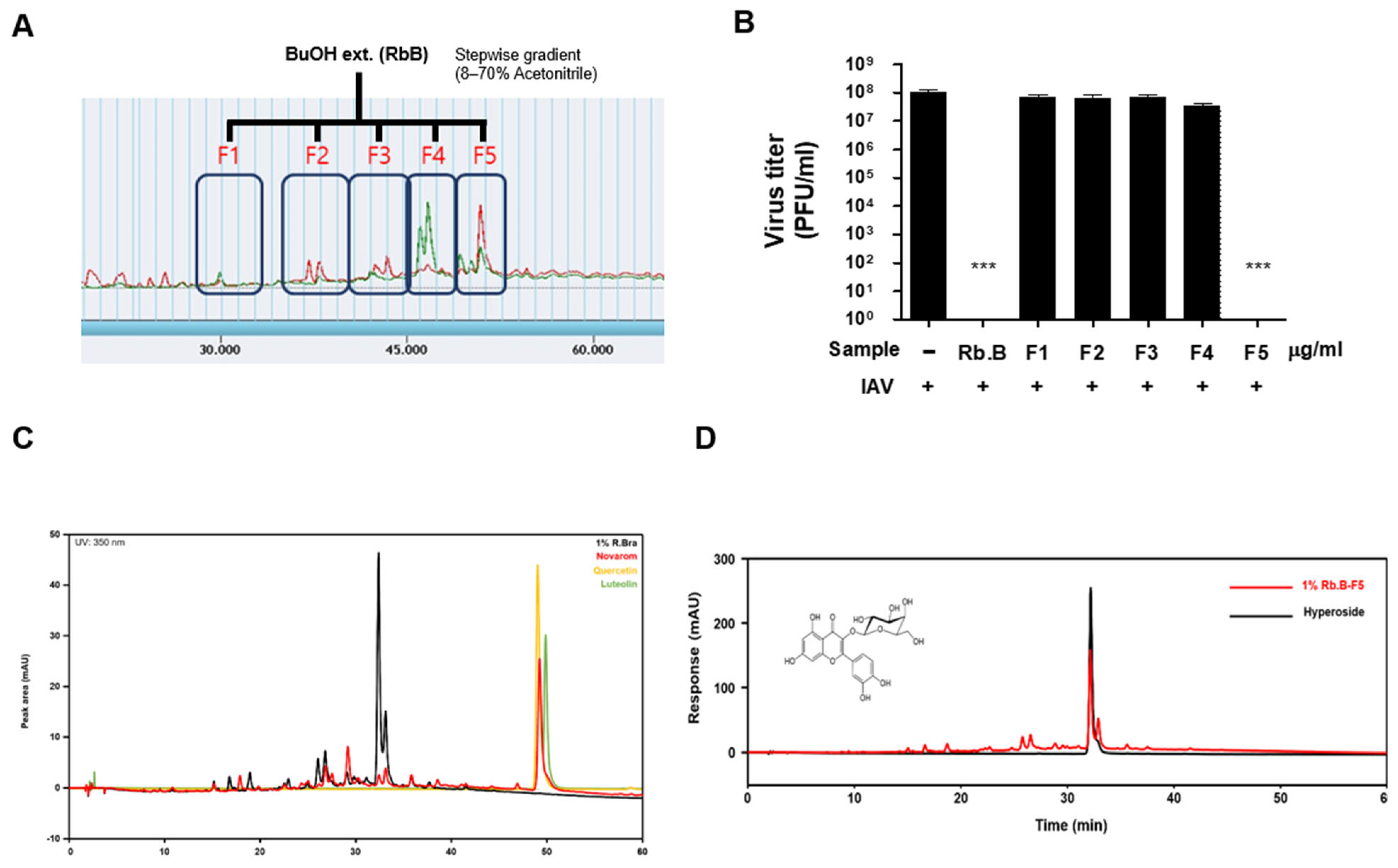
2.2. Fractionation and Active Compound Isolation

A portion of the crude extract (5 g) was suspended in 200 mL of distilled water and sequentially partitioned with equal volumes of n-hexane, dichloromethane (DCM), ethyl acetate (EA), and n-butanol. Each solvent layer was collected, concentrated under reduced pressure, and freeze-dried to yield the corresponding n-hexane, DCM, EA, n-butanol, and aqueous fractions. Among these, the n-butanol fraction, which exhibited the highest antiviral activity (see Section 3), was subjected to further purification. Active constituents were isolated from the n-butanol fraction using open-column chromatography and preparative HPLC. Compound identification was achieved by comparing retention times and UV-Vis spectra with those of an authentic standard of hyperoside. The identified hyperoside was further purified via repeated chromatography to enhance its purity, and this purified compound was used in subsequent assays.

2.3. HPLC Analysis

Analytical HPLC was performed by employing an Agilent 1200 system (Agilent Technologies, Santa Clara, CA, USA) using a C18 reverse-phase column (4.6 × 250 mm, 5 µm particle size) at 40 °C. The mobile phase consisted of water with 0.1% trifluoroacetic acid (solvent A) and acetonitrile (solvent B), applied in a gradient elution. The flow rate was 1.0 mL/min, and detection was carried out with a UV-Vis detector at 280 nm. Peaks were identified by comparing their retention times and UV absorption spectra with those of a hyperoside standard (purity ≥ 98%).

2.4. Virus and Cell Lines

Influenza virus A/Brisbane/59/2007 (H1N1) was obtained from the National Culture Collection for Pathogens (Cat. No., NCCP 42464, Osong, Republic of Korea). Madin–Darby canine kidney (MDCK) cells and human lung adenocarcinoma A549 cells were purchased from the Korean Cell Line Bank (Cat. No., 10034 and 10185, KCLB, Seoul, Republic of Korea). MDCK cells were maintained in minimum essential medium (MEM, HyClone, Logan, UT, USA) supplemented with 10% fetal bovine serum (FBS, GenDEPOT, Grand Island, NY, USA) and 1% antibiotic–antimycotic (Thermo Fisher Scientific, Waltham, MA, USA). A549 cells were cultivated in Dulbecco’s modified Eagle’s medium (DMEM) (Corning Life Science, Glendale, AZ, USA) supplemented with 10% FBS and 1% antibiotic–antimycotic. Virus stocks were prepared by propagating the H1N1 virus in MDCK cells under Biosafety Level 2 conditions. Viral titers were determined by plaque-forming unit (PFU) assays conducted on MDCK cells.

2.5. Cell Infection and Antiviral Assays

The antiviral effects of the R. brachycarpum extract and its fractions on IAV infection were evaluated in cell culture. Prior to infection assays, the cytotoxicity of the samples was assessed. MDCK cells (1 × 10⁴ cells per well) were seeded in 96-well plates and incubated for 24 h. Cells were then treated with various concentrations (0, 12.5, 25, 50, and 100 μg/mL) of the R. brachycarpum crude extract or fraction for 24 h. Cell viability was measured using water-soluble tetrazolium salt (WST)-1 (DoGenBio, Seoul, Republic of Korea) and reacted at 34 °C for 1 h. Cell viability was determined by measuring absorbance at 450 nm using a microplate reader (Thermo Fisher Scientific). After virus infection, the cytopathic effect was observed with a JuLI FL microscope (NanoEntek, Seoul, Republic of Korea).
Viral replication was further evaluated via plaque assay. MDCK cells (5 × 10⁵ cells per well) were seeded in 6-well plates and incubated for 24 h. The cells were infected with IAV at a multiplicity of infection (MOI) of 0.1. After viral adsorption for 1 h at 37 °C, the inoculum was removed, and the cell monolayers were washed with DPBS (Corning Life Science, Glendale, AZ, USA). The cells were then overlaid with MEM containing 1% agar and incubated for 3 days. After incubation, cells were fixed with 4% formaldehyde and stained with 1% crystal violet. The number of plaques (clear zones) formed by the virus was counted to quantify viral yield.

2.6. Mechanism-of-Action Analysis

To investigate the mechanism of antiviral action, MDCK cells were infected with IAV at MOI 1. Immediately after infection, cells were treated with either the R. brachycarpum crude extract or purified hyperoside (at specified concentrations) and incubated for 24 hr. For analysis of viral gene expression, total RNA was extracted from cells using TRIzol reagent (Thermo Fisher Scientific, Waltham, MA, USA), and 1 μg of RNA was reverse-transcribed to cDNA. Quantitative PCR (qPCR) was performed using ExcelTaq 2X Q-PCR Master Mix (SMOBIO, Hsinchu, Taiwan, China) to measure the mRNA levels of the IAV matrix protein 2 (M2) gene. Glyceraldehyde-3-phosphate dehydrogenase (GAPDH) served as a housekeeping control, and relative gene expression was calculated using the 2−ΔΔCt method. The forward and reverse primers for viral gene detection were as follows: M2, 5′-TGAGCCTTCTAACCGAGGTCGAAA-3′ and 5′-CCACAATATCAAGTGCACAATCCC-3′, and NS1, 5′-TCGCGGTACCTAACTGACATGACT-3′ and 5′-CTGGTCCATTCTGACACAAAGAGG-3′. As a control, canine glyceralde-hyde-3-phosphate dehydrogenase (GAPDH) gene was amplified using the following primers: 5′-AATTCCACGGCACAGTCAAGGC-3′ and 5′-AACATACTCAGCACCAGCATCACC-3′.
To examine viral protein expression, cell lysates collected 24 hr post-infection were analyzed by Western blotting for influenza A virus non-structure protein 1 (NS1). NS1 levels were detected using a specific anti-NS1 antibody (Santa Cruz, Bergheim, Germany) and chemiluminescent detection. As a control, mouse anti-β-actin antibody (CST, Danvers, MA, USA) was used for detection of host protein.

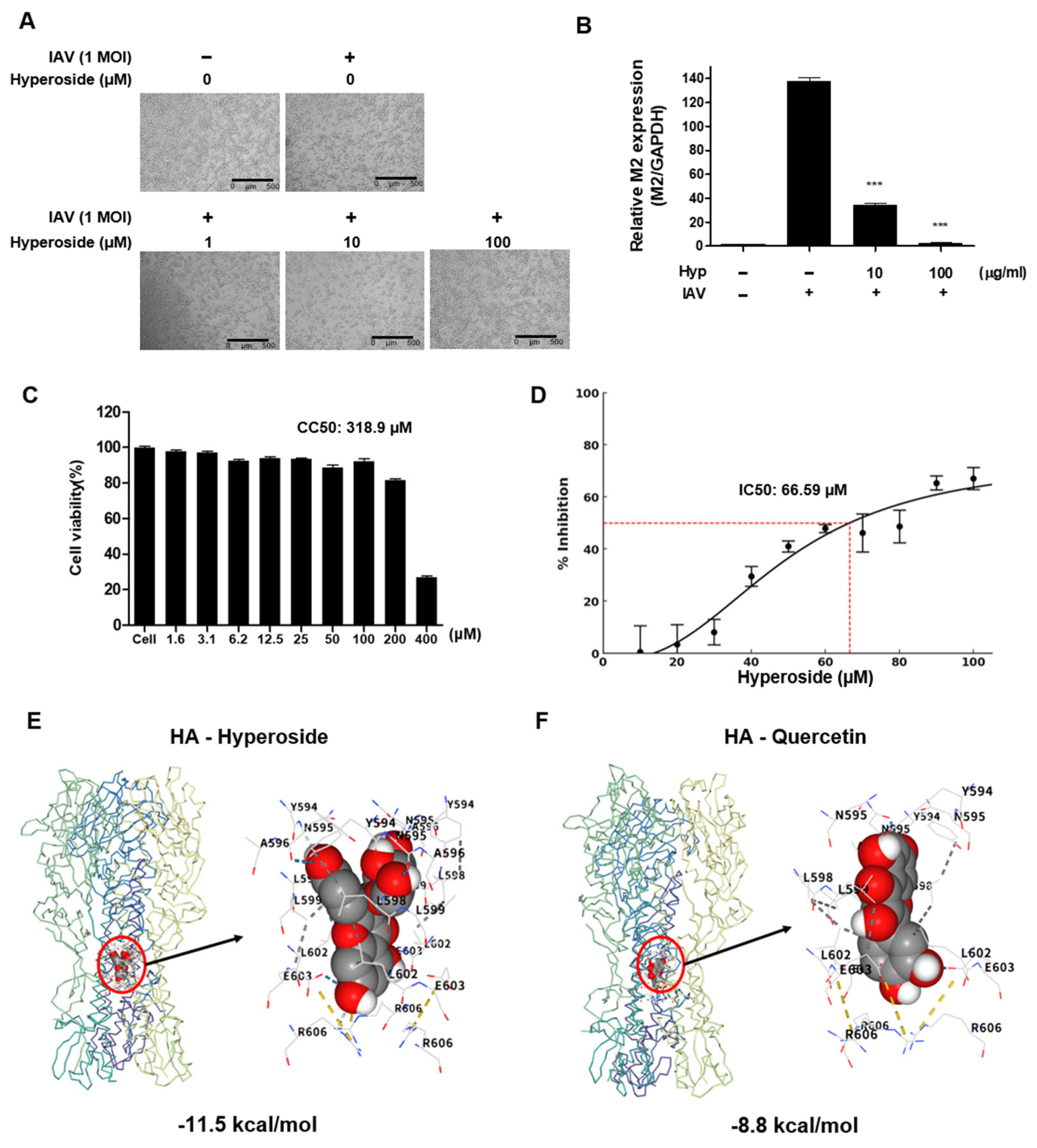
2.7. Binding Site Prediction and in Silico Molecular Docking

The binding site on the influenza A virus hemagglutinin (HA) protein was predicted using structure-based algorithms such as FTSite and FTMap, which identify ligand-binding hot spots based on structural and energetic properties [29,30,31,32]. Molecular docking simulations were performed by using CB-Dock2 [33] and FitDock [34] to evaluate the interaction between hyperoside and HA. Binding poses were analyzed and visualized using PyMOL (version 2.5), NGL Viewer (standard version integrated in CB-Dock2), and LigPlot+ (Version 2.2) to compare the binding affinity and interaction profile of hyperoside with those of quercetin. Detailed methods for binding site prediction and in silico docking are provided in the Supplementary Materials.

2.8. Statistical Analysis

All experiments were performed at least three times independently. Data are presented as the means ± standard deviations (SDs). Statistical differences between groups were evaluated using Student’s t-test or one-way analysis of variance (ANOVA) followed by students T-test. A p value < 0.05 was considered statistically significant. All statistical analyses and graphical representations were performed using GraphPad Prism software (version 10.5.0, GraphPad Software, San Diego, CA, USA).

3. Results

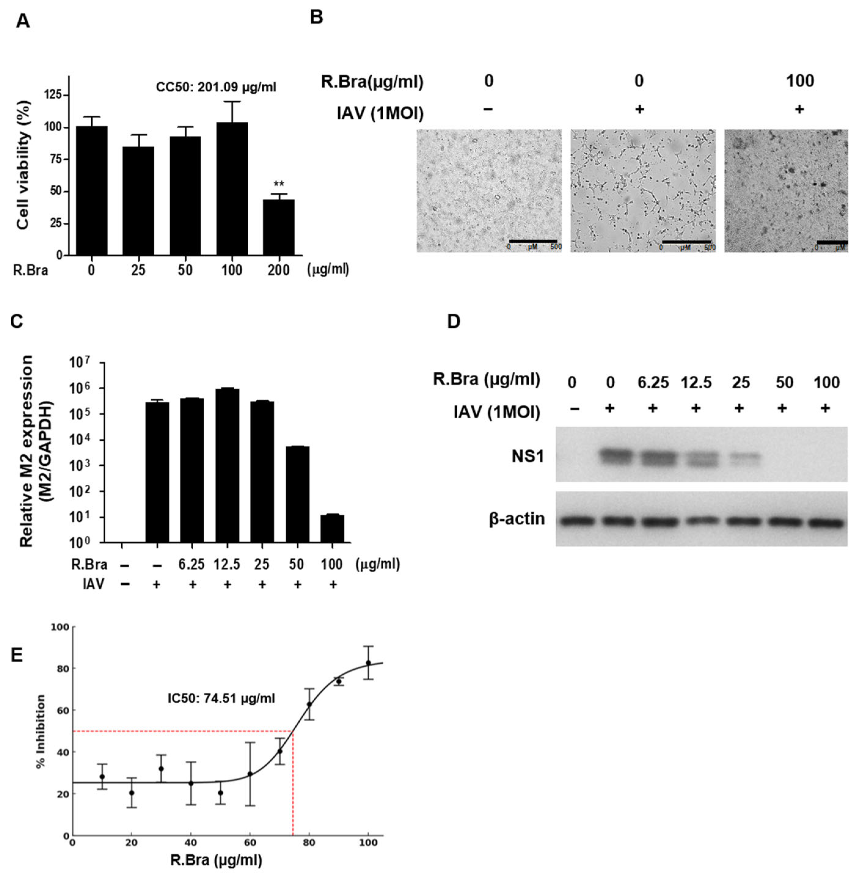
3.1. Cytotoxicity of R. brachycarpum Extract and Antiviral Activity Against IAV

The R. brachycarpum crude extract did not exhibit cytotoxicity in the MDCK cells at concentrations of up to 100 μg/mL, and it effectively suppressed the cytopathic effect (CPE) caused by IAV infection (Figure 1A,B). In IAV-infected cells treated with the extract, the viral M2 gene (a structural protein gene) mRNA level was significantly decreased at extract concentrations of 50 μg/mL and above, and the viral nonstructural protein NS1 level was significantly reduced at concentrations of 25 μg/mL and above (Figure 1C,D). In the plaque assay, the number of plaques in IAV-infected cells was reduced in a dose-dependent manner via extract treatment, and no plaques were observed at the highest concentration, 100 μg/mL (Figure 1E). From these data, the half-maximal inhibitory concentration (IC50) of the crude extract was calculated to be 74.51 μg/mL, and the 50% cytotoxic concentration (CC50) was found to be 201.09 μg/mL, indicating that the extract can effectively inhibit IAV replication without notable cytotoxicity (selectivity index SI ≈ 2.7).

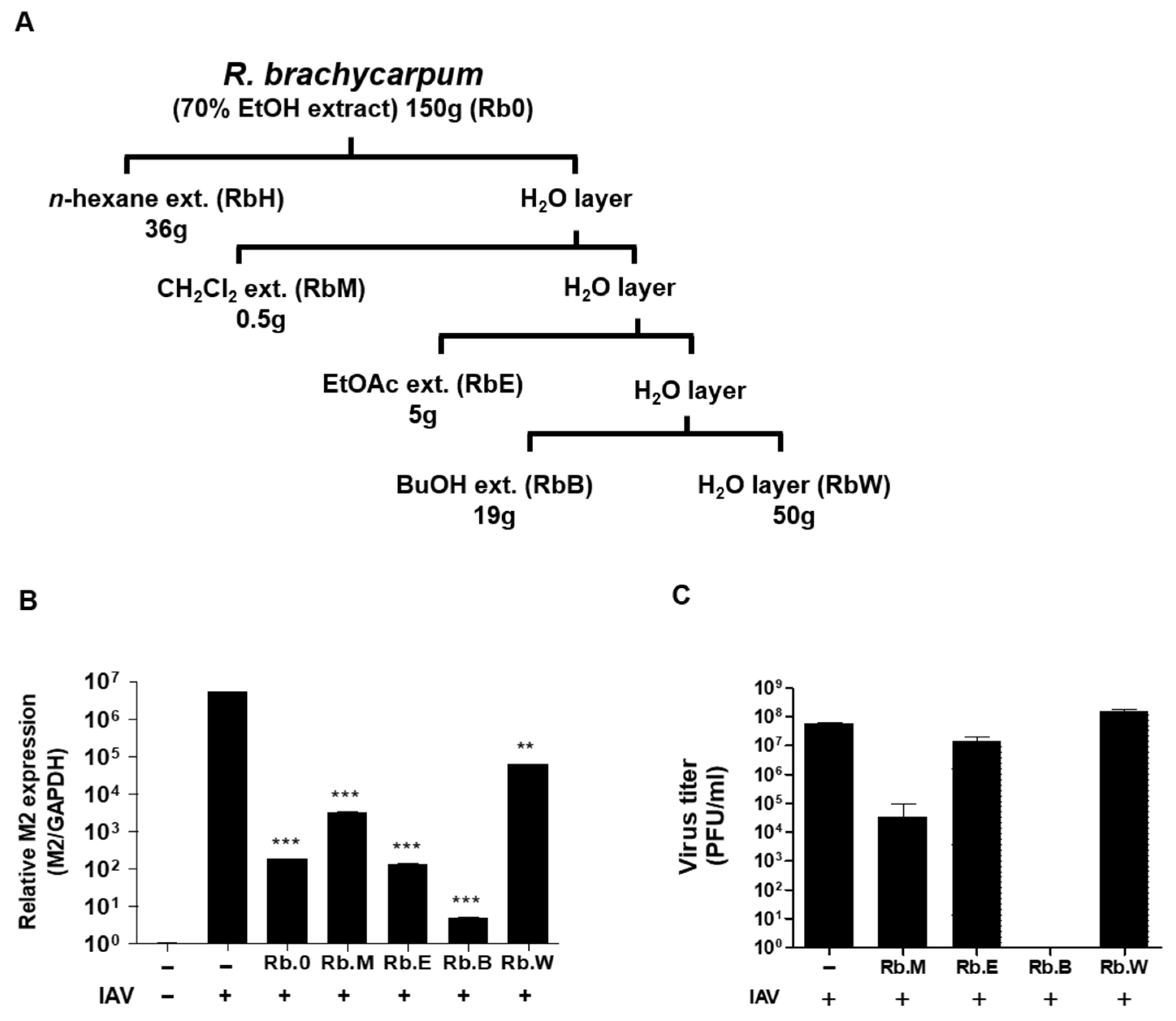
3.2. Comparison of Antiviral Activity of Solvent Fractions

The 70% ethanol extract of R. brachycarpum was fractionated sequentially into n-hexane, DCM, EA, n-butanol, and aqueous fractions, and each fraction (50 μg/mL) was tested for antiviral activity in IAV-infected MDCK cells (Figure 2A). Among these fractions, the n-butanol fraction showed the most pronounced inhibitory effect on IAV. Treatment with the butanol fraction led to a significant decrease in viral M2 mRNA levels and a marked reduction in plaque formation, whereas the EA and aqueous fractions did not exhibit statistically significant antiviral effects (Figure 2B,C). In particular, the butanol fraction reduced the number of plaques by approximately 80% compared to the untreated virus control.

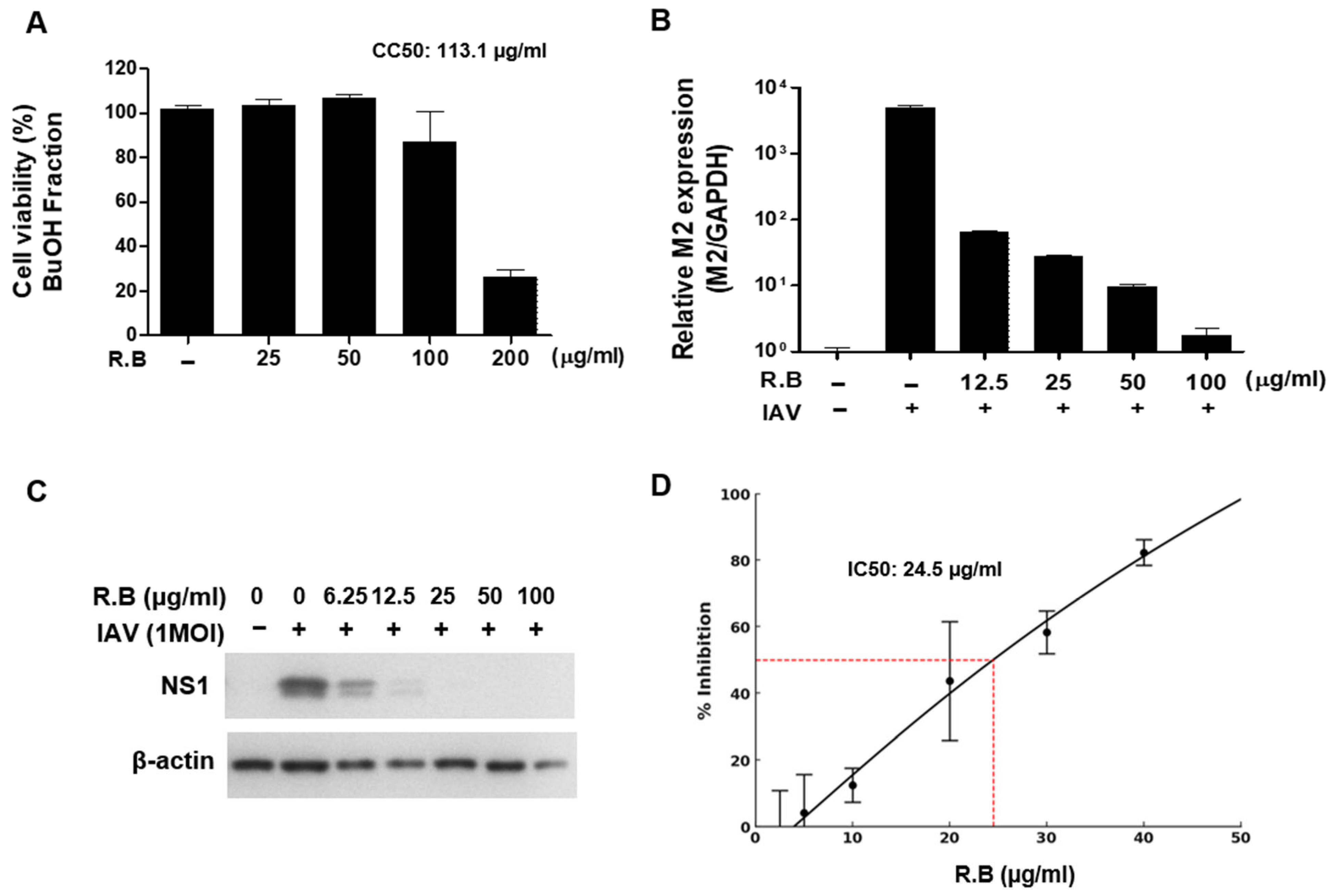
3.3. Antiviral Activity of the Butanol Fraction

Further evaluation of the n-butanol fraction confirmed its potent anti-influenza activity. According to the results of WST-1 cell viability assays, the CC50 of the butanol fraction was 113.1 μg/mL (Figure 3A). Even at the low concentration of 12.5 μg/mL, the butanol fraction significantly decreased the expression of the viral M2 gene and the NS1 protein in infected cells, indicating a strong antiviral effect (Figure 3B,C). Moreover, plaque formation was significantly suppressed at concentrations around and above 25 μg/mL, in agreement with the IC50 value of 24.5 μg/mL (Figure 3D). The IC50 of the butanol fraction was calculated to be 24.5 μg/mL, demonstrating a higher antiviral potency than the crude extract (selectivity index SI ≈ 4.6).

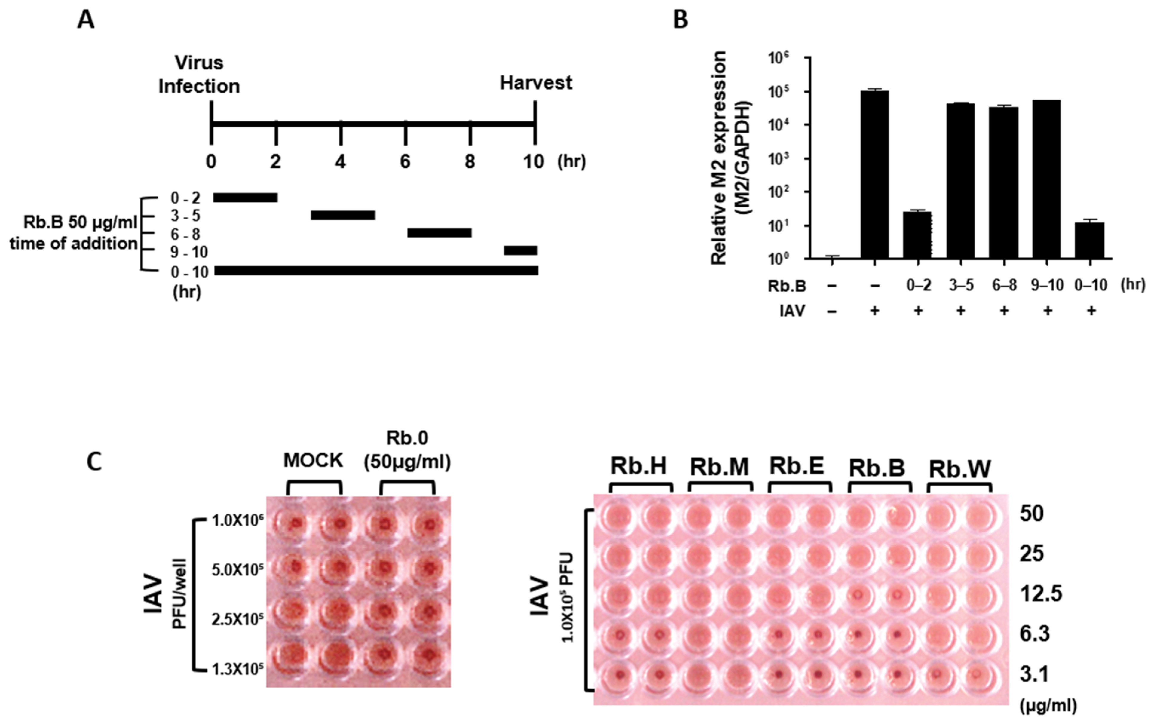
3.4. Antiviral Effect at the Early Stage of the Viral Life Cycle

We next examined the timing of the antiviral action of the R. brachycarpum samples relative to the IAV life cycle. A time-of-addition experiment showed that significant antiviral effects occurred only when the R. brachycarpum extract or fraction was present during the early stage of infection. Specifically, treatment with the sample immediately upon infection (for 0–2 h or 0–10 h post-infection) led to a significant reduction in viral M2 mRNA levels, whereas applying the treatment at later time points did not have a notable effect (Figure 4A,B). Consistently, the results of the hemagglutination inhibition (HAI) assay showed that the R. brachycarpum butanol fraction, at concentrations of 12.5 μg/mL and above, inhibited the agglutination of chicken red blood cells by the virus. This indicates that the antiviral effect of R. brachycarpum occurs primarily at the initial entry stage of the influenza virus life cycle (Figure 4C).

3.5. Identification and Analysis of Hyperoside as the Active Constituent

HPLC analysis of the R. brachycarpum n-butanol fraction revealed five major peaks. Among the corresponding sub-fractions collected, the fraction corresponding to peak F5 exhibited significant antiviral activity (Figure 5A,B). The UV-Vis absorption spectrum of the F5 sub-fraction showed strong absorbance at 254 nm and 365 nm, characteristic of a flavonoid glycoside. To further clarify the identity of this active compound, the F5 fraction was treated with the enzyme novarom (a β-glycosidase) to hydrolyze glycosidic linkages and remove minor glycosidic components. After treatment with the enzyme, the HPLC profile simplified, and the major peak’s retention time matched that of the quercetin glycoside standard. Based on this retention time and spectral comparison, the active compound was identified as quercetin-3-O-galactoside, commonly known as hyperoside (Figure 5C,D). Notably, this compound is distinct from other known constituents of R. brachycarpum such as rhododendrin, luteolin, and quercetin.
Flavonoids like quercetin and its glycosides have been reported to exhibit strong anti-influenza activity in other studies [35,36]. This literature evidence supports our finding of hyperoside as the key antiviral principle in R. brachycarpum.

3.6. Anti-Influenza Activity of Hyperoside

To confirm the role of hyperoside as the active antiviral constituent, we evaluated its effect on influenza infection in vitro. In IAV-infected cells (MOI 1) treated with hyperoside at 100 μM, virus-induced cytopathic effects were markedly reduced, and the cell monolayer appeared similar to that in uninfected control cells (Figure 6A). In addition, hyperoside treatment led to a dose-dependent decrease in viral M2 gene mRNA levels in the infected cells (Figure 6B). These results indicate that hyperoside is a major contributor to the anti-influenza activity of R. brachycarpum.
To assess the safety and efficacy profiles of hyperoside, we first evaluated its cytotoxicity in A549 cells. Hyperoside showed no significant cytotoxicity at up to 100 μM, and the calculated CC50 value was 318.9 μM (148.1 μg/mL), indicating low cellular toxicity (Figure 6C). In parallel, a plaque reduction assay was performed to quantify the inhibitory effect of hyperoside on viral replication. The compound led to a dose-dependent reduction in plaque formation, with an IC50 value of 66.59 μM (30.92 μg/mL), confirming its antiviral efficacy at the cellular level (Figure 6D).
To further investigate the mechanism underlying this antiviral effect, we performed in silico molecular docking analysis using the crystal structure of HA, with quercetin included as a reference flavonoid. Binding site prediction identified a major ligand-accessible pocket within the receptor-binding domain (RBD), which was used to define the docking region. Hyperoside exhibited a strong binding affinity (−11.5 kcal/mol) and engaged in multiple stable interactions with HA, including hydrogen bonding, hydrophobic contact, and ionic interactions (Figure 6C). In contrast, quercetin showed a weaker affinity (−8.8 kcal/mol) and engaged in fewer interactions (Figure 6D). These results suggest that hyperoside may inhibit IAV infection by directly occupying the sialic acid binding site on HA, thereby preventing viruses’ attachment and entry into host cells.

4. Discussion

Seasonal influenza, caused by influenza viruses, continues to impose a heavy health burden worldwide [2,3,11]. In particular, the high mutation rate of RNA viruses like IAV leads to the frequent emergence of variant and drug-resistant strains, necessitating continual development of new antiviral therapies [4,37,38]. In this context, discovering candidate antiviral compounds derived from natural sources (which typically have fewer side effects) offers a potentially safer and more sustainable approach compared to entirely synthetic drug development. A wide range of studies have explored natural-product-based compounds, and numerous secondary metabolites from plants and other organisms are known to possess bioactive properties. Indeed, modern drug discovery often involves validating the therapeutic efficacy of compounds isolated from traditionally used medicinal plants [14,15,39].
In this study, we investigated R. brachycarpum, an herbal medicine historically used to treat inflammatory conditions, as a source of new anti-influenza agents. The crude extract of R. brachycarpum effectively suppressed the expression of both structural and non-structural influenza viral genes in infected cells. The extract’s IC50 (29.84 μg/mL) and lack of cytotoxicity up to a high concentration (CC50 = 201.09 μg/mL) yielded a selectivity index (SI) of approximately 6.73, indicating it has a promising antiviral effect and justifying the subsequent isolation of its active components.
To efficiently identify the antiviral constituents within the extract, we fractionated the extract by solvent polarity. Flavonoids, a class of secondary metabolites known for their diverse biological activities, are often enriched in the ethyl acetate fractions of plant extracts; thus, we initially anticipated strong activity in the EA fraction. However, our results showed that the n-butanol fraction exhibited the highest anti-IAV activity, while the EA and aqueous fractions had negligible effects. The n-butanol fraction had an IC50 of 14.34 μg/mL and a CC50 of 113.07 μg/mL (SI ≈ 7.88), representing values about 17% higher SI than those of the crude extract. This finding suggests that the principal antiviral ingredient of R. brachycarpum is concentrated in the butanol fraction.
Using LC and bioactivity-guided fractionation, we separated the butanol fraction and identified five major peaks, of which one sub-fraction (F5) showed antiviral activity. Given that butanol fractions often contain glycosylated compounds (bearing both hydrophilic and hydrophobic moieties), we employed a glycosidase enzyme to simplify the mixture. This approach revealed that the active substance was a quercetin glycoside. By comparing it with reference compounds, we identified this molecule as hyperoside (quercetin-3-O-galactoside), distinct from other known R. brachycarpum constituents like rhododendrin, luteolin, or quercetin. Hyperoside is a flavonoid glycoside, and flavonoid compounds (including quercetin derivatives) have been documented to inhibit influenza virus replication through interference with viral entry and replication processes. Thus, the identification of hyperoside aligns with known antiviral mechanisms of flavonoids and highlights it as the key antiviral constituent in R. brachycarpum.
Our experimental results further demonstrated that the R. brachycarpum extract and its hyperoside-rich butanol fraction primarily exert their antiviral effects at an early stage of the influenza virus life cycle, likely during viral entry. This was evidenced via the HAI assay, where the butanol fraction blocked the hemagglutination of RBCs by the virus. Moreover, the reduction in viral RNA and protein levels in treated cells suggests that, beyond blocking entry, R. brachycarpum may also impede subsequent steps of viral replication, such as viral protein synthesis.
Hyperoside has shown antiviral activity against several viruses, including EHV-8, HSV, and HBV. In this study, we demonstrated that hyperoside also inhibits influenza A virus (IAV), acting primarily at the viral entry stage. In silico molecular docking analysis revealed strong binding affinity of hyperoside to the receptor-binding domain of hemagglutinin (HA), suggesting that it may block the attachment of the virus to host cells. This mechanism aligns with our in vitro findings, such as hemagglutination inhibition and early-stage suppression of viral gene expression.
While approved antiviral drugs like oseltamivir, zanamivir, and baloxavir show lower IC50 values, these drugs target different mechanisms and do not block HA-mediated entry. Although hyperoside showed relatively weaker potency, its low cytotoxicity (CC50 = 318.9 μM) highlights its potential as a safe, naturally derived entry inhibitor. With further structural optimization, hyperoside could serve as a promising candidate for anti-influenza drug development.
In summary, we have shown that Rhododendron brachycarpum, a plant traditionally used for various therapeutic purposes, possesses potent inhibitory activity against influenza A virus. Notably, hyperoside—an active compound known for its anti-inflammatory effects —was identified as the major antiviral ingredient of R. brachycarpum. The dual functionality of hyperoside, potentially reducing viral replication and mitigating virus-induced inflammation, makes it a promising candidate for the development of new anti-influenza therapeutics. Future studies will explore the antiviral efficacy of hyperoside derivatives, and in vivo experiments will be conducted to assess the potential of hyperoside-based treatments that concurrently suppress influenza virus proliferation and the associated inflammatory response.

5. Conclusions

In this study, we demonstrated that R. brachycarpum, a traditional medicinal plant, exhibits significant antiviral activity against influenza A virus (IAV). Our findings identified hyperoside, a flavonoid glycoside, as the primary active antiviral constituent of R. brachycarpum leaf extract. Both the crude extract and the hyperoside-rich n-butanol fraction effectively suppressed viral replication by interfering predominantly with the early stages of viral infection, particularly the entry of the virus into host cells. The purified hyperoside exhibited robust antiviral effects, significantly reducing viral mRNA and protein expression levels without exerting notable cytotoxicity. Given that hyperoside has dual functionality as an antiviral and anti-inflammatory agent, it represents a highly promising candidate for developing novel therapeutic strategies against influenza A virus infections.
Although this study confirmed the antiviral activity of hyperoside against influenza A virus, further studies are needed to validate its efficacy in vivo and against other viral subtypes. Future work will focus on synthesizing hyperoside analogues with improved potency and evaluating their activity in animal models. In addition, we plan to optimize HA-binding affinity based on our in silico findings and explore potential synergistic effects with existing antiviral drugs.

Supplementary Materials

The following supporting information can be downloaded at https://www.mdpi.com/article/10.3390/microbiolres16060132/s1. Figure S1: Predicted binding site and additional docking poses for hyperoside and quercetin. (A) Predicted ligand-binding pocket on the surface of influenza A virus hemagglutinin (HA) protein, identified using FTSite. The binding site corresponds to the receptor-binding domain (RBD), which was used as the docking region in subsequent molecular docking simulations. (B) Second-best docking pose of hyperoside showing stable interaction with the predicted HA binding pocket. The compound forms multiple hydrogen bonds (blue), hydrophobic interactions (gray), and ionic interactions (yellow) within the RBD. (C) Second-best docking pose of quercetin within the same predicted binding site. Interaction patterns are visualized using the same color scheme: hydrogen bonds (blue), hydrophobic interactions (gray), and ionic interactions (yellow).

Author Contributions

Conceptualization, S.H.J.; funding acquisition, S.H.J.; investigation, S.Y.S. and Y.H.P.; methodology S.Y.S., Y.H.P., H.C., J.H.L. and Y.J.K.; project administration, S.H.J.; resources, S.Y.S., Y.H.P. and S.J.W.; supervision, S.H.J.; visualization, Y.H.P. and W.R.I.; Writing—original draft, S.Y.S. and Y.H.P.; writing—review and editing, Y.H.P., W.R.I. and S.H.J. All authors have read and agreed to the published version of the manuscript.

Funding

This work was supported by a grant from the National Research Foundation of Korea (NRF) funded by the Korean government (MIST) (NRF-2020R1A2C1010173, S.H.J.).

Institutional Review Board Statement

Not applicable.

Informed Consent Statement

Not applicable.

Data Availability Statement

Data is contained within the article or Supplementary Material.

Conflicts of Interest

The authors declare no conflict of interest.

References

  1. Bouvier, N.M.; Palese, P. The biology of influenza viruses. Vaccine 2008, 26 (Suppl. S4), D49–D53. [Google Scholar] [CrossRef] [PubMed]
  2. Ren, H.; Jin, Y.; Hu, M.; Zhou, J.; Song, T.; Huang, Z.; Li, B.; Li, K.; Zhou, W.; Dai, H.; et al. Ecological dynamics of influenza A viruses: Cross-species transmission and global migration. Sci. Rep. 2016, 6, 36839. [Google Scholar] [CrossRef] [PubMed]
  3. Krammer, F.; Smith, G.J.D.; Fouchier, R.A.M.; Peiris, M.; Kedzierska, K.; Doherty, P.C.; Palese, P.; Shaw, M.L.; Treanor, J.; Webster, R.G.; et al. Influenza. Nat. Rev. Dis. Primers 2018, 4, 3. [Google Scholar] [CrossRef] [PubMed]
  4. Vinikoor, M.; Stevens, J.; Nawrocki, J.; Singh, K. Influenza A virus subtyping: Paradigm shift in influenza diagnosis. J. Clin. Microbiol. 2009, 47, 3055–3056. [Google Scholar] [CrossRef]
  5. Dou, D.; Revol, R.; Ostbye, H.; Wang, H.; Daniels, R. Influenza A Virus Cell Entry, Replication, Virion Assembly and Movement. Front. Immunol. 2018, 9, 1581. [Google Scholar] [CrossRef]
  6. Wang, M.; Veit, M. Hemagglutinin-esterase-fusion (HEF) protein of influenza C virus. Protein Cell 2016, 7, 28–45. [Google Scholar] [CrossRef]
  7. Asha, K.; Meseko, C.; Kumar, B. Editorial: Influenza and related viruses: Epidemiology, pathogenesis, and therapeutics. Front. Mol. Biosci. 2022, 9, 1117067. [Google Scholar] [CrossRef]
  8. Shie, J.J.; Fang, J.M. Development of effective anti-influenza drugs: Congeners and conjugates—A review. J. Biomed. Sci. 2019, 26, 84. [Google Scholar] [CrossRef]
  9. Duwe, S.C.; Milde, J.; Heider, A.; Wedde, M.; Schweiger, B.; Dürrwald, R. Increase of Synergistic Secondary Antiviral Mutations in the Evolution of A(H1N1)pdm09 Influenza Virus Neuraminidases. Viruses 2024, 16, 1109. [Google Scholar] [CrossRef]
  10. Li, Y.; Huo, S.; Yin, Z.; Tian, Z.; Huang, F.; Liu, P.; Liu, Y.; Yu, F. Retracted and republished from: “The current state of research on influenza antiviral drug development: Drugs in clinical trial and licensed drugs”. mBio 2024, 15, e00175-24. [Google Scholar] [CrossRef]
  11. Hay, A.J.; McCauley, J.W. The WHO global influenza surveillance and response system (GISRS)—A future perspective. Influenza Other Respir. Viruses 2018, 12, 551–557. [Google Scholar] [CrossRef] [PubMed]
  12. Quignon, E.; Ferhadian, D.; Hache, A.; Vivet-Boudou, V.; Isel, C.; Printz-Schweigert, A.; Donchet, A.; Crépin, T.; Marquet, R. Structural Impact of the Interaction of the Influenza A Virus Nucleoprotein with Genomic RNA Segments. Viruses 2024, 16, 421. [Google Scholar] [CrossRef]
  13. Lampejo, T. Influenza and antiviral resistance: An overview. Eur. J. Clin. Microbiol. Infect. Dis. 2020, 39, 1201–1208. [Google Scholar] [CrossRef] [PubMed]
  14. Butler, M.S. The Role of Natural Product Chemistry in Drug Discovery. J. Nat. Prod. 2004, 67, 2141–2153. [Google Scholar] [CrossRef]
  15. Lahlou, M. Screening of natural products for drug discovery. Expert Opin. Drug Discov. 2007, 2, 697–705. [Google Scholar] [CrossRef] [PubMed]
  16. Zhou, W.; Oh, J.; Wonhwa, L.; Kwak, S.; Li, W.; Chittiboyina, A.G.; Ferreira, D.; Hamann, M.T.; Lee, S.H.; Bae, J.S.; et al. The first cyclomegastigmane rhododendroside A from Rhododendron brachycarpum alleviates HMGB1-induced sepsis. Biochim. Biophys. Acta 2014, 1840, 2042–2049. [Google Scholar] [CrossRef]
  17. Hwang, T.; Noh, E.; Jeong, J.H.; Park, S.K.; Shin, D.; Kang, H. Determination of Grayanotoxins from Rhododendron brachycarpum in Dietary Supplements and Homemade Wine by Liquid Chromatography-Quadrupole Time-of-Flight-Mass Spectrometry and Liquid Chromatography-Tandem Mass Spectrometry. J. Agric. Food Chem. 2018, 66, 1935–1940. [Google Scholar] [CrossRef]
  18. Zhou, W.; Oh, J.; Li, W.; Kim, D.W.; Yang, M.H.; Jang, J.-H.; Ahn, J.S.; Lee, S.H.; Na, M. Chemical constituents of the Korean endangered species Rhododendron brachycarpum. Biochem. Syst. Ecol. 2014, 56, 231–236. [Google Scholar] [CrossRef]
  19. Chen, J.Y.; Huang, T.R.; Hsu, S.Y.; Huang, C.C.; Wang, H.S.; Chang, J.S. Effect and mechanism of quercetin or quercetin-containing formulas against COVID-19: From bench to bedside. Phytother. Res. 2024, 38, 2597–2618. [Google Scholar] [CrossRef]
  20. Ku, S.K.; Zhou, W.; Lee, W.; Han, M.S.; Na, M.; Bae, J.S. Anti-inflammatory effects of hyperoside in human endothelial cells and in mice. Inflammation 2015, 38, 784–799. [Google Scholar] [CrossRef]
  21. Wu, L.L.; Yang, X.B.; Huang, Z.M.; Liu, H.Z.; Wu, G.X. In vivo and in vitro antiviral activity of hyperoside extracted from Abelmoschus manihot (L) medik. Acta Pharmacol. Sin. 2007, 28, 404–409. [Google Scholar] [CrossRef] [PubMed]
  22. Rakers, C.; Schwerdtfeger, S.M.; Mortier, J.; Duwe, S.; Wolff, T.; Wolber, G.; Melzig, M.F. Inhibitory potency of flavonoid derivatives on influenza virus neuraminidase. Bioorg. Med. Chem. Lett. 2014, 24, 4312–4317. [Google Scholar] [CrossRef] [PubMed]
  23. Wu, W.; Li, R.; Li, X.; He, J.; Jiang, S.; Liu, S.; Yang, J. Quercetin as an Antiviral Agent Inhibits Influenza A Virus (IAV) Entry. Viruses 2015, 8, 6. [Google Scholar] [CrossRef]
  24. Yoo, H.; Ku, S.-K.; Zhou, W.; Han, M.-S.; Na, M.; Bae, J.-S. Anti-septic effects of phenolic glycosides from Rhododendron brachycarpum in vitro and in vivo. J. Funct. Foods 2015, 16, 448–459. [Google Scholar] [CrossRef]
  25. Popescu, R.; Kopp, B. The genus Rhododendron: An ethnopharmacological and toxicological review. J. Ethnopharmacol. 2013, 147, 42–62. [Google Scholar] [CrossRef]
  26. Choi, Y.H.; Zhou, W.; Oh, J.; Choe, S.; Kim, D.W.; Lee, S.H.; Na, M. Rhododendric acid A, a new ursane-type PTP1B inhibitor from the endangered plant Rhododendron brachycarpum G. Don. Bioorg. Med. Chem. Lett. 2012, 22, 6116–6119. [Google Scholar] [CrossRef]
  27. Yang, J.; Kim, M.O.; Kwon, Y.S.; Kim, M.J. Antioxidant Activity, alpha-glucosidase Inhibitory Activity and Chemoprotective Properties of Rhododendron brachycarpum Leaves Extracts. Curr. Pharm. Biotechnol. 2017, 18, 849–854. [Google Scholar] [CrossRef]
  28. Fang, F.; Bao, S.; Chen, D.; Duan, X.; Zhao, Y.; Ma, Y. Protective effects and mechanism of quercetin from Rhododendron dauricum against cerebral ischemia-reperfusion injury. Eur. J. Pharmacol. 2024, 985, 177126. [Google Scholar] [CrossRef]
  29. Ngan, C.H.; Hall, D.R.; Zerbe, B.; Grove, L.E.; Kozakov, D.; Vajda, S. FTSite: High accuracy detection of ligand binding sites on unbound protein structures. Bioinformatics 2012, 28, 286–287. [Google Scholar] [CrossRef]
  30. Kozakov, D.; Grove, L.E.; Hall, D.R.; Bohnuud, T.; Mottarella, S.E.; Luo, L.; Xia, B.; Beglov, D.; Vajda, S. The FTMap family of web servers for determining and characterizing ligand-binding hot spots of proteins. Nat. Protoc. 2015, 10, 733–755. [Google Scholar] [CrossRef]
  31. Brenke, R.; Kozakov, D.; Chuang, G.Y.; Beglov, D.; Hall, D.; Landon, M.R.; Mattos, C.; Vajda, S. Fragment-based identification of druggable ‘hot spots’ of proteins using Fourier domain correlation techniques. Bioinformatics 2009, 25, 621–627. [Google Scholar] [CrossRef] [PubMed]
  32. Jones, G.; Jindal, A.; Ghani, U.; Kotelnikov, S.; Egbert, M.; Hashemi, N.; Vajda, S.; Padhorny, D.; Kozakov, D. Elucidation of protein function using computational docking and hotspot analysis by ClusPro and FTMap. Acta Crystallogr. D Struct. Biol. 2022, 78 Pt 6, 690–697. [Google Scholar] [CrossRef] [PubMed]
  33. Liu, Y.; Yang, X.; Gan, J.; Chen, S.; Xiao, Z.X.; Cao, Y. CB-Dock2: Improved protein-ligand blind docking by integrating cavity detection, docking and homologous template fitting. Nucleic Acids Res. 2022, 50, W159–W164. [Google Scholar] [CrossRef] [PubMed]
  34. Yang, X.; Liu, Y.; Gan, J.; Xiao, Z.X.; Cao, Y. FitDock: Protein-ligand docking by template fitting. Brief. Bioinform. 2022, 23, bbac087. [Google Scholar] [CrossRef]
  35. Gansukh, E.; Nile, A.; Kim, D.H.; Oh, J.W.; Nile, S.H. New insights into antiviral and cytotoxic potential of quercetin and its derivatives—A biochemical perspective. Food Chem. 2021, 334, 127508. [Google Scholar] [CrossRef]
  36. Mehrbod, P.; Hudy, D.; Shyntum, D.; Markowski, J.; Łos, M.J.; Ghavami, S. Quercetin as a Natural Therapeutic Candidate for the Treatment of Influenza Virus. Biomolecules 2021, 11, 10. [Google Scholar] [CrossRef]
  37. Harrington, W.N.; Kackos, C.M.; Webby, R.J. The evolution and future of influenza pandemic preparedness. Exp. Mol. Med. 2021, 53, 737–749. [Google Scholar] [CrossRef]
  38. Blut, A. Influenza Virus. Transfus. Med. Hemother. 2009, 36, 32–39. [Google Scholar]
  39. Li, R.; Han, Q.; Li, X.; Liu, X.; Jiao, W. Natural Product-Derived Phytochemicals for Influenza A Virus (H1N1) Prevention and Treatment. Molecules 2024, 29, 2371. [Google Scholar] [CrossRef]
Figure 1. Antiviral activity of Rhododendron brachycarpum (R.Bra) extract in MDCK cells. (A) Cell viability assay showing R.Bra was non-toxic at up to 100 µg/mL, with CC50 at 201.09 µg/mL. (B) Cytopathic-effect (CPE) in IAV-infected MDCK cells. In comparison to the control, CPE characteristics were significantly alleviated by R.Bra treatment. (C) IAV M2 RNA levels decreased in a dose-dependent manner following R.Bra treatment. (D) Western blot analysis showing dose-dependent inhibition of viral NS1 protein expression by R.Bra. (E) Reduction in viral titers by R.Bra in IAV-infected MDCK cells, with an IC50 of 74.51 µg/mL. Values represent means ± SD (n = 3). ** p < 0.01 vs. virus control.
Microbiolres 16 00132 g001
Figure 2. Antiviral activity of Rb.0 polar and non-polar fractions. (A) Schematic representation of the fractionation of Rb.0 into five distinct layers (Rb.H, Rb.M, Rb.E, Rb.B, and Rb.W). (B) Quantitative RT-PCR analysis of IAV M2 RNA showing strongest antiviral activity with Rb.B fraction. (C) Plaque assay showing lowest viral titers in Rb.B-treated group. Values represent means ± SD (n = 3). ** p < 0.01, *** p < 0.001 vs. virus control.
Microbiolres 16 00132 g002
Figure 3. Antiviral effects of Rb.B fraction against IAV infection. (A) Results of the cell viability assay, demonstrating that the Rb.B fraction is non-toxic at up to 100 µg/mL (CC50: 113.1 µg/mL). (B,C) Dose-dependent reduction in viral NS1 expression and M2 induced by Rb.B in MDCK cells. (C) Similar antiviral activity confirmed in A549 cells with decreased NS1 protein expression and virus titers upon Rb.B treatment. (D) Reduction in viral titers induced by Rb.B in IAV-infected MDCK cells with an IC50 of 24.5 µg/mL. Values represent means ± SDs (n = 3).
Microbiolres 16 00132 g003
Figure 4. Mechanism of antiviral action of the Rb.B fraction. (A) Results of a time-course experiment demonstrating inhibition of IAV M2 RNA at various stages of viral infection by Rb.B in A549 cells. Significant inhibition was observed at the early entry stage (0–2 h and 0–10 h). (B,C) Results of a hemagglutination inhibition (HAI) assay indicating Rb.B specifically inhibits IAV entry. Values represent means ± SD (n = 3).
Microbiolres 16 00132 g004
Figure 5. Identification and antiviral assessment of active components from Rb.B fraction. (A) HPLC chromatogram shows effective separation of Rb.B into fractions F1-F5. (B) Antiviral activity assessed via plaque assay indicated fraction F5 was the active component significantly inhibiting IAV replication. (C) Novarom treatment of the Rb.B fraction resulted in a pronounced peak shift. (D) Simultaneous HPLC analysis of Rb.B and hyperoside confirmed that their respective bands coincided. Values represent means ± SDs (n = 3). *** p < 0.001 vs. virus control.
Microbiolres 16 00132 g005
Figure 6. Antiviral activity of hyperoside against influenza A virus and its molecular interaction with HA. (A) The antiviral effect of Hyperoside was confirmed by observing CPE. Although CPE was observed in IAV-infected MDCK, CPE was significantly alleviated by Hyperoside treatment. (B) Confirmation of the antiviral effect of Hyperoside in IAV-infected A549 cells. IAV M2 RNA expression decreased in a Hyperoside-concentration-dependent manner. All values are expressed as averages (n = 3). *** p < 0.001 compared to virus control. (C) Cytotoxicity of hyperoside was assessed in MDCK cell lines using an WST-1 assay. Hyperoside showed minimal cytotoxicity at up to 100 μM, and the calculated CC50 was 318.9 μM (148.1 μg/mL), indicating a favorable safety profile. (D) Antiviral efficacy of hyperoside was evaluated via plaque reduction assay. Hyperoside inhibited plaque formation in a dose-dependent manner, with an IC50 of 66.59 μM (30.92 μg/mL). (E) In silico molecular-docking model of hyperoside with the hemagglutinin (HA) protein of IAV. The best binding pose is shown, with multiple interactions/bonds visualized: hydrogen bonds (blue), hydrophobic contact (gray), and ionic interactions (yellow). Binding energy: −11.5 kcal/mol. (F) Docking model of quercetin with IAV HA, used as a reference flavonoid. The compound showed a weaker binding affinity (−8.8 kcal/mol) and fewer interactions compared to hyperoside.
Microbiolres 16 00132 g006
Disclaimer/Publisher’s Note: The statements, opinions and data contained in all publications are solely those of the individual author(s) and contributor(s) and not of MDPI and/or the editor(s). MDPI and/or the editor(s) disclaim responsibility for any injury to people or property resulting from any ideas, methods, instructions or products referred to in the content.

