Article

Emergence, Spread of Antimicrobial-Resistant Bacteria and Phylogenetic Relationships in Coastal Ecosystems—Gastropod Phorcus lineatus as a Bioindicator

1
CITAB/Inov4Agro—Centre for the Research and Technology of Agro-Environmental and Biological Sciences—Institute for Innovation, Capacity Building and Sustainability of Agri-Food Production, University of Trás-os-Montes and Alto Douro, 5000-801 Vila Real, Portugal
2
AB2Unit—Antimicrobials, Biocides & Biofilms Unit, School of Agriculture and Veterinary Sciences, University of Trás-os-Montes and Alto Douro (UTAD), 5001-801 Vila Real, Portugal
3
INIAV—National Institute for Agricultural and Veterinary Research I.P., 4485-655 Vila do Conde, Portugal
4
CECAV—Veterinary and Animal Research Centre, Associate Laboratory for Animal and Veterinary Science (AL4AnimalS), University of Trás-os-Montes and Alto Douro, 5000-801 Vila Real, Portugal
5
CIMO/SusTEC—Centro de Investigação de Montanha—Laboratório Associado para a Sustentabilidade e Tecnologia em Regiões de Montanha, IPB—Instituto Politécnico de Bragança, 5300-253 Bragança, Portugal
6
Center for Animal Science Studies (CECA-ICETA), Associated Laboratory of Animal and Veterinary Science (AL4AnimalS), University of Porto, 4099-002 Porto, Portugal
7
CIBIO/InBIO—Research Center in Biodiversity and Genetic Resources, University of Porto, Campus Agrário de Vairão, 4485-661 Vairão, Portugal
8
CIIMAR—Centro Interdisciplinar de Investigação Marinha e Ambiental, Terminal de Cruzeiros do Porto de Leixões, 4450-208 Matosinhos, Portugal
*
Author to whom correspondence should be addressed.
Microbiol. Res. 2025, 16(6), 133; https://doi.org/10.3390/microbiolres16060133
Submission received: 24 March 2025 / Revised: 13 June 2025 / Accepted: 14 June 2025 / Published: 19 June 2025

Abstract

:
Coastal environments have been recognized as key reservoirs for antibiotic-resistant bacteria. The present study evaluated marine gastropods, Phorcus lineatus, as potential bioindicators to assess the spread of antibiotic-resistant bacteria. P. lineatus was sampled in four sites, with different anthropogenic pressures, along the northwest Portuguese coastal area. From these specimens, bacteria were isolated and tested for their antimicrobial susceptibility, followed by their phylogenetic and pathotypic determination. All the Escherichia coli isolates showed resistance to at least one antimicrobial agent. The highest levels of multidrug resistance (25%) were observed in E. coli isolates obtained from SITE 2, which is impacted by the city of Porto and industrial settlements, while nearly 17% of these isolates showed a multiple antibiotic resistance (MAR) index higher than 0.2. Among the isolates, phylogroups A and B2 were the most prevalent, followed by phylogroup B1. The isolates of phylogroup A showed a higher prevalence of antimicrobial resistance. This study offers valuable insights into the antibiotic resistance risks posed to marine ecosystems and underscores the need for microbiological monitoring and the development of effective management strategies. The findings suggest P. lineatus as a potential bioindicator of antibiotic-resistant bacteria in marine environments.

Graphical Abstract

1. Introduction

The global use, and often misuse, of antibiotics in human and veterinary medicine has led to the emergence and spread of multidrug-resistant bacteria, raising serious public health concerns [1]. To address this complex challenge, the “One Health” framework has been adopted as a systems-based approach that recognizes the interdependence of human, animal, plant, and environmental health. This framework identifies antimicrobial resistance as a critical issue, as resistant pathogens and their genes can circulate across sectors and ecosystems. This approach seeks to provide effective public policies and strengthen infection control and sanitation measures [1,2]. In this context, it is crucial to understand the impacts of anthropogenic activities on the development and dissemination of antibiotic resistance.
The aquatic ecosystems constitute one of the main reservoirs and transmission pathways for antimicrobial-resistant bacteria and the horizontal transfer of antibiotic-resistance genes among microbial communities [3]. Part of the administered antibiotic dose is excreted as a parent compound in the feces and urine, eventually reaching the environment and contaminating waters as the last reservoir [4,5]. In particular, coastal waters influenced by intense human activities can play an important role in developing and disseminating antimicrobial resistance, posing serious risks to human health through consuming contaminated food or the potential transfer of resistance genes to human pathogens [1]. Several studies have reported the presence of antibiotic-resistant bacteria such as E. coli, Klebsiella pneumoniae, Vibrio spp., and Aeromonas spp., and specific integrons, leading to antibiotic resistance (e.g., blaTEM, sulI) in freshwater and marine samples [6,7,8,9]. Considerable amounts of antimicrobial-resistant bacteria and genes have been documented in various aquatic species, such as clams, mussels, fish, and marine mammals [10,11,12,13,14,15]. Nevertheless, insufficient information is still available regarding the development and dissemination of antibiotic resistance in the marine environment and coastal areas, as well as the associated risks to human health.
New bioindicators are needed to improve the detection of antibiotic-resistant bacteria and genes encoding resistance. Marine gastropods are benthic organisms sensitive to environmental changes and have been pointed out as good bioindicators for environmental monitoring programs [16,17]. In addition, gastropods have an important role as a source of seafood, acting as a transport route of pollutants and bacteria through the food chain [13,18]. Some species of marine gastropods, such as the topshells of the genus Phorcus, inhabit the rocky shores of the intertidal zone in the Northeastern Atlantic and Mediterranean Sea, being an abundant, sedentary species with a wide geographical distribution and a long lifespan [19]. Therefore, they constitute good candidates as bioindicators for evaluating anthropogenic impacts on coastal areas, such as antimicrobial resistance and pollution.
In addition to the antimicrobial resistance, investigation of E. coli presence in marine gastropods is also essential for several reasons, particularly for public health, ecology, and food safety. E. coli is commonly used as an indicator of fecal contamination in both aquatic and terrestrial environments, and its presence in gastropods may signal environmental pollution, underscoring the need for monitoring water and food sources [20]. Additionally, gastropods can act as passive carriers of pathogenic E. coli, posing a potential health risk to humans who handle or consume them. In cultures where gastropods are consumed, the presence of E. coli increases the risk of gastrointestinal illness, especially if the snails are improperly cooked or handled [21]. As gastropods play a key role in ecosystems as decomposers, studying their microbiota, including E. coli, can shed light on species interactions and the effects of contamination within habitats [22]. In agricultural settings, gastropods may come into contact with livestock and water sources, highlighting the potential for cross-contamination between the natural environment and farming operations, which has implications for animal health and food production safety [23]. In this context, analyzing both the phylogenetic groups (phylogroups) [24,25,26] and pathotypes [27] of E. coli found in gastropods provides deeper insight into their genetic diversity, pathogenic potential, and ecological adaptations. Identifying specific phylogroups and pathotypes helps determine whether certain strains are more likely to cause disease or thrive in particular environments, thereby guiding effective public health interventions and environmental management strategies [28].
In this regard, the present study aimed to (a) isolate and determine the antibiotic resistance patterns of E. coli from P. lineatus sampled in four different impacted sites of the northwest Portuguese coast; (b) identify E. coli phylogroups and assess their phylogenetic diversity; and (c) investigate the role of the marine snail P. lineatus as a bioindicator of pathogenic and antibiotic-resistant bacteria in coastal areas under anthropogenic pressure. This biomonitoring study concerning the environmental occurrence of antibiotic-resistant bacteria on the northwest Portuguese coast can help support policies and regulations to reduce antimicrobial resistance dissemination.
The present study aligns with several Sustainable Development Goals (SDGs), particularly SDG 14 (Life Below Water), by emphasizing the need to protect marine ecosystems from antibiotic resistance, and SDG 3 (Good Health and Well-being), by addressing the public health risks associated with the spread of antibiotic-resistant bacteria. Additionally, the research contributes to SDG 6 (Clean Water and Sanitation), emphasizing the importance of monitoring water quality to protect both environmental and human health.

2. Materials and Methods
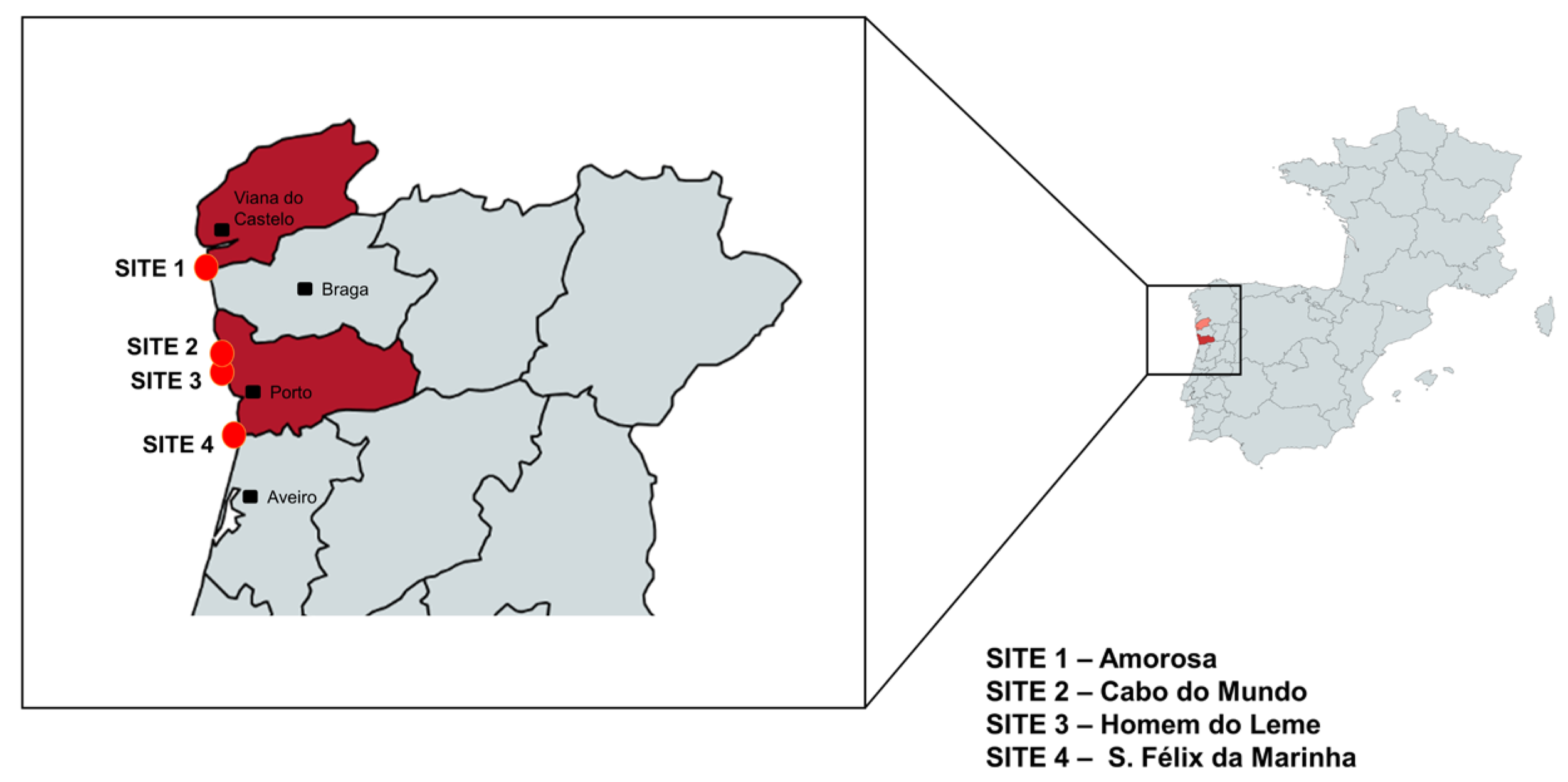
2.1. Study Area

The northwest Portuguese coast, under study, is heavily impacted by human activities from the nearby cities of Porto and Viana do Castelo, as well as by several agricultural fields and animal farms distributed along the coastline. The study area covered four sites along the northwest coast (Figure 1), selected based on different anthropogenic pressures, namely, Amorosa, 41°39′34.3″ N 8°49′29.8″ W (SITE 1); Cabo do Mundo, 41°13′15.8″ N 8°42′58.0″ W (SITE 2); Homem do Leme, 41°09′31.8″ N 8°41′10.0″ W (SITE 3); and S. Félix da Marinha, 41°02′8.91″ N 8°38′55.1″ W (SITE 4). Amorosa is a coastal area in Viana do Castelo, a city in the north of Portugal, located near the Lima River and an important seaport that comprises a maritime shipyard industry. Cabo do Mundo is situated in Porto, near industrial settlements, including a decommissioned oil refinery and the international airport Francisco Sá Carneiro. Porto also hosts the Homem do Leme beach, close to the Douro River estuary and the industrial and mercantile Leixões harbor, known for its intensive vessel traffic, which also impacts Cabo do Mundo. To the south is SITE 4—S. Félix da Marinha, a predominantly rural area with low urban anthropogenic pressure.

2.2. Sampling

Live marine gastropods were sampled in the winter of 2022. Twelve adult specimens of P. lineatus were randomly collected from the intertidal zone at each sampling site, for a total of forty-eight specimens. The gastropods were handpicked during low tide and transported alive in refrigerated flasks filled with local seawater for further analysis at the Antimicrobials, Biocides & Biofilms Unit/CITAB, Department of Veterinary Sciences, School of Agriculture and Veterinary Sciences, University of Trás-os-Montes e Alto Douro (UTAD, Vila Real, Portugal).
The harvest of gastropods in these four sites was authorized by the Portuguese Institute for Nature Conservation and Forests (ICNF), under the ATLANTIDA project—Platform for the monitoring of the North Atlantic Ocean and tools for the sustainable exploitation of the marine resources—and no ethics committee approval was needed.

2.3. Preparation of Samples and Bacterial Culture

Each specimen of P. lineatus was aseptically opened with sterilized stainless-steel blades, and soft tissues were removed. The tissues were collected in Brain Heart Infusion (BHI) (Oxoid, Lowell, MA, USA), homogenized, and subsequently incubated at 37 ± 1 °C for 24 h. After, the bacterial suspensions were inoculated onto MacConkey agar and Chromocult® Coliform Agar (CCA®) (Oxoid, Basingstoke, UK) and then incubated at 37 ± 1 °C for 24 h for E. coli and coliform bacteria detection. Isolates presumptively identified as belonging to the Enterobacteriaceae family were selected based on their growth on MacConkey agar (lactose-fermenting colonies), negative oxidase test, and Gram-negative bacilli morphology. These criteria are standard for preliminary identification of Enterobacteriaceae in environmental microbiology. A total of 103 bacterial isolates were obtained.

2.4. Identification of Pure Bacterial Isolates

From previously bacterial isolates, the presumptive E. coli colonies were identified based on their characteristic coloration on CCA®, and confirmation was carried out using lactose fermentation on MacConkey agar plates and standard biochemical tests—IMViC reactions (Indole, Methyl Red, Voges-Proskauer, Citrate Utilization). Once confirmed, isolates were stored in aliquots of BHI with 17% (v/v) glycerol at −80 °C. The E. coli were grown to obtain pure cultures for further susceptibility analysis to antimicrobial agents.

2.5. Antimicrobial Susceptibility Testing

Antimicrobial susceptibility testing was conducted using the disk diffusion method (Kirby–Bauer), following the guidelines established by the European Committee on Antimicrobial Susceptibility Testing (EUCAST). A few colonies were suspended in saline solution (NaCl) from the pure isolates, and the turbidity was adjusted to the 0.5 McFarland standard units (~cell density 1.5 × 10⁸ CFU/mL). The inoculum suspension was then spread in Mueller-Hinton (Oxoid, Basingstoke, UK), onto which antibiotic disks (Oxoid disks, UK) were placed, and incubated for approximately 24 h at 35 ± 1 °C. The diameter of the inhibition zones was measured and interpreted according to the EUCAST guidelines [29], and they were classified as susceptible or resistant. In the absence of EUCAST guidelines, the Clinical and Laboratory Standards Institute guidelines were followed [30]. Isolates with intermediate susceptibility were categorized as being susceptible. E. coli ATCC®25922 was used as the quality control strain.
The isolates were tested against 22 antibacterial agents, representing six antibiotic classes: β-lactams (penicillins: ampicillin (AMP, 10 μg), amoxicillin (AML,10 μg), amoxicillin/clavulanic acid (AMC, 20/10 μg), ticarcillin (TIC, 75 μg), ticarcillin/clavulanic acid (TIM, 85 μg), piperacillin (PRL, 100 μg), piperacillin/tazobactam (TZP, 100/10 μg)); cephalosporins of 2nd and 3rd generations: (cefoxitin (FOX, 30 μg) ceftazidime (CAZ, 30 μg) cefotaxime (CTX, 30 μg) ceftriaxone (CRO, 30 μg)); carbapenems: (imipenem (IMP, 10 μg) meropenem (MEM, 10 μg), ertapenem (ETP, 10 μg)); monobactams: (aztreonam (ATM, 30 μg); fluoroquinolones: (levofloxacin (LEV, 5 μg) ciprofloxacin (CIP, 5 μg)); aminoglycosides: (amikacin (AK, 30 μg), gentamicin (CN, 10 μg); sulphonamides: (sulfamethoxazole/trimethoprim (STX, 1.25/23.75/μg)); tetracyclines: (tetracycline (TE, 30 μg) and phosphonic acids: (fosfomycin (FOS, 50 μg). These 22 antibacterial agents were selected to represent a broad spectrum of commonly used antibiotics across six major classes, warranting a comprehensive evaluation of the antimicrobial susceptibility profile of the isolates. Also, these agents reflect those most relevant in clinical settings for the treatment of infections caused by Gram-negative bacteria, including both first-line and last-resort antibiotics. This approach aimed to assess resistance patterns and detect the presence of multidrug-resistant strains. The selection was also guided by CLSI recommendations and previous studies on antimicrobial resistance in similar bacterial populations.
Multidrug resistance (MDR) for the isolates was defined as non-susceptible to at least one agent in three or more antibiotic categories [31]. The multiple antibiotic resistance (MAR) index was calculated through the formula a/b, where a represents the number of antibiotics to which an isolate was resistant, and b represents the total number of antibiotics tested. An index value > 0.2 indicates that the sources of isolates are rendered from contamination by antibiotics, whereas < 0.2 suggests sources with less antibiotic usage or low risk [32].

2.6. Determination of E. coli Pathotypes and Phylogenetic Grouping

The determination of the pathotypes—Shiga toxin-producing E. coli (STEC), Enteroaggregative E. coli (EAEC), Enteroinvasive E. coli (EIEC), and Enterotoxigenic E. coli (ETEC)—and phylogenetic groups (A, B1, B2, D, E, F, and clade I) was performed as described by Pais et al. [33].
Briefly, for the determination of the pathotypes, primers and probes [34,35,36] were used, as described in Table S1 (see Supplementary Materials). DNA extraction was performed using a single colony of presumptive E. coli incubated at 37 ± 1 °C on Nutrient Agar (NA) plates (Bio-Rad, Hercules, CA, USA), resuspended in 250 μL of lysis reagent (NZYtech, Lisbon, Portugal), and cells were lysed by boiling for 15 min using a digital heat block (Avantor®, Radnor, PA, USA). Then, they were centrifuged for 5 min at 14,000 rpm (Bio-Rad: Model 16 Microcentrifuge). The supernatant was used as a template in the PCR real-time assay in a final volume of 25 μL, containing PCR master mix multiplex NZYSpeedy qPCR Probe Master Mix (2x) (NZYTech) and the primers and probes set aggr (20 μM), aaic (20 μM), ipah (20 μM), lt (20 μM), sth (20 μM) and stp (20 μM) (Eurofins Genomics, Ebersberg, Germany). The following strains from the European Union Reference Laboratory for Escherichia were used as positive controls: E. coli LMV-E.41 (aggr+), E. coli LMV-E.159 (aggr+; aaic+), E. coli LMV-E.40 (ipah+), E. coli LMV-E.39 (lt+; stp+) and E. coli LMV-E.166 (lt+; sth+; stp+). As a negative control, DNase-free water was used. The real-time PCR for STEC screening conditions was as follows: 95 °C, 3 min; 5 cycles of 95 °C, 15 s; 56 °C, 25 s; 65 °C, 30 s; 32 cycles of 95 °C, 15 s; 56 °C, 25 s; and 65 °C, 30 s; and for EAEC, EIEC and ETEC, screening conditions were as follows: 95 °C, 3 min; 5 cycles of 95 °C, 15 s; 52 °C, 25 s; 72 °C, 30 s; 35 cycles of 95 °C, 15 s; 52 °C, 25 s; and 72 °C, 30 s. An internal amplification control (IAC) was used simultaneously to identify potential inhibition of the PCR reactions in each sample well.
For the phylogenetic group analysis, a multiplex PCR method developed by Clermont et al. [24] was employed. The same DNA extraction used in RT-PCR for pathotype determination was used in phylogroup determination. The supernatant was used as a template in the PCR assay in a final volume of 20 µL, containing PCR master mix multiplex PCR NZYTaq 2x Green Master Mix (NZYTech, Lisbon, Portugal) and the primer set arpA (2 μM), chuA (1 μM), yjaA (1 μM) and TspE4.C2 (1 μM) (Eurofins Genomics, Porto, Portugal), as described in Table S2 (see Supplementary Materials). Each PCR reaction included both negative (DNase-free water) and positive controls. The positive controls consisted of E. coli O111 (arpA+; TspE4.C2+), E. coli O157:H7 (arpA+; chuA+), and E. coli K12 (arpA+; yjaA+). The defined PCR conditions were as follows: 95 °C, 3 min; 39 cycles of 95 °C, 30 s; 58 °C, 30 s; 72 °C, 30 s; and 72 °C, 5 min. The resulting fragments were visualized in a transilluminator under UV light (Syngene® GeneFlash, Cambridge, UK). According to the presence or absence of arpA, chuA, yjaA, and TspE4.C2 genes, a phylogroup was assigned to each isolate, as previously described by Clermont et al. [24], Table 1.

3. Results and Discussion

Antibiotic resistance represents a global public health challenge, and coastal environments have been recognized as key reservoirs and sources of antibiotic resistance dissemination [8]. On the other hand, due to their biological characteristics and their biochemical response to pollutants, marine gastropods can constitute good bioindicators for environmental monitoring [17]. However, the available data concerning antibiotic-resistant bacteria in gastropods and the potential impact on marine environments and health risks remains limited. In this context, the sampling sites were selected in this study based on different anthropogenic impacts to evaluate the influence of different human activities on the development of antibiotic-resistant bacteria in marine biota. Our findings suggest that the gastropod P. lineatus collected can mirror the antibiotic-resistant E. coli disseminated in this area, highlighting a potential “One Health” issue that warrants further investigation. Considering the bacterial isolation and identification from the sampled P. lineatus, 103 bacterial isolates were obtained (Figure 2A). Of these isolates, 83.5% (86/103) were identified as members of the Enterobacteriaceae family, whereas the remaining 16.5% (17/103) were non-Enterobacteriaceae. Among the Enterobacteriaceae, E. coli was not found in SITE 4. From the other three sites, eighteen E. coli isolates were obtained (Figure 2B). In particular, from SITE 2, at least one E. coli isolate was obtained from each of the twelve P. lineatus specimens collected, indicating a consistent presence of this bacterium among the sampled individuals. These findings suggest that the sampling locations, except SITE 4, are likely influenced by anthropogenic disturbances, probably related to high population, industry, and agricultural activities. Indeed, SITE 1 is located near the Lima River, which receives diffuse pollution from agricultural runoff and domestic sewage discharges [37], while SITES 2 and 3 are close to the Douro River estuary, which flows through extensive urban areas and is adjacent to the Matosinhos Wastewater Treatment Plant and Leixões harbor. Notably, SITE 2 stands out as the most impacted site due to its proximity to dense industrial settlements and the airport. This is consistent with the microbiological results, as all specimens collected at this location tested positive for E. coli, reinforcing the evidence of significant contamination pressure at this site. On the other hand, SITE 4 is a rural area with no relevant industries and low urban anthropogenic pressure. The presence of fecal coliform and E. coli can reflect the overall microbiological quality of both the water and aquatic biota and is, therefore, a concerning factor in these sites. Additionally, these results point out that the gastropod P. lineatus can be a suitable bioindicator species for environmental biomonitoring. Previous studies have related the presence of fecal coliforms, or E. coli, in bivalves from anthropogenically impacted areas, further supporting a bioindicator role for these mollusks in environmental monitoring studies [14,37,38,39,40].
In the present study, the 18 E. coli isolates obtained from the sampled P. lineatus were evaluated for antimicrobial susceptibility against 22 antibiotics belonging to six antibiotic classes: β-lactams (penicilins, cephalosporins, carbapenems, monobactams), fluoroquinolones, aminoglycosides, sulphonamides, tetracyclines, and phosphonic acids. The results of antimicrobial resistance of isolates are shown in Table 2.
The isolates obtained from SITE 2 exhibited higher levels of antimicrobial resistance than those from SITES 1 and 3. A high incidence of resistance was observed to amikacin (33%), followed by β-lactam antibiotics (piperacillin/tazobactam, 25%; piperacillin, 25%), tetracyclines (tetracycline, 25%), phosphonic acids (fosfomycin, 17%) and carbapenems (meropenem and ertapenem, 17% each). These results show that the E. coli isolates from these sampling sites are resistant to some of the last available resource antibiotics. It has been reported that β-lactam antibiotic resistance is considered one of the most prevalent forms of resistance in aquatic ecosystems [6,41]. In recent years, carbapenem resistance has been spreading rapidly, becoming a serious public health threat. Carbapenems, such as meropenem and ertapenem, are the latest currently available β-lactams with a broad spectrum of activity and are usually reserved for treating infections caused by multidrug-resistant pathogens [42]. In this regard, these findings illustrate the continuous dissemination of antibiotic-resistant bacteria, constituting a public health concern of extreme importance. In the present study, all the E. coli isolates showed resistance to at least one antimicrobial agent. Multidrug resistance (MDR) is defined as acquired non-susceptibility to at least one agent in three or more antimicrobial categories [31]. In SITE 2, around 25% (3/12) of the isolates demonstrated multidrug resistance (Table 2). β-lactam antibiotic resistance was present in most isolates, followed by aminoglycoside and carbapenem resistance. These results demonstrate that gastropods can act as reservoirs and vectors of antibiotic-resistant and MDR bacteria, which also constitutes a potential food safety risk. Other studies, which have shown a high prevalence and similar patterns of antibiotic resistance to those found here, have also identified seafood as a source of antibiotic-resistant bacteria [37,38,43,44,45,46]. Despite the use of the β-lactamase inhibitor tazobactam in combination with the piperacillin (broad-spectrum penicillin), resistance phenotypes continue to be observed in E. coli isolates from SITE 2. This suggests that the inhibitory activity of tazobactam may be insufficient to overcome certain resistance mechanisms, such as the production of high-level β-lactamases, porin loss, or efflux pump overexpression. Although resistance to third-generation cephalosporins and meropenem strongly suggests ESBL, AmpC, or carbapenemase activity [47], the present study relied on phenotypic methods. Low-level expression or transient silencing of β-lactamase genes in environmental enterobacteria may therefore account for some apparently discordant susceptibility profiles, emphasizing why phenotypic–genotypic discordance can occur [48,49,50].
Based on the comparison of E. coli isolates from various sources, Krumperman [32] suggested using a multiple antibiotic resistance (MAR) index of 0.2 to distinguish between low- and high-risk contamination. In the present study, nearly 17% of E. coli isolates from SITE 2 showed a MAR index higher than 0.2, indicating that this sampling site poses a high risk of contamination and harbors multiple antibiotic-resistant bacteria. The higher prevalence of antibiotic-resistant bacteria found at this site confirms that it is impacted by anthropogenic activities from urban, agro-food, and industry. Several studies have demonstrated that areas with high population density and industry are sources of antibiotic-resistant bacteria, promoting their dissemination in aquatic ecosystems [38,51,52,53]. Likewise, the presence of multiple antibiotic-resistant bacteria in marine seafood and coastal ecosystems can create selective pressures on natural bacterial strains, highlighting the dispersal and magnitude of antibiotic resistance in the marine environment and its implications for the health of both humans and marine organisms [37].
Acquisition of specific characteristics through horizontal gene transfer enables E. coli strains to encode virulence factors that make them capable of causing diarrhea or extraintestinal infections. In turn, pathogenic E. coli is divided mainly into two groups, the extraintestinal pathogenic E. coli (ExPEC) and the diarrheagenic E. coli (DEC) [54]. However, in the present study, none of the E. coli isolated from P. lineatus was classified as DEC, since the gene coding for pathotype was not detected in any of them (see Table S3, Supplementary Materials).
Evaluation of phylogenetic diversity in E. coli isolates is vital to assess the type of impact on the integrity of aquatic ecosystems [40]. In the present study, following the Clermont et al. [24] method, the eighteen E. coli isolates were categorized into phylogroups, as in Table 3.
Phylogroups A and B2 were the most predominant, with each one representing 27.7% of all E. coli isolates, followed by phylogroup B1 (22.2%). To a lesser extent, 11.1% of isolates represented the phylogroups A or C and E or D. Regarding spatial trend, the isolates from SITE 1 indicated the prevalence of phylogroup B1, while at SITE 3 all isolates showed a prevalence of phylogroup B2. At SITE 2, the isolates showed greater phylogenetic diversity, including phylogroups A, B1, B2, E or D, and A or C. It has been reported that phylogroup A strains are predominant in humans and the B1 strains in animals [55] and that phylogroups B2 and D are predominant in E. coli isolated from wastewater samples [51]. Also, a link between virulence and the phylogenetic group has been demonstrated [56], with commensal strains belonging mainly to groups A and B1 and extraintestinal pathogenic strains (ExPEC) to group B2, followed by group D [25,57,58,59,60]. Overall, the results suggest a change in the distribution of E. coli phylogroups across the sites, which could represent a change in the type of anthropogenic impacts on P. lineatus. For instance, SITE 1 is more impacted by agricultural activity pollution, while SITES 2 and 3 are mainly impacted by urban wastewater and industrial activities. Similarly, previous studies have documented a higher prevalence of commensal strains of A phylogroup in mollusks [38,40] and an association of B2 phylogroup with sampling sites close to urban areas or river estuaries under anthropogenic pressure [46,57]. Remarkably, the isolates of phylogroup A showed a higher prevalence of antimicrobial resistance, while lower antibiotic resistance was observed among isolates of groups B1, B2, E or D, and A or C. A previous report also showed a higher prevalence of antimicrobial-resistant and MDR isolates from phylogroup A in clams and water samples [46,61]. In addition to the antibiotic resistance described, considering that phylogroup B2 strains are associated with extraintestinal pathogenic E. coli and were found in SITES 2 and 3, our findings also suggest that P. lineatus can represent a reservoir of ExPEC strains from urbanized coastal areas, posing serious questions about the potential risk for humans that consume contaminated seafood.

4. Conclusions

Overall, our findings indicate that E. coli isolated from P. lineatus collected along the northwest Portuguese coast exhibited notable levels of antimicrobial phenotypic resistance, particularly to aminoglycoside (amikacin) and β-lactam antibiotics carbapenems. A spatial pattern in resistance was also observed. While the study suggests a possible association between phylogenetic groups, antibiotic resistance profiles, geographic location, and anthropogenic activity, we recognize that the limited number of isolates constrains the generalizability of this conclusion. Nevertheless, our results provide preliminary evidence of the potential use of P. lineatus as a bioindicator for monitoring antimicrobial resistance in coastal environments, particularly in areas impacted by human activity. This study contributes valuable baseline data on antibiotic-resistant E. coli and their phylogenetic diversity in edible marine gastropods, highlighting potential ecological and public health implications that warrant further investigation through expanded sampling efforts. Further studies using higher sample sizes, more locations, and sampling across various seasons and water dynamics could enhance ongoing awareness of antimicrobial resistance in the environment and ensure the effectiveness of management and mitigation strategies to improve water quality. The observed resistance patterns in phylogroups A and B2 highlight the importance of monitoring environmental contamination and the potential health risks posed by consuming seafood contaminated with pathogenic and resistant bacteria. Furthermore, the results emphasize the necessity of aligning environmental monitoring efforts with public health objectives, in line with the “One Health” approach, which links human, animal, and environmental health. This approach is essential for tackling the growing threat of antimicrobial resistance, protecting marine ecosystems, and safeguarding food safety. Finally, the present study contributes to both human and aquatic well-being, since the outcomes align with the Sustainable Development Goals (SDGs), particularly with Goals 3, 6, and 14.

Supplementary Materials

The following supporting information can be downloaded at https://www.mdpi.com/article/10.3390/microbiolres16060133/s1, Table S1. List of primers and probes used to determine E. coli isolates pathotypes; Table S2. List of primers used to determine the phylogenetic groups of E. coli isolates by Clermont et al.; Table S3. Pathotypes determination results. None of the eighteen isolates studied was classified as DEC [62,63,64,65,66].

Author Contributions

Conceptualization, E.C., S.M.M., S.V. and M.J.S.; funding acquisition, E.C., S.M.M., S.V. and M.J.S.; investigation, D.S., A.R.P. and R.B.; methodology, H.G. and G.A.; validation, C.F.; writing—original draft, D.S.; writing—review and editing, C.F., S.M.M., S.V. and M.J.S. All authors have read and agreed to the published version of the manuscript.

Funding

This work is supported by National Funds by FCT (Portuguese Foundation for Science and Technology), under the projects UID/04033/2023 Centre for the Research and Technology of Agro-Environmental and Biological Sciences and LA/P/0126/2020 (https://doi.org/10.54499/LA/P/0126/2020); UIDB/00690/2020 (CIMO); 10.54499/UIDB/00211/2020 (CECA-ICETA) and LA/P/0007/2020 (SusTEC); ATLANTIDA project (ref. NORTE-01-0145-FEDER-000 040), also supported by the Norte Portugal Regional Operational Programme (NORTE 2020), under the PORTUGAL 2020 Partnership Agreement and through the European Regional Development Fund (ERDF) and the Operational Competitiveness Programme (COMPETE); FCT-PhD grant (2023.04257.BDANA) attributed to Ana Rita Barata and FCT-PhD grant (2024.06170.BDANA) attributed to Ana Rita Pinto.

Institutional Review Board Statement

The sampling of specimens was authorized by the Instituto da Conservação da Natureza e das Florestas—the Portuguese Institute for Nature Conservation and Forests (ICNF).

Informed Consent Statement

Not applicable.

Data Availability Statement

Data is contained within the article.

Conflicts of Interest

The authors declare no conflicts of interest. The funders had no role in the design of the study; in the collection, analyses, or interpretation of data; in the writing of the manuscript; or in the decision to publish the results.

References

  1. Okoye, C.O.; Nyaruaba, R.; Ita, R.E.; Okon, S.U.; Addey, C.I.; Ebido, C.C.; Opabunmi, A.O.; Okeke, E.S.; Chukwudozie, K.I. Antibiotic Resistance in the Aquatic Environment: Analytical Techniques and Interactive Impact of Emerging Contaminants. Environ. Toxicol. Pharmacol. 2022, 96, 103995. [Google Scholar] [CrossRef] [PubMed]
  2. Velazquez-Meza, M.E.; Galarde-López, M.; Carrillo-Quiróz, B.; Alpuche-Aranda, C.M. Antimicrobial Resistance: One Health Approach. Vet. World 2022, 15, 743–749. [Google Scholar] [CrossRef]
  3. Amarasiri, M.; Sano, D.; Suzuki, S. Understanding Human Health Risks Caused by Antibiotic Resistant Bacteria (ARB) and Antibiotic Resistance Genes (ARG) in Water Environments: Current Knowledge and Questions to Be Answered. Crit. Rev. Environ. Sci. Technol. 2020, 50, 2016–2059. [Google Scholar] [CrossRef]
  4. Du, L.; Liu, W. Occurrence, Fate, and Ecotoxicity of Antibiotics in Agro-Ecosystems. A Review. Agron. Sustain. Dev. 2012, 32, 309–327. [Google Scholar] [CrossRef]
  5. Wang, M.; Shen, W.; Yan, L.; Wang, X.-H.; Xu, H. Stepwise Impact of Urban Wastewater Treatment on the Bacterial Community Structure, Antibiotic Contents, and Prevalence of Antimicrobial Resistance. Environ. Pollut. 2017, 231, 1578–1585. [Google Scholar] [CrossRef]
  6. Dasí, D.; Camaró-Sala, M.L.; González, A.; García-Ferrús, M.; Jiménez-Belenguer, A.I.; Castillo, M.Á. Antibiotic Resistance in Seawater Samples from East Coast of Spain. Appl. Sci. 2024, 14, 1965. [Google Scholar] [CrossRef]
  7. Devarajan, N.; Laffite, A.; Graham, N.D.; Meijer, M.; Prabakar, K.; Mubedi, J.I.; Elongo, V.; Mpiana, P.T.; Ibelings, B.W.; Wildi, W.; et al. Accumulation of Clinically Relevant Antibiotic-Resistance Genes, Bacterial Load, and Metals in Freshwater Lake Sediments in Central Europe. Environ. Sci. Technol. 2015, 49, 6528–6537. [Google Scholar] [CrossRef]
  8. Gambino, D.; Savoca, D.; Sucato, A.; Gargano, V.; Gentile, A.; Pantano, L.; Vicari, D.; Alduina, R. Occurrence of Antibiotic Resistance in the Mediterranean Sea. Antibiotics 2022, 11, 332. [Google Scholar] [CrossRef] [PubMed]
  9. Hladicz, A.; Kittinger, C.; Zarfel, G. Tigecycline Resistant Klebsiella Pneumoniae Isolated from Austrian River Water. Int. J. Environ. Res. Public Health 2017, 14, 1169. [Google Scholar] [CrossRef]
  10. Blackburn, J.K.; Mitchell, M.A.; Blackburn, M.-C.H.; Curtis, A.; Thompson, B.A. Evidence of Antibiotic Resistance in Free-Swimming, Top-Level Marine Predatory Fishes. J. Zoo Wildl. Med. 2010, 41, 7–16. [Google Scholar] [CrossRef]
  11. Chiesa, L.M.; Nobile, M.; Malandra, R.; Panseri, S.; Arioli, F. Occurrence of Antibiotics in Mussels and Clams from Various FAO Areas. Food Chem. 2018, 240, 16–23. [Google Scholar] [CrossRef] [PubMed]
  12. Gross, S.; Müller, A.; Seinige, D.; Wohlsein, P.; Oliveira, M.; Steinhagen, D.; Kehrenberg, C.; Siebert, U. Occurrence of Antimicrobial-Resistant Escherichia coli in Marine Mammals of the North and Baltic Seas: Sentinels for Human Health. Antibiotics 2022, 11, 1248. [Google Scholar] [CrossRef]
  13. Ozbey, G.; Tanriverdi, E.S.; Basusta, A.; Lakshmanappa, Y.S.; Otlu, B.; Zigo, F. Investigation for the Presence of Bacteria and Antimicrobial Resistance Genes in Sea Snails (Rapana venosa). Ann. Agric. Environ. Med. 2023, 30, 235–243. [Google Scholar] [CrossRef]
  14. Saavedra, M.J.; Fernandes, C.; Teixeira, A.; Álvarez, X.; Varandas, S. Multiresistant Bacteria: Invisible Enemies of Freshwater Mussels. Environ. Pollut. 2022, 295, 118671. [Google Scholar] [CrossRef] [PubMed]
  15. Unger, N.R.; Ritter, E.; Borrego, R.; Goodman, J.; Osiyemi, O.O. Antibiotic Susceptibilities of Bacteria Isolated within the Oral Flora of Florida Blacktip Sharks: Guidance for Empiric Antibiotic Therapy. PLoS ONE 2014, 9, e104577. [Google Scholar] [CrossRef] [PubMed]
  16. Coutellec, M.-A.; Caquet, T. Gastropod Ecophysiological Response to Stress. In Physiology of Molluscs; Apple Academic Press: Palm Bay, FL, USA, 2017; ISBN 978-1-315-20712-4. [Google Scholar]
  17. Santos, D.; Varandas, S.; Carrola, J.S.; Saavedra, M.J.; Luzio, A.; Monteiro, S.M.; Cabecinha, E. Environmental Health Assessment of the Northwest Portuguese Coast—Biochemical Biomarker Responses in the Marine Gastropod Phorcus Lineatus. Water 2024, 16, 5. [Google Scholar] [CrossRef]
  18. Serratore, P.; Zavatta, E.; Bignami, G.; Lorito, L. Preliminary Investigation on the Microbiological Quality of Edible Marine Gastropods of the Adriatic Sea, Italy. Ital. J. Food Saf. 2019, 8, 7691. [Google Scholar] [CrossRef]
  19. Sousa, R.; Delgado, J.; González, J.A.; Freitas, M.; Henriques, P. Marine Snails of the Genus Phorcus: Biology and Ecology of Sentinel Species for Human Impacts on the Rocky Shores; IntechOpen: London, UK, 2017; ISBN 978-1-78923-081-9. [Google Scholar]
  20. Nowicki, S.; deLaurent, Z.R.; de Villiers, E.P.; Githinji, G.; Charles, K.J. The Utility of Escherichia coli as a Contamination Indicator for Rural Drinking Water: Evidence from Whole Genome Sequencing. PLoS ONE 2021, 16, e0245910. [Google Scholar] [CrossRef]
  21. Tanyitiku, M.N.; Nicholas, G.; Sullivan, J.J.; Petcheu, I.C.N.; On, S.L.W. Survival of Escherichia coli in Edible Land Snails: Implications for Heliciculture and Public Health. Pathogens 2024, 13, 204. [Google Scholar] [CrossRef]
  22. Bankers, L.; Dahan, D.; Neiman, M.; Adrian-Tucci, C.; Frost, C.; Hurst, G.D.D.; King, K.C. Invasive Freshwater Snails Form Novel Microbial Relationships. Evol. Appl. 2021, 14, 770–780. [Google Scholar] [CrossRef]
  23. Manyi-Loh, C.; Mamphweli, S.; Meyer, E.; Okoh, A. Antibiotic Use in Agriculture and Its Consequential Resistance in Environmental Sources: Potential Public Health Implications. Molecules 2018, 23, 795. [Google Scholar] [CrossRef] [PubMed]
  24. Clermont, O.; Christenson, J.K.; Denamur, E.; Gordon, D.M. The Clermont Escherichia coli Phylo-Typing Method Revisited: Improvement of Specificity and Detection of New Phylo-Groups. Environ. Microbiol. Rep. 2013, 5, 58–65. [Google Scholar] [CrossRef] [PubMed]
  25. Clermont, O.; Bonacorsi, S.; Bingen, E. Rapid and Simple Determination of the Escherichia coli Phylogenetic Group. Appl. Environ. Microbiol. 2000, 66, 4555–4558. [Google Scholar] [CrossRef]
  26. Clermont, O.; Olier, M.; Hoede, C.; Diancourt, L.; Brisse, S.; Keroudean, M.; Glodt, J.; Picard, B.; Oswald, E.; Denamur, E. Animal and Human Pathogenic Escherichia coli Strains Share Common Genetic Backgrounds. Infect. Genet. Evol. 2011, 11, 654–662. [Google Scholar] [CrossRef] [PubMed]
  27. Geurtsen, J.; de Been, M.; Weerdenburg, E.; Zomer, A.; McNally, A.; Poolman, J. Genomics and Pathotypes of the Many Faces of Escherichia coli. FEMS Microbiol. Rev. 2022, 46, fuac031. [Google Scholar] [CrossRef]
  28. Croxen, M.A.; Law, R.J.; Scholz, R.; Keeney, K.M.; Wlodarska, M.; Finlay, B.B. Recent Advances in Understanding Enteric Pathogenic Escherichia coli. Clin. Microbiol. Rev. 2013, 26, 822–880. [Google Scholar] [CrossRef]
  29. European Committee on Antimicrobial Susceptibility Testing (EUCAST)—Breakpoint Tables for Interpretation of MICs and Zone Diameters. Version 13. 2023. Available online: http://www.eucast.org/clinical_breakpoints/ (accessed on 12 June 2025).
  30. Clinical and Laboratory Standards Institute (CLSI). Performance Standards for Antimicrobial Susceptibility Testing, 30th ed.; CLSI supplement M100; Clinical and Laboratory Standards Institute: Wayne, PA, USA, 2020; ISBN 978-1-68440-067-6. [Google Scholar]
  31. Magiorakos, A.-P.; Srinivasan, A.; Carey, R.B.; Carmeli, Y.; Falagas, M.E.; Giske, C.G.; Harbarth, S.; Hindler, J.F.; Kahlmeter, G.; Olsson-Liljequist, B.; et al. Multidrug-Resistant, Extensively Drug-Resistant and Pandrug-Resistant Bacteria: An International Expert Proposal for Interim Standard Definitions for Acquired Resistance. Clin. Microbiol. Infect. 2012, 18, 268–281. [Google Scholar] [CrossRef]
  32. Krumperman, P.H. Multiple Antibiotic Resistance Indexing of Escherichia coli to Identify High-Risk Sources of Fecal Contamination of Foods. Appl. Environ. Microbiol. 1983, 46, 165–170. [Google Scholar] [CrossRef]
  33. Pais, S.; Costa, M.; Barata, A.R.; Rodrigues, L.; Afonso, I.M.; Almeida, G. Evaluation of Antimicrobial Resistance of Different Phylogroups of Escherichia coli Isolates from Feces of Breeding and Laying Hens. Antibiotics 2022, 12, 20. [Google Scholar] [CrossRef]
  34. Aranda, K.R.S.; Fabbricotti, S.H.; Fagundes-Neto, U.; Scaletsky, I.C.A. Single Multiplex Assay to Identify Simultaneously Enteropathogenic, Enteroaggregative, Enterotoxigenic, Enteroinvasive and Shiga Toxin-Producing Escherichia coli Strains in Brazilian Children. FEMS Microbiol. Lett. 2007, 267, 145–150. [Google Scholar] [CrossRef]
  35. Schmidt, H.; Knop, C.; Franke, S.; Aleksic, S.; Heesemann, J.; Karch, H. Development of PCR for Screening of Enteroaggregative Escherichia coli. J. Clin. Microbiol. 1995, 33, 701–705. [Google Scholar] [CrossRef]
  36. ISO/TS 13136:2012. Available online: https://www.iso.org/standard/53328.html (accessed on 2 September 2024).
  37. Bighiu, M.A.; Norman Haldén, A.; Goedkoop, W.; Ottoson, J. Assessing Microbial Contamination and Antibiotic Resistant Bacteria Using Zebra Mussels (Dreissena polymorpha). Sci. Total Environ. 2019, 650, 2141–2149. [Google Scholar] [CrossRef] [PubMed]
  38. Giacometti, F.; Pezzi, A.; Galletti, G.; Tamba, M.; Merialdi, G.; Piva, S.; Serraino, A.; Rubini, S. Antimicrobial Resistance Patterns in Salmonella Enterica Subsp. Enterica and Escherichia coli Isolated from Bivalve Molluscs and Marine Environment. Food Control 2021, 121, 107590. [Google Scholar] [CrossRef]
  39. Tabanelli, G.; Montanari, C.; Gardini, A.; Maffei, M.; Prioli, C.; Gardini, F. Environmental Factors Affecting Escherichia coli Concentrations in Striped Venus Clam (Chamelea gallina L.) Harvested in the North Adriatic Sea. J. Food Prot. 2017, 80, 1429–1435. [Google Scholar] [CrossRef]
  40. Varandas, S.; Fernandes, C.; Cabecinha, E.; Gomes, S.; da Silva, G.J.; Saavedra, M.J. Escherichia coli Phylogenetic and Antimicrobial Pattern as an Indicator of Anthropogenic Impact on Threatened Freshwater Mussels. Antibiotics 2023, 12, 1401. [Google Scholar] [CrossRef]
  41. Tahri, L.; Hafiane, F.Z.; Fekhaoui, M. Prevalence and Antibiotic Resistance of the Escherichia coli in the Groundwater (Tadla-Morocco). Groundw. Sustain. Dev. 2021, 13, 100572. [Google Scholar] [CrossRef]
  42. Elshamy, A.A.; Aboshanab, K.M. A Review on Bacterial Resistance to Carbapenems: Epidemiology, Detection and Treatment Options. Future Sci. OA 2020, 6, FSO438. [Google Scholar] [CrossRef]
  43. Guedes, B.; Godinho, O.; Lage, O.M.; Quinteira, S. Microbiological Quality, Antibiotic Resistant Bacteria and Relevant Resistance Genes in Ready-to-Eat Pacific Oysters (Magallana gigas). FEMS Microbiol. Lett. 2023, 370, fnad053. [Google Scholar] [CrossRef] [PubMed]
  44. Krahulcová, M.; Cverenkárová, K.; Koreneková, J.; Oravcová, A.; Koščová, J.; Bírošová, L. Occurrence of Antibiotic-Resistant Bacteria in Fish and Seafood from Slovak Market. Foods 2023, 12, 3912. [Google Scholar] [CrossRef]
  45. Scarano, C.; Piras, F.; Virdis, S.; Ziino, G.; Nuvoloni, R.; Dalmasso, A.; De Santis, E.P.L.; Spanu, C. Antibiotic Resistance of Aeromonas ssp. Strains Isolated from Sparus aurata Reared in Italian Mariculture Farms. Int. J. Food Microbiol. 2018, 284, 91–97. [Google Scholar] [CrossRef]
  46. Vignaroli, C.; Di Sante, L.; Leoni, F.; Chierichetti, S.; Ottaviani, D.; Citterio, B.; Biavasco, F. Multidrug-Resistant and Epidemic Clones of Escherichia coli from Natural Beds of Venus Clam. Food Microbiol. 2016, 59, 1–6. [Google Scholar] [CrossRef] [PubMed]
  47. Nasrollahian, S.; Graham, J.P.; Halaji, M. A review of the mechanisms that confer antibiotic resistance in pathotypes of E. coli. Front Cell Infect Microbiol 2024, 14, 1387497. [Google Scholar] [CrossRef]
  48. Enne, V.I.; Delsol, A.A.; Roe, J.M.; Bennett, P.M. Evidence of antibiotic resistance gene silencing in Escherichia coli. Antimicrob Agents Chemother 2006, 50, 3003–3010. [Google Scholar] [CrossRef]
  49. Bilozor, A.; Balode, A.; Chakhunashvili, G.; Chumachenko, T.; Egorova, S.; Ivanova, M.; Kaftyreva, L.; Kõljalg, S.; Kõressaar, T.; Lysenko, O.; et al. Application of Molecular Methods for Carbapenemase Detection. Front. Microbiol. 2019, 10, 1755. [Google Scholar] [CrossRef]
  50. Bodendoerfer, E.; Marchesi, M.; Imkamp, F.; Courvalin, P.; Böttger, E.C.; Mancini, S. Co-occurrence of aminoglycoside and β-lactam resistance mechanisms in aminoglycoside- non-susceptible Escherichia coli isolated in the Zurich area, Switzerland. Int J Antimicrob Agents. 2020, 56, 106019. [Google Scholar] [CrossRef]
  51. Anastasi, E.M.; Matthews, B.; Stratton, H.M.; Katouli, M. Pathogenic Escherichia coli Found in Sewage Treatment Plants and Environmental Waters. Appl. Environ. Microbiol. 2012, 78, 5536–5541. [Google Scholar] [CrossRef] [PubMed]
  52. Bong, C.W.; Low, K.Y.; Chai, L.C.; Lee, C.W. Prevalence and Diversity of Antibiotic Resistant Escherichia coli from Anthropogenic-Impacted Larut River. Front. Public Health 2022, 10, 794513. [Google Scholar] [CrossRef] [PubMed]
  53. Titilawo, Y.; Sibanda, T.; Obi, L.; Okoh, A. Multiple Antibiotic Resistance Indexing of Escherichia coli to Identify High-Risk Sources of Faecal Contamination of Water. Environ. Sci. Pollut. Res. 2015, 22, 10969–10980. [Google Scholar] [CrossRef]
  54. Pokharel, P.; Dhakal, S.; Dozois, C.M. The Diversity of Escherichia coli Pathotypes and Vaccination Strategies against This Versatile Bacterial Pathogen. Microorganisms 2023, 11, 344. [Google Scholar] [CrossRef]
  55. Tenaillon, O.; Skurnik, D.; Picard, B.; Denamur, E. The Population Genetics of Commensal Escherichia coli. Nat. Rev. Microbiol. 2010, 8, 207–217. [Google Scholar] [CrossRef]
  56. Picard, B.; Garcia, J.S.; Gouriou, S.; Duriez, P.; Brahimi, N.; Bingen, E.; Elion, J.; Denamur, E. The Link between Phylogeny and Virulence in Escherichia coli Extraintestinal Infection. Infect. Immun. 1999, 67, 546–553. [Google Scholar] [CrossRef] [PubMed]
  57. Carlos, C.; Pires, M.M.; Stoppe, N.C.; Hachich, E.M.; Sato, M.I.; Gomes, T.A.; Amaral, L.A.; Ottoboni, L.M. Escherichia coli Phylogenetic Group Determination and Its Application in the Identification of the Major Animal Source of Fecal Contamination. BMC Microbiol. 2010, 10, 161. [Google Scholar] [CrossRef]
  58. Chakraborty, A.; Saralaya, V.; Adhikari, P.; Shenoy, S.; Baliga, S.; Hegde, A. Characterization of Escherichia coli Phylogenetic Groups Associated with Extraintestinal Infections in South Indian Population. Ann. Med. Health Sci. Res. 2015, 5, 241–246. [Google Scholar] [CrossRef] [PubMed]
  59. Girardeau, J.P.; Dalmasso, A.; Bertin, Y.; Ducrot, C.; Bord, S.; Livrelli, V.; Vernozy-Rozand, C.; Martin, C. Association of Virulence Genotype with Phylogenetic Background in Comparison to Different Seropathotypes of Shiga Toxin-Producing Escherichia coli Isolates. J. Clin. Microbiol. 2005, 43, 6098–6107. [Google Scholar] [CrossRef] [PubMed]
  60. Nowrouzian, F.L.; Adlerberth, I.; Wold, A.E. Enhanced Persistence in the Colonic Microbiota of Escherichia coli Strains Belonging to Phylogenetic Group B2: Role of Virulence Factors and Adherence to Colonic Cells. Microbes Infect. 2006, 8, 834–840. [Google Scholar] [CrossRef]
  61. Pereira, A.; Santos, A.; Tacão, M.; Alves, A.; Henriques, I.; Correia, A. Genetic Diversity and Antimicrobial Resistance of Escherichia coli from Tagus Estuary (Portugal). Sci. Total Environ. 2013, 461–462, 65–71. [Google Scholar] [CrossRef]
  62. EU Reference Laboratory for E. coli, Identification and Characterization of SHIGA Toxin (Verocytotoxin)-Producing Escherichia coli (STEC/VTEC) by Real Time PCR Amplification of the Main Virulence Genes and the Genes Associated with the Serogroups Mainly Associated with Severe Human Infection. EURL-VTEC_Method 02_Rev1. Available online: https://www.iss.it/documents/20126/0/EURL-VTEC_Method_02_Rev_1.pdf/b5173cbd-5789-c729-b0c3-81da039e88c7?t=1644309049719 (accessed on 12 June 2025).
  63. EU Reference Laboratory for E. coli, Detection of Enteroaggregative Escherichia coli in Food by Real Time PCR Amplification of the aggR and aaiC Genes. EURL-VTEC_Method_05_Rev 2. Available online: https://www.iss.it/documents/20126/0/EURL-VTEC_Method_05_Rev+2.pdf/c0e054b4-0fbc-1189-8f3d-063d000af712?t=1644309220376 (accessed on 12 June 2025).
  64. EU Reference Laboratory for E. coli, Detection of Enteroinvasive Escherichia coli in Food by Real Time PCR Amplification of the ipaH Gene. EURL-VTEC_Method_07_Rev 0. Available online: https://www.iss.it/documents/20126/0/EURL-VTEC_Method_07_Rev+0.pdf/c4da4642-00ab-d350-d942-8d4469225ede?t=1644309249941 (accessed on 12 June 2025).
  65. EU Reference Laboratory for E. coli, Detection of Enterotoxigenic Escherichia coli in Food by Real Time PCR Amplification of the lt, sth, and stp Genes, Encoding the Heat-Labile and Heat-Stable Enterotoxins. EURL-VTEC_Method_08_Rev 0. Available online: https://www.iss.it/documents/20126/0/EURL-VTEC_Method_08_Rev+0.pdf/e82174f2-ea21-a67d-10e9-70df683940a8?t=1644309265000 (accessed on 12 June 2025).
  66. Fricker, M.; Messelhäußer, U.; Busch, U.; Scherer, S.; Ehling-Schulz, M. Diagnostic real-time PCR assays for the detection of emetic Bacillus cereus strains in foods and recent food-borne outbreaks. Appl. Environ. Microbiol. 2007, 73, 1892–1898. [Google Scholar] [CrossRef]
Figure 1. Map of sampling sites and their relation with the northwest Portuguese coast: S1—Amorosa (41°39′34.3″ N 8°49′29.8″ W), S2—Cabo do Mundo (41°13′15.8″ N 8°42′58.0″ W), S3—Homem do Leme (41°09′31.8″ N 8°41′10.0″ W), S4—S. Félix da Marinha (41°02′8.91″ N 8°38′55.1″ W).
Microbiolres 16 00133 g001
Figure 2. (A) Total number of bacteria isolates obtained from Phorcus lineatus, on each sampling site; (B) number of isolates, per sampling site, of coliform bacteria, Escherichia coli, and non-Enterobacteriaceae.
Microbiolres 16 00133 g002
Table 1. Phylogroups of E. coli isolates based on the presence (+) or absence (-) of the genes arpA, chuA, yjaA, and TspE4.C2.
PhylogroupTarget Gene
arpAchuAyjaATspE4.C2
A+---
A or C+-+-
B1+--+
B2-++-
B2-+-+
B2-+++
E or D++--
E or D++-+
E or Clade I+++-
F-+--
Unknown (a) +-++
(a) Multi-locus sequence typing (MLST) is necessary to identify the phylogenetic group.
Table 2. Percentage of E. coli resistant to six antibiotic classes—β-lactams (1) (penicilins, cephalosporins, carbapenems, monobactams); fluoroquinolones (2); aminoglycosides (3); sulphonamides (4); tetracyclines (5); and phosphonic acids (6)—isolated from Phorcus lineatus—the multidrug-resistant (MDR) bacteria, and the multiple antibiotic resistance (MAR) index.
Antimicrobial
Agent
SITE 1—AmorosaSITE 2—Cabo do MundoSITE 3—Homem do Leme
ResistantSusceptibleResistantSusceptibleResistantSusceptible
% (No. Isolates/Total)% (No. Isolates/Total)% (No. Isolates/Total)
AMP (1)--100 (2/2)--100 (12/12)--100 (4/4)
AML (1)--100 (2/2)--100 (12/12)--100 (4/4)
AMC (1)--100 (2/2)--100 (12/12)--100 (4/4)
TIC (1)--100 (2/2)--100 (12/12)--100 (4/4)
TIM (1)--100 (2/2)--100 (12/12)--100 (4/4)
PRL (1)--100 (2/2)25 (3/12)75 (9/12)--100 (4/4)
TZP (1)--100 (2/2)25 (3/12)75 (9/12)--100 (4/4)
FOX (1)--100 (2/2)--100 (12/12)--100 (4/4)
CAZ (1)--100 (2/2)8 (1/12)92 (11/12)--100 (4/4)
CTX (1)--100 (2/2)8 (1/12)92 (11/12)--100 (4/4)
CRO (1)--100 (2/2)8 (1/12)92 (11/12)--100 (4/4)
IMP (1)--100 (2/2)--100 (12/12)--100 (4/4)
MEM (1)--100 (2/2)17 (2/12)83 (10/12)--100 (4/4)
ETP (1)--100 (2/2)17 (2/12)83 (10/12)--100 (4/4)
ATM (1)--100 (2/2)8 (1/12)92 (11/12)--100 (4/4)
LEV (2)--100 (2/2)--100 (12/12)--100 (4/4)
CIP (2)--100 (2/2)--100 (12/12)--100 (4/4)
CN (3)--100 (2/2)25 (3/12)75 (9/12)--100 (4/4)
AK (3)--100 (2/2)33 (4/12)67 (8/12)--100 (4/4)
STX (4)--100 (2/2)8 (1/12)92 (11/12)--100 (4/4)
TE (5)--100 (2/2)25 (3/12)75 (9/12))--100 (4/4)
FOS (6)--100 (2/2)17 (2/12)83 (10/12)--100 (4/4)
Multidrug-resistant (MDR)025% (3/12)0
Multiple antibiotic resistance (MAR) index<0.216.7% > 0.2<0.2
Notes: ampicillin—AMP, amoxicillin—AML, amoxicillin/clavulanic acid—AMC, ticarcillin—TIC, ticarcillin/clavulanic acid—TIM, piperacillin—PRL, piperacillin/tazobactam—TZP, cefoxitin—FOX, ceftazidime—CAZ, cefotaxime—CTX, ceftriaxone—CRO, imipenem—IMP, meropenem—MEN, ertapenem—ETP, aztreonam—ATM, levofloxacin—LEV, ciprofloxacin—CIP, amikacin—AK, gentamicin—CN, sulfamethoxazole/trimethoprim—STX, tetracycline—TE, and fosfomycin—FOS.
Table 3. Phylogroups of E. coli isolates (N = 18) from the gastropods Phorcus lineatus sampled along the northwest Portuguese coast * and their antibiotic resistance profile.
Phylogroup
AB1B2A or CE or D
All Sites/Total5 (27.7%)4 (22.2%)5 (27.7%)2 (11.1%)2 (11.1%)
Site 1—Amorosa0 (0%)2 (11.1%)0 (0%)0 (0%)0 (0%)
Site 2—Cabo do Mundo5 (27.7%)2 (11.1%)1 (5.5%)2 (11.1%)2 (11.1%)
Site 3—Homem do Leme0 (0%)0 (0%)4 (22.2%)0 (0%)0 (0%)
Antibiotic resistance patternPRL/TZP/CAZ/
CTX/CRO/MEM/ETP/
ATM/CN/AK/STX/TE/FOS
AKAKAKCN/AK/TE
* P. lineatus sampled from Site 4 did not show E. coli.
Disclaimer/Publisher’s Note: The statements, opinions and data contained in all publications are solely those of the individual author(s) and contributor(s) and not of MDPI and/or the editor(s). MDPI and/or the editor(s) disclaim responsibility for any injury to people or property resulting from any ideas, methods, instructions or products referred to in the content.

