Article

Genotypic Characterisation and Risk Assessment of Virulent ESBL-Producing E. coli in Chicken Meat in Tunisia: Insights from Multi-Omics Machine Learning Perspective

1
University Manouba, Service de Microbiologie et Immunologie, Ecole Nationale de Médecine Vétérinaire, IRESA, Sidi Thabet 2020, Tunisia
2
University Manouba, Service d’Aquaculture et Ichtyopathologie, Ecole Nationale de Médecine Vétérinaire, IRESA, Sidi Thabet 2020, Tunisia
3
DICK Company, Poulina Group Holding, Ben Arous 2097, Tunisia
4
Department of Food Science, Laval University, Quebec City, QC G1V 0A6, Canada
*
Author to whom correspondence should be addressed.
These authors contributed equally to this work.
Microbiol. Res. 2025, 16(6), 131; https://doi.org/10.3390/microbiolres16060131
Submission received: 16 February 2025 / Revised: 19 April 2025 / Accepted: 28 April 2025 / Published: 18 June 2025

Abstract

:
Antibiotics are frequently used in the poultry industry, which has led to the emergence of bacterial strains that are resistant to antimicrobial treatments. The main objectives of this research were to conduct a multimodal risk assessment, to determine the extent of contamination of chicken meat with Escherichia coli, assess the prevalence of strains resistant to extended-spectrum cephalosporins (ESC), and characterise the genes associated with resistance and virulence. A standardised procedure involving enrichment in buffered peptone water and isolation of E. coli on MacConkey agar was carried out on 100 chicken carcasses. Subsequently, the sensitivity of the strains was tested against 21 antibiotic discs. Additionally, ESBL production was detected using a double synergy test. Specific PCRs were employed to identify resistance to critical antibiotics in human medicine (such as cephalosporins, carbapenems, fluoroquinolones, and colistin), as well as the presence of virulence genes. The contamination rate of chicken meat with E. coli was 82%. The prevalence of ESC-resistant isolates was 91.2%. Furthermore, 76.5% of the isolates exhibited ESBL production, with the different beta-lactamase genes (blaCTXM, blaTEM, and blaSHV). The mcr-1 gene, associated with colistin resistance, was detected in four strains (5.9%). Some isolates also carried resistance genes such as sul1, sul2, sul3, tetA, tetB, qnrB, and qnrS. In addition, several virulence genes were detected. In our study, we were able to link the expression of AMR to the iron metabolic regulatory elements using a multimodal machine learning approach; this mechanism could be targeted to mitigate the bacteria virulence and resistance. The high prevalence of ESBL-producing and multi-resistant E. coli strains in poultry presents significant human health risks, with the focus on antibiotic-resistant uropathogenic strains since poultry meat could be an important source of uropathogenic strains, underscoring the danger of hard-to-treat urinary tract infections, stressing the need for controlled antibiotic use and thorough monitoring.

1. Introduction

Chicken meat constitutes an indispensable source of proteins, boasting an annual production of 122 million tons, corresponding to a substantial 35% of global meat production [1]. Over the past decades, the poultry industry has firmly entrenched itself as a cornerstone of the global food security strategy, particularly pronounced in developing nations such as Tunisia. This paradigm shift is exemplified by Tunisian consumers’ embrace of industrial poultry products, catalysing an exponential surge in production and consumption trends, outpacing alternative meat sources in consumer preferences [2].
This escalation in meat consumption has propelled the poultry sector’s relentless pursuit of modernisation, achieved through the meticulous refining of breeding conditions and the implementation of rigorous infectious disease containment strategies [3]. However, the recurrent and often unwarranted use of antibiotics to combat animal infections presents a substantial menace to public health, as it fosters the emergence of antimicrobial-resistant (AMR) bacterial strains [4]. Bacterial infections inflict millions of fatalities annually, according to the Review on Antimicrobial Resistance published in 2016 [5]. If left unchecked, projections forecast a harrowing escalation of deaths to 10 million individuals by 2050, accompanied by an estimated global economic burden of a staggering 100 trillion USD. These alarming statistics underscore the gravity and immediacy of the antimicrobial resistance crisis [6].
The genesis of antimicrobial resistance unfolds through horizontal gene transfer or genetic mutations, with multidrug-resistant (MDR) bacteria often harbouring a constellation of antimicrobial-resistance genes [7]. The rising prevalence of these AMR bacteria poses a formidable challenge to healthcare systems. E. coli, a prominent member of the Enterobacterales Order, resides as a commensal microorganism in the gastrointestinal tracts of poultry, mammals, and humans. It notably contributes to a significant proportion of foodborne infections, yielding disproportionate economic repercussions [5]. The Center for Disease Control and Prevention (CDC) highlights the substantial financial burden of this threat, estimating annual healthcare losses to be in the billions [8].
Inadequate hygiene practices within the poultry sector have precipitated the emergence of diverse biological hazards on farms, exerting dire consequences on avian and human health [9]. The injudicious employment of antimicrobials in developing countries for animal disease prevention and treatment has provided fertile ground for the emergence of resistant strains [10], thereby exacerbating the antimicrobial resistance crisis.
Escherichia coli, the most common Enterobacterales species, is a part of the commensal microbiota, naturally residing as harmless bacteria in the intestines of humans and animals. However, numerous pathogenic strains can potentially cause intestinal and extraintestinal infections in humans and animals [11]. Several virulence factors increase E. coli’s ability to induce extra-intestinal infections. For instance, flagella-mediated swimming motility significantly impacts the ability of bacteria to adhere and stick to the host epithelium, and a number of adhesins, including FimH, Afa, Foc, and PapG, play a key role in the physiopathology of E. coli. By secreting toxins including hemolysin (HlyA) and cytotoxic necrotising factor (Cnf1), E. coli can exacerbate an infection. These virulence-induced disruptions promote bacterial survival and infection progression, highlighting the interaction between virulence factors and host responses [12]. E. coli strains are categorised into seven phylogenetic groups (A, B1, B2, C, D, E, F) and an additional Escherichia cryptic clade I using a multiplex PCR assay developed by Clermont et al. (2013) [13]. Studies indicate that extraintestinal pathogenic E. coli (ExPEC) primarily belong to phylogroups B2 and D, while commensal and diarrheagenic strains are associated with other phylogroups.
Antimicrobial agents have long been used in food-producing animals. However, the rise of antimicrobial-resistant bacteria in both humans and animals has raised significant food safety concerns. Chicken products are now considered potential sources of foodborne pathogens and antimicrobial-resistant bacteria in humans [14]. Multidrug-resistant (MDR) bacteria are frequently detected in poultry, likely as a response to selective pressure from the widespread and indiscriminate use of antimicrobials in poultry farming, either as feed additives or for therapeutic purposes [15]. Antibiotic resistance is a major concern to global health and is present in many bacterial strains across the globe. Overuse and/or misuse of antibiotics in people, animals, and agriculture may be the cause of the development of antibiotic resistance. These days, managing infections is becoming more difficult due to the rise of bacterial diseases that cannot be treated. Additionally, the prevalence of MDR-E. coli is globally increasing, and E. coli isolates are becoming more resistant to wide-spectrum cephalosporins [16]. Extended-spectrum beta-lactamases (ESBLs) are enzymes that hydrolyse extended-spectrum cephalosporins, rendering beta-lactam antibiotics ineffective. They can be chromosomal or plasmid-mediated, facilitating bacterial conjugation and resistance spread [17]. Poultry and other food-producing animals have been identified as potential sources of ESBL-producing bacteria transmission to humans. This can occur through direct contact or consumption of contaminated meat products, potentially leading to intestinal colonisation and, in some cases, severe infections [18].
Understanding the dynamics of E. coli’s antimicrobial resistance assumes paramount importance, given its pivotal role in propagating resistance genes [19]. Beta-lactamases hold a significant among the array of resistance mechanisms, conferring resistance to beta-lactam antibiotics, including penicillins and cephalosporins. Recent times have witnessed the emergence of diverse beta-lactamase enzymes, such as extended-spectrum beta-lactamases (ESBLs) and AmpC beta-lactamases, posing a formidable challenge. The widespread prevalence of these enzymes TEM, SHV, OXA, CMY, and CTX-M among Gram-negative bacteria is deeply concerning. ESBLs and AmpC genes often reside within mobile genetic elements, facilitating their horizontal transfer through conjugation, transformation, and transduction [17,20].
The contamination of chicken meat with ESBL and AmpC-producing E. coli is an unmitigated concern for food safety [21,22]. The World Health Organization [6] has classified ESBL-producing bacteria as a critical threat necessitating immediate attention and prioritisation.
Data from multimodalities were subjected to a machine learning risk assessment framework to improve our comprehension of the behaviour of resistant dynamics and assess its root causes. Machine learning models have shown themselves to be effective instruments for spotting patterns in intricate biological data, enabling more comprehensive risk assessments and providing fresh approaches to the fight against AMR. With machine learning and cross data modalities techniques, this project seeks to advance knowledge of how E. coli resistance arises in the chicken sector and investigate novel strategies for addressing this urgent problem. The research findings could inform policy and intervention measures that could lessen the burden of AMR in humans and animals [23,24].
Addressing this challenge demands comprehensive investigations and concerted international collaboration. In this context, our study endeavours to elucidate the prevalence of E. coli contamination in fresh chicken meat, evaluate the extent of extended-spectrum cephalosporin-resistant strains (ESC), and delineate profiles of antimicrobial resistance genes (AMGs) and virulence genes (VGs) using an innovative multimodal machine learning approach. By delving into the genotypic attributes of antibiotic-resistant E. coli strains, invaluable insights can be gleaned to formulate effective strategies against antimicrobial resistance (AMR). While the current landscape is undoubtedly disconcerting, the fusion of rigorous research and collaborative endeavours with the insightful prospect of artificial intelligence harbours the potential to chart a healthier trajectory for the future.
The direct spread of antibiotic-resistant bacteria in the environment and population, which has an impact on human health, is thought to be largely caused by poultry. Assessing the frequency of antibiotic resistance profiles in E. coli isolates from broiler meat was the aim of this investigation. Additionally, it sought to describe these E. coli isolates’ integrons, virulence factors, phylogenetic groups, and antibiotic resistance genotypes.

2. Material and Methods

2.1. Sampling

From March to June 2018, 100 chicken carcasses intended for human consumption were studied. These carcasses were obtained from two chicken meat production and distribution companies in Tunisia and distributed as follows: 50 samples from supplier A in northern Tunisia (slaughterhouse N) and 50 samples from supplier B in southern Tunisia (slaughterhouse S). The carcasses were transported in separate bags under cold conditions to Microbiology and Immunology Laboratory of the National School of Veterinary Medicine of Sidi Thabet to study and isolate E. coli. While other pathogens such as Salmonella may have been assessed during the broader study, the present report focuses solely on the findings related to E. coli. Noteworthy, information about use of antibiotics was not accessible and broilers were apparently healthy.

2.2. Isolation and Identification of E. coli

From each chicken, 25 g of breast meat and thigh meat were aseptically removed, then placed in a sterile package containing 250 mL of buffered peptone water and homogenised by a stomacher machine (Gosselin™ S-BLENDER-1) then incubated for 18–22 h at 37 °C. After homogenisation, 10 μL of the suspension was inoculated in parallel on MacConkey agar and MacConkey agar supplemented with cefotaxime (CTX; 1 g/L) to maximise the chances of selecting ESBL-producing strains [25]. Out of the 100 samples, 82 E. coli isolates (82%) were successfully recovered on MCC medium, while 68 isolates (68%) were obtained on MacConkey medium supplemented with cefotaxime (MCC + CTX). Isolates exhibiting resistance to cefotaxime were selected for further investigation. One presumptive E. coli colony was picked up from MCC + CTX culture and was streaked on brain heart infusion agar, and API 20E galleries (bioMérieux, Marcy-l’Étoile, France) were used to identify isolates.

2.3. Antibiotic Susceptibility Testing

The susceptibility of 68 E. coli strains isolated in MacConkey agar supplemented with cefotaxime (CTX; 1 g/L) against 21 antibiotics was estimated by the disk diffusion method according to the recommendations of CLSI [26] and, for certain agents used in the veterinary sector, the CA-SFM/EUCAST 2021 guidelines [27]. The following panel of antibiotic discs (μg/disk) was used: trimethoprim-sulfamethoxazole (1.25/23.75 μg), chloramphenicol (30 μg), florfenicol (30 μg), gentamicin (10 μg), streptomycin (10 μg), tetracycline (30 μg), amoxicillin (25 μg), piperacillin (100 μg), ticarcillin-clavulanic acid (75/10 μg), amoxicillin-clavulanic acid (20/10 μg), cephalothin (30 μg), cefuroxime (30 μg), cefoxitin (30 μg), cefotaxime (30 μg), ceftazidime (30 μg), aztreonam (30 μg), ertapenem (10 μg), nalidixic acid (30 μg), and enrofloxacin (5 μg). Furthermore, the sensitivity of colistin was tested using the colispot® test. After 18–24 h of incubation at 37 °C, the diameter of the zone of inhibition surrounding antibiotic discs was measured and translated into Sensitive (S), Intermediate (I), and Resistant (R) categories. The E. coli ATCC 25922 reference strain was used as a negative control.

2.4. Screening and Confirmation of ESBLs and Colistin Resistance

The phenotypic detection of ESBL production was carried out by the double synergy test using a disk of cefotaxime, ceftazidime, and cefepime around a disk containing clavulanate, which was placed on Mueller–Hinton agar plates with a distance of 30 mm between the two discs [28]
After the 18–22 h incubation at 37 °C, the increasing inhibition zone by ≥5 mm of any cephalosporin disc with clavulanic acid was counted as ESBL-positive according to CLSI criteria. Regarding AmpC-producing E. coli resistance, this phenotype was not directly assessed. However, isolates exhibiting resistance to cefoxitin were selected for the detection of cephalosporinase genes. The colispot test was used to test the susceptibility to colistin (Mast group Ltd., Pool, UK) with a solution of colistin at a concentration of 8 μg/μL [29]. An inhibition zone expressed the susceptibility of the bacteria. The method for determining the MIC in a liquid medium concerning colistin was carried out by microdilution according to standard ISO 20776-1 on Mueller–Hinton broth with a turbidity inoculum equal to 5 × 10⁵ CFU/mL incubated in a normal atmosphere at 35 ± 2 °C, 20 ± 4 h in a sterile microplate containing a range of antibiotic dilutions (0.5; 1; 2; 4; 6; 8; 12; 16; 24; 32; 64 μg/mL).

2.5. DNA Extraction PCR Condition

DNA extraction was performed using the boiling method. Before DNA extraction, the E. coli isolates were cultured in nutrient agar at 37 °C for 18 h. Bacteria were suspended in 1 mL of sterile deionised water and the bacterial suspension was centrifuged at 13,000 rpm for 5 min. Then, 100 µL of deionised water was added to the pellet, and the suspensions were incubated at 95 °C for 10 min. The bacterial suspensions were then diluted in 500–600 µL of distilled water and stored at −20 °C to be used as DNA template for polymerase chain reaction (PCR) amplification in a DNA thermal cycler (2720 thermal cycler, Applied Biosystem by Life Technologies, Foster City, CA, USA).
The PCR mix contained 10 mM of each dNTP, 2 mM MgCl2, 1× PCR buffer, 10 μM (0.5 μL/primer) of forward and reverse primers, 1.0 U Taq polymerase (Bio Basic, Markham, ON, Canada), and DNA template (2.0 μL). The primer sequences and annealing temperature are listed in Table 1. The gel electrophoresis was used to separate PCR products by using (0.8–1.5%) agarose gel in a Tris/Borate/EDTA buffer (TBE) containing ethidium bromide as a fluorescent dye to visualise the PCR segments under ultraviolet (UV).
Bacterial strains which carried the target genes and were previously isolated from poultry and cattle in the Laboratory of Microbiology and Immunology at the National School of Veterinary Medicine of Sidi Thabet (Tunisia) were used in this study as positive controls.
All primers used to amplify resistance genes, virulence genes, and phylogenetic clustering in E. coli are represented in Table S1.

2.6. Molecular Study of Antimicrobial β-Lactamase and Carbapenemase Genes

The presence of genes encoding β-lactamases TEM, SHV, CTX-M-G1, CMY, CTX-M-15, and CTX-M-G9 was searched by PCR in all E. coli strains obtained on MacConkey agar supplemented with cefotaxime. The presence of the carbapenemase genes was determined using gene-specific primers targeting blaIMP, blaVIM, blaNDM, blaKPC, and blaOXA-48 (Table S1). PCR was performed with a DNA thermal cycle (applied biosystems thermal cycler). The PCR reaction condition was as follows: initial denaturation at 94 °C for 4 min; 30 cycles of denaturation at 94 °C for 30 s, annealing for 45 s at specific temperature (Table S1), extension at 72 °C for 60 s; and a final extension (72 °C, 5 min).

2.7. Molecular Study of Non-β-Lactam Antibiotic Resistance Genes

The presence of genes encoding resistance to sulfamethoxazole (sul1, sul2, sul3), tetracycline (tet(A), tet(B)), quinolones (qnrA, qnrB, qnrS), and colistin (mcr-1) was determined by PCR (Table S1).

2.8. Virulence Genotyping of E. coli Strains

The PCR assay was used to detect the presence of 16 virulence genes, namely, fimH, traT, ibeA, aer, sfa/foc, cnf1, iutA, fyuA, papGIII, papA, cdt3, hly, eae, stx1, stx2, and ehxA (Table S1).

2.9. Phylogenetic Analysis

The phylogenetic groups were determined by a multiplex PCR assay. The detection of A, B1, B2, and D groups was performed by amplification of arpA, chuA, yjaA, and TspE4.C2 genes, while C and E groups were further identified using specific primer sets. The interpretation of PCR products to determine phylogenetic groups of E. coli isolates was realised by the method described by Clermont et al. (2013) [13] (Table S1). The interpretation of PCR based on the presence or absence of these markers allows classification into phylogenetic groups (A, B1, B2, C, D, E, F) or cryptic clades. For example, group A is positive for arpA only, while group B2 is positive for chuA and yjaA. Group D is positive for chuA and arpA but negative for yjaA, while group B1 is negative for all targets. Other groups (C, E, and F) are identified based on additional markers specific for each one.

2.10. Data Mining and Modelling Analysis

We conducted a comprehensive multimodal machine learning analysis, employing a blend of supervised and unsupervised learning methodologies. Through this approach, we aimed to unveil concealed patterns within AMR dynamics. To achieve this, we constructed a multifaceted clustering and classification test, leveraging a collection of cutting-edge algorithms.
We started by running an unsupervised model using linear and non-linear clustering algorithms, namely, hierarchical clustering using ward D2 distance, principal component analysis (PCA), t-distributed stochastic neighbour embedding (t-nse) clustering, and the uniform manifold approximation and projection for dimension reduction (UMAP) clustering approach. Then, we ran a supervised classification model for AMR prediction using logistic regression and random forest algorithms. Our dataset underwent a feature engineering process, ultimately enabling us to identify and choose the most optimal model for extracting risk factors and their prediction powers on AMR occurrence both at the genotypic and phenotypic levels.
Machine learning models were designed and run on Qomics® cloud computing platform (https://github.com/mosmvai, creation date and last visit on 15 April 2025).

3. Results

The origins, antibiotic resistance profiles, number of antibiotic families affected by resistance, ESBL phenotype, phylogroup, detected genes encoding antibiotic resistance, and virulence characteristics were examined, and all the results are presented in Table S1.

3.1. Phenotype Evaluations

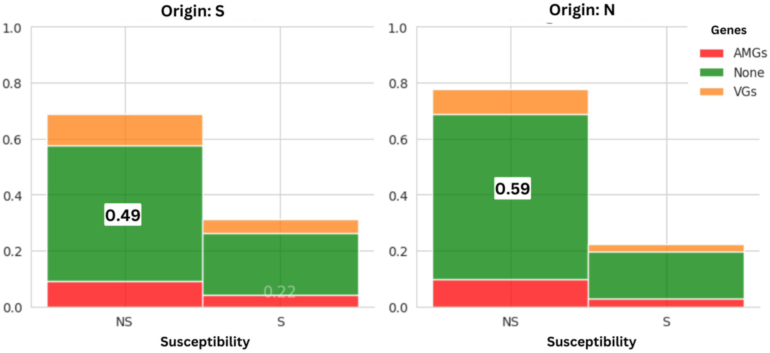
3.1.1. Prevalence of AMR Between Suppliers

Out of the 100 samples, 82 isolates of E. coli (82%) were isolated on the MCC medium, and 68 strains were isolated on the MacConkey medium with cefotaxime (MCC + CTX) (68%) (Table 1). There was no statistically significant difference in the prevalence of E. coli on MCC and MCC + CTX between chicken samples from both suppliers.
In fact, the strains from both suppliers had similar AMR prevalence rates and harboured virulence and resistance genes (Figure 1).
The analysis of antimicrobial resistance in isolates from both slaughterhouses revealed a statistically significant difference in resistance levels for 10 antimicrobial agents (p < 0.05). Notably, all isolates displayed lower resistance to ertapenem and colistin compared to other tested antibiotics. Specifically, strains from Slaughterhouse S exhibited higher resistance to penicillins (amoxicillin, piperacillin) and second-generation cephalosporins (cephalothin, cefuroxime) compared to strains from Slaughterhouse N. However, resistance was lower for quinolones, sulfonamides, and aminoglycosides among strains from Slaughterhouse S. This highlights a distinctive resistance pattern between the two slaughterhouses (Figure 2).

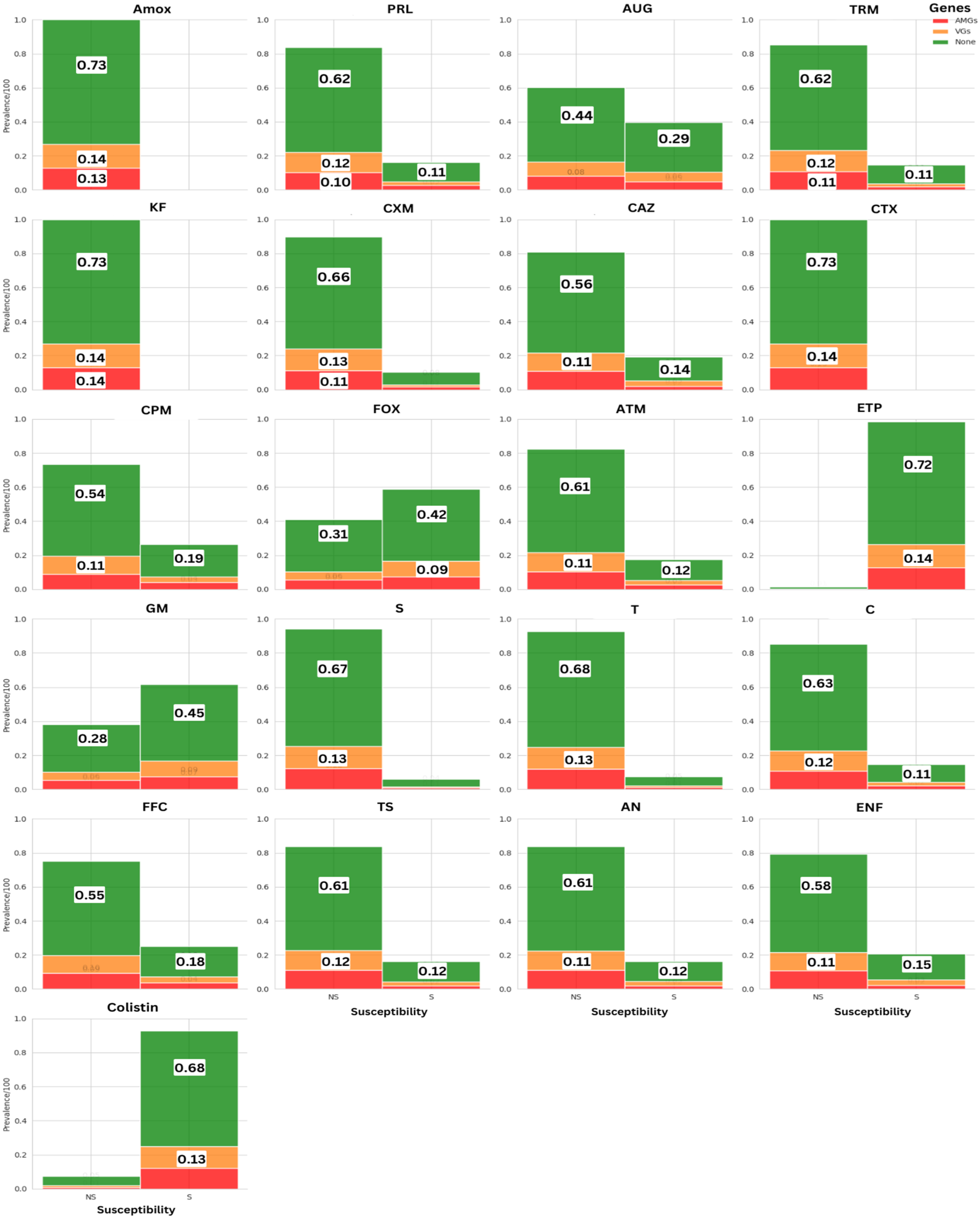
3.1.2. Prevalence of AMR Across Antimicrobial Molecules

All E. coli isolates that grew on MCC medium supplemented with cefotaxime (n = 68) were tested for resistance to 21 different antimicrobial agents, and 67 (98.52%) of the isolates were MDR. Of all 68 isolates studied, no strain (0%) was susceptible to all the 21 tested antibiotics, while 100% of the strains were resistant to at least 11 antibiotics. The non-susceptibility to beta-lactamines was most prevalent in isolates where the resistance rate was greater than 90% for amoxicillin, piperacillin, cephalothin, and cefotaxime. Significant resistance to β-lactams was associated with associations of amoxicillin-clavulanic acid (60.3%) and ticarcillin-clavulanic acid (85.3%) (Table S2).
In addition, 41.2% of E. coli strains were resistant to cefoxitin, suggesting the possible production of a plasmid cephalosporinase type AmpC. On the other hand, the strains have a low level of resistance to ertapenem (1.5%) and colistin (7.4%) (Table S2).
The rate of non-susceptible strains for other antimicrobials was as follows: tetracycline (92.6%), nalidixic acid (83.8%), trimethoprim-sulfamethoxazole (83.3%), chloramphenicol (80.15%), enrofloxacin (79.4%), and gentamicin (38.3%), just as shown in Figure 3 where AMR proportion is categorised by gene presence.
The quantitative assessment of AMR between antibiotics showed that 98.5% of all bacterial strains exhibited resistance to at least three antibiotic families. Notably, the most prevalent resistances for amoxicillin, cephalothin, and cefotaxime were observed. At the same time, ertapenem and colistin conserved their activity {Figure 3}, and the ESBL secretion ability of most strains did not influence the overall susceptibility pattern (Figure 4).
The MIC (minimal inhibitory concentration) of colistin was determined in all colistin-resistant E. coli isolates. The results are presented in Table 2 and interpreted according to the recommendations of CA-SFM/EUCAST [27]. Escherichia coli ATCC 25922 was used as a control for each run of the test.

3.2. Genetic Assessment

3.2.1. Detection of Beta-Lactamase Coding Genes

A total of 66 (97.05%) of the 68 E. coli isolates harboured β-lactamase genes, among which the blaCTX-M-G1 gene was the most predominant, found in 44 (64.7%) strains, followed by blaTEM in 33 (48.5%) strains. Furthermore, blaCMY and blaSHV genes were detected in 16 (23.5%) and 10 (14.7%), respectively. Based on PCR assay, no strain carried carbapenem resistance genes.

3.2.2. Detection of Non-β-Lactams Resistance Coding Genes

The tetracycline resistance genes (tetA, tetB) were detected in 38 strains at 41.2% and 14.7%, respectively. The mcr-1 gene was confirmed in four colistin-resistant strains out of five colistin-resistant isolates. The presence of sul genes linked with sulfamethoxazole resistance was observed in 66 sulfonamide resistance isolates [sul1 (13.2%), sul2 (70.6%), and sul3 (13.2%). The frequencies of qnr genes were qnrB (36.8%) and qnrS (4.4%), while the qnrA gene was not detected in our collection.

3.2.3. Detection of Virulence Coding Genes

The presence of 16 virulence genes (fimH, traT, IbeA, aer, sfa/foc, cnf1, iutA, fyuA, papGIII, papA, cdt3, hly, eae, stx1, stx2, and ehxA) was investigated in the 68 E. coli strains. Only nine virulence genes (fimH, traT, IbeA, aer, cnf1, iutA, fyuA, papGIII, and cdt3) were detected. Two strains lacked virulence genes, and no strain harboured Shiga-toxin-coding genes.

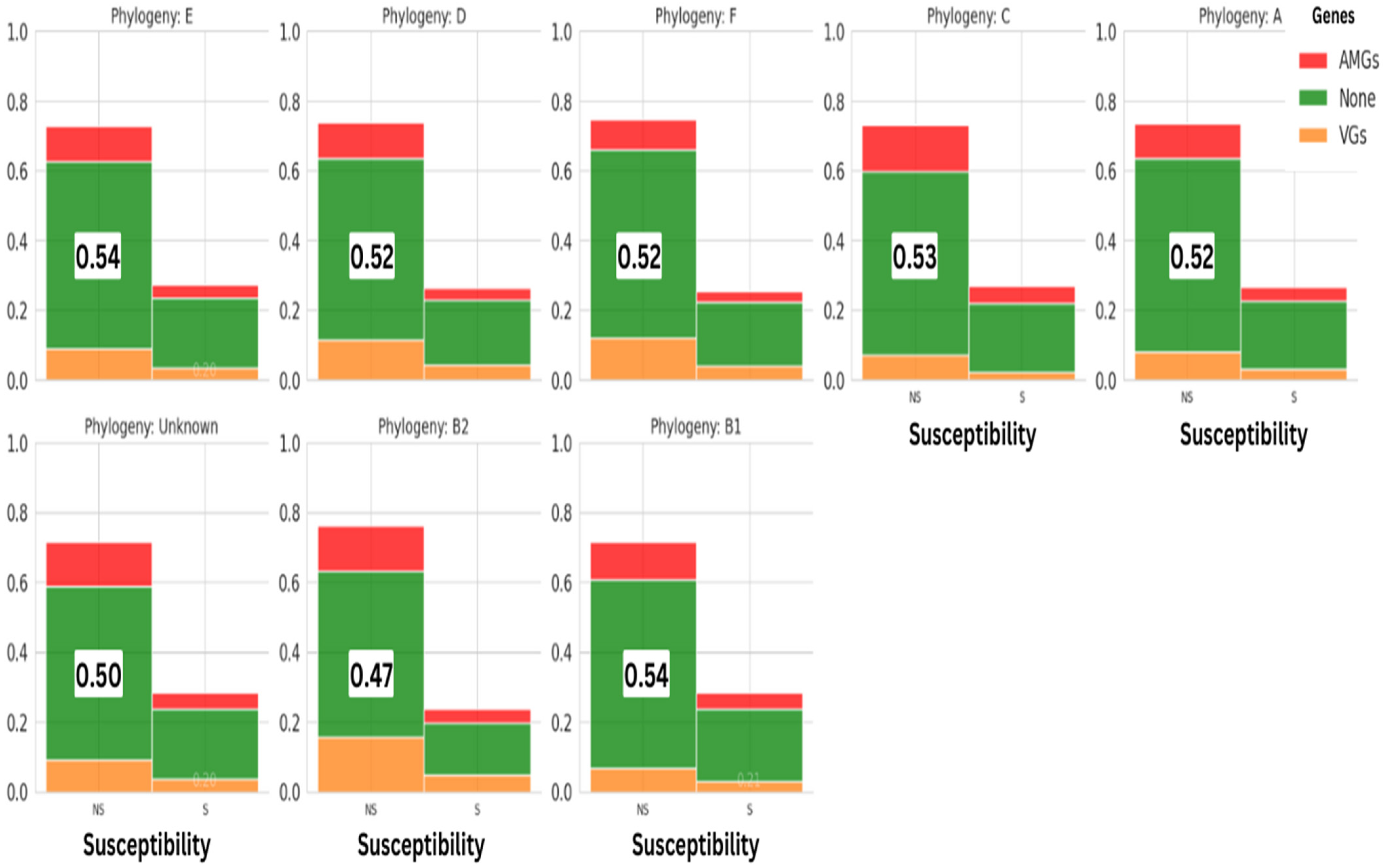
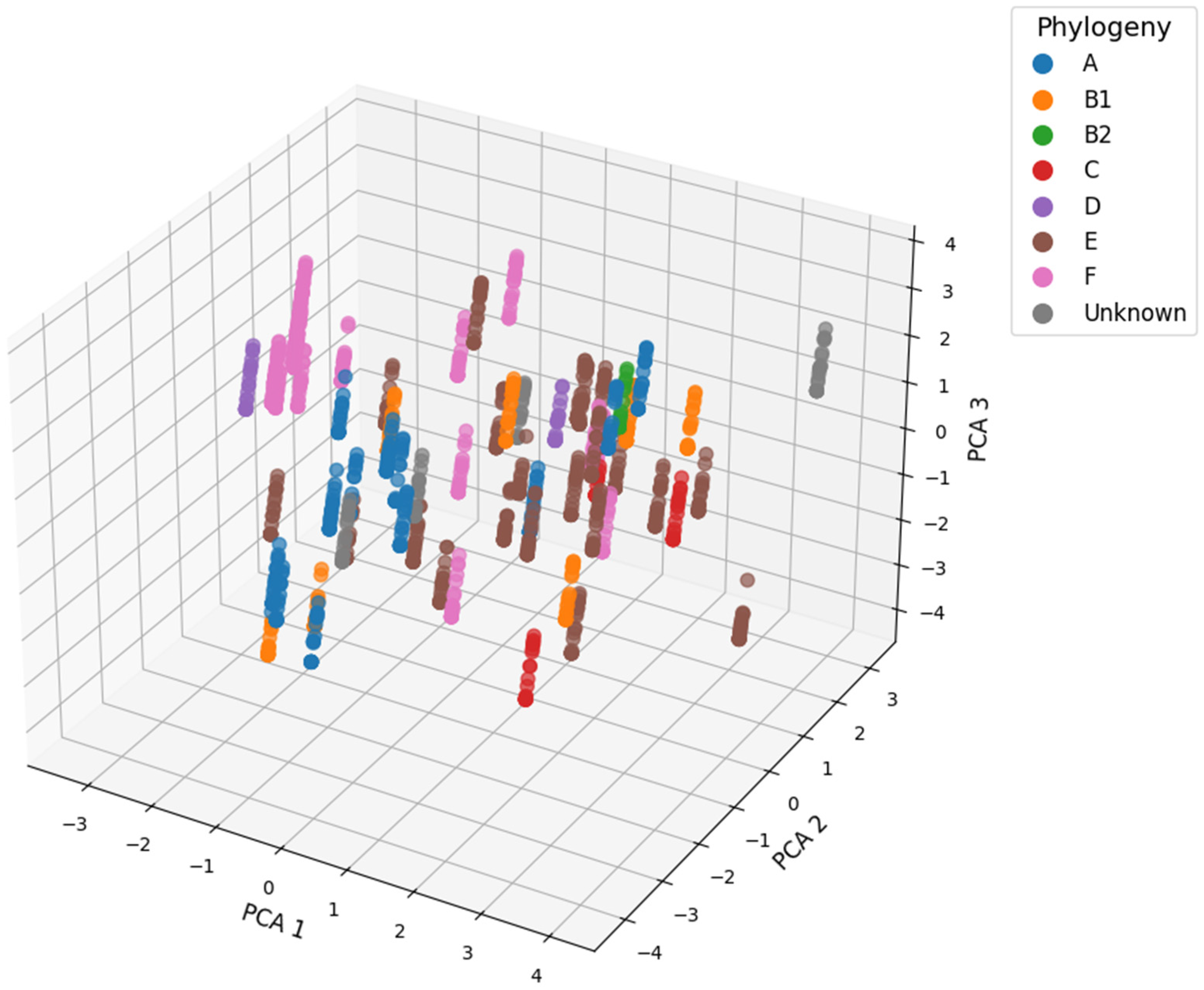
3.2.4. Phylogroups Assessment of E. coli Strains

The frequency distributions of the different E. coli phylogroups among the investigated isolates showed that pathogenic-associated phylogroups, namely, phylogroups E, D, B2, and F, were dominant in E. coli strains with a prevalence of 61.8% (42/68), whereas commensal-associated phylogroups A, B1, and C had a prevalence of 32.3% (22/68).
All isolated strains had a relatively high resistance rate, regardless of the phylogroups they belonged to (Figure 5).

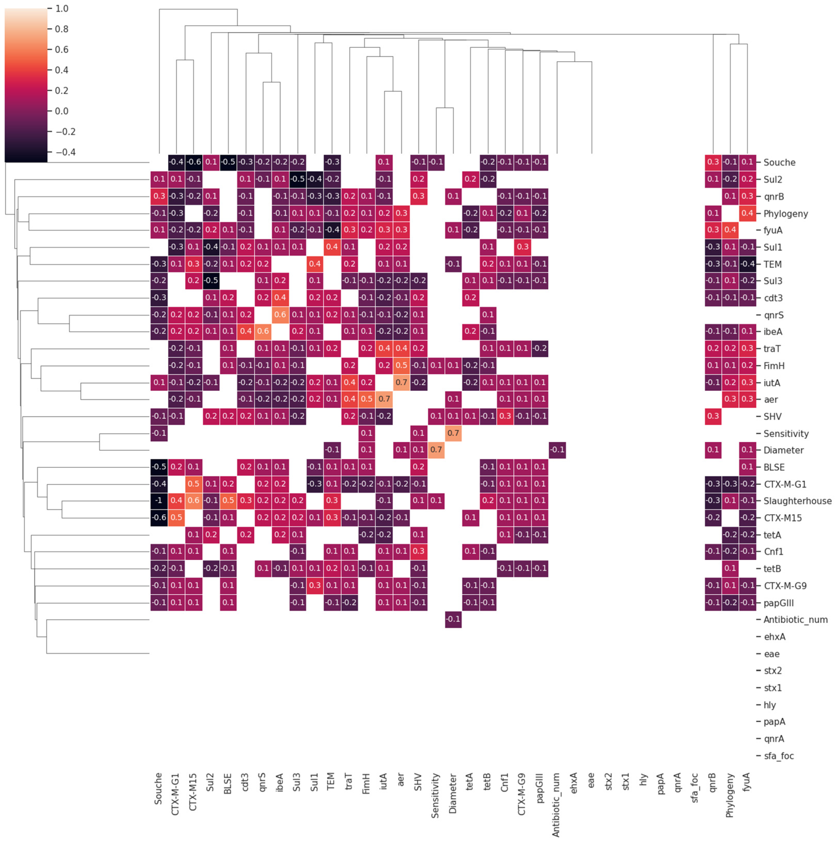
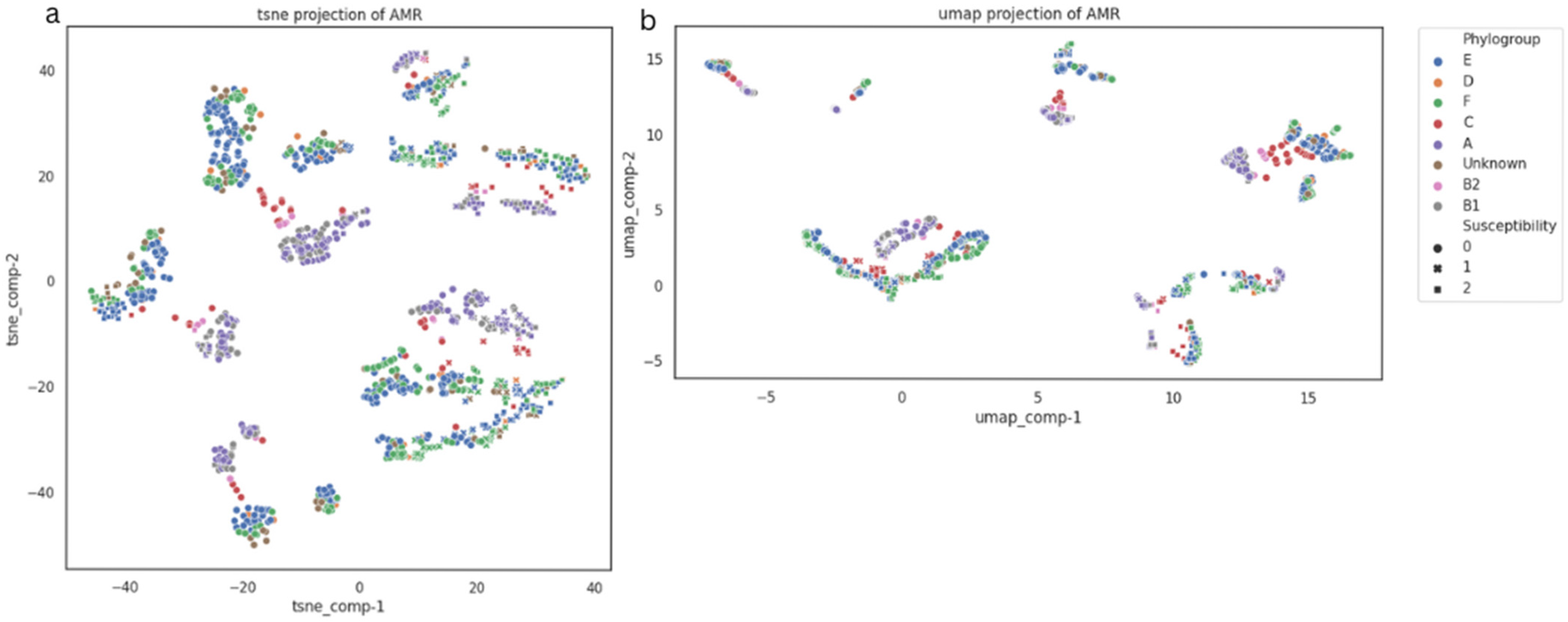
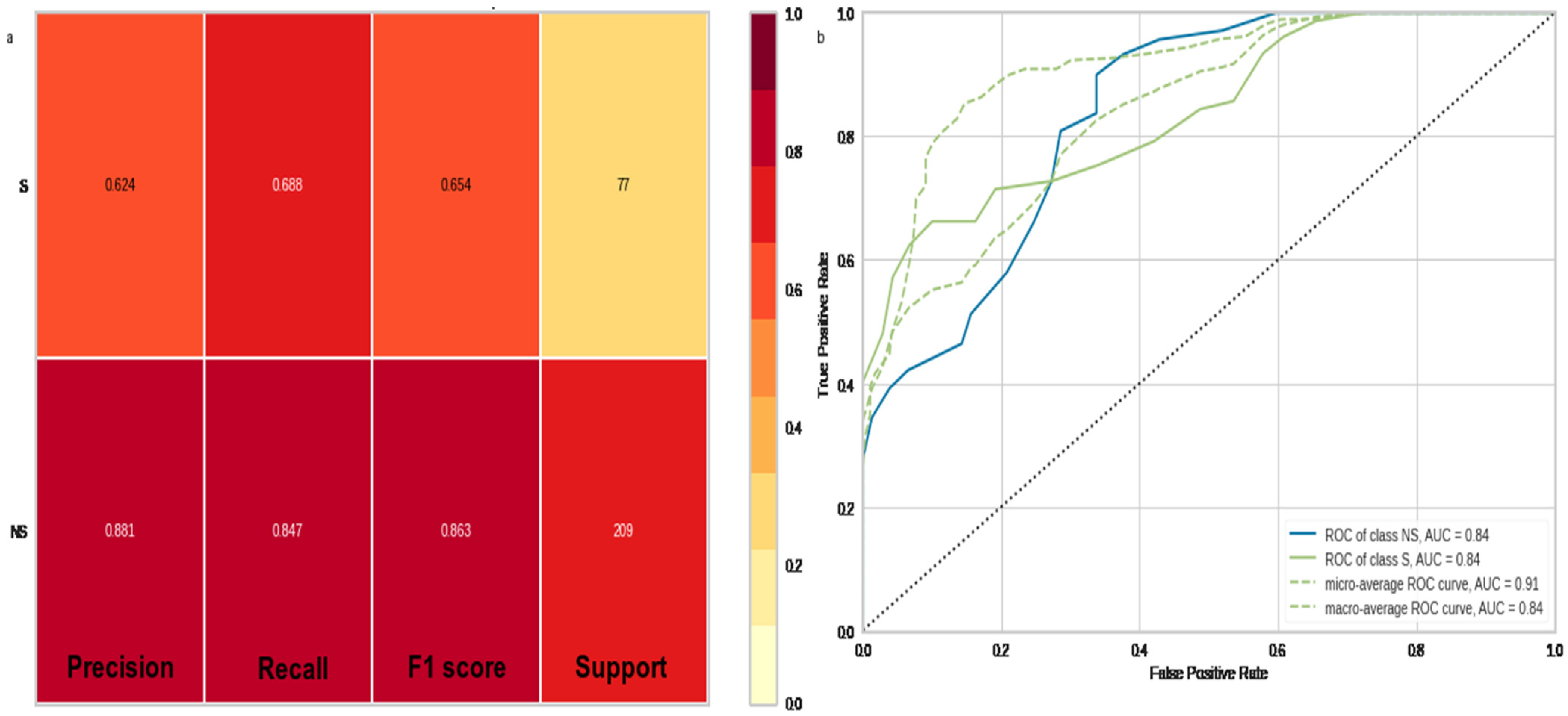
3.3. Data Mining and Machine Learning Evaluations

The distribution of AMR bacteria among phylogenetic groups showed uniformity, with no significant differences observed between genomic features, namely, antimicrobial-resistant genes (AMGs) and virulence genes (VGs). These genes were present in both susceptible and non-susceptible samples (Figure 5). This uniformity was also conserved among samples coming from slaughterhouses (Figure 2) and across the ESBL-producing capacity of bacteria isolates (Figure 4) as well. Interestingly, a substantial proportion of the PCR evaluation (>40%) did not detect any of the screened genes (Figure 3).
At the genomic level, there was no direct correlation between the presence of resistance genes and the expression of the resistance phenotype in the general non-stratified assessment. Remarkably, in certain extreme cases, susceptible isolates exhibited a higher frequency of resistance gene compared to their resistant counterparts (Figure 3).
This compelling discovery has catalysed further inquiries into the dynamics of antimicrobial resistant gene expressions and the potential risk factors that underlie the occurrence of antimicrobial resistance in chicken meat.
In fact, both linear (Figure 6, Figure 7 and Figure 8) and non-linear clustering analyses (Figure 9) revealed a homogeneous distribution of AMR data. It is impossible to separate between data points in the clustering assessments.
Meanwhile, we were able to place the unknown phylogroup into the E and F phylogroups cluster, both of which were geometrically close, and Figure 9 clearly shows the clustering conclusions.
The intricacies of the AMR dynamic are beyond the scope of basic statistical approaches. To gain deeper insights into the underlying patterns governing AMR and potential risk factors, we harnessed the power of multimodal machine learning (MML) models by subjecting our dataset to train–test analysis.
Throughout our modelling endeavour, diverse analyses were executed, revealing the logistic regression model as the top-performing one. The training phase converged after 350 epochs, as illustrated in Figure 10a,b, attaining a notable area under the receiver operating characteristic curve (ROC-AUC) of 84%, as depicted in Figure 10. Furthermore, critical metrics such as Accuracy, Precision, Recall, and F1-Score all surpassed 79%, as shown in Figure 10. Our model underwent comprehensive training, using all available features, as depicted in Figure 11a. Subsequent cross-validation enabled us to pinpoint the most influential features impacting antimicrobial resistance (AMR) expression (Figure 11). Notably, among all factors, our analysis revealed that the application of particular antimicrobial compounds critically shaped resistance patterns, with the choice of specific agents emerging as the principal driver behind escalating antimicrobial resistance incidence. The data demonstrates that the use of most molecules directly correlates with amplified AMR prevalence.
While on the other hand ertapenem and colistin had odds ratios of 347 (CI between 236 and 510) and 65 (CI between 35 and 80), respectively, making them highly conserved active molecules among all screened ones (Figure 11a), these findings are consistent with results reported previously. Conversely, our analysis revealed that none of the other factors under study, including ESBL secretion, meat origin, AMGs and VGs, had any significant influence over AMR expression. These findings are further supported by the absence of a significant correlation between these examined risk factors and the AMR profile (Figure 5).
Meanwhile, a final evaluation employing the adjusted F1 score of the random forest classifier led to the determination of the potential prediction of meat origin through the integration of ESBL, phylogroup, CTM-X-G1, and CTMX-M-15 profiles, indicating a possible geographical nuance of AMR distribution.
In addition, TEM gene presence was significantly predicted by CTX-M-15, sul1, fyuA, iutA, and qnrB gene presence and the phylogroup as well.
Furthermore, the same assessment established associations, linking aer and iutA genes together, and phylogroup to both fyuA and qnrB. All of these genes code for important resistance factors and iron transport systems.
This model demonstrates the interrelation among various multidrug resistance genes, which confer protection against beta-lactams, sulfamides, and quinolones to the evaluated strains; one of the most prominent features is the presence of CTX-M-15, TEM, sul1, and qnrB genes, along with genes responsible for acquiring iron from the host environment.
fyuA codes for ferric yersiniabactin receptor in biofilm-forming E. coli strains [30,31], and iutA codes for aerobactin siderophore ferric receptor protein; both genes facilitate the iron acquisition by mediating the uptake of siderophores [32] and are major contributors to the expression of AMR in our strains. These findings highlight the complex network of genetic determinants influencing bacterial resistance patterns.
In this evaluation, we also deduced the absence of a clear correlation between AMR gene presence and the susceptibility test (Figure 6). This observation emphasises the intricate and multifaceted characteristics of AMR within food matrices (Figure 11b), which need a comprehensive and in-depth investigation.

4. Discussion

In this study, we evaluated 100 chicken carcasses obtained from two distinct suppliers located in separate geographical regions in Tunisia (north and south). The aim was to determine the rate of contamination of chicken meat with E. coli, to assess the prevalence of extended-spectrum β-lactamases (ESBLs) and to characterise resistance and virulence genes patterns. For these objectives, a quantitative risk assessment of E. coli in chicken meat was developed using multi omics machine learning through the compilation of genotypic and phenotypic features.
Our examination revealed a contamination rate of 82% of E. coli in chicken meat, in alignment with comparable studies in Turkey, Spain, and India, where prevalence ranged between 80% and 90% [33,34,35]. These observations highlight the extensive distribution of E. coli in the environment and its capacity to spoil the agricultural and food supply chain, thus posing a significant threat to consumers. Notably, E. coli has consistently posed a challenge to the food processing sector, particularly without robust control measures.
In this study, the isolated strains exhibited significant resistance to a wide array of antibiotics, encompassing β-lactams, with notable emphasis on broad-spectrum cephalosporins. E. coli, distinguished by its remarkable enzymatic adaptability against antibiotics, particularly ESBLs, has garnered attention. In this study, 76.5% of the tested isolates were ESBL-producers.
Similar prevalence rates have been documented in other regions, with figures such as 79.17% in Thailand [36], 79.8% in the Netherlands [37], 81% in Italy [38], and 91.7% in France [39] aligning with our findings.
In Tunisia, a rate of 63.8% was reported [40], while in 2017, 28.8% of isolates exhibited ESBL production in chicken meat [41]. However, certain investigations [42,43] failed to identify ESBL-producing strains of E. coli in poultry meat. These escalating ESBL-producing E. coli prevalence emphasise the necessity for judicious antibiotic usage among veterinarians, grounded in an evidence-based approach.
Recently, plasmid-mediated colistin resistance has raised significant global concerns, jeopardising the efficacy of a critical antimicrobial agent for human health. The initial identification of the plasmid-mediated colistin resistance gene mcr-1 in China in 2015 marked a pivotal discovery in the AMR dynamic, detected in numerous countries ever since [44]. The present investigation disclosed a 7.4% resistance rate to colistin, with four out of five colistin-resistant strains also exhibiting ESBL production patterns. These results align with findings from other Tunisian researchers and international studies [45,46,47,48]. The association of colistin-resistant+ ESBL is attributed to the common presence of plasmid-mediated genes governing both colistin resistance and ESBL production [49]. Resistance to colistin and other last-resort antibiotics frequently aligns with the presence of multidrug resistance (MDR) and carbapenemase-producing Enterobacterales (CPE), with ESBLs frequently implicated in MDR mechanisms [50].
Another noteworthy facet pertains to MDR, characterised by resistance to three or more antibiotic families, which is highly prevalent in E. coli [51]. Notably, the current study indicated a striking 98.5% prevalence of a multi-resistance phenotype among the strains examined. MDR strains pose a significant concern as they limit our capacity to control infections, potentially leading to therapeutic failure and horizontal gene transfer to other pathogenic bacteria [52].
Our study also indicated substantial resistance rates among the isolates to various antibiotics, including amoxicillin, tetracycline, chloramphenicol, trimethoprim-sulfamethoxazole, and nalidixic acid. These outcomes concur with data provided by other studies [40,53]. In contrast, a notably elevated resistance rate (94.1%) was observed for streptomycin, surpassing the 78% reported in Tunisia [42] and the 61.9% reported in Bangladesh [54]. The notably high rate of streptomycin resistance in our study (94.1%) can likely be attributed to the frequent use of amoxicillin, tetracycline, and aminoglycosides in industrial husbandry.
Many antibiotics, including tetracyclines, aminoglycosides, sulfonamides, cefuroxime, and penicillins, are commonly used in poultry production and disease treatment. This extensive use has led to high resistance levels against these agents like streptomycin for example (94.1%) that is probably due to overuse of this antibiotic, increasing the risk of resistant bacteria transmission from food-producing animals to humans. Given the prohibition of chloramphenicol in animal agriculture due to the risk of bone marrow aplasia in humans, surveillance of resistance to the phenicol family, specifically chloramphenicol, remains crucial [55]. The association between resistance to chloramphenicol and florfenicol, the second molecule being a permissible alternative for treating E. coli infections in animals, was explored. The cross-resistance potential between these antibiotics stems from their shared target enzyme and has been attributed to a homolog of the chloramphenicol resistance efflux gene, cmlA, which may lead to multidrug resistance due to overuse [56]. Results indicated resistance rates of 85.3% for chloramphenicol and 75% for florfenicol, which aligns with a study carried out in Romania [57]. In European Union countries, particularly Poland, the United Kingdom, Germany, France, and Spain, the rate of resistance to chloramphenicol ranges from 4% to 25%, which is relatively low compared to our findings [58].
Our study revealed an 85.3% resistance rate to chloramphenicol compared to 75% for florfenicol. Cross-resistance between chloramphenicol and florfenicol may arise because both antibiotics target the same bacterial enzyme, the 50S ribosomal subunit. Resistance to one antibiotic can lead to resistance to the other, and bacteria resistant to both are often resistant to other antibiotic classes, fostering multidrug resistance. The overuse of antibiotics for infection prevention or treatment exerts selection pressure, driving the development of antibiotic-resistant bacteria.
The blaCTX-M gene predominates as the ESBL type in E. coli within the intestinal microbiota of chickens and chicken meat [59]. In this study, the most prevalent was blaCTX-M-G1, followed by the blaTEM gene and blaSHV gene. Additionally, one isolate carried the blaCTX-M-G9 group. These findings parallel those reported in domestically produced and imported chicken meat in Japan [60]. Genotypic analysis of all ESBL-positive isolates indicated that the majority (79%) were of the CTX-M type, with blaCTX-M-15 being the most frequent. CTX-M-15 is recognised as the predominant ESBL type globally among humans [61].
Notably, in France, CTX-M-15 was found in a chicken isolate therefore from food-producing animals for the first time [62]. The initial reports of the blaCTX-M-15 gene in E. coli strains of animal origin in Tunisia, as well as the presence of CTX-M β-lactamases in live broiler chickens, were documented by Mnif et al. (2012), then confirmed by Jouini et al. (2021) in diarrheic poultry [63,64]. CTX-M-15 prevalence has also been documented in other animals, including healthy animals and retail meat, in Spain [65], Japan [66], and Korea [67].
Most CTX-M-producing E. coli isolates carry more than one β-lactamase gene. The coexistence of multiple β-lactamase genes in the same strain raises concerns, as it can amplify co-resistance and resistance to various antibiotic classes. The presence of the mcr-1 gene has been demonstrated in Enterobacterales isolated from animals, animal-derived foods, and humans [68]. Among the colistin-resistant isolates, the mcr-1 gene was identified in four out of five cases. In prior Tunisian studies, the mcr-1 gene was also detected in chickens [69,70]. Numerous studies have established the presence of mcr-1 in European poultry and other meat products [71].
The resistance rates of our samples to quinolones and fluoroquinolones antibiotics indicate that 83.8% of isolates were resistant to nalidixic acid, and 79.4% were resistant to enrofloxacin. Several plasmid-mediated quinolone resistance (PMQR) mechanisms have been identified, including Qnr proteins (encoded by qnr genes), aminoglycoside acetyltransferase AAC(6′)-Ib-cr [aac(6′)-Ib-cr gene], quinolone efflux pump QepA (qepA genes), and multidrug resistance pump OqxAB (oqxAB genes) [72].
In parallel with PMQR mechanisms, our study also focused on plasmid-mediated β-lactam resistance. However, one limitation of this study is the absence of an evaluation of AmpC β-lactamase production. Although the primary objective was to investigate plasmid-mediated β-lactam resistance, it is recognised that exposure to β-lactam antibiotics can induce chromosomal AmpC expression, potentially contributing to increased resistance even among initially susceptible isolates. While current data suggest a low prevalence of AmpC-hyperproducing strains within the study population, the possibility of their presence cannot be definitively excluded. Accordingly, future investigations should incorporate targeted assessments of AmpC induction and expression to elucidate its contribution more thoroughly to resistance development during β-lactam exposure.
In our study, we detected qnr genes (qnrA, qnrB, qnrS) and revealed that qnrB to be the most prevalent. Multiple researchers in Tunisia have documented the presence of these genes in E. coli of animal origin [73]. Earlier studies in Italy [74,75] and other countries [76,77] have reported the presence of plasmid-mediated resistance qnrB and qnrS genes.
Regarding phylogroups, extraintestinal pathogenic E. coli primarily belongs to B2 and D groups, while commensal strains are associated with the A and B1 groups [78,79]. Findings from this investigation revealed that most E. coli strains belonged to phylogroup E (30.9%), followed by F (26.5%) and A (17.6%), with phylogroup B2 and D identified in 4.4% of E. coli strains.
Pathogenic strains typically possess an array of virulence genes encoding factors directly involved in their ability to induce colibacillosis [80]. The pathogenic potential of E. coli is augmented by numerous virulence determinants influencing interactions with host cells, such as adhesins (attachment factors), fimbriae-mediated invasion, various toxins (cytonecrotising and hemolysin factors), and iron acquisition systems. While most E. coli isolates are commensals, pathogenic strains diverge from non-pathogenic strains based on the acquisition of virulence factors [81].
In this study, we analysed the presence of 16 distinct virulence factors. Collectively, a total of 17 unique gene combinations or virulotypes were identified. The gene encoding the adhesin, fimH, emerged as the most prevalent virulence determinant, being detected in 86% of the isolates. This was followed by genes encoding aerobactin (iutA), invasion (aer), serum resistance factor (traT), and yersiniabactin (fyuA), which were found in 84%, 77%, 74%, and 39% of the isolates, respectively. Our findings closely align with the profiles of virulence genes observed in E. coli strains isolated from broiler chickens in Algeria [82].
These identified genes (fimH, iutA, fyuA, traT) have been verified to be associated with pathogenicity islands linked to virulence plasmids in Avian Pathogenic E. coli (APEC) strains [83]. These virulence-associated genes on plasmids play a pivotal role in promoting pathogenicity, facilitating the evolution of pathogenic strains from commensal ones, and contributing to the risk of zoonotic infection [84].
Furthermore, our study detected a prevalent gene combination, “fimH, iutA, aer, traT”, in most isolates. This gene combination is implicated in the extraintestinal translocation and invasion of the intestinal epithelium by E. coli [85].
The application of multimodal machine learning has revealed a nuanced perspective on antibiotic resistance (AMR) dynamics of E. coli, indicating that the mere presence of resistance genes is insufficient to fully explain AMR occurrence in chicken meat. Many scientific reports highlight the significant role of multiple biological processes on AMG expression including gene regulation networks, horizontal gene transfer, microbiome interactions, environmental factors, and co-selection mechanisms [86,87,88]. The comprehensive interplay of these factors contributes AMR landscape [89,90,91], underscoring the need for a holistic understanding beyond gene presence.
Our study provides critical insights into the complex dynamics of antimicrobial resistance (AMR) in poultry by leveraging advanced machine learning techniques. We uncover a strong link between resistance factor genes and iron acquisition genes, both essential for bacterial fitness and adaptation. This connection not only deepens our understanding of AMR networks but also highlights potential therapeutic targets to mitigate resistance and enhance drug efficacy, a fact documented recently by [92]. Additionally, we identify the “Eagle effect”, a phenomenon explaining bacterial survival under high antibiotic pressure, which underscores the importance of functional assessments [93,94].
In fact, our analysis employs established machine learning (ML) techniques, including supervised and unsupervised methods such as logistic regression, random forests, principal component analysis (PCA), t-distributed stochastic neighbour embedding (t-SNE), and uniform manifold approximation and projection (UMAP). These approaches provide interpretable insights into AMR dynamics, unlike deep learning models, which are often criticised for being “black boxes”. By leveraging these multimodal ML methods, we maintain transparency and explainability in our findings, ensuring that risk factors and predictions are both understandable and actionable.
Our approach facilitates the development of targeted interventions to mitigate antimicrobial resistance (AMR) in poultry, underscoring the necessity for an intelligent, unified platform to effectively address this escalating public health threat. SmartSim© (https://huggingface.co/collections?p=0&search=Smartsim&sort=trending, accessed on 11 May 2022) is an open-source intelligent agent that exemplifies the integration of human expertise and artificial intelligence by enabling real-time drug optimisation through the incorporation of pharmacodynamic and pharmacokinetic indices. This advanced tool could be leveraged for the detection, prediction, and mitigation of AMR risks.

5. Conclusions

In conclusion, an alarming rate of ESBL-producing strains (76.5%) and a very high rate of multi-resistant strains (98.5%) were observed. Significant resistance to 3rdGC and 4thGC have been also noted, which must be monitored because some of them may be reserved for human use, in addition to the risk of contamination by these resistant bacteria for the consumer and any person in contact with these animals. The results showed that E. coli virulence genes, particularly fimH, hlyA, traT, aer, and cdt3, are widely distributed in poultry meat in the studied regions. Eating chicken meat is considered a very plausible source of human contamination by strains of E. coli that are extra-intestinal and resistant to wide spectrum cephalosporins, particularly uropathogenic strains. Faced with the threat of antibiotic resistance, the awareness of human and animal health professionals on the use of antibiotics leads to a common conclusion: it is essential to reason and limit the use of recommended drugs to maintain their effectiveness as part of the One Smart Health approach. This kind of strategy may benefit from the many advantages that artificial intelligence offers to monitor and mitigate public health risk at scale.

Supplementary Materials

References [95,96,97,98,99,100,101,102,103,104,105,106,107,108,109] are cited in the Supplementary Materials. The following supporting information can be downloaded at: https://www.mdpi.com/article/10.3390/microbiolres16060131/s1, Table S1. Primers employed to amplify the resistance genes, virulence genes, and phylogenetic grouping in E. coli isolates; Table S2. Origins, antibiotic resistance profiles, number of antibiotic families, ESBL phenotype, phylogroup, detected genes encoding antibiotic resistance and virulence characteristics of the examined E. coli isolates.

Author Contributions

L.M. designed the study, secured the funds, and supervised the work. I.F. participated in the interpretation of the results and their discussion. K.A. collected the samples and carried out the whole laboratory work. O.A. contributed to the collection of samples. A.M. performed the artificial intelligence (AI) treatment of data. G.T. and E.M. contributed to the laboratory work. K.A. wrote the original draft of the manuscript. K.A. and G.T. performed the data analysis and interpretation. L.M., I.F. and G.T. critically reviewed and edited the manuscript. All authors have read and agreed to the published version of the manuscript.

Funding

This work was financially supported by the project PEER cycle 7-349 “Monitoring of bacterial antimicrobial resistance for a better health of animals in Tunisia” [2019–2023] funded by the United States Agency for International Development (USAID). This research was supported by the International Development Research Center (IDRC)-Innovet-Initiative [Avibiocin project].

Institutional Review Board Statement

Not applicable.

Informed Consent Statement

Not applicable.

Data Availability Statement

The datasets generated during and analysed during the current study are available in this manuscript.

Conflicts of Interest

The authors declare that they have no conflicts of interest. For affiliation 3, no commercial or other conflicts of interest were involved. The collaboration was strictly scientific. All authors have read and approved the final version of the article.

References

  1. FAO. 2021. Available online: https://www.fao.org/home/en/ (accessed on 11 May 2022).
  2. Cunicoles LGIdPAe. Produits Avicole GIPAC. 2018. Available online: http://www.gipac.tn (accessed on 11 May 2022).
  3. Hafez, H.M.; Attia, Y.A. Challenges to the poultry industry: Current perspectives and strategic future after the COVID-19 outbreak. Front. Vet. Sci. 2020, 7, 516. [Google Scholar] [CrossRef]
  4. Espinosa, R.; Tago, D.; Treich, N. Infectious diseases and meat production. Environ. Resour. Econ. 2020, 76, 1019–1044. [Google Scholar] [CrossRef]
  5. Murray, C.J.; Ikuta, K.S.; Sharara, F.; Swetschinski, L.; Aguilar, G.R.; Gray, A.; Han, C.; Bisignano, C.; Rao, P.; Wool, E.; et al. Global burden of bacterial antimicrobial resistance in 2019: A systematic analysis. Lancet 2022, 399, 629–655. [Google Scholar] [CrossRef]
  6. WHO. Antimicrobial Resistance: WHO. 2023. Available online: https://www.who.int/health-topics/antimicrobial-resistance (accessed on 13 June 2024).
  7. Manyi-Loh, C.; Mamphweli, S.; Meyer, E.; Okoh, A. Antibiotic use in agriculture and its consequential resistance in environmental sources: Potential public health implications. Molecules 2018, 23, 795. [Google Scholar] [CrossRef] [PubMed]
  8. Redfield, R.F. Centers for Disease Control and Prevention Justification of Appropriation Estimates for Appropriations Committees Fiscal Year 2021; Communicable Disease Center: Atlanta, GA, USA, 2020.
  9. Pym, R. Poultry genetics and breeding in developing countries. In Poultry Development Review; FAO: Rome, Italy, 2013; pp. 80–83. [Google Scholar]
  10. Ayukekbong, J.A.; Ntemgwa, M.; Atabe, A.N. The threat of antimicrobial resistance in developing countries: Causes and control strategies. Antimicrob. Resist. Infect. Control 2017, 6, 47. [Google Scholar] [CrossRef] [PubMed]
  11. Ramos, S.; Silva, V.; de Lurdes Enes Dapkevicius, M.; Caniça, M.; Tejedor-Junco, M.; Igrejas, G. Escherichia coli as commensal and pathogenic bacteria among food-producing animals: Health implications of extended spectrum β-lactamase (ESBL) production. Animals 2020, 10, 2239. [Google Scholar] [CrossRef] [PubMed]
  12. Massella, E.; Giacometti, F.; Bonilauri, P.; Reid, C.J.; Djordjevic, S.P.; Merialdi, G.; Bacci, C.; Fiorentini, L.; Massi, P.; Bardasi, L.; et al. Antimicrobial resistance profile and ExPEC virulence potential in commensal Escherichia coli of multiple sources. Antibiotics 2021, 10, 351. [Google Scholar] [CrossRef]
  13. Clermont, O.; Christenson, J.K.; Denamur, E.; Gordon, D.M. The Clermont Escherichia coli phylo-typing method revisited: Improvement of specificity and detection of new phylo-groups. Environ. Microbiol. Rep. 2013, 5, 58–65. [Google Scholar] [CrossRef]
  14. Agostinho, J.M.A.; Cardozo, M.V.; Borzi, M.M.; Marin, J.M. Antibiotic resistance and virulence factors among Escherichia coli isolates from avian organic fertilizer. Ciênc. Rural 2020, 50, e20180849. [Google Scholar] [CrossRef]
  15. Marshall, B.M.; Levy, S.B. Food animals and antimicrobials: Impacts on human health. Clin. Microbiol. Rev. 2011, 24, 718–733. [Google Scholar] [CrossRef] [PubMed]
  16. Sherchan, J.B.; Hayakawa, K.; Miyoshi-Akiyama, T.; Ohmagari, N.; Kirikae, T.; Nagamatsu, M.; Tojo, M.; Ohara, H.; Tandukar, S. Clinical epidemiology and molecular analysis of extended-spectrum-β-lactamase-producing Escherichia coli in Nepal: Characteristics of sequence types 131 and 648. Antimicrob. Agents Chemother. 2015, 59, 3424–3432. [Google Scholar] [CrossRef] [PubMed]
  17. Ghafourian, S.; Sadeghifard, N.; Soheili, S.; Sekawi, Z. Extended spectrum beta-lactamases: Definition, classification and epidemiology. Curr. Issues Mol. Biol. 2015, 17, 11–22. [Google Scholar]
  18. Ribeiro, L.F.; Nespolo, N.M.; Rossi, G.A.M.; Fairbrother, J.M. Exploring extended-spectrum beta-lactamase (ESBL)-producing Escherichia coli in food-producing animals and animal-derived foods. Pathogens 2024, 13, 346. [Google Scholar] [CrossRef]
  19. Bintsis, T. Foodborne pathogens. AIMS Microbiol. 2017, 3, 529. [Google Scholar] [CrossRef]
  20. Bush, K.; Bradford, P.A. β-Lactams and β-lactamase inhibitors: An overview. Cold Spring Harb. Perspect. Med. 2016, 6, a025247. [Google Scholar] [CrossRef] [PubMed]
  21. Dierikx, C.M.; van der Goot, J.A.; Smith, H.E.; Kant, A.; Mevius, D.J. Presence of ESBL/AmpC-producing Escherichia coli in the broiler production pyramid: A descriptive study. PLoS ONE 2013, 8, e79005. [Google Scholar] [CrossRef]
  22. Gazal, L.E.d.S.; Medeiros, L.P.; Dibo, M.; Nishio, E.K.; Koga, V.L.; Gonçalves, B.C.; Grassotti, T.T.; de Camargo, T.C.L.; Pinheiro, J.J.; Vespero, E.C.; et al. Detection of ESBL/AmpC-producing and fosfomycin-resistant Escherichia coli from different sources in poultry production in Southern Brazil. Front. Microbiol. 2021, 11, 604544. [Google Scholar] [CrossRef]
  23. Nsubuga, M.; Galiwango, R.; Jjingo, D.; Mboowa, G. Generalizability of machine learning in predicting antimicrobial resistance in E. Coli: A multi-country case study in Africa. BMC Genom. 2024, 25, 287. [Google Scholar] [CrossRef]
  24. Moradigaravand, D.; Palm, M.; Farewell, A.; Mustonen, V.; Warringer, J.; Parts, L. Prediction of antibiotic resistance in Escherichia coli from large-scale pan-genome data. PLoS Comput. Biol. 2018, 14, e1006258. [Google Scholar] [CrossRef]
  25. EURL-AR. Quantification of ESBL/AmpC-Producing Escherichia coli in Caecal Content and Fresh Meat Samples. 2017. Available online: https://www.food.dtu.dk/english/-/media/institutter/foedevareinstituttet/temaer/antibiotikaresistens/eurl-ar/protocols/esbl-ampc-and-camrbapenemase-producing-e-coli/399_esbl-ampc-quantification-protocol-19-03-2018.pdf (accessed on 27 April 2025).
  26. CLSI. Erformance Standards for Antimicrobial Susceptibility Testing. 2020. Available online: https://www.nih.org.pk/wp-content/uploads/2021/02/CLSI-2020.pdf (accessed on 27 April 2025).
  27. EUCAST ECoAST. Clinical Breakpoints—Breakpoints and Guidance. 2021. Available online: https://www.eucast.org/clinical_breakpoints (accessed on 27 April 2025).
  28. Dortet, L.; Poirel, L.; Nordmann, P. Rapid detection of ESBL-producing Enterobacteriaceae in blood cultures. Emerg. Infect. Dis. 2015, 21, 504–507. [Google Scholar] [CrossRef] [PubMed] [PubMed Central]
  29. Jouy, E.; Haenni, M.; Le Devendec, L.; Le Roux, A.; Châtre, P.; Madec, J.-Y.; Kempf, I. Improvement in routine detection of colistin resistance in E. coli isolated in veterinary diagnostic laboratories. J. Microbiol. Methods 2017, 132, 125–127. [Google Scholar] [CrossRef] [PubMed]
  30. Hancock, V.; Ferrieres, L.; Klemm, P. The ferric yersiniabactin uptake receptor FyuA is required for efficient biofilm formation by urinary tract infectious Escherichia coli in human urine. Microbiology 2008, 154, 167–175. [Google Scholar] [CrossRef]
  31. Tu, J.; Hue, T.; Qi, K.; Shao, Y.; Huang, B.; Wang, X.; Zhou, X. The irp2 and fyuA genes in High Pathogenicity Islands are involved in the pathogenesis of infections caused by avian pathogenic Escherichia coli (APEC). Pol. J. Vet. Sci. 2016, 19, 21–29. [Google Scholar] [CrossRef] [PubMed]
  32. Ikeda, M.; Kobayashi, T.; Fujimoto, F.; Okada, Y.; Higurashi, Y.; Tatsuno, K.; Okugawa, S.; Moriya, K. The prevalence of the iutA and ibeA genes in Escherichia coli isolates from severe and non-severe patients with bacteremic acute biliary tract infection is significantly different. Gut Pathog. 2021, 13, 32. [Google Scholar] [CrossRef]
  33. Pehlivanlar Önen, S.; Aslantaş, Ö.; Şebnem Yılmaz, E.; Kürekci, C. Prevalence of β-lactamase producing Escherichia coli from retail meat in Turkey. J. Food Sci. 2015, 80, M2023–M2029. [Google Scholar] [CrossRef]
  34. Hussain, A.; Shaik, S.; Ranjan, A.; Nandanwar, N.; Tiwari, S.K.; Majid, M.; Baddam, R.; Qureshi, I.A.; Semmler, T.; Wieler, L.H.; et al. Risk of transmission of antimicrobial resistant Escherichia coli from commercial broiler and free-range retail chicken in India. Front. Microbiol. 2017, 8, 2120. [Google Scholar] [CrossRef]
  35. García-Béjar, B.; García de Blas Martín, I.; Arévalo-Villena, M.; Briones Pérez, A. High prevalence of antibiotic-resistant Escherichia coli isolates from retail poultry products in Spain. Animals 2021, 11, 3197. [Google Scholar] [CrossRef]
  36. Sornsenee, P.; Chimplee, S.; Arbubaker, A.; Kongchai, S.; Madimong, H.; Romyasamit, C. Occurrence, Antimicrobial Resistance Profile, and Characterization of Extended-Spectrum β-Lactamase–Producing Escherichia coli Isolates from Minced Meat at Local Markets in Thailand. Foodborne Pathog. Dis. 2022, 19, 232–240. [Google Scholar] [CrossRef]
  37. Overdevest, I.; Willemsen, I.; Rijnsburger, M.; Eustace, A.; Xu, L.; Hawkey, P.; Heck, M.; Savelkoul, P.; Vandenbroucke-Grauls, C.; van der Zwaluw, K.; et al. Extended-spectrum β-lactamase genes of Escherichia coli in chicken meat and humans, The Netherlands. Emerg. Infect. Dis. 2011, 17, 1216. [Google Scholar] [CrossRef]
  38. Ghodousi, A.; Bonura, C.; Di Carlo, P.; van Leeuwen, W.B.; Mammina, C. Extraintestinal pathogenic Escherichia coli sequence type 131 H30-R and H30-Rx subclones in retail chicken meat, Italy. Int. J. Food Microbiol. 2016, 228, 10–13. [Google Scholar] [CrossRef]
  39. Casella, T.; Nogueira, M.C.L.; Saras, E.; Haenni, M.; Madec, J.-Y. High prevalence of ESBLs in retail chicken meat despite reduced use of antimicrobials in chicken production, France. Int. J. Food Microbiol. 2017, 257, 271–275. [Google Scholar] [CrossRef] [PubMed]
  40. Hassen, B.; Abbassi, M.S.; Ruiz-Ripa, L.; Mama, O.M.; Hassen, A.; Torres, C.; Hammami, S. High prevalence of mcr-1 encoding colistin resistance and first identification of blaCTX-M-55 in ESBL/CMY-2-producing Escherichia coli isolated from chicken faeces and retail meat in Tunisia. Int. J. Food Microbiol. 2020, 318, 108478. [Google Scholar] [CrossRef] [PubMed]
  41. Salah, A.M.; Mohamed, Z.; Bilel, H.; Souhir, Z.; Asma, D.; Riadh, M. ESBL/Cephalosporinase-producing Escherichia coli from retail poultry meat in Tunisia: Predominance of blaCTX-M gene and multidrug resistance. J. Microbes Microbio. Tech. 2017, 1, 101. [Google Scholar]
  42. Soufi, L.; Abbassi, M.S.; Sáenz, Y.; Vinué, L.; Somalo, S.; Zarazaga, M.; Abbas, A.; Dbaya, R.; Khanfir, L.; Ben Hassen, A.; et al. Prevalence and diversity of integrons and associated resistance genes in Escherichia coli isolates from poultry meat in Tunisia. Foodborne Pathog. Dis. 2009, 6, 1067–1073. [Google Scholar] [CrossRef] [PubMed]
  43. Badi, S.; Salah Abbassi, M.; Snoussi, M.; Werheni, R.; Hammami, S.; Maal-Bared, R.; Hassen, A. High rates of antibiotic resistance and biofilm production in Escherichia coli isolates from food products of animal and vegetable origins in Tunisia: A real threat to human health. Int. J. Environ. Health Res. 2022, 32, 406–416. [Google Scholar] [CrossRef] [PubMed]
  44. Liu, Y.-Y.; Wang, Y.; Walsh, T.R.; Yi, L.-X.; Zhang, R.; Spencer, J.; Doi, Y.; Tian, G.; Dong, B.; Huang, X.; et al. Emergence of plasmid-mediated colistin resistance mechanism MCR-1 in animals and human beings in China: A microbiological and molecular biological study. Lancet Infect. Dis. 2016, 16, 161–168. [Google Scholar] [CrossRef]
  45. Dhaouadi, S.; Soufi, L.; Hamza, A.; Fedida, D.; Zied, C.; Awadhi, E.; Mtibaa, M.; Hassen, B.; Cherif, A.; Torres, C.; et al. Co-occurrence of mcr-1 mediated colistin resistance and β-lactamase-encoding genes in multidrug-resistant Escherichia coli from broiler chickens with colibacillosis in Tunisia. J. Glob. Antimicrob. Resist. 2020, 22, 538–545. [Google Scholar] [CrossRef]
  46. Ferjani, S.; Maamar, E.; Ferjani, A.; Meftah, K.; Battikh, H.; Mnif, B.; Hamdoun, M.; Chebbi, Y.; Kanzari, L.; Achour, W.; et al. Tunisian multicenter study on the prevalence of colistin resistance in clinical isolates of Gram negative bacilli: Emergence of Escherichia coli harbouring the mcr-1 gene. Antibiotics 2022, 11, 1390. [Google Scholar] [CrossRef]
  47. Devan, S.; Aklilu, E.; Lemlem, M.; Zakaria, Z.; Hamdan, R. Detection of colistin-resistant Escherichia coli isolated from broiler chickens in Kelantan, Malaysia. Trop. Biomed. 2022, 39, 197–202. [Google Scholar]
  48. Nath, C.; Das, T.; Islam, M.S.; Hasib, F.Y.; Singha, S.; Dutta, A.; Barua, H.; Islam, Z. Colistin Resistance in Multidrug-Resistant Escherichia coli Isolated from Retail Broiler Meat in Bangladesh. Microb. Drug Resist. 2023, 29, 523–532. [Google Scholar] [CrossRef]
  49. Hatrongjit, R.; Kerdsin, A.; Akeda, Y.; Hamada, S. Detection of plasmid-mediated colistin-resistant and carbapenem-resistant genes by multiplex PCR. MethodsX 2018, 5, 532–536. [Google Scholar] [CrossRef] [PubMed]
  50. Karaiskos, I.; Giamarellou, H. Multidrug-resistant and extensively drug-resistant Gram-negative pathogens: Current and emerging therapeutic approaches. Expert Opin. Pharmacother. 2014, 15, 1351–1370. [Google Scholar] [CrossRef] [PubMed]
  51. Catalano, A.; Iacopetta, D.; Ceramella, J.; Scumaci, D.; Giuzio, F.; Saturnino, C.; Aquaro, S.; Rosano, C.; Sinicropi, M.S. Multidrug resistance (MDR): A widespread phenomenon in pharmacological therapies. Molecules 2022, 27, 616. [Google Scholar] [CrossRef] [PubMed]
  52. Soufi, L.; Sáenz, Y.; Vinué, L.; Abbassi, M.S.; Ruiz, E.; Zarazaga, M.; Ben Hassen, A.; Hammami, S.; Torres, C. Escherichia coli of poultry food origin as reservoir of sulphonamide resistance genes and integrons. Int. J. Food Microbiol. 2011, 144, 497–502. [Google Scholar] [CrossRef]
  53. Rafiq, K.; Sani, A.A.; Hossain, M.T.; Hossain, M.T.; Hadiuzzaman, M.; Bhuiyan, M.A.S. Assessment of the presence of multidrug-resistant Escherichia coli, Salmonella and Staphylococcus in chicken meat, eggs and faeces in Mymensingh division of Bangladesh. Heliyon 2024, 10, e36690. [Google Scholar] [CrossRef]
  54. Rahman, M.M.; Husna, A.; Elshabrawy, H.A.; Alam, J.; Runa, N.Y.; Badruzzaman, A.T.M.; Banu, N.A.; Al Mamun, M.; Paul, B.; Das, S.; et al. Isolation and molecular characterization of multidrug-resistant Escherichia coli from chicken meat. Sci. Rep. 2020, 10, 21999. [Google Scholar] [CrossRef] [PubMed] [PubMed Central]
  55. Mensah, S.; Koudandé, O.; Sanders, P.; Laurentie, M.; Mensah, G.; Abiola, F. Antimicrobial residues in foods of animal origin in Africa: Public health risks. Rev. Sci. Tech. 2014, 33, 987–996. [Google Scholar]
  56. White, D.G.; Hudson, C.; Maurer, J.J.; Ayers, S.; Zhao, S.; Lee, M.D.; Bolton, L.; Foley, T.; Sherwood, J. Characterization of chloramphenicol and florfenicol resistance in Escherichia coli associated with bovine diarrhea. J. Clin. Microbiol. 2000, 38, 4593–4598. [Google Scholar] [CrossRef]
  57. Brătfelan, D.O.; Tabaran, A.; Colobatiu, L.; Mihaiu, R.; Mihaiu, M. Prevalence and Antimicrobial Resistance of Escherichia coli Isolates from Chicken Meat in Romania. Animals 2023, 13, 3488. [Google Scholar] [CrossRef]
  58. Roth, N.; Käsbohrer, A.; Mayrhofer, S.; Zitz, U.; Hofacre, C.; Domig, K.J. The application of antibiotics in broiler production and the resulting antibiotic resistance in Escherichia coli: A global overview. Poult. Sci. 2019, 98, 1791–1804. [Google Scholar] [CrossRef] [PubMed]
  59. Grami, R.; Mansour, W.; Dahmen, S.; Mehri, W.; Haenni, M.; Aouni, M.; Madec, J.-Y. The blaCTX-M-1 IncI1/ST3 plasmid is dominant in chickens and pets in Tunisia. J. Antimicrob. Chemother. 2013, 68, 2950–2952. [Google Scholar] [CrossRef] [PubMed]
  60. Nahar, A.; Awasthi, S.P.; Hatanaka, N.; Okuno, K.; Hoang, P.H.; Hassan, J.; Hinenoya, A.; Yamasaki, S. Prevalence and characteristics of extended-spectrum β-lactamase-producing Escherichia coli in domestic and imported chicken meats in Japan. J. Vet. Med. Sci. 2018, 80, 510–517. [Google Scholar] [CrossRef]
  61. Carattoli, A. Animal reservoirs for extended spectrum β-lactamase producers. Clin. Microbiol. Infect. 2008, 14, 117–123. [Google Scholar] [CrossRef] [PubMed]
  62. Meunier, D.; Jouy, E.; Lazizzera, C.; Kobisch, M.; Madec, J.-Y. CTX-M-1-and CTX-M-15-type β-lactamases in clinical Escherichia coli isolates recovered from food-producing animals in France. Int. J. Antimicrob. Agents 2006, 28, 402–407. [Google Scholar] [CrossRef]
  63. Mnif, B.; Ktari, S.; Rhimi, F.; Hammami, A. Extensive dissemination of CTX-M-1-and CMY-2-producing Escherichia coli in poultry farms in Tunisia. Lett. Appl. Microbiol. 2012, 55, 407–413. [Google Scholar] [CrossRef]
  64. Jouini, A.; Klibi, A.; Elarbi, I.; Chaabene, M.B.; Hamrouni, S.; Souiai, O.; Hanachi, M.; Ghram, A.; Maaroufi, A. First detection of human ST131-CTX-M-15-O25-B2 clone and high-risk clonal lineages of ESBL/pAmpC-producing E. coli isolates from diarrheic poultry in Tunisia. Antibiotics 2021, 10, 670. [Google Scholar] [CrossRef] [PubMed]
  65. Blanc, V.; Mesa, R.; Saco, M.; Lavilla, S.; Prats, G.; Miró, E.; Navarro, F.; Cortés, P.; Llagostera, M. ESBL-and plasmidic class C β-lactamase-producing E. coli strains isolated from poultry, pig and rabbit farms. Vet. Microbiol. 2006, 118, 299–304. [Google Scholar] [CrossRef]
  66. Hiroi, M.; Yamazaki, F.; Harada, T.; Takahashi, N.; Iida, N.; Noda, Y.; Yagi, M.; Nishio, T.; Kanda, T.; Kawamori, F.; et al. Prevalence of extended-spectrum β-lactamase-producing Escherichia coli and Klebsiella pneumoniae in food-producing animals. J. Vet. Med. Sci. 2012, 74, 189–195. [Google Scholar] [CrossRef]
  67. Seo, K.W.; Kim, Y.B.; Jeon, H.Y.; Lim, S.-K.; Lee, Y.J. Comparative genetic characterization of third-generation cephalosporin-resistant Escherichia coli from chicken meat produced by integrated broiler operations in South Korea. Poult. Sci. 2018, 97, 2871–2879. [Google Scholar] [CrossRef]
  68. Zhao, Q.; Li, Y.; Tian, Y.; Shen, Y.; Wang, S.; Zhang, Y. Clinical impact of colistin banning in food animal on mcr-1-positive Enterobacteriaceae in patients from Beijing, China, 2009–2019: A long-term longitudinal observational study. Front. Microbiol. 2022, 13, 826624. [Google Scholar] [CrossRef] [PubMed]
  69. Grami, R.; Mansour, W.; Mehri, W.; Bouallègue, O.; Boujaâfar, N.; Madec, J.-Y.; Haenni, M. Impact of food animal trade on the spread of mcr-1-mediated colistin resistance, Tunisia, July 2015. Eurosurveillance 2016, 21, 30144. [Google Scholar] [CrossRef] [PubMed]
  70. Maamar, E.; Alonso, C.A.; Hamzaoui, Z.; Dakhli, N.; Abbassi, M.S.; Ferjani, S.; Saidani, M.; Boubaker, I.B.-B.; Torres, C. Emergence of plasmid-mediated colistin-resistance in CMY-2-producing Escherichia coli of lineage ST2197 in a Tunisian poultry farm. Int. J. Food Microbiol. 2018, 269, 60–63. [Google Scholar] [CrossRef]
  71. Skov, R.L.; Monnet, D.L. Plasmid-mediated colistin resistance (mcr-1 gene): Three months later, the story unfolds. Eurosurveillance 2016, 21, 30155. [Google Scholar] [CrossRef]
  72. Ferjani, S.; Saidani, M.; Amine, F.S.; Boutiba-Ben Boubaker, I. Prevalence and characterization of plasmid-mediated quinolone resistance genes in extended-spectrum β-lactamase-producing Enterobacteriaceae in a Tunisian hospital. Microb. Drug Resist. 2015, 21, 158–166. [Google Scholar] [CrossRef] [PubMed]
  73. Sallem, R.B.; Gharsa, H.; Slama, K.B.; Rojo-Bezares, B.; Estepa, V.; Porres-Osante, N.; Jouini, A.; Klibi, N.; Sáenz, Y.; Boudabous, A.; et al. First detection of CTX-M-1, CMY-2, and QnrB19 resistance mechanisms in fecal Escherichia coli isolates from healthy pets in Tunisia. Vector-Borne Zoonotic Dis. 2013, 13, 98–102. [Google Scholar] [CrossRef]
  74. Cerquetti, M.; García-Fernández, A.; Giufrè, M.; Fortini, D.; Accogli, M.; Graziani, C.; Luzzi, I.; Caprioli, A.; Carattoli, A. First report of plasmid-mediated quinolone resistance determinant qnrS1 in an Escherichia coli strain of animal origin in Italy. Antimicrob. Agents Chemother. 2009, 53, 3112–3114. [Google Scholar] [CrossRef]
  75. Niero, G.; Bortolaia, V.; Vanni, M.; Intorre, L.; Guardabassi, L.; Piccirillo, A. High diversity of genes and plasmids encoding resistance to third-generation cephalosporins and quinolones in clinical Escherichia coli from commercial poultry flocks in Italy. Vet. Microbiol. 2018, 216, 93–98. [Google Scholar] [CrossRef]
  76. Fortini, D.; Fashae, K.; García-Fernández, A.; Villa, L.; Carattoli, A. Plasmid-mediated quinolone resistance and β-lactamases in Escherichia coli from healthy animals from Nigeria. J. Antimicrob. Chemother. 2011, 66, 1269–1272. [Google Scholar] [CrossRef]
  77. Veldman, K.; van Essen-Zandbergen, A.; Kant, A.; Mevius, D. Characterization of qnr-positive Escherichia coli isolates from food-producing animals in the Netherlands. J. Antimicrob. Chemother. 2012, 67, 239–240. [Google Scholar] [CrossRef]
  78. Smith, J.L.; Fratamico, P.M.; Gunther, N.W. Extraintestinal pathogenic Escherichia coli. Foodborne Pathog. Dis. 2007, 4, 134–163. [Google Scholar] [CrossRef] [PubMed]
  79. Chakraborty, A.; Saralaya, V.; Adhikari, P.; Shenoy, S.; Baliga, S.; Hegde, A. Characterization of Escherichia coli phylogenetic groups associated with extraintestinal infections in South Indian population. Ann. Med. Health Sci. Res. 2015, 5, 241–246. [Google Scholar] [PubMed]
  80. De Carli, S.; Ikuta, N.; Lehmann, F.K.M.; da Silveira, V.P.; de Melo Predebon, G.; Fonseca, A.S.K.; Lunge, V.R. Virulence gene content in Escherichia coli isolates from poultry flocks with clinical signs of colibacillosis in Brazil. Poult. Sci. 2015, 94, 2635–2640. [Google Scholar] [CrossRef]
  81. Tayh, G.; Nagarjuna, D.; Sallem, R.B.; Verma, V.; Yahia, H.B.; Gharsa, H.; Yadav, M.; Ben Slama, K. Identification of Virulence Factors Among ESBL-Producing Escherichia coli Clinical Isolates from GAZA Strip, Palestine: Virulence Genes among ESBL-producing E. coli. J. Microbiol. Biotechnol. Food Sci. 2021, 11, e2865. [Google Scholar] [CrossRef]
  82. Messaili, C.; Messai, Y.; Bakour, R. Virulence gene profiles, antimicrobial resistance and phylogenetic groups of fecal Escherichia coli strains isolated from broiler chickens in Algeria. Vet. Ital. 2019, 55, 35–46. [Google Scholar]
  83. Johnson, T.J.; Nolan, L.K. Pathogenomics of the virulence plasmids of Escherichia coli. Microbiol. Mol. Biol. Rev. 2009, 73, 750–774. [Google Scholar] [CrossRef]
  84. Mellata, M.; Ameiss, K.; Mo, H.; Curtiss, R., III. Characterization of the contribution to virulence of three large plasmids of avian pathogenic Escherichia coli χ7122 (O78: K80: H9). Infect. Immun. 2010, 78, 1528–1541. [Google Scholar] [CrossRef]
  85. Poole, N.M.; Green, S.I.; Rajan, A.; Vela, L.E.; Zeng, X.-L.; Estes, M.K.; Maresso, A.W. Role for FimH in extraintestinal pathogenic Escherichia coli invasion and translocation through the intestinal epithelium. Infect. Immun. 2017, 85, 10-1128. [Google Scholar] [CrossRef]
  86. Fajardo, A.; Martínez-Martín, N.; Mercadillo, M.; Galán, J.C.; Ghysels, B.; Matthijs, S.; Cornelis, P.; Wiehlmann, L.; Tümmler, B.; Baquero, F.; et al. The neglected intrinsic resistome of bacterial pathogens. PLoS ONE 2008, 3, e1619. [Google Scholar] [CrossRef]
  87. Palmer, A.C.; Kishony, R. Understanding, predicting and manipulating the genotypic evolution of antibiotic resistance. Nat. Rev. Genet. 2013, 14, 243–248. [Google Scholar] [CrossRef]
  88. Wozniak, R.A.; Waldor, M.K. A toxin–antitoxin system promotes the maintenance of an integrative conjugative element. PLoS Genet. 2009, 5, e1000439. [Google Scholar] [CrossRef] [PubMed]
  89. Bengtsson-Palme, J.; Hammaren, R.; Pal, C.; Östman, M.; Björlenius, B.; Flach, C.-F.; Fick, J.; Kristiansson, E.; Tysklind, M.; Larsson, D.J. Elucidating selection processes for antibiotic resistance in sewage treatment plants using metagenomics. Sci. Total Environ. 2016, 572, 697–712. [Google Scholar] [CrossRef]
  90. Van Schaik, W. The human gut resistome. Philos. Trans. R. Soc. B Biol. Sci. 2015, 370, 20140087. [Google Scholar] [CrossRef] [PubMed]
  91. Stokes, H.W.; Gillings, M.R. Gene flow, mobile genetic elements and the recruitment of antibiotic resistance genes into Gram-negative pathogens. FEMS Microbiol. Rev. 2011, 35, 790–819. [Google Scholar] [CrossRef] [PubMed]
  92. Andersson, D.I.; Hughes, D. Antibiotic resistance and its cost: Is it possible to reverse resistance? Nat. Rev. Microbiol. 2010, 8, 260–271. [Google Scholar] [CrossRef]
  93. Levy, S.B. The Antibiotic Paradox: How Miracle Drugs Are Destroying the Miracle; Springer: Berlin/Heidelberg, Germany, 2013. [Google Scholar]
  94. Jackson, L.K.; Dailey, T.A.; Anderle, B.; Warren, M.J.; Bergonia, H.A.; Dailey, H.A.; Phillips, J.D. Exploiting differences in heme biosynthesis between bacterial species to screen for novel antimicrobials. Biomolecules 2023, 13, 1485. [Google Scholar] [CrossRef]
  95. Poirel, L.; Walsh, T.R.; Cuvillier, V.; Nordmann, P. Multiplex PCR for detection of acquired carbapenemase genes. Diagn. Microbiol. Infect. Dis. 2011, 70, 119–123. [Google Scholar] [CrossRef]
  96. Jouini, A.; Vinué, L.; Slama, K.B.; Sáenz, Y.; Klibi, N.; Hammami, S.; Boudabous, A.; Torres, C. Characterization of CTX-M and SHV extended-spectrum β-lactamases and associated resistance genes in Escherichia coli strains of food samples in Tunisia. J. Antimicrob. Chemother. 2007, 60, 1137–1141. [Google Scholar] [CrossRef]
  97. Batchelor, M.; Hopkins, K.; Threlfall, E.; Clifton-Hadley, F.; Stallwood, A.; Davies, R.; Liebana, E. bla CTX-M genes in clinical Salmonella isolates recovered from humans in England and Wales from 1992 to 2003. Antimicrob. Agents Chemother. 2005, 49, 1319–1322. [Google Scholar] [CrossRef]
  98. Muzaheed Doi, Y.; Adams-Haduch, J.M.; Endimiani, A.; Sidjabat, H.E.; Gaddad, S.M.; Paterson, D.L. High prevalence of CTX-M-15-producing Klebsiella pneumoniae among inpatients and outpatients with urinary tract infection in Southern India. J. Antimicrob. Chemother. 2008, 61, 1393–1394. [Google Scholar] [CrossRef]
  99. Literacka, E.; Empel, J.; Baraniak, A.; Sadowy, E.; Hryniewicz, W.; Gniadkowski, M. Four variants of the Citrobacter freundii AmpC-type cephalosporinases, including novel enzymes CMY-14 and CMY-15, in a Proteus mirabilis clone widespread in Poland. Antimicrob. Agents Chemother. 2004, 48, 4136–4143. [Google Scholar] [CrossRef] [PubMed]
  100. Mazel, D.; Dychinco, B.; Webb, V.A.; Davies, J. Antibiotic resistance in the ECOR collection: Integrons and identification of a novel aad gene. Antimicrob. Agents Chemother. 2000, 44, 1568–1574. [Google Scholar] [CrossRef] [PubMed]
  101. Sáenz, Y.; Brinas, L.; Domínguez, E.; Ruiz, J.; Zarazaga, M.; Vila, J.; Torres, C. Mechanisms of resistance in multiple-antibiotic-resistant Escherichia coli strains of human, animal, and food origins. Antimicrob. Agents Chemother. 2004, 48, 3996–4001. [Google Scholar] [CrossRef]
  102. Perreten, V.; Boerlin, P. A new sulfonamide resistance gene (sul3) in Escherichia coli is widespread in the pig population of Switzerland. Antimicrob. Agents Chemother. 2003, 47, 1169–1172. [Google Scholar] [CrossRef]
  103. Cattoir, V.; Poirel, L.; Rotimi, V.; Soussy, C.-J.; Nordmann, P. Multiplex PCR for detection of plasmid-mediated quinolone resistance qnr genes in ESBL-producing enterobacterial isolates. J. Antimicrob. Chemother. 2007, 60, 394–397. [Google Scholar] [CrossRef] [PubMed]
  104. Johnson, J.R.; Stell, A.L. Extended virulence genotypes of Escherichia coli strains from patients with urosepsis in relation to phylogeny and host compromise. J. Infect. Dis. 2000, 181, 261–272. [Google Scholar] [CrossRef]
  105. Tóth, I.; Hérault, F.; Beutin, L.; Oswald, E. Production of Cytolethal Distending Toxins by Pathogenic Escherichia coli Strains Isolated from Human and Animal Sources: Establishment of the Existence of a New cdt Variant (Type IV). J. Clin. Microbiol. 2003, 41, 4285–4291. [Google Scholar] [CrossRef]
  106. Ruiz, J.; Simon, K.; Horcajada, J.P.; Velasco, M.; Barranco, M.; Roig, G.; Moreno-Martínez, A.; Martínez, J.A.; de Anta, T.J.; Mensa, J.; et al. Differences in Virulence Factors among Clinical Isolates of Escherichia coli Causing Cystitis and Pyelonephritis in Women and Prostatitis in Men. J. Clin. Microbiol. 2002, 40, 4445–4449. [Google Scholar] [CrossRef]
  107. Vidal, M.; Kruger, E.; Durán, C.; Lagos, R.; Levine, M.; Prado, V.; Toro, C.; Vidal, R. Single multiplex PCR assay to identify simultaneously the six categories of diarrheagenic Escherichia coli associated with enteric infections. J. Clin. Microbiol. 2005, 43, 5362–5365. [Google Scholar] [CrossRef]
  108. Paton, A.W.; Paton, J.C. Detection and characterization of Shiga toxigenic Escherichia coli by using multiplex PCR assays for stx 1, stx 2, eaeA, enterohemorrhagic E. coli hlyA, rfb O111, and rfb O157. J. Clin. Microbiol. 1998, 36, 598–602. [Google Scholar]
  109. Yamamoto, S.; Terai, A.; Yuri, K.; Kurazono, H.; Takeda, Y.; Yoshida, O. Detection of urovirulence factors in Escherichia coli by multiplex polymerase chain reaction. FEMS Immunol. Med. Microbiol. 1995, 12, 85–90. [Google Scholar] [CrossRef] [PubMed]
Figure 1. Prevalence of AMR between suppliers N and S. Abbreviation: NS: non susceptible, S: Susceptible, VGs: Virulence genes, AMG: Antimicrobial resistance genes, None: absence of virulence and antimicrobial resistance genes.
Microbiolres 16 00131 g001
Figure 2. Prevalence of AMR across antimicrobial molecules between slaughterhouses N and S. Abbreviation: Amox: amoxicillin, PRL: piperacillin, AUG: amoxicillin + clavulanic acid, TIM: ticarcillin + clavulanic acid, KF: cephalothin, CXM: cefuroxime, FOX: cefoxitin, CTX: cefotaxime, CPM: cefepime, CAZ: ceftazidime, ATM: aztreonam, ETP: ertapenem, TS: trimethoprim-sulfamethoxazole, NA: nalidixic acid, ENF: enrofloxacin, C: chloramphenicol, FFC: florfenicol, GM: gentamicin, S: streptomycin, T: tetracycline, and Colispot: colistin.
Microbiolres 16 00131 g002
Figure 3. Prevalence of AMR across antimicrobial molecules. Abbreviations: VGs: virulence genes, AMG: antimicrobial genes, None: absence of virulence and antimicrobial genes. Amox: amoxicillin, PRL: piperacillin, AUG: amoxicillin + clavulanic acid, TIM: ticarcillin + clavulanic acid, KF: cephalothin, CXM: cefuroxime, FOX: cefoxitin, CTX: cefotaxime, CPM: cefepime, CAZ: ceftazidime, ATM: aztreonam, ETP: ertapenem, TS: trimethoprim-sulfamethoxazole, NA: nalidixic acid, ENF: enrofloxacin, C: chloramphenicol, FFC: florfenicol, GM: gentamicin, S: streptomycin, T: tetracycline, and Colispot: colistin.
Microbiolres 16 00131 g003
Figure 4. Prevalence of AMR between negative and positive ESBL-producing strains across antimicrobial molecules. Note: green susceptible strains, yellow: intermediary susceptible strains, and orange: resistant strains. Abbreviation: Amox: amoxicillin, PRL: piperacillin, AUG: amoxicillin + clavulanic acid, TIM: ticarcillin + clavulanic acid, KF: cephalothin, CXM: cefuroxime, FOX: cefoxitin, CTX: cefotaxime, CPM: cefepime, CAZ: ceftazidime, ATM: aztreonam, ETP: ertapenem, TS: trimethoprim-sulfamethoxazole, NA: nalidixic acid, ENF: enrofloxacin, C: chloramphenicol, FFC: florfenicol, GM: gentamicin, S: streptomycin, T: tetracycline, and Colispot: colistin.
Microbiolres 16 00131 g004
Figure 5. Prevalence of AMR across phylogroups. Abbreviation: NS: non-susceptible, S: susceptible. VGs: virulence genes, AMG: antimicrobial genes, None: absence of virulence and antimicrobial genes.
Microbiolres 16 00131 g005
Figure 6. Pearson correlation and hierarchical clustering analysis of genomic and phenotypic features of AMR.
Microbiolres 16 00131 g006
Figure 7. Principal components analysis of AMR data by ESBL secretion feature. Note: green points: positive ESBL strain, blue points: negative ESBL strain.
Microbiolres 16 00131 g007
Figure 8. Principal components analysis of AMR data by phylogroups.
Microbiolres 16 00131 g008
Figure 9. Non-linear clustering analysis of AMR.(a), tsne distribution. (b), umap distribution Abbreviation: tsne: t-distributed stochastic neighbour embedding clustering approach; umap: uniform manifold approximation and projection for dimension reduction clustering approach.
Microbiolres 16 00131 g009
Figure 10. Illustration of the training performances of a multi-omics machine learning model for the assessment of antimicrobial resistance risks. (a), machine learning performances for predicting AMR expression. (b), the area under the receiver operating characteristic curve. Abbreviation: S: susceptible strain, NS: non susceptible strain.
Microbiolres 16 00131 g010
Figure 11. Outputs of multimodal machine learning risk assessment. (a) ML feature importance ranking; (b) Odds ratios of AMR expression by antibiotic.
Microbiolres 16 00131 g011
Table 1. Contamination rate of chicken carcasses by E. coli.
Number of Samples Contamination Rate E. coli (MCC)Contamination Rate E. coli (MCC + CTX)p-Value E. coli
Supplier N5076% (38/50)68% (34/50)0.157
Supplier S5088% (44/50)68% (34/50)
Total 10082% (82/100)68% (68/100)-
Note: p values > 0.05 were statistically non-significant; MCC: MacConkey agar and MCC + CTX: MacConkey agar supplemented with cefotaxime.
Table 2. Colistin minimum inhibitory concentration (MIC) of E. coli strains.
Strain B14CTXB28CTXB44CTXB45CTXA10CTX
MIC (µg/mL)2<2466
ESBL++++
mcr-1 gene++++
Abbreviations: MIC: minimum inhibitory concentration, +: gene detected −: gene not detected, ESBL: extended-spectrum beta-lactamase.
Disclaimer/Publisher’s Note: The statements, opinions and data contained in all publications are solely those of the individual author(s) and contributor(s) and not of MDPI and/or the editor(s). MDPI and/or the editor(s) disclaim responsibility for any injury to people or property resulting from any ideas, methods, instructions or products referred to in the content.

