Article

In Vitro Evaluation of the Ovicidal Potential of Proteases from Beauveria bassiana Against Eurytrema pancreaticum Eggs

by
Lisseth Bibiana Puentes Figueroa
1,
Amanda do Carmo Alves
1,
Adriane Toledo da Silva
1,
Debora Castro de Souza
1,
Nivia Kelly Lima Sales
1,
Lorrana Verdi Flores
2,
Tiago Facury Moreira
3,
Fabio Ribeiro Braga
4 and
Filippe Elias de Freitas Soares
1,*
1
Laboratório de Biotecnologia e Bioquímica Aplicada, Departamento de Química, Universidade Federal de Lavras, Lavras 37200-000, MG, Brazil
2
Laboratório Central de Biologia Molecular, Departamento de Química, Universidade Federal de Lavras, Lavras 37200-000, MG, Brazil
3
Escola de Veterinária, Departamento de Clínica e Cirurgia Veterinária, Escola de Veterinária, Universidade Federal de Minas Gerais, Belo Horizonte 31270-800, MG, Brazil
4
Laboratório de Parasitologia Experimental e Controle Biológico, Universidade Vila-Velha, Vila Velha 29102-920, ES, Brazil
*
Author to whom correspondence should be addressed.
Microbiol. Res. 2025, 16(6), 127; https://doi.org/10.3390/microbiolres16060127
Submission received: 21 March 2025 / Revised: 11 June 2025 / Accepted: 11 June 2025 / Published: 13 June 2025

Abstract

:
In the search for new alternatives for controlling parasitic agents, proteases from Beauveria bassiana stand out. The aim of this study was to evaluate in silico and in vitro the ovicidal potential of B. bassiana proteases on Eurytrema pancreaticum Janson, 1889 (Dicrocoeliidae) eggs. Beauveria bassiana Bals. -Criv., 1835 (Cordycipitaceae) (IP 361) was cultivated for enzymatic production. Proteins were precipitated with acetone (1:4 ratio), and specific activity was determined. Protease profiles were assessed via zymography, and inhibition by phenylmethylsulfonyl fluoride (PMSF) and ethylenediaminetetraacetic acid (EDTA) was tested. Three-dimensional models of the proteases were generated. Eurytrema pancreaticum eggs were used for the in vitro anthelmintic evaluation of the proteases. The results showed that precipitation significantly concentrated proteolytic activity (p < 0.01) compared to the crude extract. However, no chitinase activity was detected. The proteolytic profile of the precipitate revealed five bands with molecular weights from 25.6 to 66.9 kDa. In the in vitro tests, the proteases significantly (p < 0.01) reduced the number of intact E. pancreaticum eggs by 53% compared to the control with denatured enzymes. These findings highlight the ovicidal potential of B. bassiana proteases, though further studies are needed to confirm their application in parasite control.

1. Introduction

Advances in the global industry and in-depth knowledge of enzymes have boosted their use in the creation of high-quality products, such as bioinsecticides, making them relevant tools in biological control [1]. Enzymes, biological catalysts found in all living things, are taking on a crucial role in green chemistry as promising tools for more sustainable and economically viable industrial processes, given their high specificity and efficiency [2,3,4].
Entomopathogenic fungi are well-established biological control agents that are gradually replacing synthetic chemicals in pest control due to their biocontrol and enzyme production capabilities [5,6]. As a result, many studies have been dedicated to the potential of various entomopathogenic fungi for the production of important enzymes and other metabolites, with Beauveria bassiana standing out [7,8,9]. The main enzymes produced by B. bassiana are lipases (EC 3.1.1.3), proteases (EC. 3.4), and chitinases (EC 3.2.1.14) [10,11].
Proteases, hydrolytic enzymes that catalyze the cleavage of proteins into smaller peptide chains and amino acid groups, are important due to their physicochemical properties, mechanisms of action, and fundamental participation in physiological processes in biological control [12,13,14].
The action of extracellular proteases is a crucial factor in the infection of entomopathogenic fungi such as B. bassiana in arthropods. In this context, the most frequently studied proteolytic enzymes for this fungus are the subtilisin-like serine protease Pr1 and the trypsin-like protease Pr2 [13,15]. Although the nematicidal activity of B. bassiana has been known since the 1990s, the action of its proteases has been demonstrated recently on infective larvae (L3) of the nematode Haemonchus spp. [16]. Thus, new scientific aspects could be outlined, opening up the possibility for an anthelmintic action of proteases from this fungus. However, there are no reports in the literature about the interaction of this fungus and its metabolites and helminth eggs.
The genus Eurytrema belongs to the family Dicrocoeliidae, class Trematoda, which includes the main parasites of the pancreatic ducts of ruminants [17]. Bovine eurythrematosis is associated with a drop in the productive performance of animals and also leads to losses in the insulin extraction industry, as it causes the pancreas to be identified as problematic during routine inspections [18,19,20]. The literature describes that the problems caused by this helminth are irreversible. In addition, effective anthelmintic drugs against Eurytrema are unknown [21,22]. Reports of this disease in humans have also been described [23,24].
Thus, the aim of this study was to characterize in silico and in vitro the ovicidal potential of B. bassiana proteases on E. pancreaticum eggs.

2. Materials and Methods

2.1. Obtaining and Extracting Extracellular Enzymes

The isolate IP 361 of B. bassiana s.l. used in this study was kindly provided by Professor Christian Luz. Beauveria bassiana s.l. The isolate was first isolated in 2010 from Amblyomma sculptum Berlese, 1888 (Ixodidae) adults in Central Brazil. This fungus belongs to the collection of the Invertebrate Pathology Laboratory of the Institute of Tropical Pathology and Public Health (IPTSP) of the Federal University of Goiás. It was grown in Petri dishes containing 2.0% Potato Dextrose Agar (PDA) at 25 ± 1 °C in the dark for 10 days.
The conidia were obtained from the culture plates and suspended in a sterile aqueous solution of 0.04% (v/v) Tween 80. Then, an inoculum at a concentration of 1 × 10⁷ conidia/mL was prepared. The number of conidia was counted in a Neubauer chamber. From this solution of conidia, 0.5 mL was added to flasks containing 40 g of pre-cooked rice and 20 mL of whey, previously autoclaved. The media were kept in a Biochemical Oxygen Demand Incubator (BOD) under light conditions at 27 ± 1 °C for 10 days. Samples were manually shaken every three days to homogenize the conidia in order to improve contact and germination [16].
After the solid fermentation period, 30 mL of sterile distilled water was added to the culture media. The flasks were shaken for 2 h at 120 RPM. The extracts were vacuum filtered using Whatman No. 4 paper and centrifuged at 10,000× g for 15 min. The supernatant obtained, containing enzymes and free of fungal cells, was referred to as a crude extract, as established by Figueroa et al. [16].

2.2. Enzyme Concentration

The enzymes were concentrated by precipitation with acetone, following the methodology of Doonan [25]. A portion of the supernatant was mixed with the solvent in a 1:4 (v/v) ratio for protein concentration. The samples were incubated at 4 °C for 1 h and centrifuged at 10,000× g for 10 min. The supernatant was discarded, and the precipitate obtained was resuspended in 100 µL of distilled water for later use.

2.3. Total Protease Activity

To determine total protease activity, 20 µL of the sample was used and incubated at 37 °C in the presence of 500 µL of 1.0% (w/v) casein and 480 µL of 50 mM Tris-HCl buffer (pH 7.15) for 30 min. After this period, the reaction was stopped by adding 1 mL of 10.0% (w/v) trichloroacetic acid (TCA, P.A., ACS, Dinâmica). The suspensions were centrifuged at 10,000× g for 10 min, and the absorbance of the resulting supernatant was measured at 280 nm using a spectrophotometer. One unit of protease enzyme activity was defined as the amount of enzyme that catalyzes the release of 1 µg of tyrosine per minute under the aforementioned experimental conditions [26].

2.4. Total Chitinase Activity

To determine the activity of total chitinase, 20 µL of the sample was incubated at 37 °C in the presence of 500 µL of 1.0% (w/v) colloidal chitin, and 480 µL of 50 mM Tris-HCl buffer (pH 7.15) was used for 60 min. After this period, the reaction was stopped with the addition of 1 mL of DNS and brought to a boil for 5 min. The suspensions were centrifuged at 10,000× g for 10 min, and the supernatant was used to read the absorbance at 540 nm. One unit of chitinase activity was defined as the amount of enzyme required to catalyze the release of 1.0 μM of N-acetylglucosamine per minute under the assay conditions [27].

2.5. Determination of Total Protein and Specific Activity

The total protein concentration was determined using bovine serum albumin (BSA) as a standard, following the protocol described by Bradford [28]. From the data obtained, the specific activity of the protease and chitinase was calculated by dividing the enzymatic activity by the concentration of total proteins (U mg−1 of protein).

2.6. Enzyme Characterization

2.6.1. Zymogram

In order to analyze the profile of the proteases present in the active precipitate extract of the fungus B. bassiana (IP 361), a zymogram was produced using casein as a substrate (Casein-SDS-PAGE), as described by Braga et al. [29], with some modifications. The samples were subjected to electrophoresis on a 10% polyacrylamide gel containing 1% casein. The samples were mixed with sample buffer (without β-mercaptoethanol) and applied without prior heating. After electrophoresis, the gel was incubated in a 2.5% solution of Triton X-100 for one hour, followed by three washes with distilled water. The gel was then incubated in 50 mM Tris-HCl reaction buffer (pH 7.15) for 2 h at 37 °C. For further development, the gel was stained with 0.5 g of Coomassie Brilliant Blue R-250 in an aqueous solution containing 10% acetic acid and 30% methanol. The presence of enzymatic action was evidenced by the formation of white halos on the gel.

2.6.2. Effects of Inhibitors on Proteolytic Activity

Proteolytic activity was determined using 20 µL of the precipitated extract in the presence of casein as a substrate and the following inhibitors at a concentration of 10 mM: iodoacetamide, phenylmethylsulfonyl fluoride (PMSF, as serine inhibitors), and ethylendiaminetetraacetic acid (EDTA, as a metalloprotease inhibitor). The conditions used in the assay were: pH 7.15 (50 mM Tris-HCl buffer) at 37 °C for 30 min. For the proteolytic assay, a control group was set up without the presence of inhibitors. All groups were carried out in triplicate [30].

2.6.3. Structural Prediction

The FASTA sequence was selected from the National Center for Biotechnology Information (NCBI) database, using as precursors the proteases of B. bassiana ARSEF 2860 (GCF_000280675). Pepstats and DeepLoc 2.0 (https://www.ebi.ac.uk/Tools/seqstats/emboss_pepstats/) were used to predict protein characteristics such as theoretical isoelectric point (pI), molecular weight, and localization (https://services.healthtech.dtu.dk/services/DeepLoc-2.0/).
In silico prediction of the three-dimensional (3D) structure of B. bassiana proteases was carried out by protein modeling using the ColabFold tool (https://colab.research.google.com/github/sokrypton/ColabFold/blob/main/AlphaFold2.ipynb). The FASTA sequences of the identified B. bassiana proteases were edited, after eliminating the amino acid residues encoding the signal peptide, using the SignalP-5.0 platform (https://services.healthtech.dtu.dk/services/SignalP-5.0/).

2.7. Obtaining Eurytrema Pancreaticum Eggs

The adults of E. pancreaticum were manually removed from the pancreas of cattle after death at the farm of the Veterinary School of the Federal University of Minas Gerais (UFMG), Belo Horizonte, Brazil. The helminths were then homogenized in water and filtered through 250-mesh per-inch metal sieves. The sedimentation method was then used to recover the eggs [31]. The eggshell integrity was assessed by optical microscopy using a 10× objective.

2.8. In Vitro Ovicidal Effect

Two experimental groups were defined to evaluate the ovicidal effect of IP 361 proteases on E. pancreaticum in vitro. The first group consisted of the active precipitated extract (“APE”), and the second group contained the denatured precipitated extract (“DPE,” an extract previously boiled for 2 h). Each group contained 100 µL of the active and denatured precipitated extracts and 100 µL of approximately 70 ± 7.20 eggs of E. pancreaticum, respectively. Six replicates were made for each experimental group. The experiment was kept at 28 °C ± 1 in the dark for 24 h. After this period, the number of intact eggs present in each replicate was counted. The count was made using an optical microscope with a 10× objective.

2.9. Statistical Analysis

One-way analysis of variance (ANOVA) was carried out with significance levels of 1 to 5%. The efficiency of egg destruction by the APE-treated groups compared to the DPE group was evaluated using the Tukey test at 1 to 5% significance levels [32]. The following equation was used to calculate the average percentage reduction in the number of intact eggs [33]:
% R e d u c t i o n = x _ i n t a c t   e g g s   D P E x _ i n t a c t   e g g s A P E x _ i n t a c t   e g g s   D P E   ×   100

3. Results

3.1. Specific Activity of Extracellular Enzymes

The proteolytic activity obtained from the production of the crude extract in the solid medium of rice with whey was 15 Umg−1, showing a significant difference (p < 0.01) in relation to the activity after precipitation, where it was 180 U mg−1. The proteolytic activity value after optimization was approximately 12 times higher. The chitinolytic activity of the crude extract was 0.282 Umg−1. However, after precipitation, no chitinase activity was evident. From this precipitated enzyme extract, enriched in proteases, the enzymatic characterization and ovicidal activity analyses on E. pancreaticum eggs were carried out.

3.2. Enzyme Characterization

3.2.1. Zymogram

From the results identified in Figure 1, the active precipitate extract (APE) showed five proteases. The molecular weights of the proteases visualized in the zymogram were approximately 25.6, 37.2, 46.0, 57.0, and 66.9 kDa. The molecular weights of the proteins identified were compared with the sequences of B. bassiana ARSEF 2860 proteases deposited in the NCBI. The weights coincided with the following proteases: trypsin-related protease (~25.6 kDa), subtilisin-like protease Pr1F (~37.2 kDa), subtilisin-like protease Pr1A (~46.0 kDa), subtilisin-like protease (~57.0 kDa), and alkaline serine protease (~66.9 kDa). Proteolytic activity was observed as clear bands on the gel containing casein (1% m/v) as a substrate.

3.2.2. Effects of Inhibitors on Proteolytic Activity

Several inhibitors were evaluated for their effects on the proteases of B. bassiana IP 361. The serine protease inhibitor PMSF (10 mM) showed significant inhibition of enzyme activity (100%) compared to the control (no inhibitors), while EDTA (10 mM) showed no inhibition with a value of 0%.
The presence of serine proteases in the precipitated extract of B. bassiana was confirmed by this analysis, providing essential data for the construction of the phylogenetic tree.

3.2.3. Structural Prediction

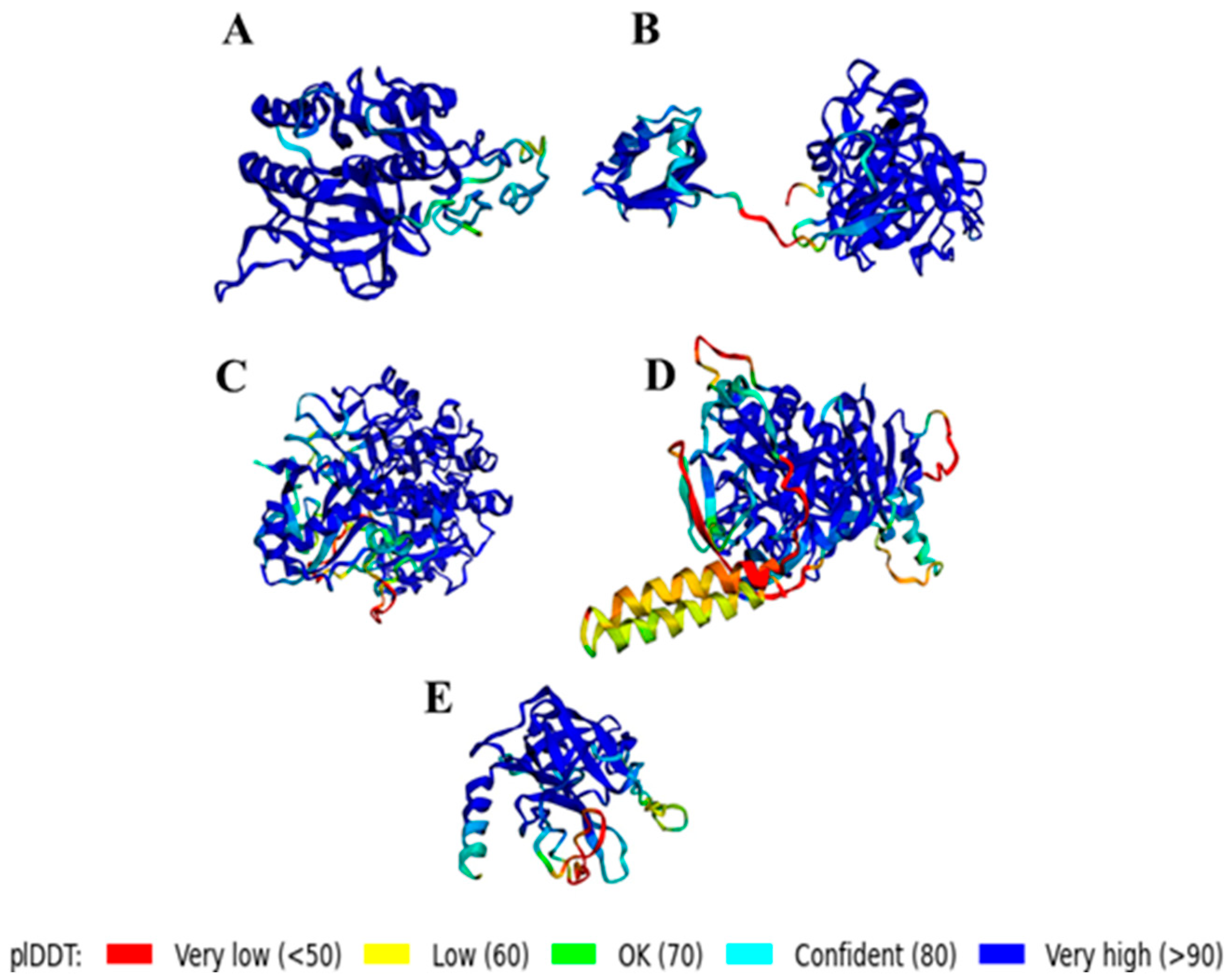
The three-dimensional structure (Figure 2) of B. bassiana serine proteases was modeled using the ColabFold software for protein structure prediction. This tool generates a model confidence score per residue (pLDDT), ranging from 0 to 100. Regions with a score of less than 50 pLDDT may not have a well-defined structure in isolation. Figure 2A–D shows the modeled structures for the sequences with a conserved domain in Family: Peptidase_S8, including Subtilisin-like protease Pr1F, Subtilisin-like protease Pr1A, Alkaline serine protease, and Sublitisin-like protease. Additionally, the structure of the serine protease belonging to the Trypsin Family is shown in Figure 2E.

3.3. In Vitro Ovicidal Effect

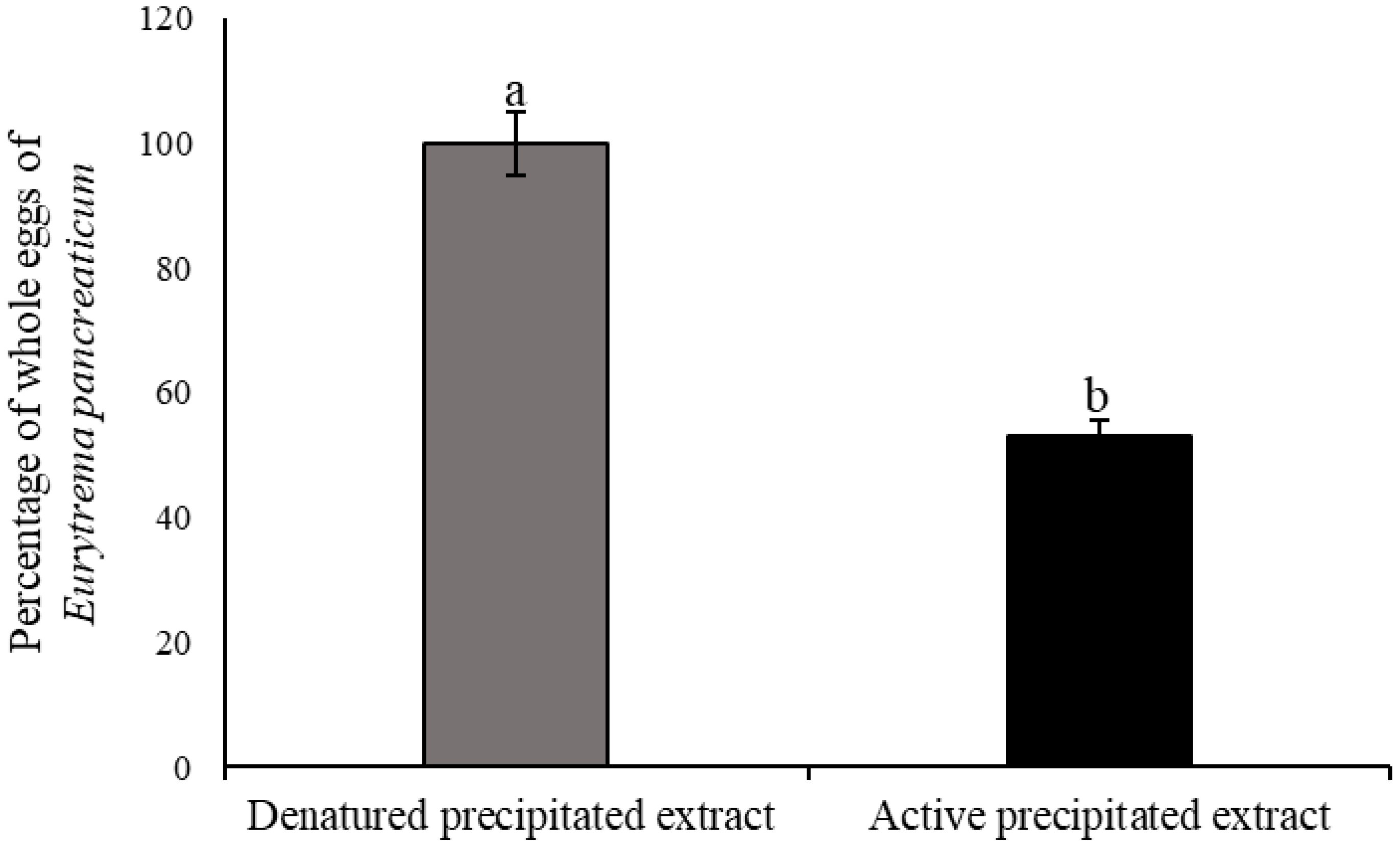
The proteases present in the APE produced by B. bassiana caused a significant reduction (p < 0.01) in the number of intact eggs of E. pancreaticum compared to the DPE group, indicating its ovicidal effect in vitro. The percentage reduction compared to the DPE group was 53% (Figure 3). Figure 4C,D show the digestive action of the proteases on the eggshell and subsequently on the internal contents of the helminth.

4. Discussion

To obtain optimum levels of extracellular enzyme production from B. bassiana, rice culture medium supplemented with whey was used. By containing sugars and nitrogen-containing compounds, this culture medium can increase conidia production, improve persistence, and increase germination under optimum relative humidity conditions. The production of microbial biomass from cheese whey has been considered as an option [16,34].
Several studies have demonstrated the feasibility of enzyme production through solid fermentation, showing promising results. Alves et al. [35] optimized the production of extracellular enzymes from B. bassiana in solid fermentation, obtaining a significant increase in the activity of exocellulase (10%), endocellulase (63%), chitinase (60%), and β-1,3-glucanase (61%). However, the results of this study show the potential of solid fermentation as an effective and sustainable strategy for enzyme production.
Proteolytic activity was enhanced by the precipitation process compared to the cell-free crude extract (p < 0.01). Moreover, no chitinolytic activity was evident in the precipitated extract. This result is based on the role of acetone as an organic solvent, which increased the degree of purification about twelvefold and reduced the activity of the chitinases, in addition to the residual impurities present in the culture medium.
Acetone precipitation stands out as an effective technique for fractionating, concentrating, and purifying proteins in complex biological systems [36]. Studies have shown the selectivity of acetone precipitation, as it favors the precipitation of high molecular weight proteins, which makes it a valuable tool for concentrating specific proteins [37].
The zymogram stood out as a valuable tool for identifying and characterizing the proteases present in the active precipitate extract. In our study, the presence of five bands expressed by proteolytic activity was evidenced by the formation of casein digestion halos in the precipitated extract of B. bassiana isolate IP 361 (Figure 1). The estimated molecular weight of these bands agrees with the molecular weight of subtilisin- and trypsin-type serine proteases previously reported for the B. bassiana isolate ARSEF 2860 in the NCBI database [38]. Monte et al. [39] reported that this technique allows the detection of latent and active forms of enzymes in different samples, such as cells, extracts, tissues, or biological fluids, making it a robust and versatile technique.
Arias-Aravena et al. [40] used the zymogram technique to identify exoenzymes secreted by the entomopathogenic fungus B. pseudobassiana RGM 2184 in different culture media. This technique revealed the presence of three proteases in the supernatant, with estimated molecular weights of approximately 36, 105, and 113 kDa. The authors suggested that these proteases belong to the Pr1 protease complex, similar to subtilisin, and may play a crucial role in the pathogenicity of the fungus.
Correlating with the results of our study, we can suggest the presence of two isoforms of serine proteases in the IP 361 isolate of B. bassiana, the subtilisin-like protease Pr1F and the subtilisin-like protease Pr1A (Figure 1). The subtilisin-like (Pr1) and trypsin-like (Pr2) serine proteases are the most studied proteolytic enzymes of entomopathogenic fungi, with eleven isoforms related to the gene encoding Pr1 in B. bassiana [41,42]. The study by Gao et al. [43] showed that the conserved Pr1 proteases act in the degradation of insect cuticle, with five of them (Pr1C, Pr1G, Pr1A2, Pr1B1, and Pr1B2) individually contributing to 19–29% of the virulence of B. bassiana.
There is a wide variation in the molecular weight of proteases from different strains of B. bassiana, from 32 kDa to 105 kDa. In addition, these proteases have been shown to exhibit proteolytic activities on a wide range of substrates, from collagen to casein and elastin [11,44,45,46].
The effect of inhibitors of serine proteases and metalloproteases on the proteolytic activity of the precipitate of B. bassiana (IP 361) is described. The inhibition by PMSF (100%) is related to serine proteases, and the lack of effect of EDTA (0%) suggests the absence of active metalloproteases in the extract. Studies such as that reported by AlGhanimi et al. [42] mentioned that concentrations of 1 and 5 mM of PMSF completely inhibited the activity of proteases, indicating the presence of serine proteases, where the amino acid residues in the active site are sulfonated by the inhibitor. However, in this same study, EDTA induced activity to 144.13% and 163.75% using 1 and 5 mM, respectively.
Similarly, studies reported that alkaline-type proteases may be present in the strain of Beauveria sp. (MTCC 5184), showing the existence of alkaline proteases [47]. The proteolytic enzymes of the subtilisin S8 family are mostly endopeptidases that are best known for their thermostability and nonspecificity and are particularly active at neutral and slightly alkaline pHs [48].
The different histories of the lineages in the evolution of insect pathogenicity suggest the possibility that Pr1 is fundamental for the degradation of the host cuticle and is possibly more conserved in B. bassiana than in the M. anisopliae complex, in which the Peptidase_S8 family has been diversified through molecular evolution [49]. Likewise, Xiao et al. [38] demonstrated that the genome of B. bassiana has a similar number of proteases to that of M. robertsii.
In this context, Sánchez-Pérez et al. [50] identified that the catalytic activity in the degradation of the cuticle by Pr1 and Pr2 in the integument of insects occurred through microorganisms such as B. bassiana and M. anisopliae. However, the biochemical characterization of Pr2 has not yet been elucidated [51].
The three-dimensional structures of the subtilisin-like protease Pr1F, subtilisin-like protease Pr1A, alkaline serine protease, subtilisin-like protease, and trypsin-related protease from ARSEF 2860 were elucidated and modeled in silico using FASTA protein sequences obtained from the NCBI database. This modeling allowed the predictive generation of the three-dimensional conformation of each protease (Figure 2). Further investigation is necessary to determine nucleotide sequences of the protease and confirm the structural prediction in the present study based on the sequences.
In principle, it is suggested that all serine peptidases could be cataloged as homologous to chymotrypsin. Subtilisin enzymes are proteases that contain the catalytic triad in the order of aspartic acid (Asp39), histidine (His69), and serine (Ser224) residues, which have evolved independently [52,53]. Likewise, Dhawan et al. [15] described that the molecular structure of the subtilisin-like protease Pr1 consists of five cysteines forming two disulfide bridges.
The trypsin-like peptidase Pr2 catalyzes the cleavage of peptide chains mainly on the carboxylic side of the amino acids Arginine and Lysine, and the bonds formed by hydrophobic residues are catalyzed in the cleavage by Pr1 [13] (Figure 2E).
Ramos-Llorca et al. [54] found that the catalytic activity of trypsin family aminopeptidases is dependent on an aspartic acid residue located at the base of the primary substrate binding site. The results of the predictive characterization of the proteases of B. bassiana IP 361 reinforce the importance of subtilisin-like (Pr1) and trypsin-like (Pr2) in the pathogenic process of entomopathogenic fungi. Pr1 acts in the comprehensive catalysis of the insect cuticular protein and is secreted in the initial stages of fungal penetration. Pr2 works as a supplementary protease, collaborating with Pr1 in the catalysis of protein degradation [55,56].
Pinheiro et al. [57] detailed that Eurytrema spp. eggs, in general, have a rich composition of structural proteins such as sclerotin and keratin. In the present study, the proteases of B. bassiana showed ovicidal activity on the eggs (Figure 3) and a percentage reduction in the number of intact eggs compared to the denatured extract (p < 0.01), showing the release of their internal contents (Figure 4). However, the challenge for the future is to better elucidate possible ways of applying the enzymatic precipitate of the B. bassiana fungus with a view to sustainable parasite control [58,59]. Another future possibility is the applicability of these entomopathogenic fungi in the environmental control of Eurytrema, which, in turn, has an arthropod (grasshopper) in its evolutionary cycle [60].
Based on the results obtained, we suggested that further experimental studies need to be carried out with a view to the applicability of B. bassiana proteases in the control of pre-parasitic forms of gastrointestinal helminth parasites.

5. Conclusions

This study represents the first report of the in vitro action of Beauveria bassiana proteases on Eurytrema pancreaticum eggs.
Precipitation significantly concentrated proteolytic activity. However, no chitinase activity was detected. The proteolytic profile of the precipitate revealed at least five enzymes. Moreover, B. bassiana proteases had a destructive action on E. pancreaticum eggs.
The results obtained provide valuable information that paves the way for further research into the in vitro use of cell-free enzyme extracts. These findings could boost the development of new biochemical control sustainable technologies, with a focus on environmental mitigation of neglected diseases.

Author Contributions

Conceptualization, L.B.P.F., F.R.B., T.F.M. and F.E.d.F.S.; methodology and validation, L.B.P.F., A.d.C.A., A.T.d.S., D.C.d.S., N.K.L.S. and L.V.F.; investigation, L.B.P.F., L.V.F.; T.F.M. and F.E.d.F.S.; formal analysis, L.B.P.F. and F.E.d.F.S.; resources, L.V.F., T.F.M. and F.E.d.F.S.; data curation, L.B.P.F. and F.E.d.F.S.; writing—original draft preparation, L.B.P.F. and F.E.d.F.S.; writing—review and editing, L.B.P.F., A.d.C.A., A.T.d.S., D.C.d.S., F.R.B., T.F.M. and F.E.d.F.S., supervision, T.F.M. and F.E.d.F.S., project administration and funding acquisition, F.E.d.F.S. All authors have read and agreed to the published version of the manuscript.

Funding

This research was supported by grants from the Minas Gerais Research Funding Foundation (FAPEMIG) (RED-00161-23).

Institutional Review Board Statement

Not applicable.

Informed Consent Statement

Not applicable.

Data Availability Statement

All data related to this study are available from the corresponding authors and will be provided upon request.

Acknowledgments

The authors thank CAPES (Coordination for the Improvement of Higher Education Personnel), FAPEMIG (Minas Gerais Research Funding Foundation), and FAPES (Fundação de Apoio à Pesquisa do Espírito Santo) for the financial support. The authors also thank the Invertebrate Pathology Laboratory of the Institute of Tropical Pathology and Public Health (IPTSP) of the Federal University of Goiás (kindly provided by Professor Christian Luz).

Conflicts of Interest

The authors declare that they have no conflicts of interest in the publication.

References

  1. Sandoval, B.A.; Hyster, T.K. Emerging strategies for expanding the toolbox of enzymes in biocatalysis. Curr. Opin. Chem. Biol. 2020, 55, 45–51. [Google Scholar] [CrossRef] [PubMed]
  2. Page, M.J.; Di Cera, E. Serine peptidases: Classification, structure and function. Cell. Mol. Life Sci. 2008, 65, 1220–1236. [Google Scholar] [CrossRef] [PubMed]
  3. Rekik, H.; Jaouadi, N.Z.; Gargouri, F.; Bejar, W.; Frikha, F.; Jmal, N.; Jaouadi, B. Production, purification and biochemical characterization of a novel detergent-stable serine alkaline protease from Bacillus safensis strain RH12. Int. J. Biol. Macromol. 2019, 121, 1227–1239. [Google Scholar] [CrossRef]
  4. Naveed, M.; Nadeem, F.; Mehmood, T.; Bilal, M.; Anwar, Z.; Amjad, F. Protease—A versatile and ecofriendly biocatalyst with multi-industrial applications: An updated review. Catal. Lett. 2021, 151, 307–323. [Google Scholar] [CrossRef]
  5. Dhawan, M.; Joshi, N. Enzymatic comparison and mortality of Beauveria bassiana against cabbage caterpillar Pieris brassicae LINN. Braz. J. Microbiol. 2017, 48, 522–529. [Google Scholar] [CrossRef]
  6. Bara, G.T.; Laing, M.D. Entomopathogens: Potential to control thrips in avocado, with special reference to Beauveria bassiana. Hortic. Rev. 2020, 47, 325–368. [Google Scholar] [CrossRef]
  7. dos Reis, C.B.; Sobucki, L.; Mazutti, M.A.; Guedes, J.V. Production of chitinase from Metarhizium anisopliae by solid-state fermentation using sugarcane bagasse as substrate. Ind. Biotechnol. 2018, 14, 230–234. [Google Scholar] [CrossRef]
  8. Rojas-Osnaya, J.; Rocha-Pino, Z.; Nájera, H.; González-Márquez, H.; Shirai, K. Novel transglycosylation activity of β-N-acetylglucosaminidase of Lecanicillium lecanii produced by submerged culture. Int. J. Biol. Macromol. 2020, 145, 759–767. [Google Scholar] [CrossRef] [PubMed]
  9. Liu, Y.; Li, Y.; Tong, S.; Yuan, M.; Wang, X.; Wang, J.; Fan, Y. Expression of a Beauveria bassiana chitosanase (BbCSN-1) in Pichia pastoris and enzymatic analysis of the recombinant protein. Protein Expr. Purif. 2020, 166, 105519. [Google Scholar] [CrossRef]
  10. Grogan, G.J.; Holland, H.L. The biocatalytic reactions of Beauveria spp. J. Mol. Catal. B Enzym. 2000, 9, 1–32. [Google Scholar] [CrossRef]
  11. Amobonye, A.; Bhagwat, P.; Pandey, A.; Singh, S.; Pillai, S. Biotechnological potential of Beauveria bassiana as a source of novel biocatalysts and metabolites. Crit. Rev. Biotechnol. 2020, 40, 1019–1034. [Google Scholar] [CrossRef] [PubMed]
  12. Dar, S.A.; Rather, B.A.; Kandoo, A.A. Insect pest management by entomopathogenic fungi. J. Entomol. Zool. Stud. 2017, 5, 1185–1190. [Google Scholar]
  13. Semenova, T.A.; Dunaevsky, Y.E.; Beljakova, G.A.; Belozersky, M.A. Extracellular peptidases of insect-associated fungi and their possible use in biological control programs and as pathogenicity markers. Fungal Biol. 2020, 124, 65–72. [Google Scholar] [CrossRef] [PubMed]
  14. Negi, R.; Sharma, B.; Kaur, S.; Kaur, T.; Khan, S.S.; Kumar, S.; Yadav, A.N. Microbial antagonists: Diversity, formulation and applications for management of pest–pathogens. Egypt. J. Biol. Pest Control. 2023, 33, 105. [Google Scholar] [CrossRef]
  15. Dhawan, M.; Joshi, N.; Ghuman, S.; Sandhu, S.; Sharma, M. Deciphering the relationships among enzymatic systems and virulence of Beauveria bassiana: A Review. J. Exp. Biol. Agric. Sci. 2020, 8, 730–742. [Google Scholar] [CrossRef]
  16. Figueroa, L.B.P.; Mamani, R.C.C.; de Souza, D.C.; de Souza Alves, J.C.; de Souza, S.A.; Ferreira, C.B.; Soares, F.E.F. Enzyme production by the entomopathogenic fungi Beauveria bassiana and Metarhizium anisopliae and their application in the control of nematodes (Haemonchus spp. and Meloidogyne incognita) in vitro. J. Nat. Pestic. Res. 2024, 8, 100077. [Google Scholar] [CrossRef]
  17. de Sousa, D.E.; de Castro, M.B. Pancreatic eurytrematosis in small ruminants: A forgotten disease or an untold history? Vet. Parasitol. 2022, 311, 109794. [Google Scholar] [CrossRef]
  18. Rachid, M.A.; Aquino, H.M.; Facury-Filho, E.J.; Carvalho, A.U.; Valle, G.R.; Vasconcelos, A.C. Chronic interstitial pancreatitis and chronic wasting disease caused by Eurytrema coelomaticum in Nelore cow. Arq. Bras. Med. Vet. Zootec. 2011, 63, 741–743. [Google Scholar] [CrossRef]
  19. Rojo-Vázquez, F.A.; Meana, A.; Valcárcel, F.; Martínez-Valladares, M. Update on trematode infections in sheep. Vet. Parasitol. 2012, 189, 15–38. [Google Scholar] [CrossRef]
  20. de Sousa, D.E.; Barbosa, E.D.; Wilson, T.M.; Machado, M.; Oliveira, W.J.; Duarte, M.A.; de Castro, M.B. Eurytrema coelomaticum natural infection in small ruminants: A neglected condition. Parasitology 2021, 148, 576–583. [Google Scholar] [CrossRef]
  21. Ilha, M.R.; Loretti, A.P.; Reis, A.C. Wasting and mortality in beef cattle parasitized by Eurytrema coelomaticum in the State of Paraná, southern Brazil. Vet. Parasitol. 2005, 133, 49–60. [Google Scholar] [CrossRef] [PubMed]
  22. Bassani, C.A.; Sangioni, L.A.; Saut, J.P.; Yamamura, M.H.; Headley, S.A. Epidemiology of eurytrematosis (Eurytrema spp. Trematoda: Dicrocoeliidae) in slaughtered beef cattle from the central-west region of the State of Paraná, Brazil. Vet. Parasitol. 2006, 141, 356–361. [Google Scholar] [CrossRef]
  23. Tandon, V.; Shylla, J.A.; Ghatani, S.; Athokpam, V.D.; Sahu, R. Neglected tropical diseases: Trematodiases—The Indian scenario. Proc. Natl. Acad. Sci. India Sect. B Biol. Sci. 2015, 85, 901–907. [Google Scholar] [CrossRef]
  24. Toledo, R.; Cortés, A.; Álvarez-Izquierdo, M.; Muñoz-Antoli, C.; Esteban, J.G. New Insights into the Treatment of Foodborne Trematode Infections. In Neglected Tropical Disease: Drug Discovery and Development; Swinney, D., Pollastri, M., Mannhold, R., Buschmann, H., Holenz, J., Eds.; John Wiley & Sons, Ltd.: Hoboken, NJ, USA, 2019; pp. 289–323. [Google Scholar] [CrossRef]
  25. Doonan, S. Bulk purification by fractional precipitation. In Protein Purification Protocols: Methods in Molecular Biology; Cutler, P., Ed.; Humana Press: New York, NY, USA, 2004; pp. 117–124. [Google Scholar] [CrossRef]
  26. Braga, F.R.; Araújo, J.V.; Soares, F.E.F.; Araujo, J.M.; Geniêr, H.L.A.; Silva, A.R.; Ferreira, S.R. Optimizing protease production from an isolate of the nematophagous fungus Duddingtonia flagrans using response surface methodology and its larvicidal activity on horse cyathostomins. J. helminthol. 2011, 85, 164–170. [Google Scholar] [CrossRef]
  27. Miller, G.L. Use of dinitrosalicylic acid reagent for determination of reducing sugar. J. Anal. Chem. 1959, 31, 426–428. [Google Scholar] [CrossRef]
  28. Bradford, M. A rapid and sensitive method for the quantitation of microgram quantities of protein utilizing the principle of protein-dye binding. Anal. Biochem. 1976, 72, 248–254. [Google Scholar] [CrossRef] [PubMed]
  29. Braga, F.R.; Araújo, J.V.; Soares, F.E.D.; Araujo, J.M.; Ferreira, S.R.; Tavela, A.D.; Queiroz, J.H. Proteolytic action of the crude extract of Duddingtonia flagrans on cyathostomins (Nematoda: Cyathostominae) in coprocultures. Rev. Bras. Parasitol. Vet. 2013, 22, 143–146. [Google Scholar] [CrossRef]
  30. Xiao, Y.Z.; Wu, D.K.; Zhao, S.Y.; Lin, W.M.; Gao, X.Y. Statistical optimization of alkaline protease production from Penicillium citrinum YL-1 under solid-state fermentation. Prep. Biochem. Biotechnol. 2015, 45, 447–462. [Google Scholar] [CrossRef]
  31. Dennis, W.R.; Stone, W.M.; Swanson, L.E. A new laboratory and field diagnostic test for fluke ova in feces. J. Am. Vet. Med. Assoc. 1954, 124, 47–50. [Google Scholar] [PubMed]
  32. Ayres, M.; Ayres Júnior, M.; Ayres, D.L.; Santos, A.A. Bioestat—Aplicações Estatísticas nas Áreas das Ciências Bio-Médicas; Ong Mamirauá: Belém, Brazil, 2007. [Google Scholar]
  33. Mendoza-De Gives, P.; Vazquez-Prats, V.M. Reduction of Haemonchus contortus infective larvae by three nematophagous fungi in sheep faecal cultures. Vet. Parasitol. 1994, 55, 197–203. [Google Scholar] [CrossRef]
  34. Kassa, A.; Brownbridge, M.; Parker, B.L.; Skinner, M.; Gouli, V.; Gouli, S.; Hata, T. Whey for mass production of Beauveria bassiana and Metarhizium anisopliae. Mycol. Res. 2008, 112, 583–591. [Google Scholar] [CrossRef] [PubMed]
  35. Alves, E.A.; Schmaltz, S.; Tres, M.V.; Zabot, G.L.; Kuhn, R.C.; Mazutti, M.A. Process development to obtain a cocktail containing cell-wall degrading enzymes with insecticidal activity from Beauveria bassiana. Biochem. Eng. J. 2020, 156, 107484. [Google Scholar] [CrossRef]
  36. Bryjak, J.; Rekuć, A. Effective purification of Cerrena unicolor laccase using microfiltration, ultrafiltration and acetone precipitation. Appl. Biochem. Biotechnol. 2010, 160, 2219–2235. [Google Scholar] [CrossRef]
  37. Baghalabadi, V.; Doucette, A.A. Mass spectrometry profiling of low molecular weight proteins and peptides isolated by acetone precipitation. Anal. Chim. Acta. 2020, 1138, 38–48. [Google Scholar] [CrossRef]
  38. Xiao, G.; Ying, S.H.; Zheng, P.; Wang, Z.L.; Zhang, S.; Xie, X.Q.; Shang, Y.; St Leger, R.J.; Zhao, G.P.; Wang, C.; et al. Genomic perspectives on the evolution of fungal entomopathogenicity in Beauveria bassiana. Sci. Rep. 2012, 2, 483. [Google Scholar] [CrossRef]
  39. Monte, J.F.; Moreno, C.J.; Monteiro, J.P.; de Oliveira Rocha, H.A.; Ribeiro, A.R.; Silva, M.S. Use of Zymography in Trypanosomiasis Studies, In Zymography. Methods in Molecular Biology; Wilkesman, J., Kurz, L., Eds.; Humana Press: New York, NY, USA, 2017; pp. 213–220. [Google Scholar] [CrossRef]
  40. Arias-Aravena, M.; Altimira, F.; Gutiérrez, D.; Ling, J.; Tapia, E. Identification of exoenzymes secreted by entomopathogenic fungus Beauveria pseudobassiana RGM 2184 and their effect on the degradation of cocoons and pupae of quarantine pest Lobesia botrana. J. Fungi. 2022, 8, 1083. [Google Scholar] [CrossRef] [PubMed]
  41. Vikhe, A.G.; Dale, N.S.; Umbarkar, R.B.; Labade, G.B.; Savant, A.R.; Walunj, A.A. Isolation and Purification of Cuticle Degrading Extra Cellular Proteases from Entomopathogenic Fungal Species of Beauveria bassiana. Int. J. Pure Appl. Biosci. 2016, 4, 130–135. [Google Scholar] [CrossRef]
  42. Alghanimi, A.A.; Alebadi, S.M.; Al-ethari, A.Y. Partial purification and characterization of protease from local isolate of Beauveria bassiana. Sci. J. Med. Res. 2020, 4, 17–22. [Google Scholar]
  43. Gao, B.J.; Mou, Y.N.; Tong, S.M.; Ying, S.H.; Feng, M.G. Subtilisin-like Pr1 proteases marking the evolution of pathogenicity in a wide-spectrum insect-pathogenic fungus. Virulence 2020, 11, 365–380. [Google Scholar] [CrossRef]
  44. Urtz, B.E.; Rice, W.C. Purification and characterization of a novel extracellular protease from Beauveria bassiana. Mycol. Res. 2000, 104, 180–186. [Google Scholar] [CrossRef]
  45. Firouzbakht, H.; Zibaee, A.; Hoda, H.; Sohani, M.M. Purification and characterization of the cuticle-degrading proteases produced by an isolate of Beauveria bassiana using the cuticle of the predatory bug, Andrallus spinidens Fabricius (Hemiptera: Pentatomidae). J. Plant Prot. Res. 2015, 55, 179–186. [Google Scholar] [CrossRef]
  46. Borgi, I.; Dupuy, J.W.; Blibech, I.; Lapaillerie, D.; Lomenech, A.M.; Rebai, A.; Gargouri, A. Hyper-proteolytic mutant of Beauveria bassiana, a new biological control agent against the tomato borer. Agron. Sustain. Dev. 2016, 36, 60. [Google Scholar] [CrossRef]
  47. Shankar, S.; Laxman, R.S. Biophysicochemical characterization of an alkaline protease from Beauveria sp. MTCC 5184 with multiple applications. Appl. Biochem. Biotechnol. 2015, 175, 589–602. [Google Scholar] [CrossRef] [PubMed]
  48. Rawlings, N.D.; Barrett, A.J.; Thomas, P.D.; Huang, X.; Bateman, A.; Finn, R.D. The MEROPS database of proteolytic enzymes, their substrates and inhibitors in 2017 and a comparison with peptidases in the PANTHER database. Nucleic Acids Res. 2017, 46, 624–632. [Google Scholar] [CrossRef]
  49. Andreis, F.C.; Schrank, A.; Thompson, C.E. Molecular evolution of Pr1 proteases depicts ongoing diversification in Metarhizium spp. Mol. Genet. Genom. 2019, 294, 901–917. [Google Scholar] [CrossRef]
  50. Sánchez-Pérez, L.D.C.; Barranco-Florido, J.E.; Rodríguez-Navarro, S.; Cervantes-Mayagoitia, J.F.; Ramos-López, M.Á. Enzymes of entomopathogenic fungi, advances and insights. Adv. Enzym. Res. 2014, 2, 65–76. [Google Scholar] [CrossRef]
  51. Shin, T.Y.; Lee, M.R.; Park, S.E.; Lee, S.J.; Kim, W.J.; Kim, J.S. Pathogenesis-related genes of entomopathogenic fungi. Arch. Insect Biochem. Physiol. 2020, 105, e21747. [Google Scholar] [CrossRef]
  52. Gilliland, G.L.; Teplyakov, A. Structural calcium (trypsin, subtilisin). In Encyclopedia of Inorganic and Bioinorganic Chemistry; Scott, R.A., Ed.; John Wiley & Sons, Ltd.: Hoboken, NJ, USA, 2011; pp. 1–14. [Google Scholar] [CrossRef]
  53. Azrin, N.A.; Ali, M.S.; Rahman, R.N.; Oslan, S.N.; Noor, N.D. Versatility of subtilisin: A review on structure, characteristics, and applications. Biotechnol. Appl. Biochem. 2022, 69, 2599–2616. [Google Scholar] [CrossRef]
  54. Ramos-Llorca, A.; Decraecker, L.; Cacheux, V.M.; Zeiburlina, I.; De Bruyn, M.; Battut, L.; Augustyns, K. Chemically diverse activity-based probes with unexpected inhibitory mechanisms targeting trypsin-like serine proteases. Front. Chem. 2023, 10, 1089959. [Google Scholar] [CrossRef]
  55. Dias, B.A.; Neves, P.M.; Furlaneto-Maia, L.; Furlaneto, M.C. Cuticale degrading proteases produced by the entomopathogenic fungus Beauveria bassiana in the presence of coffee berry borer cuticle. Braz. J. Microbiol. 2008, 39, 301–306. [Google Scholar] [CrossRef]
  56. Pour, S.A.; Zibaee, A.; Rostami, M.G.; Hoda, H.; Shahriari, M. Mortality and immune challenge of a native isolate of Beauveria bassina against the larvae of Glyphodes pyloalis Walker (Lepidoptera: Pyralidae). Egypt. J. Biol. Pest Control 2021, 31, 37. [Google Scholar] [CrossRef]
  57. Pinheiro, J.; Franco-Acuña, D.O.; Oliveira-Menezes, A.; Brandolini, S.V.; Adnet, F.A.; Torres, E.L.; Damatta, R.A. Additional study of the morphology of eggs and miracidia of Eurytrema coelomaticum (Trematoda). Helminthologia 2015, 52, 244–251. [Google Scholar] [CrossRef]
  58. Fissiha, W.; Kinde, M.Z. Anthelmintic resistance and its mechanism: A review. Infect. Drug Resist. 2021, 14, 5403–5410. [Google Scholar] [CrossRef]
  59. Charlier, J.; Bartley, D.J.; Sotiraki, S.; Martinez-Valladares, M.; Claerebout, E.; von Samson-Himmelstjerna, G.; Rinaldi, L. Anthelmintic resistance in ruminants: Challenges and solutions. Adv. Parasitol. 2022, 115, 171–227. [Google Scholar] [CrossRef] [PubMed]
  60. Jubb, K.V. The pancreas. In Pathology of Domestic Animals; Jubb, K.V., Kennedy, P.C., Palmer, N., Eds.; Academic Press: San Diego, CA, USA, 1993; pp. 407–424. [Google Scholar] [CrossRef]
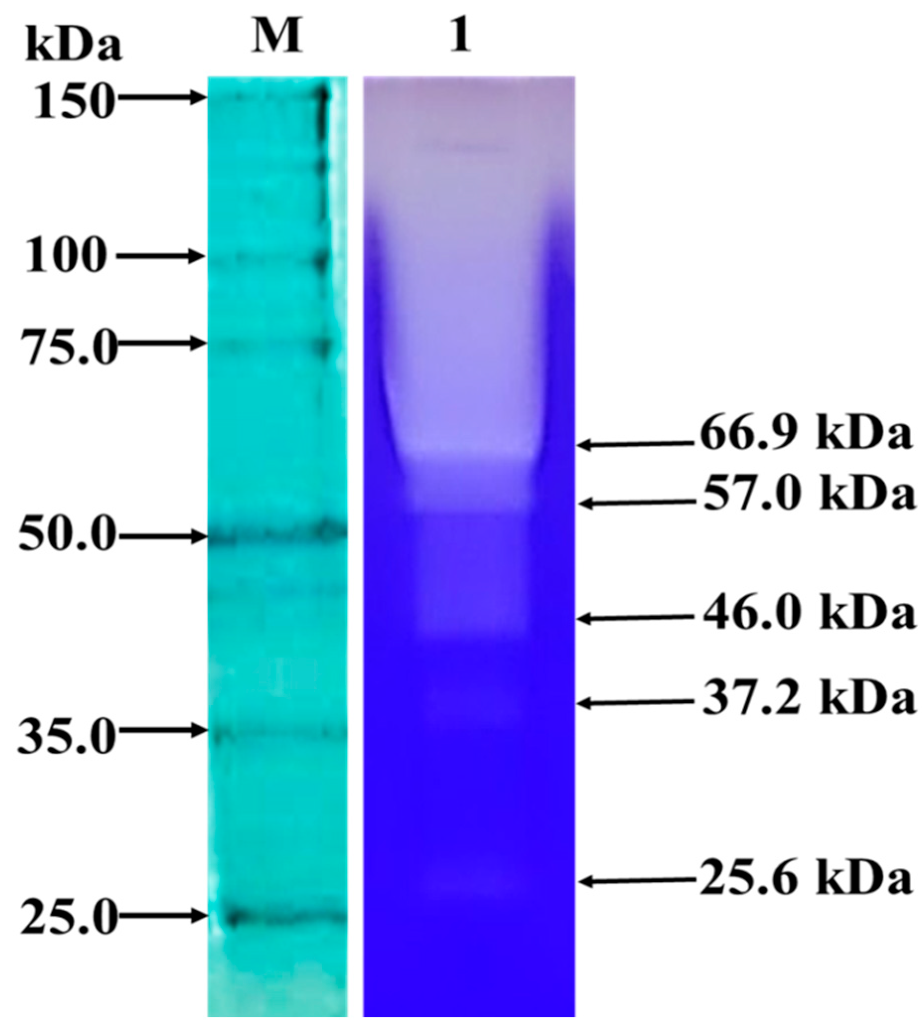
Figure 1. Zymogram of the active precipitated extract produced by Beauveria bassiana IP 361 s.l., in rice culture medium with whey. Lane M. Protein molecular mass marker (Thermo Scientific). Lane 1. Active precipitated extract. Clear bands, indicated by black arrows, showing protease activity on a 10% native PAGE gel containing 1% (w/v) casein.
Microbiolres 16 00127 g001
Figure 2. Three-dimensional (3D) structures of Beauveria bassiana proteases by ColabFold modeling tool, using the model confidence score per residue (pLDDT). (A) Subtilisin-like protease Pr1F. (B) Subtilisin-like protease Pr1A. (C) Alkaline serine protease. (D) Sublitisin-like protease. (E) Trypsin-related protease.
Microbiolres 16 00127 g002
Figure 3. Evaluation of the ovicidal effect of Beauveria bassiana proteases on Eurytrema pancreaticum, after 24 h of incubation at 28 ± 1 °C in the dark. Different letters indicate that the control group with denatured extract and the group treated with the active extract showed a significant difference (p < 0.01).
Microbiolres 16 00127 g003
Figure 4. In vitro ovicidal effect of Beauveria bassiana proteases on Eurytrema pancreaticum eggs. (A) Denatured precipitated extract, (BD) Active precipitated extract. The black arrows indicate the digestion of the shell and internal contents due to the action of the proteases.
Microbiolres 16 00127 g004
Disclaimer/Publisher’s Note: The statements, opinions and data contained in all publications are solely those of the individual author(s) and contributor(s) and not of MDPI and/or the editor(s). MDPI and/or the editor(s) disclaim responsibility for any injury to people or property resulting from any ideas, methods, instructions or products referred to in the content.