Article

Retrospective Study 2019–2021 of Antimicrobial Resistance in Escherichia coli, Klebsiella pneumoniae, and Proteus mirabilis in Mexicali, Mexico

by
Dolores A. Márquez-Salazar
1,
Ricardo Delgadillo-Valles
2,
Gerson N. Hernández-Acevedo
2,
Edwin Barrios-Villa
3,
Raquel Muñiz-Salazar
4,
Gilberto López-Valencia
5,
Rafael Martínez-Miranda
6 and
Jonathan Arauz-Cabrera
1,*
1
Departamento de Farmacología, Facultad de Medicina Mexicali, Universidad Autónoma de Baja California, Humberto Torres Sanginés SN, Centro Cívico, Mexicali C.P. 21000, BC, Mexico
2
Departamento de Microbiología y Parasitología Clínica, Facultad de Medicina Mexicali, Universidad Autónoma de Baja California, Humberto Torres Sanginés SN, Centro Cívico, Mexicali C.P. 21000, BC, Mexico
3
Laboratorio de Biología Molecular y Genómica, Departamento de Ciencias Químico Biológicas y Agropecuarias, Universidad de Sonora, Campus Caborca, Av. Universidad e Irigoyen S/N, H. Caborca C.P. 83621, SO, Mexico
4
Laboratorio de Epidemiología y Ecología Molecular, Escuela de Ciencias de la Salud, Unidad Valle Dorado, Campus Ensenada, Universidad Autónoma de Baja California, Blvd. Zertuche y Blvd. de los Lagos s/n Fracc. Valle Dorado, Ensenada C.P. 22890, BC, Mexico
5
Instituto de Investigaciones en Ciencias Veterinarias, Universidad Autónoma de Baja California, Carr. San Felipe km 3.5, Fracc, Campestre, Mexicali C.P. 21386, BC, Mexico
6
Departamento de Microbiología, Hospital Almater, Avenida Francisco I. Madero 1060, Colonia Nueva, Mexicali C.P. 21000, BC, Mexico
*
Author to whom correspondence should be addressed.
Microbiol. Res. 2025, 16(6), 126; https://doi.org/10.3390/microbiolres16060126
Submission received: 28 April 2025 / Revised: 5 June 2025 / Accepted: 6 June 2025 / Published: 11 June 2025

Abstract

:
Community-acquired infections caused by Enterobacterales are a growing public health concern, particularly in border regions where patient mobility may influence resistance patterns. Antimicrobial resistance (AMR) surveillance is critical for establishing local treatment guidelines. The aim of this study was to investigate AMR rates in clinical isolates of Escherichia coli, Klebsiella pneumoniae, and Proteus mirabilis obtained from community-acquired infections in Mexicali between 2019 and 2021. A retrospective study was conducted, analyzing 2871 Enterobacterales isolates (E. coli, K. pneumoniae, P. mirabilis). Species identification and antimicrobial susceptibility testing were performed using MALDI-TOF and VITEK 2 systems, interpreted according to CLSI and EUCAST breakpoints. ESBL production was detected in 37.6% of E. coli, 27.7% of K. pneumoniae, and none of the P. mirabilis isolates. Among ESBL producers, ciprofloxacin resistance reached 90.4% in E. coli and 81.0% in K. pneumoniae, indicating a significant level of co-resistance. Carbapenem-resistant K. pneumoniae (n = 13) and one E. coli isolate were also identified, all from community-acquired infections. Resistance patterns varied by infection site, with UTIs accounting for the majority of isolates. The high rates of ESBLs and co-resistance to ciprofloxacin among Enterobacterales highlight the urgent need for targeted AMR surveillance and site-specific empirical treatment strategies.

1. Introduction

The rise of antimicrobial resistance (AMR) represents a critical global public health concern. Although AMR is a natural evolutionary process by which microorganisms develop resistance to antimicrobial agents, its acceleration has been largely driven by the widespread misuse and overuse of antibiotics in humans, animals, and agricultural practices [1]. AMR complicates the management of infectious diseases, increases the risk of surgical complications, and poses serious threats to immunosuppressed individuals. According to the World Health Organization (WHO), AMR may result in 10 million deaths annually by 2050 [2], potentially surpassing cancer and diabetes as the leading causes of mortality [3]. Of the estimated 7.7 million annual deaths associated with bacterial infections, 4.95 million are linked to drug-resistant pathogens, with 1.25 million deaths directly attributed to bacteria resistant to available antibiotics [1]. The COVID-19 pandemic may have further exacerbated AMR [4,5], as broad-spectrum antibiotics were often prescribed for patients suspected of having bacterial co-infections, despite the viral etiology of SARS-CoV-2 [6,7].
Among the most concerning AMR-related pathogens are the members of the order Enterobacterales, particularly including Escherichia coli, Klebsiella pneumoniae, and Proteus mirabilis. These gram-negative bacteria, typically gastrointestinal commensals, can cause intra-abdominal infections, bloodstream infections, and urinary tract infections (UTIs) [8,9,10]. Notably, they are among the leading contributors to AMR-related mortality worldwide [11]. These pathogens can acquire resistance through chromosomal mutations, horizontal gene transfer, or plasmid-mediated mechanisms [12]. A particularly alarming mechanism involves the production of extended-spectrum beta-lactamases (ESBLs), enzymes that confer resistance to third- and fourth-generation cephalosporins and penicillins [2], thereby complicating the selection of empirical treatment [13].
In response to this growing challenge, global surveillance initiatives have been established to monitor and curb the spread of AMR, supporting the development of evidence-based antimicrobial stewardship policies. The WHO’s “One Health” approach encourages interdisciplinary collaboration and coordinated actions at local, national, and international levels. As part of its Global Action Plan, the WHO launched the Global Antimicrobial Resistance and Use Surveillance System (GLASS) in 2015. In alignment with this initiative, Mexico launched the “National Strategy against Antimicrobial Resistance” in 2018 [3]. Nevertheless, despite these efforts, Mexico still lacks an official AMR surveillance network and has yet to formally participate in the GLASS program [14].
Most AMR studies in Mexico have focused on hospital environments [15,16,17,18] with few addressing community settings [15,18,19,20]. Moreover, the northwestern border region, despite being a focal point for medical tourism, remains underrepresented in current AMR data. Cities such as Mexicali are popular destinations for international patients seeking affordable and high-quality healthcare services. However, increased cross-border patient mobility also heightens the risk of acquiring and disseminating resistant pathogens. As such, targeted AMR surveillance in this region is crucial for detecting trends and informing public health interventions.
In Mexico, UTIs rank as the third most common cause of morbidity, following respiratory and gastrointestinal infections [21], and are associated with increased hospital stays and mortality. Given the distinct dynamics of the border population and the scarcity of updated regional AMR data, this study aimed to describe resistance patterns in E. coli, K. pneumoniae, and P. mirabilis from community-acquired infections in Mexicali between 2019 and 2021. The findings are intended to inform regional AMR monitoring strategies and support data-driven public health planning.

2. Materials and Methods

2.1. Study Design and Data Collection

This retrospective study was conducted in Mexicali, Mexico (32°37′40.1″ N 115°27′16.1″ W), using data collected between January 2019 and December 2021. A total of 2871 non-duplicate bacterial isolates were obtained from community-acquired infections and provided by a clinical diagnostic laboratory. The dataset included 2234 E. coli, 433 K. pneumoniae, and 204 P. mirabilis isolates. Data were curated to include only isolates recovered from urinary tract and lower respiratory tract infections with ≥100,000 colony-forming units (CFUs), as well as isolates from blood and other body fluids. Exclusion criteria comprised isolates from vaginal swabs, fecal samples, environmental or non-clinical sources, and duplicated entries from the same patient or infection episode.

2.2. Bacterial Identification and Antimicrobial Susceptibility Testing

Species identification was performed from isolated colonies cultured on MacConkey Agar (MCD Lab, Tultitlán de Mariano Escobedo, Mexico) from each sample. Identification was carried out using matrix-assisted laser desorption/ionization time-of-flight mass spectrometry (MALDI-TOF) (Bruker Daltonics, Bremen, Germany), with the ethanol–formic acid protein extraction method [22].
Antimicrobial susceptibility testing (AST) was conducted using the Vitek 2 Compact System (BioMerieux, Marcy-l’Étoile, France) employing the broth microdilution method to determine minimum inhibitory concentrations (MICs). The AST procedure utilized the reagent cards AST-N71, AST-N72, and AST-X05 (BioMerieux, France). Detection of ESBL production was also performed using the Vitek 2 Compact System (BioMerieux, France), following the Clinical and Laboratory Standards Institute (CLSI) guidelines [23].
MIC results were recorded in a Microsoft Excel spreadsheet and interpreted according to the CLSI breakpoints [23] for the following agents: ampicillin (AMP), amikacin (AMK), aztreonam (AZT), chloramphenicol (CL), ceftazidime (CAZ), cefixime (CFM), ciprofloxacin (CIP), cefpodoxime (CPD), ceftriaxone (CRO), ceftolozane/tazobactam (CT), cefotetan (CTT), cefotaxime (CTX), cefuroxime (CXM), cefuroxime axetil (CXMA), doripenem (DOR), ertapenem (ETP), cefepime (FEP), fosfomycin (FOS), cefoxitin (FOX), nitrofurantoin (FT), gentamicin (GM), imipenem (IPM), levofloxacin (LEV), meropenem (MEM), minocycline (MNO), nalidixic acid (NA), norfloxacin (NOR), piperacillin (PIP), ampicillin/sulbactam (SAM), trimethoprim/sulfamethoxazole (STX), ticarcillin/clavulanic acid (TCC), tetracycline (TE), trimethoprim (TMP), and piperacillin/tazobactam (TZP). For colistin (CS), moxifloxacin (MXF), and tigecycline (TGC), MICs were interpreted using the European Committee on Antimicrobial Susceptibility Testing (EUCAST) guidelines [24] as CLSI breakpoints were not available for these agents at the time of analysis.

2.3. Statistical Analysis

The frequency of ESBL-producing isolates was determined by year and by infection site. Antimicrobial resistance rates were expressed as percentages. Statistical differences in ESBL prevalence over the study period were assessed using the chi-square test. Statistical significance was defined as p < 0.05. All statistical analyses were performed using Statistix 9.0 software (Analytical Software, Tallahassee, FL, USA).

3. Results

A total of 2871 isolates were collected from community-acquired infections between 2019 and 2021, comprising 2234 E. coli, 433 K. pneumoniae, and 204 P. mirabilis isolates. Among these, 960 (33.4%) isolates were identified as ESBL producers. Specifically, ESBL production was detected in 840 (37.6%) E. coli isolates and 120 (27.7%) K. pneumoniae isolates, whereas none of the P. mirabilis isolates exhibited ESBL production (Table 1). Among ESBL-positive isolates, 90.4% of the E. coli and 81.0% of K. pneumoniae also showed resistance to CIP.
An increase in the total number of isolates was observed throughout the study period. In 2019, 671 E. coli, 87 K. pneumoniae, and 54 P. mirabilis were collected. This number rose to 642 E. coli, 120 K. pneumoniae, and 72 P. mirabilis in 2020, and further increased in 2021, with 921 E. coli, 226 K. pneumoniae, and 78 P. mirabilis isolates.

3.1. Antimicrobial Resistance Profile by Infection Site

  • Urinary Tract Infections (UTIs)
UTIs accounted for the majority of collected isolates (2148/2871). Resistance rates to commonly prescribed antimicrobials for UTIs varied by species. E. coli exhibited the highest resistance to CIP (55.5%), followed by P. mirabilis (26.9%) and K. pneumoniae (14.2%) (Figure 1a). A similar pattern was observed for CRO, with E. coli showing the highest resistance (38.9%), followed by K. pneumoniae (29.7%) and P. mirabilis (7.4%). Despite the observed trends, no statistically significant year-to-year differences in antimicrobial resistance rates were found between 2019 and 2021 (p > 0.05). Among ESBL-producing strains isolated from UTIs, CIP resistance was observed in 90.6% of E. coli and 88.1% of K. pneumoniae.
  • Lower Respiratory Tract Infections (LRTIs)
K. pneumoniae was the most frequent species isolated from lower respiratory tract samples (n = 164), followed by E. coli (n = 114) and P. mirabilis (n = 8). Among LRTI isolates, ESBL production was more common in E. coli (65.8%) compared to K. pneumoniae (29.3%). E. coli isolates showed resistance rates exceeding 60% to CRO, LEV, and CXM, while K. pneumoniae exhibited rates of 30.4%, 28.4%, and 29.6%, respectively (Figure 1b). Three of the eight P. mirabilis isolates (37.5%) were resistant to LEV. Among ESBL-producing LRTI isolates, CIP resistance was observed in 90.5% of E. coli and 77.1% of K. pneumoniae.
  • Soft Tissue Infections
Soft tissue infections were the second most frequent source of P. mirabilis isolates (24/204) and E. coli (123/2234) isolates, and the third most frequent for K. pneumoniae isolates (40/433). All three microorganisms exhibited resistance to CRO and LEV at different rates (Figure 1c). Among ESBL producers, CIP resistance was detected in 87.7% of E. coli and 71.4% of K. pneumoniae isolates from soft tissue infections.
  • Bloodstream Infections
A total of 46 E. coli isolates (2.1%), 17 K. pneumoniae isolates (3.9%), and one P. mirabilis isolate were recovered from blood samples. Resistance to quinolones (CIP) was observed in all three pathogens at different rates. (Figure 1d). Among ESBL-producing blood isolates, CIP resistance was observed in 86.7% of E. coli and 100% of K. pneumoniae isolates. Complete resistance profiles for these isolates are provided in the Supplementary Materials.
Table 1 shows that ESBL-producing isolates were most frequently detected in E. coli from lower respiratory tract (65.8%) and soft tissue infections (55.3%), while K. pneumoniae showed the highest ESBL proportions in soft tissue (35.0%) and “other” sources (28.0%). Bloodstream and sterile body fluid isolates also carried ESBLs in a non-negligible proportion, particularly in E. coli (34.0% and 47.7%, respectively). No P. mirabilis isolates exhibited ESBL production in any site or year. Importantly, the annual increase in both total isolates and ESBL-positive cases was consistent across nearly all sample types, with the most pronounced rise observed in 2021.

3.2. Carbapenem Resistance

Carbapenem resistance detected in one E. coli isolate was identified from a UTI case in 2021. In contrast, 13 carbapenem-resistant K. pneumoniae isolates were identified: seven from UTIs, three from LRTIs, two from soft tissue infections, and one from a pharyngeal swab. Chronologically, two isolates were isolated in 2019, three in 2020, and eight in 2021.
Among the carbapenem-resistant K. pneumoniae isolates, two (one from a UTI and one from an LRTI) were resistant to all four carbapenems tested (MEM, ETP, IPM, and DOR). The remaining isolates exhibited resistance to two of the four analyzed carbapenems. Notably, four of the carbapenem-resistant isolates were also classified as ESBL producers. None of the P. mirabilis isolates exhibited resistance to carbapenems.

4. Discussion

Enterobacterales are among the most common pathogens implicated in both community- and healthcare-associated infections worldwide [16]. Their resistance patterns often vary depending on the infection site [3]. These bacteria are mostly associated with UTIs [25]. In line with global reports, UTIs were the predominant infection type in our study. Resistance patterns differed across species and infection sites, underscoring the importance of developing and implementing up-to-date, site-specific treatment guidelines and pathogen-targeted therapeutic strategies.
Among the isolates analyzed, E. coli exhibited the highest resistance to CIP, with an overall rate of 55.5%, and a similar rate observed in UTIs (52.6%). These results align with national reports indicating that CIP resistance in E. coli surpasses 50% in UTIs [3,9,16]. Additionally, over 90% of ESBL-producing E. coli and more than 80% of ESBL-producing K. pneumoniae isolates were resistant to CIP across all infection sites, reflecting a high prevalence of co-resistance. Given that CIP is often employed for treating uncomplicated UTIs caused by multidrug-resistant (MDR) organisms [13], these co-resistance patterns may significantly limit its clinical efficacy. A relevant shortcoming of this study is the inability to distinguish between complicated and uncomplicated UTIs, due to the lack of accompanying clinical information. This distinction is essential for guiding appropriated antimicrobial therapy and should be addressed in future studies that integrate microbiological findings with clinical data.
Although K. pneumoniae and P. mirabilis showed lower resistance to CIP compared to E. coli, rates were still significant, particularly for P. mirabilis (26.9%). Notably, K. pneumoniae was almost equally represented in urinary (n = 181) and lower respiratory tract infections (n = 164) and was the predominant species isolated from the latter. In these respiratory specimens, 29.3% of isolates were identified as ESBL producers, consistent with previous findings that associate K. pneumoniae with pneumonia [26].
While K. pneumoniae has not traditionally been a leading cause of community-acquired pneumonia, it can colonize the nasopharynx [8]. Rising resistance to first-line agents such as beta-lactams and fluoroquinolones complicates empirical treatment. Current clinical guidelines from the Infectious Diseases Society of America (IDSA) and Mexico’s National Center for Technological Excellence in Health (CENETEC) recommend initial therapy with a beta-lactam combined with a fluoroquinolone [27,28]. Our findings suggest that antimicrobial susceptibility profiles should be carefully reviewed before initiating empirical therapy in suspected K. pneumoniae pneumonia, especially in light of high fluoroquinolone resistance among ESBL-producing isolates from LRTIs.
High resistance to CRO, a third-generation cephalosporin, was also observed: 38.9% in E. coli and 29.7% in K. pneumoniae. These findings are consistent with previous studies from northern Mexico reporting resistance rates up to 98% in E. coli [16]. ESBL production likely accounts for a significant portion of this resistance, as ESBL prevalence paralleled CRO resistance in both species.
Conversely P. mirabilis showed a lower CRO resistance rate (7.4%), possibly due to different mechanisms. Although P. mirabilis isolates in this study did not exhibit ESBL production or carbapenem resistance, emerging evidence indicates that species can employ alternative resistance mechanisms that may escape conventional phenotypic detection. For example, exposure to amikacin and bacteriophages has been shown to alter outer membrane protein (OMP) profiles in P. mirabilis biofilms, affecting proteins related to membrane permeability and structural remodeling, which may enhance AMR and phage evasion [2]. Moreover, resistance to carbapenems in P. mirabilis has been linked to gene amplification events involving blaVIM-1, driven by insertion sequence IS26, which increase gene dosage and carbapenemase activity [29]. This suggests that even in the absence of traditional resistance phenotypes, P. mirabilis can exhibit clinically significant resistance through complex and often overlooked molecular mechanisms.
Furthermore, the presence of AmpC-type beta-lactamases may confer resistance to certain beta-lactam antibiotics. Additionally, resistance to quinolones exhibited by these microorganisms may involve chromosomal mutations affecting DNA gyrase, as well as overexpression of efflux pumps [12]. These mechanisms may also coexist with ESBLs in some strains, complicating treatment decisions [13].
Fosfomycin (FOS) retained high in vitro activity against E. coli (resistance: 3.9%) and P. mirabilis (resistance: 9.6%), suggesting FOS could remain as a viable option for uncomplicated cystitis [13]. Nitrofurantoin (FT) is another treatment option for cystitis caused by E. coli (resistance: 6.5%). FT resistance was high in P. mirabilis, consistent with its intrinsic resistance [30]. Similar intrinsic patterns were observed for TGC, further limiting treatment options for P. mirabilis infections. The susceptibility rates for K. pneumoniae to FOS (18.6%) and FT (26.6%) suggest that these agents may still be viable in select cases of cystitis. Similar intrinsic resistance patterns were observed with TGC, further limiting treatment options for P. mirabilis infections.
Carbapenem-resistant Enterobacterales (CREs) were also identified in this study. Thirteen K. pneumoniae and one E. coli exhibited resistance to carbapenems, raising concern as these antibiotics are considered last-resort treatments for MDR infections [13]. Although CREs are usually associated with hospital-acquired infections [31], our data confirm their presence in community-acquired infections in Mexicali. Prior local reports have also documented carbapenem-resistant Pseudomonas aeruginosa [32] and K. pneumoniae [33].
The number of carbapenem-resistant K. pneumoniae isolates increased over the study period (two in 2019, three in 2020, and eight in 2021). Alongside this trend, we also observed a gradual increase in the total number of isolates, suggesting a post-COVID-19 pandemic acceleration in resistance, potentially driven by increased antibiotic use [4]. Although no CREs were recovered from blood in this study, three were isolated from respiratory samples, with one detected before and two after the onset of the pandemic. These findings suggest a possible shift in resistance patterns post-pandemic. In Mexico, carbapenems are restricted to hospital settings, to prevent the spread of resistant strains. Nonetheless, the detection of CREs in community-acquired infections highlights the need for comprehensive surveillance. Furthermore, four CRE isolates were also ESBL producers, suggesting the coexistence of multiple resistance mechanisms. The frequent coexistence of ESBLs and fluoroquinolone resistance, as well as carbapenem resistance in some cases, underscores the increasing complexity of managing community-acquired infections in a highly mobile region like the United States–Mexico border.
In addition to UTIs and respiratory infections, Enterobacterales can cause infections in other body sites, mainly in immunosuppressed patients or individuals with comorbidities [34]. Although such patient-level data were unavailable in this study, E. coli remained the predominant gram-negative organisms in blood and other sterile fluids isolates, in line with prior reports [34]. Despite the overall large sample size, the number of isolates from blood (n = 64) and sterile fluids (n = 71) was limited, reducing the generalizability of findings for these infection types. Nevertheless, these proportions reflect the actual distribution of community-acquired infections across body sites. Increased sampling would help refine site-specific therapeutic recommendations.
This study represents one of the few investigations centered on community-acquired infections in northwestern Mexico, a region with significant patient mobility due to medical tourism. However, several limitations must be acknowledged. First, demographic and clinical data (such as patient age, sex, and comorbidities) were not available, which limits the ability to identify potential risk factors associated with resistant infections. This limitation stems from the retrospective design of the study and the use of de-identified microbial isolates obtained through routine clinical care. Because no personal identifiers or clinical records were linked to the samples, it was not possible to assess associations between patient characteristics and resistance patterns. While this approach ensured patient confidentiality, it restricts a more detailed understanding of host-related drivers of antimicrobial resistance. Moreover, the lack of detailed metadata, such as patient hospitalization status, recent healthcare exposure, or timing of symptom onset relative to admission, precludes a definitive exclusion of healthcare-associated infections. It is therefore possible that some of the isolates included in the dataset (particularly those from sterile body fluids or respiratory samples) may have originated from patients with prior contact with healthcare settings. Future research incorporating clinical and demographic data is encouraged to better characterize at-risk populations and guide targeted interventions. Incorporating clinical background information applying Centers for Disease Control and Prevention (CDC) or European Centre for Disease Prevention and Control (ECDC) criteria for healthcare-associated infections is warranted to more accurately delineate infection origin and better inform local epidemiological surveillance efforts. Second, phenotypic testing did not include full co-resistance profiling (e.g., multidrug- or extensively drug-resistant determination), nor were molecular assays performed to characterize ESBLs or carbapenemase genes. This limits the ability to determine the genetic basis of resistance and to assess potential transmission dynamics. While molecular characterization was beyond the scope of this study, we consider these findings a valuable foundation for future research incorporating genomic and epidemiological methods. Incorporating genotypic data would improve our understanding of resistance transmission and inform local treatment protocols. Lastly, antimicrobial susceptibility testing was not standardized across all isolates; testing panels varied based on specimen type and reagent availability. Some agents were tested in fewer isolates due to limited use or availability and the region. Despite these constraints, our findings highlight the circulation of resistant Enterobacterales in the community and the need for ongoing, standardized AMR surveillance.
The growing global threat posed by multidrug-resistant gram-negative bacteria underscores the urgent need to explore novel antimicrobial strategies beyond traditional targets. One such promising strategy is the “Trojan horse” approach, which involves conjugating antibiotics to siderophores—bacterial iron-chelating molecules that facilitate active transport into the periplasmic space. This strategy enables the antibiotic to bypass permeability barriers and enzymatic degradation. Cefiderocol, a siderophore–cephalosporin conjugate approved by the FDA, exemplifies this method by showing potent efficacy against carbapenem-resistant Enterobacteriaceae and Pseudomonas aeruginosa, even in the presence of multiple beta-lactamases [35].
Moreover, targeting bacterial metallophores, which include siderophores and other metal-binding small molecules like staphylopine and pseudopaline, is emerging as a novel and viable strategy. These metabolites are essential for bacterial metal acquisition, virulence, and survival during infection. Interfering with their biosynthesis, secretion, or transport can limit bacterial fitness and virulence. Additionally, metallophore–antibiotic conjugates can be engineered to exploit metal uptake pathways for intracellular delivery, as seen in the case of cefiderocol. Strategies aimed at disrupting metallophore activity or blocking their metal complexes hold promise for next-generation antimicrobial therapies [36].
To effectively address the escalating challenge of AMR in E. coli, K. pneumoniae, and P. mirabilis, it is imperative to implement comprehensive genotypic characterization and establish robust prospective surveillance systems. These measures are crucial for understanding the molecular mechanisms driving resistance and for informing targeted interventions. Advanced techniques, such as whole-genome sequencing (WGS), have been employed to characterize carbapenemase-producing K. pneumoniae strains, revealing the dissemination of specific resistance genes and clonal lineages across healthcare settings [37]. Similarly, the detection of ESBLs and AmpC beta-lactamase genes in E. coli and K. pneumoniae isolates underscores the need of molecular diagnostics for accurate resistance profiling [38]. In the case of P. mirabilis genotypic analyses have identified the presence of multiple beta-lactamase genes, including blaSHV and blaTEM, highlighting the species’ potential as a reservoir for resistance determinants [30]. On the other hand, implementing longitudinal surveillance programs is essential for monitoring AMR trends and guiding empirical therapy. Genomic surveillance has proven effective in tracking the transmission dynamics of multidrug-resistant E. coli within hospitals environments [39], enabling early detection of emerging resistance patterns and supporting timely infection control measures. Additionally, integrating surveillance data onto national and international databases facilitates a coordinated response to the global AMR threat.
To enhance the effectiveness of AMR management, the following actions are recommended: First, integrating molecular diagnostics, such as WGS and other molecular tools, into routine diagnostic workflows to enable precise identification of resistance genes and clonal relationships. Second, developing and maintaining surveillance systems that collect and analyze AMR data across different healthcare institutions would ensure timely dissemination of the findings’ stakeholders. Third, adopting standardized methodologies for sample collection, testing, and data reporting would ensure comparability and reliability of surveillance data. Fourth, capacity building through investment in training and infrastructure to support molecular diagnostics and data analysis, particularly in resource-limited settings. Finally, using surveillance data to inform antimicrobial stewardship initiatives and public health policies aimed at curbing the spread of AMR.

5. Conclusions

To adapt empirical treatments to each region and community requires ongoing monitoring of AMR. This study provides valuable insights into the resistance profiles of community-acquired infections in a high-mobility region along the United States–Mexico border, where Enterobacterales (E. coli, K. pneumoniae, and P. mirabilis) are important etiologic agents.
Our findings reveal high rates of resistance to CIP and third-generation cephalosporins, especially among E. coli and K. pneumoniae. More than 90% of ESBL-producing E. coli and over 80% of ESBL-producing K. pneumoniae isolates were also resistant to CIP, indicating a high level of co-resistance that severely limits therapeutic options. Furthermore, the detection of carbapenem-resistant isolates in community-acquired infections raises concern and suggests the potential dissemination of MDR strains outside hospital settings.
Considering the resistance rates reported in the present study, it is recommended to prioritize antimicrobials with lower resistance rates, and to tailor empiric treatments to the specific infection site and pathogen involved. The frequent coexistence of resistance mechanisms, including ESBL and fluoroquinolone resistance, emphasizes the urgent need for continued local surveillance and robust antimicrobial stewardship. Rational use of antimicrobials, such as third-generation cephalosporins and fluoroquinolones, is essential to mitigate the continued rise of AMR.

Supplementary Materials

The following supporting information can be downloaded at https://www.mdpi.com/article/10.3390/microbiolres16060126/s1: Table S1: Antimicrobial resistance profile.

Author Contributions

D.A.M.-S.: data curation, formal analysis, investigation, methodology, visualization, writing—original draft. R.D.-V.: investigation, methodology, writing—review and editing. G.N.H.-A.: formal analysis, investigation, methodology, writing—review and editing. E.B.-V.: investigation, validation, writing—review and editing. R.M.-S.: investigation, formal analysis, writing—review and editing. G.L.-V.: methodology, data curation, formal analysis, writing—review and editing. R.M.-M.: conceptualization, resources, supervision, validation, writing—review and editing. J.A.-C.: conceptualization, project administration, resources, supervision, validation, writing—review and editing. All authors have read and agreed to the published version of the manuscript.

Funding

This research received no external funding.

Institutional Review Board Statement

The study was performed following the guidelines of the General Health Law regulating human subject research and following the principles of ethics established by the Declaration of Helsinki (1964) developed by the World Medical Association.

Informed Consent Statement

Not applicable.

Data Availability Statement

All the data used in this study are available within the main article and Supplementary Materials.

Acknowledgments

The study is register in UABC SICASPI 106/3979. Dolores A. Marquez-Salazar is a SECITHTI fellow, number 739111.

Conflicts of Interest

The authors declare no conflicts of interest.

References

  1. Okeke, I.N.; de Kraker, M.E.A.; Van Boeckel, T.P.; Kumar, C.K.; Schmitt, H.; Gales, A.C.; Bertagnolio, S.; Sharland, M.; Laxminarayan, R. The Scope of the Antimicrobial Resistance Challenge. Lancet 2024, 403, 2426–2438. [Google Scholar] [CrossRef] [PubMed]
  2. Gharavi, M.J.; Zarei, J.; Roshani-Asl, P.; Yazdanyar, Z.; Sharif, M.; Rashidi, N. Comprehensive Study of Antimicrobial Susceptibility Pattern and Extended Spectrum Beta-Lactamase (ESBL) Prevalence in Bacteria Isolated from Urine Samples. Sci. Rep. 2021, 11, 578. [Google Scholar] [CrossRef] [PubMed]
  3. Garza-González, E.; Franco-Cendejas, R.; Morfín-Otero, R.; Echaniz-Aviles, G.; Rojas-Larios, F.; Bocanegra-Ibarias, P.; Flores-Treviño, S.; Ponce-De-León, A.; Rodríguez-Noriega, E.; Alavez-Ramírez, N.; et al. The Evolution of Antimicrobial Resistance in Mexico During the Last Decade: Results from the INVIFAR Group. Microb. Drug Resist. 2020, 26, 1372–1382. [Google Scholar] [CrossRef]
  4. López-Jácome, L.E.; Fernández-Rodríguez, D.; Franco-Cendejas, R.; Camacho-Ortiz, A.; Morfin-Otero, M.D.R.; Rodríguez-Noriega, E.; Ponce-De-León, A.; Ortiz-Brizuela, E.; Rojas-Larios, F.; Velázquez-Acosta, M.D.C.; et al. Increment Antimicrobial Resistance during the COVID-19 Pandemic: Results from the Invifar Network. Microb. Drug Resist. 2022, 28, 338–345. [Google Scholar] [CrossRef]
  5. Chaaban, T.; Ezzeddine, Z.; Ghssein, G. Antibiotic Misuse during the COVID-19 Pandemic in Lebanon: A Cross-Sectional Study. COVID 2024, 4, 921–929. [Google Scholar] [CrossRef]
  6. Langford, B.J.; So, M.; Raybardhan, S.; Leung, V.; Soucy, J.P.R.; Westwood, D.; Daneman, N.; MacFadden, D.R. Antibiotic Prescribing in Patients with COVID-19: Rapid Review and Meta-Analysis. Clin. Microbiol. Infect. 2021, 27, 520–531. [Google Scholar] [CrossRef] [PubMed]
  7. Calderón-Parra, J.; Muiño-Miguez, A.; Bendala-Estrada, A.D.; Ramos-Martínez, A.; Muñez-Rubio, E.; Carracedo, E.F.; Montes, J.T.; Rubio-Rivas, M.; Arnalich-Fernandez, F.; Pérez, J.L.B.; et al. Inappropriate Antibiotic Use in the COVID-19 Era: Factors Associated with Inappropriate Prescribing and Secondary Complications. Analysis of the Registry SEMI-COVID. PLoS ONE 2021, 16, e0251340. [Google Scholar] [CrossRef]
  8. Palusiak, A. Proteus mirabilis and Klebsiella pneumoniae as Pathogens Capable of Causing Co-Infections and Exhibiting Similarities in Their Virulence Factors. Front. Cell. Infect. Microbiol. 2022, 12, 991657. [Google Scholar] [CrossRef]
  9. Sokhn, E.S.; Salami, A.; El Roz, A.; Salloum, L.; Bahmad, H.F.; Ghssein, G. Antimicrobial Susceptibilities and Laboratory Profiles of Escherichia coli, Klebsiella pneumoniae, and Proteus mirabilis Isolates as Agents of Urinary Tract Infection in Lebanon: Paving the Way for Better Diagnostics. Med. Sci. 2020, 8, 32. [Google Scholar] [CrossRef]
  10. Vance, M.K.; Cretella, D.A.; Ward, L.M.; Vijayvargiya, P.; Garrigos, Z.E.; Wingler, M.J.B. Risk Factors for Bloodstream Infections Due to ESBL-Producing Escherichia coli, Klebsiella Spp., and Proteus mirabilis. Pharmacy 2023, 11, 74. [Google Scholar] [CrossRef]
  11. Murray, C.J.; Ikuta, K.S.; Sharara, F.; Swetschinski, L.; Robles Aguilar, G.; Gray, A.; Han, C.; Bisignano, C.; Rao, P.; Wool, E.; et al. Global Burden of Bacterial Antimicrobial Resistance in 2019: A Systematic Analysis. Lancet 2022, 399, 629–655. [Google Scholar] [CrossRef] [PubMed]
  12. Macesic, N.; Uhlemann, A.C.; Peleg, A.Y. Multidrug-Resistant Gram-Negative Bacterial Infections. Lancet 2025, 405, 257–272. [Google Scholar] [CrossRef] [PubMed]
  13. Tamma, P.D.; Heil, E.L.; Justo, J.A.; Mathers, A.J.; Satlin, M.J.; Bonomo, R.A. Infectious Diseases Society of America 2024 Guidance on the Treatment of Antimicrobial-Resistant Gram-Negative Infections. Clin. Infect. Dis. 2024, ciae403. [Google Scholar] [CrossRef]
  14. Torumkuney, D.; De La Torre, C.; Langfeld, K.; Lopez-Turrent, N.P.; Ossaille Beltrame, C. Country Data on AMR in Mexico in the Context of Community-Acquired Respiratory Tract Infections: Links between Antibiotic Susceptibility, Local and International Antibiotic Prescribing Guidelines, Access to Medicine and Clinical Outcome. J. Antimicrob. Chemother. 2022, 77, i43–i50. [Google Scholar] [CrossRef]
  15. Silva-Sanchez, J.; Barrios, H.; Reyna-Flores, F.; Bello-Diaz, M.; Sanchez-Perez, A.; Rojas, T.; Consortium, B.R.; Garza-Ramos, U. Prevalence and Characterization of Plasmid-Mediated Quinolone Resistance Genes in Extended-Spectrum β-Lactamase-Producing Enterobacteriaceae Isolates in Mexico. Microb. Drug Resist. 2011, 17, 497–505. [Google Scholar] [CrossRef]
  16. Gallegos-Miranda, V.; Garza-Ramos, U.; Bolado-Martínez, E.; Navarro-Navarro, M.; Félix-Murray, K.R.; Candia-Plata, M.D.C.; Sanchez-Martinez, G.; Dúran-Bedolla, J.; Silva-Sánchez, J. ESBL-Producing Escherichia Coli and Klebsiella Pneumoniae from Health-Care Institutions in Mexico. J. Chemother. 2021, 33, 122–127. [Google Scholar] [CrossRef]
  17. Silva-Sánchez, J.; Cruz-Trujillo, E.; Barrios, H.; Reyna-Flores, F.; Sánchez-Pérez, A.; Garza-Ramos, U.; Morfin-Otero, R.; Rodríguez-Noriega, E.; Novales, G.M.; Solórzano, F.; et al. Characterization of Plasmid-Mediated Quinolone Resistance (PMQR) Genes in Extended-Spectrum β-Lactamase-Producing Enterobacteriaceae Pediatric Clinical Isolates in Mexico. PLoS ONE 2013, 8, e77968. [Google Scholar] [CrossRef]
  18. Miranda-Romero, A.L.; Silva-Sanchez, J.; Garza–Ramos, U.; Barrios, H.; Sánchez-Pérez, A.; Reyna-Flores, F. Molecular Characterization of ESBL-Producing Escherichia coli Isolates from Hospital- and Community-Acquired Infections in NW Mexico. Diagn. Microbiol. Infect. Dis. 2017, 87, 49–52. [Google Scholar] [CrossRef]
  19. Ballesteros-Monrreal, M.G.; Mendez-Pfeiffer, P.; Ortíz, B.; Bolado-Martínez, E.; Álvarez-Ainza, M.L.; Enciso-Martínez, Y.; Arenas-Hernández, M.M.P.; Diaz-Murrieta, B.; Barrios-Villa, E.; Valencia, D. Uropathogenic E. coli and Hybrid Pathotypes in Mexican Women with Urinary Tract Infections: A Comprehensive Molecular and Phenotypic Overview. Curr. Issues Mol. Biol. 2024, 46, 5909–5928. [Google Scholar] [CrossRef]
  20. Reyna-Flores, F.; Barrios, H.; Garza-Ramos, U.; Sánchez-Pérez, A.; Rojas-Moreno, T.; Uribe-Salas, F.J.; Fagundo-Sierra, R.; Silva-Sanchez, J. Molecular Epidemiology of Escherichia coli O25b-ST131 Isolates Causing Community-Acquired UTIs in Mexico. Diagn. Microbiol. Infect. Dis. 2013, 76, 396–398. [Google Scholar] [CrossRef]
  21. Robles-Torres, J.I.; Ocaña-Munguía, M.A.; Madero Morales, P.A.; Ruiz-Galindo, E.; Garza-González, E.; Gómez-Guerra, L. Antimicrobial Resistance and Extended Spectrum Beta-Lactamases in Urinary Tract Infections: A Serious Problem in Northern Mexico. Rev. Mex. Urología 2020, 80, 1–12. [Google Scholar] [CrossRef]
  22. Freiwald, A.; Sauer, S. Phylogenetic Classification and Identification of Bacteria by Mass Spectrometry. Nat. Protoc. 2009, 4, 732–742. [Google Scholar] [CrossRef]
  23. CLSI. Performance Standards for Antimicrobial Susceptibility Testing. M100 (Ed 32); Clinical and Laboratory Standards Institute: Berwyn, PA, USA, 2022. [Google Scholar]
  24. European Committee on Antimicrobial Susceptibility Testing Breakpoint Tables for Interpretation of MICs and Zone Diameters, Version 13.0. Available online: https://www.eucast.org/eucast_news/news_singleview?tx_ttnews%5Btt_news%5D=518&cHash=2509b0db92646dffba041406dcc9f20c (accessed on 8 January 2024).
  25. Ballesteros-Monrreal, M.G.; Mendez-Pfeiffer, P.; Barrios-Villa, E.; Arenas-Hernández, M.M.P.; Enciso-Martínez, Y.; Sepúlveda-Moreno, C.O.; Bolado-Martínez, E.; Valencia, D. Uropathogenic Escherichia coli in Mexico, an Overview of Virulence and Resistance Determinants: Systematic Review and Meta-Analysis. Arch. Med. Res. 2023, 54, 247–260. [Google Scholar] [CrossRef]
  26. Wyres, K.L.; Lam, M.M.C.; Holt, K.E. Population Genomics of Klebsiella Pneumoniae. Nat. Rev. Microbiol. 2020, 18, 344–359. [Google Scholar] [CrossRef]
  27. CENETEC. Diagnóstico y Tratamiento de La Neumonía Bacteriana Adquirida En La Comunidad En Población Menor a 18 Años. Available online: http://www.cenetec-difusion.com/CMGPC/GPC-SS-120-21/ER.pdf (accessed on 17 September 2024).
  28. Metlay, J.P.; Waterer, G.W.; Long, A.C.; Anzueto, A.; Brozek, J.; Crothers, K.; Cooley, L.A.; Dean, N.C.; Fine, M.J.; Flanders, S.A.; et al. Diagnosis and Treatment of Adults with Community-Acquired Pneumonia. In An Official Clinical Practice Guideline of the American Thoracic Society and Infectious Diseases Society of America; American Thoracic Society Documents: San Francisco, CA, USA, 2019; Volume 200, pp. E45–E67. [Google Scholar] [CrossRef]
  29. Bontron, S.; Poirel, L.; Kieffer, N.; Savov, E.; Trifonova, A.; Todorova, I.; Kueffer, G.; Nordmann, P. Increased Resistance to Carbapenems in Proteus mirabilis Mediated by Amplification of the blaVIM-1-Carrying and IS26-Associated Class 1 Integron. Microb. Drug Resist. 2019, 25, 663–667. [Google Scholar] [CrossRef]
  30. Girlich, D.; Bonnin, R.A.; Dortet, L.; Naas, T. Genetics of Acquired Antibiotic Resistance Genes in Proteus Spp. Front. Microbiol. 2020, 11, 500668. [Google Scholar] [CrossRef] [PubMed]
  31. Rojas-Larios, F.; Martínez-Guerra, B.A.; López-Jácome, L.E.; Bolado-Martínez, E.; Vázquez-Larios, M.D.R.; Velázquez-Acosta, M.D.C.; Romero-Romero, D.; Mireles-Dávalos, C.D.; Quintana-Ponce, S.; Feliciano-Guzmán, J.M.; et al. Active Surveillance of Antimicrobial Resistance and Carbapenemase-Encoding Genes According to Sites of Care and Age Groups in Mexico: Results from the INVIFAR Network. Pathogens 2023, 12, 1144. [Google Scholar] [CrossRef] [PubMed]
  32. Delgadillo-Valles, R.; Marquez-Salazar, D.A.; Rechy-Iruretagoyena, D.A.; Hernandez-Acevedo, G.N.; Arauz-Cabrera, J.I.; Barrios-Villa, E. Investigation of the Beta-Lactam Resistance Profile in Pseudomonas Aeruginosa Strains in Mexicali: 2019–2021. Rev. Argent. Microbiol. 2024, 56, 368–372. [Google Scholar] [CrossRef]
  33. Arauz-Cabrera, J.; Marquez-Salazar, D.; Delgadillo-Valles, R.; Caporal-Hernandez, L.; Hernandez-Acevedo, G.N.; Barrios-Villa, E. Genomic Profile of a Multidrug-Resistant Klebsiella Pneumoniae Strain Isolated from a Urine Specimen. Curr. Microbiol. 2024, 81, 276. [Google Scholar] [CrossRef]
  34. Garza-González, E.; Camacho-Ortiz, A.; Ponce-De-Leon, A.; Ortiz-Brizuela, E.; López-Jácome, L.E.; Colin, C.; Rojas-Larios, F.; Newton-Sánchez, O.A.; Echaniz-Aviles, G.; Carnalla-Barajas, M.N.; et al. Bacterial Incidence and Drug Resistance from Pathogens Recovered from Blood, Cerebrospinal and Pleural Fluids in 2019–2020. Results of the Invifar Network. PeerJ 2023, 11, e14411. [Google Scholar] [CrossRef]
  35. Tillotson, G.S. Trojan Horse Antibiotics–A Novel Way to Circumvent Gram-Negative Bacterial Resistance? Infect. Dis. 2016, 9, 45–52. [Google Scholar] [CrossRef] [PubMed]
  36. Ezzeddine, Z.; Ghssein, G. Towards New Antibiotics Classes Targeting Bacterial Metallophores. Microb. Pathog. 2023, 182, 106221. [Google Scholar] [CrossRef] [PubMed]
  37. Fox, V.; Mangioni, D.; Renica, S.; Comelli, A.; Teri, A.; Zatelli, M.; Orena, B.S.; Scuderi, C.; Cavallero, A.; Rossi, M.; et al. Genomic Characterization of Klebsiella pneumoniae Carbapenemase-Producing Klebsiella pneumoniae (KPC-Kp) Strains Circulating in Three University Hospitals in Northern Italy over Three Years. Antimicrob. Resist. Infect. Control. 2024, 13, 70. [Google Scholar] [CrossRef] [PubMed]
  38. Bakr, K.I.; Abdul-Rahman, S.M.; Muhammad Hamasalih, R. Molecular Detection of β-Lactamase Genes in Klebsiella pneumoniae and Escherichia coli Isolated from Different Clinical Sources. Cell. Mol. Biol. 2022, 67, 170–180. [Google Scholar] [CrossRef]
  39. Ludden, C.; Coll, F.; Gouliouris, T.; Restif, O.; Blane, B.; Blackwell, G.A.; Kumar, N.; Naydenova, P.; Crawley, C.; Brown, N.M.; et al. Defining Nosocomial Transmission of Escherichia Coli and Antimicrobial Resistance Genes: A Genomic Surveillance Study. Lancet Microbe 2021, 2, e472–e480. [Google Scholar] [CrossRef]
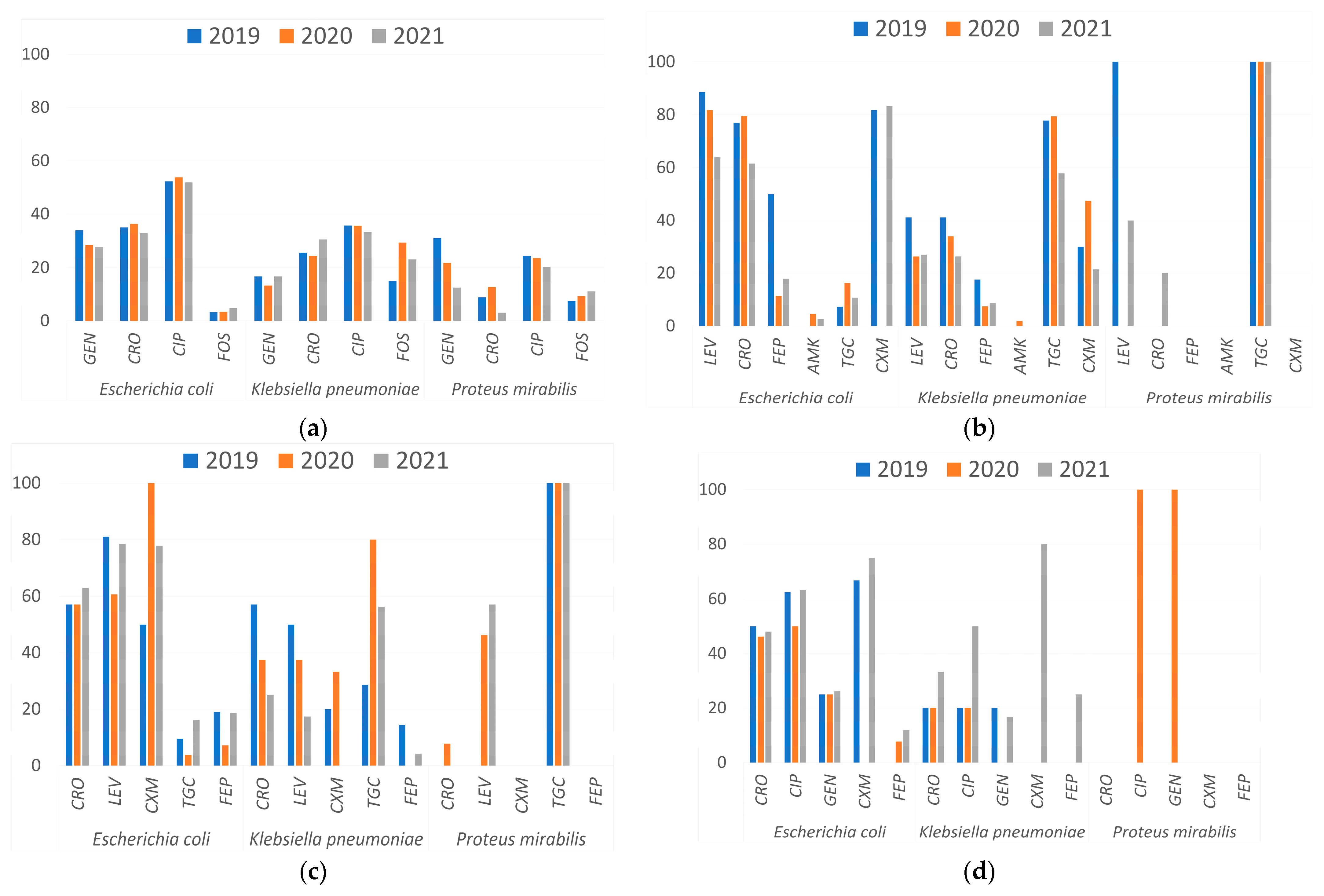
Figure 1. Antimicrobial resistance rates by year, microorganism, and infection site. (a) Urinary tract, (b) lower respiratory tract, (c) soft tissue, and (d) blood. Amikacin (AMK), cefuroxime (CXM), ciprofloxacin (CIP), ceftriaxone (CRO), cefepime (FEP), fosfomycin (FOS), gentamicin (GEN), levofloxacin (LEV), tigecycline (TGC).
Microbiolres 16 00126 g001
Table 1. Distribution of ESBL-producing isolates, by infection site and year, of Escherichia coli, Klebsiella pneumoniae, and Proteus mirabilis.
Sample SourceMicroorganismNumber of Isolates (%)ESBL-Producing Isolates
201920202021Total (%)
Lower respiratory tractE. coli114 (5.1)2034 *21 *75 (65.8)
K. pneumoniae164 (37.9)6 18 2448 (29.3)
P. mirabilis **8 (3.9)00 00 (0)
Soft tissueE. coli123 (5.5)11 1542 68 (55.3)
K. pneumoniae40 (9.2)4 3714 (35.0)
P. mirabilis **24 (11.8)0 0 0 0 (0)
Sterile body fluids 1E. coli65 (2.9) 8 5 18 31 (47.7)
K. pneumoniae6 (1.4)11 0 2 (33.3)
P. mirabilis **0 (0)00 0 0 (0)
BloodE. coli46 (2.1)2 6 9 17 (34.0)
K. pneumoniae17 (3.9)1 1 2 4 (23.5)
P. mirabilis **1 (0.005)00 0 0 (0)
Urinary tractE. coli1803 (80.7)198 179 237 614 (34.1)
K. pneumoniae181 (41.8)10 11 24 46 (25.4)
P. mirabilis **164 (80.4)0 0 0 0 (0)
Other 2E. coli83 (3.7)11 10 14 35 (42.2)
K. pneumoniae25 (5.7)2 2 3 7 (28.0)
P. mirabilis **7 (3.4)0 00 0 (0)
TotalE. coli2234250 249 341 840 (37.6)
K. pneumoniae43324 3660 120 (27.7).
P. mirabilis **2040 0 0 0 (0)
1: Peritoneal, cerebrospinal, synovial fluid, etc. 2: Biopsy, pharyngeal exudate, feces, etc. *: p < 0.05. ** No ESBL-producing P. mirabilis were found.
Disclaimer/Publisher’s Note: The statements, opinions and data contained in all publications are solely those of the individual author(s) and contributor(s) and not of MDPI and/or the editor(s). MDPI and/or the editor(s) disclaim responsibility for any injury to people or property resulting from any ideas, methods, instructions or products referred to in the content.