Article

Energy Metabolism and Aerobic Respiratory Chain of Vitreoscilla sp. C1: Comparison with β-Proteobacteria

by
Paul T. Nguyen
1,2,
Yuyao Hu
1,
Anne Caroline Mascarenhas dos Santos
1,
Pingdong Liang
1,3,
Benjamin C. Stark
1,
Karina Tuz
1 and
Oscar Juárez
1,*
1
Department of Biological Sciences, Illinois Institute of Technology, Chicago, IL 60616, USA
2
R and F Laboratories, Downers Grove, IL 60515, USA
3
Department of Cell and Developmental Biology, Northwestern University, Chicago, IL 60611, USA
*
Author to whom correspondence should be addressed.
Microbiol. Res. 2025, 16(5), 94; https://doi.org/10.3390/microbiolres16050094
Submission received: 27 March 2025 / Revised: 23 April 2025 / Accepted: 30 April 2025 / Published: 4 May 2025
(This article belongs to the Topic Redox in Microorganisms, 2nd Edition)

Abstract

:
As the source of the first reported class of non-mammalian hemoglobin, Vitreoscilla sp. C1 is a historically important microorganism that has offered important clues to understanding how bacteria can thrive at low oxygen tension, with potential applications to wastewater and sludge bioengineering. However, the processes that enable this bacterium to thrive in such environments remain unclear. In this study, we analyzed the published Vitreoscilla sp. C1 genome to predict the core metabolic pathways used by this microorganism to support cell growth under hypoxic conditions, compared them with the predicted metabolism of other important β-proteobacteria, and tested Vitreoscilla’s respiratory activity in vitro in the presence of various substrates and inhibitors. Vitreoscilla sp. C1 carries a functional Krebs cycle and the genes for a branched aerobic respiratory chain, minus the genes for complexes III and IV, and our results show that Vitreoscilla sp. C1 sugar metabolism is carried out through a unique pathway that shunts intermediaries from glycolysis, bypassing phosphofructokinase-I, into the non-oxidative section of the pentose phosphate pathway, reducing its oxygen dependency, which appears as an adaptation to the microaerophilic environment that this organism inhabits. Although Vitreoscilla sp. C1 features a simplified respiratory chain, experimental data demonstrate that all predicted branches are functional, with two main dehydrogenases and two terminal oxidases.

1. Introduction

Vitreoscilla sp. C1 is a filamentous, Gram-negative β-proteobacterium inhabiting aerobic and microaerophilic environments such as soil, stagnant freshwater sediments, and decaying plant matter [1,2]. Vitreoscilla sp. C1 possesses exceptional respiratory adaptations allowing it to survive at low oxygen tension. The most well-known and studied of these adaptations are the Vitreoscilla hemoglobin (VHb) and the redox-driven sodium-pumping cytochrome bo3 oxidase [2,3,4]. VHb was originally reported as a ‘soluble cytochrome o’, and it was not until the determination of its amino acid sequence, two decades later in the 1960s, that the protein was revealed as the first reported type of bacterial hemoglobin [3,5]. Following VHb’s identification, a myriad of other bacterial hemoglobins have been identified, and their presence among microbes is now recognized as ubiquitous, with two-thirds of bacterial species possessing one or more hemoglobins [6,7]. Despite possessing a single globin domain, VHb is a polyvalent multifunctional protein with roles in oxygen delivery (notably to cytochrome bo3, a terminal respiratory oxidase especially relevant at low oxygen concentrations) in transcription mediation under oxidative stress and in nitric oxide detoxification [8,9,10,11,12]. This versatility made VHb a prime target for important biotechnological applications in bioremediation, cell growth, stress resistance, and the production of various biomolecules (e.g., enzymes, antimicrobials, valuable metabolites, and biofuels) [6,13,14,15,16,17].
For example, VHb-engineered bacterial strains used in bioremediation processes have increased efficacy in the breakdown of organic contaminants [15,17,18,19] due to the increased availability of oxygen. The improvement has been shown for degradation of aromatic compounds including benzoic acid, 2,4-dinitrotoluene, and 2-chlorobenzoic acid by species of Pseudomonas, Xanthomonas, and Burkholderia engineered to express VHb, compared with the unengineered strains. This enhancement is presumed to be the result of VHb increasing the availability of oxygen to the cells both for increased respiration and thus ATP production for growth and for the use by oxygenases in the early steps of aromatic compound metabolism. Regarding possible uses of VHb technology in wastewater treatment, engineering of Nitrosomonas europaea to express VHb resulted in enhanced oxygen uptake and conversion of ammonia to nitrite compared with the unengineered strain [17].
Although Vitreoscilla sp. C1 has been considered historically important with a significant biotechnological impact as a result of its hemoglobin, it was never used as a model organism itself because no genetic systems exist that could be used for its engineering. Aside from VHb and its respiratory chain, relatively little is known about the metabolic strategies that it uses to survive and adapt to its own environment. A deeper understanding of this metabolism may in turn point to strategies that could enhance the beneficial effects of VHb in heterologous species engineered to express VHb.
In this work, we used a combination of comparative genomic and biochemical approaches to characterize the metabolism used by this microorganism and compare it with other important β-proteobacteria. The data show that while Vitreoscilla sp. C1 carries the genetic complement for the Krebs cycle and glyoxylate cycles, it remarkably lacks phosphofructokinase-1 (PFK-I), the Entner–Doudoroff pathway (EDP), and the oxidative branch of the pentose phosphate pathway. Its carbon metabolism is mediated by a modification of the glycolytic pathway that bypasses PFK-I by shunting intermediates through the non-oxidative branch of the pentose phosphate pathway, which is a unique metabolic strategy not found in any other β-proteobacteria and might represent an adaptation to the microaerophilic environment that this organism inhabits. Moreover, the results indicate that under aerobic conditions, Vitreoscilla sp. C1 uses two main types of dehydrogenases, succinate dehydrogenase and complex I, as the only NADH dehydrogenases. Interestingly, this microorganism uses two terminal oxidases in the respiratory chain, cytochrome bd oxidase and cytochrome bo3 oxidase, which allow the operation of the aerobic metabolism at low oxygen tensions. This information is crucial in order to understand the metabolic adaptations of this important microorganism to its environment.

2. Materials and Methods

2.1. Metabolic Profiling of Carbon/Energy Pathways and Respiratory Chains in β-Proteobacteria

The Vitreoscilla sp. C1 (NZ_CP019644.2), Neisseria gonorrhoeae FA 1090 (NC_002946.2), Nitrosomonas europaea ATCC 19718 (NC_004757.1), and Burkholderia cepacia ATCC 25416 (NZ_CP034553.1, NZ_CP034554.1, NZ_CP034555.1, NZ_CP034556.1, NZ_CP034557.1) proteomes were downloaded from the NCBI database, and their metabolic profiles were reconstructed as follows. Proteins were queried against the KEGG database [20] using GhostKOALA (Supplementary Data S1) [21]; KO components of the glycolysis, pentose phosphate, and Entner–Duodoroff (phosphorylated, non-phosphorylated, and semi-phosphorylated) pathways, TCA, glyoxylate, and methylcitrate cycles, were retrieved from the KEGG Pathways database (Supplementary Data S2) [20,22]. Respiratory chain components (complex I, type II NADH:quinone oxidoreductase, succinate dehydrogenase, NADH:ubiquinone reductase, bc1 complex, caa3-cytochrome c oxidase, cbb3-type cytochrome c oxidase, cyanide-insensitive quinol oxidase, cytochrome bo3 ubiquinol oxidase, and F-type ATP synthase) KO modules were also downloaded from KEGG (Supplementary Data S2) [20,22]. Then, the presence/absence of proteins from the aforementioned pathways was inferred from the KO numbers retrieved with GhostKOALA searches using get_KO.pl v0.1 (Supplementary Data S3). Core proteins with no KO matches were further searched for in the proteome of each β-proteobacterium with BLASTP and TBLASTN [23,24] homology searches using an E-value of 1e-10 as a cutoff. The metabolic profiling results of the carbon/energy pathway and respiratory chain for the selected β-proteobacteria are found in Table S1 and Table S2, respectively. Supplementary Datas S1–S3 are accessible at https://doi.org/10.5281/zenodo.7754389.

2.2. Vitreoscilla Membrane Preparation

Vitreoscilla sp. C1 cells were cultured under agitation (250 rpm) conditions at 37 °C in the medium previously reported by Webster et al. [25,26]. Cells were harvested by centrifugation at the early stationary phase of growth. Cells were washed twice with KHE buffer (150 mM KCl, 20 mM HEPES, 1 mM EDTA, pH 7.5) and stored at −80 °C. Frozen cell pellets were thawed, resuspended in KHE buffer with the addition of 1 mg/mL of DNAase and 1 mM PMSF. The suspension was sonicated at 4 °C using five 1 min cycles (with a one min pause between each cycle) using a Branson Sonifier Cell Disruptor 350 (50% duty cycle, output control 6). Cell debris was eliminated by centrifugation at 6 × 10³× g for 30 min at 4 °C. The supernatant was collected and centrifuged at 105× g for 5 h. The pellet containing the membranes was washed and resuspended in KHE buffer and stored at −80 °C.

2.3. Oximetry

The respiratory activity of Vitreoscilla sp. C1 membranes (0.2 mg/mL) was measured at 25 °C in KHE buffer in a 2 mL custom-made glass chamber, adapted to a Clark-type oxygen electrode (YSI 5300), as described previously [27]. Assays were carried out in the presence of specific substrates and inhibitors for each of the respiratory complexes. NADH oxidase activity was tested using 200 µM NADH. Succinate dehydrogenase was tested in the presence of 10 mM succinate. KCN titrations (0–2.5 mM) were performed to analyze the role of the different terminal oxidases.

3. Results and Discussion

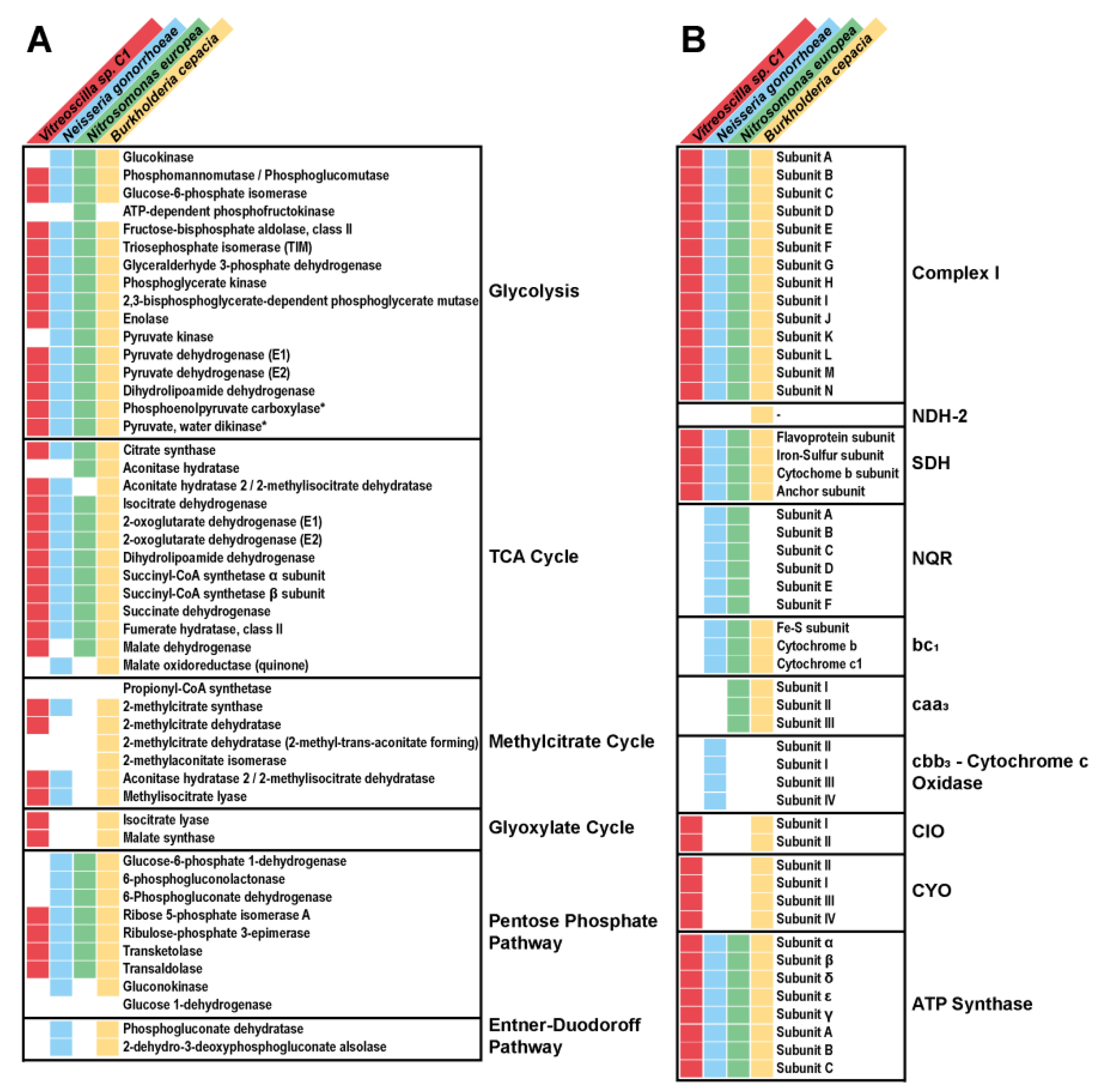
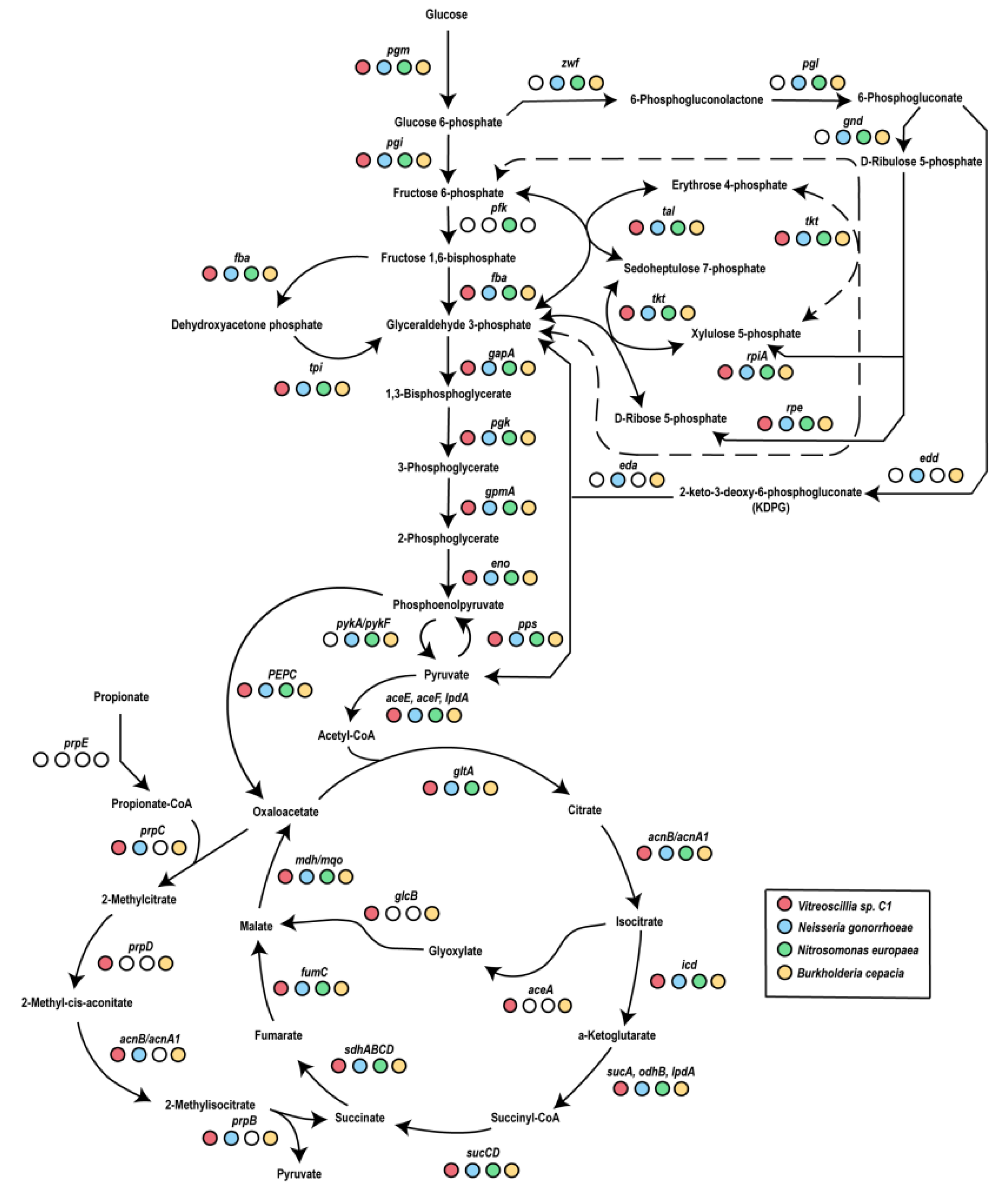
3.1. Vitreoscilla sp. C1 Central Carbon Metabolism

To understand the central metabolic pathways used by Vitreoscilla sp. C1, we carried out analysis of the genes for the following:
  • Glycolysis.
  • The Entner–Doudoroff pathway.
  • The pentose phosphate pathway.
  • The Krebs cycle.
  • The glyoxylate pathway.
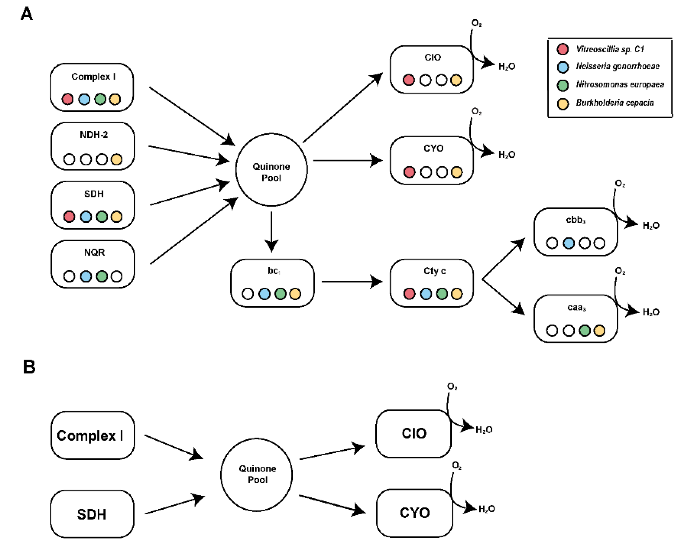
  • The aerobic respiratory chain.
Genomic searches were performed on the updated genome of Vitreoscilla sp. C1 (NZ_CP019644.2) [28,29] and three other important ω-proteobacteria representatives:
  • The human pathogen Neisseria gonorrhoeae FA 1090 [30,31,32].
  • The ammonia and nitrite-oxidizing chemolithoautotroph Nitrosomonas europaea ATCC 19718 [33,34].
  • The opportunistic pathogen Burkholderia cepacia ATCC 25416 [35,36,37].
These bacteria are metabolically divergent and have adapted to their specific environments. This analysis offers clues to understanding the common metabolic strategies used by β-proteobacteria.
In our initial analysis, we found that glycolysis, the Entner–Doudoroff pathway, the glyoxylate cycle, and the Krebs cycle were mostly complete and fully annotated in the published genome of Vitreoscilla sp. C1 (NZ_CP019644.2) (Figure 1), allowing us to reconstruct the full metabolic pathways (Figure 2). While most glycolytic enzymes are found in Vitreoscilla sp. C1 and all β-proteobacteria, Vitreoscilla sp. C1 lacks the following:
(1)
Phosphofructokinase-I, which is essential for glycolysis.
(2)
The enzymes required for the Entner–Doudoroff pathway.
(3)
The entire oxidative branch of the pentose phosphate pathway (Figure 2).
Remarkably, the data show that this microorganism is able to carry out the full oxidation of glucose through an uncommon pathway. This involves the Krebs cycle and an alternative pathway that uses glycolysis bypassing PFK-I, re-routing fructose-6 phosphate into the non-oxidative branch of the pentose phosphate pathway to produce glyceraldehyde 3-phosphate for pyruvate metabolism. This pathway has also been reported in Ralstonia solanacearum [38]. Interestingly, analysis of the genome data indicates that some species of Vitreoscilla, such as V. filliformis, contain a PFK-1 gene and genes for the oxidative section of the pentose phosphate pathway, indicating a high degree of metabolic and genetic variability in the genus, which may be a reflection of the specific niches that these organisms inhabit.
Another unusual feature we found in Vitreoscilla sp. C1 is that it does not encode a pyruvate kinase and instead uses phosphoenolpyruvate kinase to synthesize ATP from AMP, PEP, and inorganic phosphate. Interestingly, we also found a functional methylisocitrate pathway that may allow the metabolism of propionate. However, its role in Vitreoscilla sp. C1 is currently unknown.

3.2. Identification and Functional Characterization of Vitreoscilla sp. C1 Respiratory Chain Complexes

In addition to the analysis of the central carbon metabolism in Vitreoscilla, we carried out an analysis of the genes of all subunits of the main respiratory enzymes that are found in other aerobic microorganisms such as Escherichia coli [39] and Pseudomonas aeruginosa [40] (Figure 3). Vitreoscilla sp. C1 contains the genes for the main subunits of complex I as the main NADH:ubiquinone oxidoreductase. β-proteobacteria are remarkably diverse regarding the types of respiratory NADH dehydrogenases that they use, while Vitreoscilla sp. C1 only carries complex I. B. cepacia carries complex I and the non-ion pumping NDH-2 NADH dehydrogenase. Neisseria and Nitrosomonas bacteria carry complex I, as well as the Na+/ H+ pumping NQR complex. Interestingly, the presence of NQR is associated with pathogenesis or commensalism [41], and its activity in the pathogenic Neisseria corroborates the trend. The presence of NQR in Nitrosomonas does not seem to fit into this common strategy, indicating that NQR could have other roles in the cell. Vitreoscilla sp. C1 also carries the genes for succinate dehydrogenase, which is required for the functioning of both the Krebs cycle and the respiratory chain. In contrast with most proteobacteria, including E. coli and P. aeruginosa, Vitreoscilla sp. C1 lacks complex III (ubiquinol:cytochrome c reductase; cytochrome bc1) and complex IV (cytochrome c oxidase type cbb3 or aa3). Instead, this microorganism carries the genes for two terminal ubiquinol oxidases, cytochrome bo3 oxidase and the cyanide-resistant cytochrome bd oxidase.

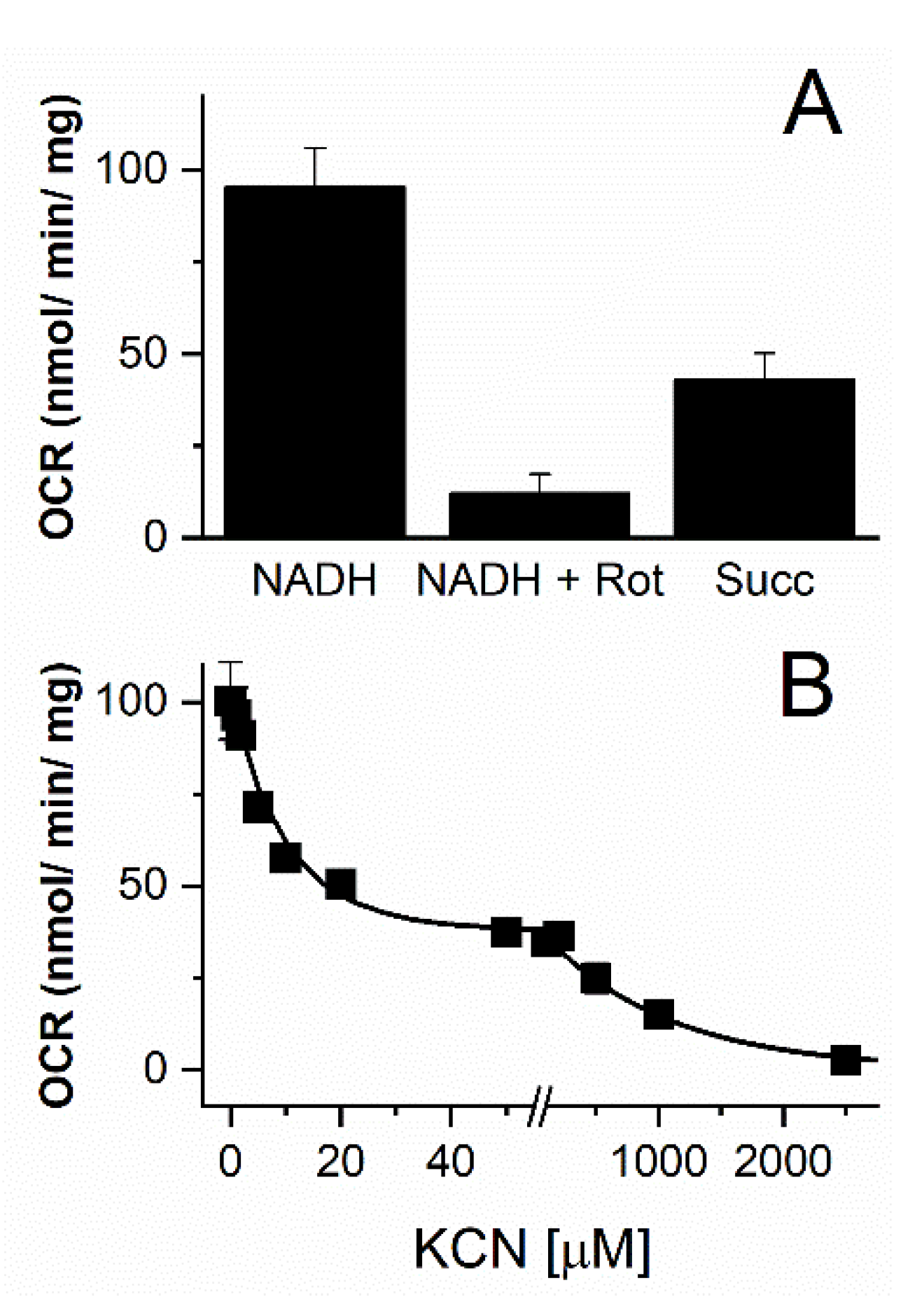
3.3. Functional Characterization of the Vitreoscilla sp. C1 Respiratory Chain

The genomic analysis provided a clear view of the metabolic pathways that Vitreoscilla uses to survive in the environment; for the most part, these are straightforward and similar to those in other bacteria. The analysis of Vitreoscilla sp. C1 electron transfer pathways indicates that they are branched, both at the level of the dehydrogenases and at the level of the terminal oxidases. In other bacteria with branched respiratory chains, such as P. aeruginosa [42], it has been observed that specific pathways are selected under certain conditions (i.e., not all branches are used at the same time); this allows more efficient use of the resources in the environment.
To clarify the role of the respiratory pathways used by Vitreoscilla sp. C1, the activity of the respiratory complexes was measured, following the oxygen consumption rate (OCR) in the presence of specific substrates and inhibitors of the different enzymes. Significant NADH and succinate-dependent activities reveal that the NADH and succinate dehydrogenases, as well as the respiratory chain in general, are functional in the growth conditions tested and that they probably play a major role in cell physiology. To corroborate that complex I is the only respiratory NADH dehydrogenase found in the genome, we tested the effects of 1 µM rotenone, a specific inhibitor of complex I [43,44]. As can be observed in Figure 4, rotenone inhibited the NADH-dependent activity by more than 90%, indicating that complex I is the main NADH dehydrogenase in Vitreoscilla sp. C1.
In addition to examining the dehydrogenases, we explored the terminal oxidases used by Vitreoscilla using KCN titrations. The two types of oxidases have different sensitivities to KCN, with inhibition constants separated by more than one order of magnitude. Cytochrome bo3 oxidase has an apparent Ki of 10–30 µM [45], while the cytochrome bd oxidase Ki is in the mM range. The titration curve (Figure 4B) showed two kinetic components with different inhibitory sensitivities. The first component is responsible for 60% of the respiratory activity and has an IC50 of 5–10 µM, consistent with cytochrome bo3 oxidase, which is associated with Vitreoscilla hemoglobin [46]. The second component contributes 40% of the activity, has an inhibition constant of 0.5–1 mM, and likely corresponds to cytochrome bd oxidase. Interestingly, bd oxidase has a high affinity for oxygen [47]. The presence of bo3–hemoglobin and the high affinity bd oxidase guarantee the operation of the respiratory chain at low oxygen tension.

4. Conclusions

In summary, this manuscript offers a clear picture of the type of metabolism used by Vitreoscilla sp. C1 to support its growth and survival. Moreover, it shows the common strategies used by β-proteobacteria and their specific adaptations to the niches that they inhabit. A comprehensive picture of Vitreoscilla’s intermediary metabolism is important for a more complete understanding of how its hemoglobin and respiratory chain are integrated into its overall ability to thrive aerobically in conditions of low oxygen. The large number of bacterial species for which engineering to express VHb has resulted in improvement in a similarly large number of important biotechnological applications (the production of numerous valuable compounds and biofuels, bioremediation of aromatic compounds, and enhancement of nitrification in wastewater) has proven the usefulness of VHb technology. This is particularly true of applications in which oxygen availability is limited (such as in dense fermentor cultures and subsurface soil). The knowledge of Vitreoscilla’s overall metabolism may provide guidance in designing more complex genetic engineering strategies to improve the advantages of VHb technology even further.
Moreover, this study reveals novel metabolic adaptations in Vitreoscilla sp. C1, particularly its unique glycolytic bypass and simplified aerobic respiratory chain, providing insights into its survival mechanisms under hypoxic conditions and its potential for biotechnological applications. In particular, we propose that other organisms could be optimized to be used in bioremediation by overexpressing bd oxidase and the bo3–hemoglobin pair, which can allow for effective growth under microaerophilic conditions. Due to the low oxygen tensions and likely low oxidative stress, the central carbon metabolism can be engineered to resemble that of Vitreoscilla sp C1 by passing the oxidative branch of the pentose phosphate pathway, which would allow for higher yields as the decarboxylation steps would not be involved in metabolism.

Supplementary Materials

The following supporting information can be downloaded at https://www.mdpi.com/article/10.3390/microbiolres16050094/s1, Supplementary Table S1: Central metabolism of select β-proteobacteria; Supplementary Table S2: Respiratory chain of select β-proteobacteria. The Supplementary Data S1, Data files containing Ghostkoala analysis; S2, Data files containing KO components for the carbon/energy pathway and respiratory chain; and S3, The Perl script written as part of this manuscript, are accessible at https://doi.org/10.5281/zenodo.7754389.

Author Contributions

Conceptualization, K.T. and O.J.; methodology, P.T.N. and A.C.M.d.S.; validation, P.T.N. and O.J.; formal analysis, P.T.N., K.T. and O.J.; investigation, P.T.N., Y.H., A.C.M.d.S. and P.L.; resources, K.T. and O.J.; data curation, P.T.N. and A.C.M.d.S.; writing—original draft preparation, P.T.N., A.C.M.d.S. and O.J.; writing—review and editing, P.L., B.C.S., K.T. and O.J.; visualization, P.T.N., Y.H., P.L. and O.J.; supervision, K.T. and O.J.; project administration, K.T. and O.J.; funding acquisition, O.J. All authors have read and agreed to the published version of the manuscript.

Funding

This research was funded by Illinois Institute of Technology, startup funds, and NIH, grant R15GM131292 to O.J.

Institutional Review Board Statement

Not applicable.

Informed Consent Statement

Not applicable.

Data Availability Statement

Data are contained within the article and Supplementary Material.

Conflicts of Interest

The authors declare no conflicts of interest.

Abbreviations

The following abbreviations are used in this manuscript:
EDTAEthylenediaminetetraacetic acid
HEPES4-(2-Hydroxyethyl)piperazine-1-ethanesulfonic acid
KEGGKyoto Encyclopedia of Genes and Genomes
KOKEGG Orthology
NADHβ-Nicotinamide adenine dinucleotide
TCATricarboxylic acid cycle

References

  1. Pringsheim, E.G. The Vitreoscillaceae: A Family of Colourless, Gliding, Filamentous Organisms. J. Gen. Microbiol. 1951, 5, 124–149. [Google Scholar] [CrossRef]
  2. Strohl, W.R.; Schmidt, T.M.; Lawry, N.H.; Mezzino, M.J.; Larkin, J.M. Characterization of Vitreoscilla Beggiatoides and Vitreoscilla Filiformis Sp. Nov., Nom. Rev., and Comparison with Vitreoscilla Stercoraria and Beggiatoa Alba. Int. J. Syst. Bacteriol. 1986, 36, 302–313. [Google Scholar] [CrossRef]
  3. Wakabayashi, S.; Matsubara, H.; Webster, D.A. Primary Sequence of a Dimeric Bacterial Haemoglobin from Vitreoscilla. Nature 1986, 322, 481–483. [Google Scholar] [CrossRef] [PubMed]
  4. Efiok, B.J.; Webster, D.A. A Cytochrome That Can Pump Sodium Ion. Biochem. Biophys. Res. Commun. 1990, 173, 370–375. [Google Scholar] [CrossRef]
  5. Webster, D.A.; Hackett, D.P. The Purification and Properties of Cytochrome o from Vitreoscilla. J. Biol. Chem. 1966, 241, 3308–3315. [Google Scholar] [CrossRef] [PubMed]
  6. Frey, A.D.; Kallio, P.T. Bacterial Hemoglobins and Flavohemoglobins: Versatile Proteins and Their Impact on Microbiology and Biotechnology. FEMS Microbiol. Rev. 2003, 27, 525–545. [Google Scholar] [CrossRef]
  7. Vinogradov, S.N.; Hoogewijs, D.; Bailly, X.; Arredondo-Peter, R.; Gough, J.; Dewilde, S.; Moens, L.; Vanfleteren, J.R. A Phylogenomic Profile of Globins. BMC Evol. Biol. 2006, 6, 31. [Google Scholar] [CrossRef]
  8. Ramandeep; Hwang, K.W.; Raje, M.; Kim, K.J.; Stark, B.C.; Dikshit, K.L.; Webster, D.A. Vitreoscilla Hemoglobin. Intracellular Localization and Binding to Membranes. J. Biol. Chem. 2001, 276, 24781–24789. [Google Scholar] [CrossRef]
  9. Park, K.-W.; Kim, K.-J.; Howard, A.J.; Stark, B.C.; Webster, D.A. Vitreoscilla Hemoglobin Binds to Subunit I of Cytochrome Bo Ubiquinol Oxidases. J. Biol. Chem. 2002, 277, 33334–33337. [Google Scholar] [CrossRef]
  10. Lin, J.-M.; Stark, B.C.; Webster, D.A. Effects of Vitreoscilla Hemoglobin on the 2,4-Dinitrotoluene (2,4-DNT) Dioxygenase Activity of Burkholderia and on 2,4-DNT Degradation in Two-Phase Bioreactors. J. Ind. Microbiol. Biotechnol. 2003, 30, 362–368. [Google Scholar] [CrossRef]
  11. Kaur, R.; Pathania, R.; Sharma, V.; Mande, S.C.; Dikshit, K.L. Chimeric Vitreoscilla Hemoglobin (Vhb) Carrying a Flavoreductase Domain Relieves Nitrosative Stress in Escherichia Coli: New Insight into the Functional Role of Vhb. Appl. Environ. Microbiol. 2002, 68, 152–160. [Google Scholar] [CrossRef] [PubMed]
  12. Anand, A.; Duk, B.T.; Singh, S.; Akbas, M.Y.; Webster, D.A.; Stark, B.C.; Dikshit, K.L. Redox-Mediated Interactions of VHb (Vitreoscilla Haemoglobin) with OxyR: Novel Regulation of VHb Biosynthesis under Oxidative Stress. Biochem. J. 2010, 426, 271–280. [Google Scholar] [CrossRef]
  13. Khosla, C.; Bailey, J.E. Heterologous Expression of a Bacterial Haemoglobin Improves the Growth Properties of Recombinant Escherichia Coli. Nature 1988, 331, 633–635. [Google Scholar] [CrossRef]
  14. Khosravi, M.; Webster, D.A.; Stark, B.C. Presence of the Bacterial Hemoglobin Gene Improves Alpha-Amylase Production of a Recombinant Escherichia Coli Strain. Plasmid 1990, 24, 190–194. [Google Scholar] [CrossRef] [PubMed]
  15. Stark, B.C.; Pagilla, K.R.; Dikshit, K.L. Recent Applications of Vitreoscilla Hemoglobin Technology in Bioproduct Synthesis and Bioremediation. Appl. Microbiol. Biotechnol. 2015, 99, 1627–1636. [Google Scholar] [CrossRef]
  16. Abanoz, K.; Stark, B.C.; Akbas, M.Y. Enhancement of Ethanol Production from Potato-Processing Wastewater by Engineering Escherichia Coli Using Vitreoscilla Haemoglobin. Lett. Appl. Microbiol. 2012, 55, 436–443. [Google Scholar] [CrossRef] [PubMed]
  17. Kunkel, S.A.; Pagilla, K.R.; Stark, B.C. Engineering of Nitrosomonas Europaea to Express Vitreoscilla Hemoglobin Enhances Oxygen Uptake and Conversion of Ammonia to Nitrite. AMB Express 2015, 5, 43. [Google Scholar] [CrossRef]
  18. Arnaldos, M.; Kunkel, S.A.; Stark, B.C.; Pagilla, K.R. Enhanced Heme Protein Expression by Ammonia-Oxidizing Communities Acclimated to Low Dissolved Oxygen Conditions. Appl. Microbiol. Biotechnol. 2013, 97, 10211–10221. [Google Scholar] [CrossRef]
  19. Arnaldos, M.; Kunkel, S.A.; Stark, B.C.; Pagilla, K.R. Characterization of Heme Protein Expressed by Ammonia-Oxidizing Bacteria under Low Dissolved Oxygen Conditions. Appl. Microbiol. Biotechnol. 2014, 98, 3231–3239. [Google Scholar] [CrossRef]
  20. Kanehisa, M. The KEGG Database. In ‘In Silico’ Simulation of Biological Processes; John Wiley & Sons: Hoboken, NJ, USA, 2002; pp. 91–103. ISBN 978-0-470-85789-2. [Google Scholar]
  21. Kanehisa, M.; Sato, Y.; Morishima, K. BlastKOALA and GhostKOALA: KEGG Tools for Functional Characterization of Genome and Metagenome Sequences. J. Mol. Biol. 2016, 428, 726–731. [Google Scholar] [CrossRef]
  22. Qiu, Y.-Q. KEGG Pathway Database. In Encyclopedia of Systems Biology; Dubitzky, W., Wolkenhauer, O., Cho, K.-H., Yokota, H., Eds.; Springer: New York, NY, USA, 2013; pp. 1068–1069. ISBN 978-1-4419-9863-7. [Google Scholar]
  23. Altschul, S.F.; Gish, W.; Miller, W.; Myers, E.W.; Lipman, D.J. Basic Local Alignment Search Tool. J. Mol. Biol. 1990, 215, 403–410. [Google Scholar] [CrossRef]
  24. Johnson, M.; Zaretskaya, I.; Raytselis, Y.; Merezhuk, Y.; McGinnis, S.; Madden, T.L. NCBI BLAST: A Better Web Interface. Nucleic Acids Res. 2008, 36, W5–W9. [Google Scholar] [CrossRef] [PubMed]
  25. Webster, D.A.; Hackett, D.P. Respiratory Chain of Colorless Algae II. Cyanophyta. Plant Physiol. 1966, 41, 599–605. [Google Scholar] [CrossRef]
  26. Gonzales-Prevatt, V.; Webster, D.A. Purification and Properties of NADH-Cytochrome o Reductase from Vitreoscilla. J. Biol. Chem. 1980, 255, 1478–1482. [Google Scholar] [CrossRef] [PubMed]
  27. Liang, P.; Rosas-Lemus, M.; Patel, D.; Fang, X.; Tuz, K.; Juárez, O. Dynamic Energy Dependency of Chlamydia Trachomatis on Host Cell Metabolism during Intracellular Growth: Role of Sodium-Based Energetics in Chlamydial ATP Generation. J. Biol. Chem. 2018, 293, 510–522. [Google Scholar] [CrossRef]
  28. Veseli, I.A.; Mascarenhas dos Santos, A.C.; Juárez, O.; Stark, B.C.; Pombert, J.-F. Complete Genome Sequence of Vitreoscilla Sp. Strain C1, Source of the First Bacterial Hemoglobin. Microbiol. Resour. Announc. 2018, 7, e00922-18. [Google Scholar] [CrossRef] [PubMed]
  29. Kolmogorov, M.; Yuan, J.; Lin, Y.; Pevzner, P.A. Assembly of Long, Error-Prone Reads Using Repeat Graphs. Nat. Biotechnol. 2019, 37, 540–546. [Google Scholar] [CrossRef]
  30. Hebeler, B.H.; Morse, S.A. Physiology and Metabolism of Pathogenic Neisseria: Tricarboxylic Acid Cycle Activity in Neisseria Gonorrhoeae. J. Bacteriol. 1976, 128, 192–201. [Google Scholar] [CrossRef]
  31. Morse, S.A.; Stein, S.; Hines, J. Glucose Metabolism in Neisseria Gonorrhoeae. J. Bacteriol. 1974, 120, 702–714. [Google Scholar] [CrossRef]
  32. Winter, D.B.; Morse, S.A. Physiology and Metabolism of Pathogenic Neisseria: Partial Characterization of the Respiratory Chain of Neisseria Gonorrhoeae. J. Bacteriol. 1975, 123, 631–636. [Google Scholar] [CrossRef]
  33. Corregido, M.C.; Asención Diez, M.D.; Iglesias, A.Á.; Piattoni, C.V. New Pieces to the Carbon Metabolism Puzzle of Nitrosomonas Europaea: Kinetic Characterization of Glyceraldehyde-3 Phosphate and Succinate Semialdehyde Dehydrogenases. Biochimie 2019, 158, 238–245. [Google Scholar] [CrossRef] [PubMed]
  34. Whittaker, M.; Bergmann, D.; Arciero, D.; Hooper, A.B. Electron Transfer during the Oxidation of Ammonia by the Chemolithotrophic Bacterium Nitrosomonas Europaea. Biochim. Biophys. Acta 2000, 1459, 346–355. [Google Scholar] [CrossRef] [PubMed]
  35. Van Acker, H.; Sass, A.; Bazzini, S.; De Roy, K.; Udine, C.; Messiaen, T.; Riccardi, G.; Boon, N.; Nelis, H.J.; Mahenthiralingam, E.; et al. Biofilm-Grown Burkholderia Cepacia Complex Cells Survive Antibiotic Treatment by Avoiding Production of Reactive Oxygen Species. PLoS ONE 2013, 8, e58943. [Google Scholar] [CrossRef]
  36. Lefebre, M.D.; Flannagan, R.S.; Valvano, M.A. A Minor Catalase/Peroxidase from Burkholderia Cenocepacia Is Required for Normal Aconitase Activity. Microbiology 2005, 151, 1975–1985. [Google Scholar] [CrossRef] [PubMed]
  37. Pessi, G.; Braunwalder, R.; Grunau, A.; Omasits, U.; Ahrens, C.H.; Eberl, L. Response of Burkholderia Cenocepacia H111 to Micro-Oxia. PLoS ONE 2013, 8, e72939. [Google Scholar] [CrossRef]
  38. Jyoti, P.; Shree, M.; Joshi, C.; Prakash, T.; Ray, S.K.; Satapathy, S.S.; Masakapalli, S.K. The Entner-Doudoroff and Nonoxidative Pentose Phosphate Pathways Bypass Glycolysis and the Oxidative Pentose Phosphate Pathway in Ralstonia Solanacearum. mSystems 2020, 5, 10–1128. [Google Scholar] [CrossRef]
  39. Blattner, F.R.; Plunkett, G.; Bloch, C.A.; Perna, N.T.; Burland, V.; Riley, M.; Collado-Vides, J.; Glasner, J.D.; Rode, C.K.; Mayhew, G.F.; et al. The Complete Genome Sequence of Escherichia Coli K-12. Science 1997, 277, 1453–1462. [Google Scholar] [CrossRef]
  40. Stover, C.K.; Pham, X.Q.; Erwin, A.L.; Mizoguchi, S.D.; Warrener, P.; Hickey, M.J.; Brinkman, F.S.; Hufnagle, W.O.; Kowalik, D.J.; Lagrou, M.; et al. Complete Genome Sequence of Pseudomonas Aeruginosa PAO1, an Opportunistic Pathogen. Nature 2000, 406, 959–964. [Google Scholar] [CrossRef]
  41. Reyes-Prieto, A.; Barquera, B.; Juárez, O. Origin and Evolution of the Sodium -Pumping NADH: Ubiquinone Oxidoreductase. PLoS ONE 2014, 9, e96696. [Google Scholar] [CrossRef]
  42. Liang, P.; Fang, X.; Hu, Y.; Yuan, M.; Raba, D.A.; Ding, J.; Bunn, D.C.; Sanjana, K.; Yang, J.; Rosas-Lemus, M.; et al. The Aerobic Respiratory Chain of Pseudomonas Aeruginosa Cultured in Artificial Urine Media: Role of NQR and Terminal Oxidases. PLoS ONE 2020, 15, e0231965. [Google Scholar] [CrossRef]
  43. Lümmen, P. Complex I Inhibitors as Insecticides and Acaricides. Biochim. Et Biophys. Acta (BBA) Bioenerg. 1998, 1364, 287–296. [Google Scholar] [CrossRef]
  44. Ernster, L.; Dallner, G.; Azzone, G.F. Differential Effects of Rotenone and Amytal on Mitochondrial Electron and Energy Transfer. J. Biol. Chem. 1963, 238, 1124–1131. [Google Scholar] [CrossRef]
  45. Di Tomaso, G.; Fedi, S.; Carnevali, M.; Manegatti, M.; Taddei, C.; Zannoni, D. The Membrane-Bound Respiratory Chain of Pseudomonas Pseudoalcaligenes KF707 Cells Grown in the Presence or Absence of Potassium Tellurite. Microbiology 2002, 148, 1699–1708. [Google Scholar] [CrossRef] [PubMed]
  46. Graf, S.; Brzezinski, P.; von Ballmoos, C. The Proton Pumping Bo Oxidase from Vitreoscilla. Sci. Rep. 2019, 9, 4766. [Google Scholar] [CrossRef]
  47. Grund, T.N.; Radloff, M.; Wu, D.; Goojani, H.G.; Witte, L.F.; Jösting, W.; Buschmann, S.; Müller, H.; Elamri, I.; Welsch, S.; et al. Mechanistic and Structural Diversity between Cytochrome Bd Isoforms of Escherichia Coli. Proc. Natl. Acad. Sci. USA 2021, 118, e2114013118. [Google Scholar] [CrossRef]
Figure 1. Metabolic pathways (A) and respiratory complex subunits (B) of Vitreoscilla sp. C1 and other β-proteobacteria species. Colored boxes indicate the presence of these genes in Vitreoscillia sp. C1 (red), Neisseria gonorrohoea FA 1090 (blue), Nitrosomonas europaea ATCC 19718 (green), and Burkholderia cepacia ATCC 25416 (yellow). *, presumptive.
Microbiolres 16 00094 g001
Figure 2. Central metabolic pathway map of β-proteobacteria species. Colored circles indicate the presence of enzymes in Vitreoscillia sp. C1 (red), Neisseria gonorrhoeae FA 1090 (blue), Nitrosomonas europaea ATCC 19718 (green), and Burkholderia cepacia ATCC 25416 (yellow).
Microbiolres 16 00094 g002
Figure 3. Aerobic respiratory chain pathway of β-proteobacteria species. Colored circles indicate the presence of enzymes in Vitreoscillia sp. C1 (red), Neisseria gonorrhoeae FA 1090 (blue), Nitrosomonas europaea ATCC 19718 (green), and Burkholderia cepacia ATCC 25416 (yellow). (A) Diversity of respiratory enzymes in β-proteobacteria. (B) Vitreoscilla sp. C1 respiratory chain.
Microbiolres 16 00094 g003
Figure 4. Respiratory activities of Vitreoscilla sp. C1 membranes in the presence of respiratory substrates and inhibitors. (A) Oxygen consumption rates (OCR) of Vitreoscilla membranes in the presence of 250 µM NADH with and without 1µM rotenone (Rot) and with 1 mM succinate (Succ). Values are averages of >4 independent trials (error bars represent standard deviations). (B) NADH-dependent oxygen consumption rates of Vitreoscilla membranes in the presence of increasing concentrations of potassium cyanide (KCN). Each point is the average of >3 independent trials.
Microbiolres 16 00094 g004
Disclaimer/Publisher’s Note: The statements, opinions and data contained in all publications are solely those of the individual author(s) and contributor(s) and not of MDPI and/or the editor(s). MDPI and/or the editor(s) disclaim responsibility for any injury to people or property resulting from any ideas, methods, instructions or products referred to in the content.

