Article

Detoxification of Ustiloxin A by Hydroxylation of Endophytic Fungus Petriella setifera Nitaf10

Department of Plant Pathology, College of Plant Protection, China Agricultural University, Beijing 100193, China
*
Author to whom correspondence should be addressed.
These authors contributed equally to this work.
Microbiol. Res. 2025, 16(5), 93; https://doi.org/10.3390/microbiolres16050093
Submission received: 17 February 2025 / Revised: 25 April 2025 / Accepted: 28 April 2025 / Published: 29 April 2025

Abstract

:
Ustiloxins are a kind of cyclopeptide mycotoxins produced by rice false smut pathogen Villosiclava virens, which seriously threatens the safe production of rice and health of humans and animals. Hydroxylation, a biotransformation reaction that regio- and stereoselectively introduces a hydroxyl group into the molecule catalyzed by the hydroxylase produced by organisms, has been considered an efficient way to detoxify mycotoxins. In this study, the endophytic fungus Petriella setifera Nitaf10 was found to be able to detoxify ustiloxin A, the main toxic component in V. virens. In addition to the two main transformed products previously identified, ustiloxins A1 and A2, an additional transformed product was obtained by using cell-free extract (CFE) of P. setifera Nitaf10 prepared with 5 mmol/L of pH 9.0 carbonate-buffered solution (CBS). It was structurally characterized as a novel ustiloxin analog named 13-hydroxy ustiloxin A (1) by analysis of the 1D and 2D NMR and HRESIMS spectra as well as by comparison with known ustiloxins. Biotransformation reaction of ustiloxin A was found to proceed via hydroxylation, and was possibly catalyzed by the intracellular hydroxylase in the CFE. The cytotoxic and phytotoxic activities of 13-hydroxy ustiloxin A (1) were much weaker than those of ustiloxin A. Detoxification of ustiloxin A by hydroxylation of P. setifera will be an efficient strategy.

1. Introduction

Rice false smut (RFS), caused by the pathogenic fungus Villosiclava virens (anamorph: Ustilaginoidea virens), is one of the most destructive fungal diseases in rice production [1]. RFS can result in decreased production and severe economic loses in rice-growing areas around the world. Furthermore, the RFS pathogen can produce ustiloxins which are harmful mycotoxins to rice plants as well as to humans and animals. Therefore, RFS seriously threatens the high-yield and safe production of rice [1,2]. Six ustiloxins have been isolated from the water extract of RFS balls and the cultured RFS pathogen V. virens [3,4,5]. Among them, ustiloxin A was the main toxic component [6]. Ustiloxins exhibit a variety of toxic activities such as renal toxicity [7,8,9], hepatotoxicity [10,11,12], phytotoxicity [4,13,14,15], and cytotoxicity [3,4,16]. It is necessary to remove or detoxify them from rice food and feed.
Some strategies such as physical, chemical, and biological detoxification methods can be employed to decrease or detoxify the toxicity of mycotoxins [17]. Mycotoxins are usually transformed into less toxic or nontoxic products through bioconversion by either living organisms or their biosynthesized enzymes as biocatalysts [18,19,20]. In order to detoxify ustiloxins in rice food and feed, detoxification through bioconversion might be an efficient strategy.
Previously, we identified two detoxified products, ustiloxins A1 and A2 of ustiloxin A, by using the cell-free extract (CFE, pH 7.0) containing intracellular amine oxidase and oxidative decarboxylase of plant endophytic fungus Petriella setifera Nitaf10 [16]. In our ongoing search for new transformed products of ustiloxin A from the incubated reaction solution, a new oxidation product was identified. In the present study, the CFE (pH 9.0) of P. setifera Nitaf10 was employed to convert ustiloxin A into a novel ustiloxin analog named 13-hydroxy ustiloxin A (1) (Figure 1). The cytotoxic and phytotoxic activities of 13-hydroxy ustiloxin A (1) were greatly decreased. The details of analysis, structural elucidation, and cytotoxic activity of the transformed product 13-hydroxy ustiloxin A (1) are reported herein. The transformed mechanism of ustiloxin A as well as the potential application of hydroxylase are also discussed.

2. Materials and Methods

2.1. General Experimental Procedures

Column chromatography (CC) was performed on Sephadex G15 (40–70 μm; Amersham Pharmacia Biotech, Uppsala, Sweden). HPLC-DAD analysis was performed using a Shimadzu LC-20A instrument with an SPD-M20A photodiode array detector (Shimadzu Corp., Tokyo, Japan) and an analytic C18 column (250 mm × 4.6 mm i.d., 5 μm; Phenomenex Inc., Torrance, CA, USA).
The analytical HPLC elution conditions were as follows: 0–30 min, methanol from 10 to 100%, 30–40 min, 100% methanol-flushing column, 40–50 min, 10% methanol balance, flow rate was 1.0 mL/min, detection wavelength was 210 nm.
Semi-preparative HPLC separation was carried out on a Lumtech instrument (Lumiere Tech. Ltd., Beijing, China) equipped with a K-501 pump (flow rate: 3 mL/min) and a K-2501 UV detector using a Luna-C18 column (250 mm × 10 mm i.d., 5 μm, Phenomenex Inc., Torrance, CA, USA). UV spectra were recorded on a TU-1810 UV-vis spectrophotometer (Beijing Persee General Instrument Co., Ltd., Beijing, China). Ustiloxin A was prepared according to the previous report [4].
The liquid chromatography-high-resolution electrospray ionization mass spectrometry (LC-HRESIMS) analyses were performed on an Agilent 1260 HPLC system coupled to the Q-TOF/MS 6520 spectrometer (Agilent Technologies, Santa Clara, CA, USA), equipped with a Phenomene × C18 column (i.d., 100 mm × 2.0 mm, 3 μm). The mobile phase employed chromatographic-grade acetonitrile (containing 0.1% formic acid) and water (containing 0.1% formic acid). Compound 1 was analyzed in both positive and negative ion modes using an isocratic elution with acetonitrile/water (15:85, v/v), and the detection wavelength was set at 210 nm. The HRESIMS spectra were then recorded.
Compound 1 was dissolved in D2O for NMR measurement. 1H and 2D NMR (HSQC, HMBC, and COSY) spectra were measured on an Avance 500 NMR spectrometer (Bruker BioSpin, Zurich, Switzerland). Chemical shifts were expressed in δ (ppm), referring to the solvent residual peaks at δH 4.79, and coupling constants (J) were expressed in hertz (Hz).
All solvents including methanol, acetonitrile, and formic acid were of HPLC grade (Fisher Scientific, Loughborough, UK). Water was purified by a Milli-Q system (TTL-30C, Tongtai, Beijing, China). The solvent D2O used for NMR spectroscopic measurement of compound 1 was purchased from Actis Technology Co., Ltd., London, UK.

2.2. Endophytic Fungus Petriella setifera Nitaf10

The plant endophytic fungus Petriella setifera Nitaf10 (GenBank accession number KM095515) was isolated from the healthy roots of Nicotiana tabacum (Solanilaceae) [21].

2.3. Preparation of Buffer Solution

Next, 5 mmol/L of buffered solutions with pH values 7.0 and 9.0 were prepared. They were pH 7.0 phosphate-buffered solution (PBS), and pH 9.0 carbonate-buffered solution (CBS). Then, 200 µL ustiloxin A solution (500 µg/mL) was added in 800 µL of pH 7.0 or pH 9.0 buffered solution at 28 °C and 180 rpm for 3 days. The reaction was stopped with an equal volume of methanol.

2.4. Preparation of the Cell-Free Extract of P. setifera

The activated fungus P. setifera Nitaf10 was cultured in PDB (potato dextrose broth) medium at 180 rpm for 3 to 5 days. The liquid fungal suspension was centrifuged at 12,000× g at 4 °C for 20 min. The supernatant containing extracellular enzymes was proved to be incapable of converting ustiloxin A [16]. After the supernatant was removed, 10 mL of 0.5 mol/L pH 7.0 PBS or pH 9.0 CBS was added to 10 g of the fresh precipitated mycelia, which was in an ice bath and subjected to ultrasonic treatment 150 times (2 s oscillation and 2 s of intermittent for each time). Then, centrifugation was performed at 12,000× g and 4 °C for 20 min. The supernatant, which was called the cell-free extract (CFE) containing intracellular crude enzymes, was collected.

2.5. Incubation of Ustiloxin A in the Cell-Free Extract of P. setifera

The incubation of ustiloxin A in the cell-free extract (CFE) of P. setifera was previously described [16]. Briefly, 800 µL of the CFE containing intracellular crude enzymes was added into the vial. Then, 200 µL of 500 µg/mL ustiloxin A was added; the control was the crude enzyme solution without ustiloxin A. The conversion reaction lasted for 3 days at 28 °C, 180 rpm.

2.6. Isolation and Structural Identification of the Transformed Product 1 of Ustiloxin A

After ustiloxin A (30 mg) was incubated for 3 days in the CFE prepared with 5 mmol/L of pH 9.0 CBS, an additional product (1.8 mg), which was subsequently identified as 13-hydroxy ustiloxin (1), was produced as well as the two main transformed products previously identified, ustiloxins A1 and A2 [16]. The biotransformation reaction was performed in 0.5 mol/L of pH 9.0 CBS, the reaction solution was concentrated on Sephadex G-15 (Bio-Resin, Beijing, China) with ultrapure water as the mobile phase to obtain crude transformed products, which were analyzed and compared by HPLC, and the pure compounds including 13-hydroxy ustiloxin A (1), ustiloxins A1 and A2, were prepared by semi-preparative HPLC with the methanol–water (20:80, v/v) as the mobile phase. The detection wavelength was set at 210 nm. The purified compounds were structurally identified by analysis of UV, MS, 1D and 2D NMR data.

2.7. Cytotoxic Activity Assay

The cytotoxic activity of the compounds was tested against human carcinoma cells using the microculture methyl-thiazolyl-tetrazolium (MTT) assay as previously described [4]. The human carcinoma cell lines including colon cancer cells (HCT-8), pancreatic cancer cells (PANC-1), gastric cancer cells (HGC-27), liver hepatocellular carcinoma cells (HepG2), and lung cancer cells (PC9) were tested on ustiloxin A and 13-hydroxy ustiloxin A (1). Taxol was used as the positive control. All experiments were performed with three replicates. The median inhibitory concentration (IC50) values of the compounds were determined by linear regression.

2.8. Phytotoxic Activity Assay

The phytotoxic activity of the compounds was evaluated by their inhibitory activities on the radicle elongation of rice (Oryza sativa) seeds. The variety Danluo No. 2 of rice seeds was used. The bioassay was performed using the method described previously [4] with some modifications. Briefly, five 3-day germinated rice seeds were sown onto each well of a 24-well plate, containing 200 μL of working solution. For preparing working solution, 1 mg of each purified compound was dissolved in 1 mL of ultrapure water to obtain the stock solution (1 mg/mL), which was further diluted to a series of concentrations (400, 200, 100, 50, and 25 μg/mL) with ultrapure water. The ultrapure water was used as the negative control, and glyphosate (N-(phosphonomethyl)glycine) at the same concentrations as the positive control. Plates were incubated in a moist chamber at 25 °C in the dark. The length of each radicle or germ was measured after treatment for 48 h. The inhibition of radicle or germ elongation was calculated as follows: Inhibition (%) = [(Lc − Lt)/Lc] × 100, where Lc is the radicle or germ length of the control group, and Lt is the length of the treated group. All experiments were performed with three replicates. The IC50 values of the compounds were determined by linear regression.

3. Results

3.1. HPLC Analysis of the Transformed Products of Ustiloxin A

The cell-free extract (CFE) containing intracellular enzymes of P. setifera Nitaf10 was previously proved to be able to convert ustiloxin A into ustiloxins A1 and A2 [16]. If ustiloxin A was incubated in the CFE prepared with 5 mmol/L of pH 7.0 phosphate-buffered solution (PBS), only two products, ustiloxins A1 and A2, were monitored by HPLC analysis (Figure 2B). If ustiloxin A was incubated in the CFE prepared with 5 mmol/L of pH 9.0 carbonate-buffered solution (CBS), an additional product (the peak d in Figure 2C) was monitored by HPLC analysis. The UV spectrum (Figure 2E) of the additional peak d was identical to that of ustiloxin A (Figure 2D), indicating that this additional transformed product (1) could be an analog of ustiloxin A. Hence, 1 should also be a transformed product of ustiloxin A.

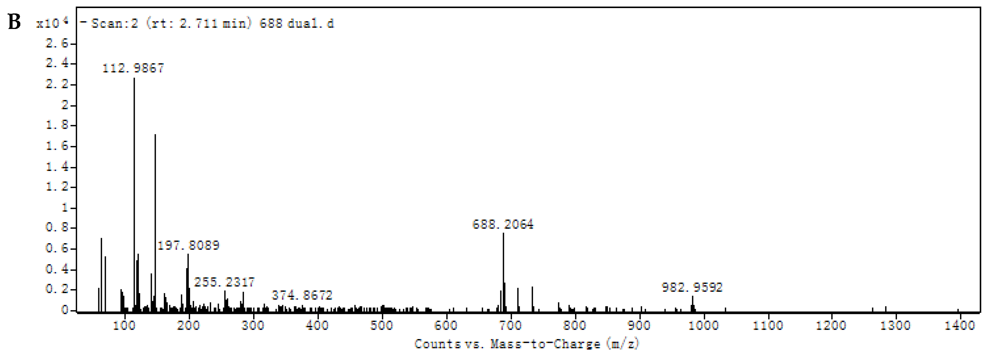
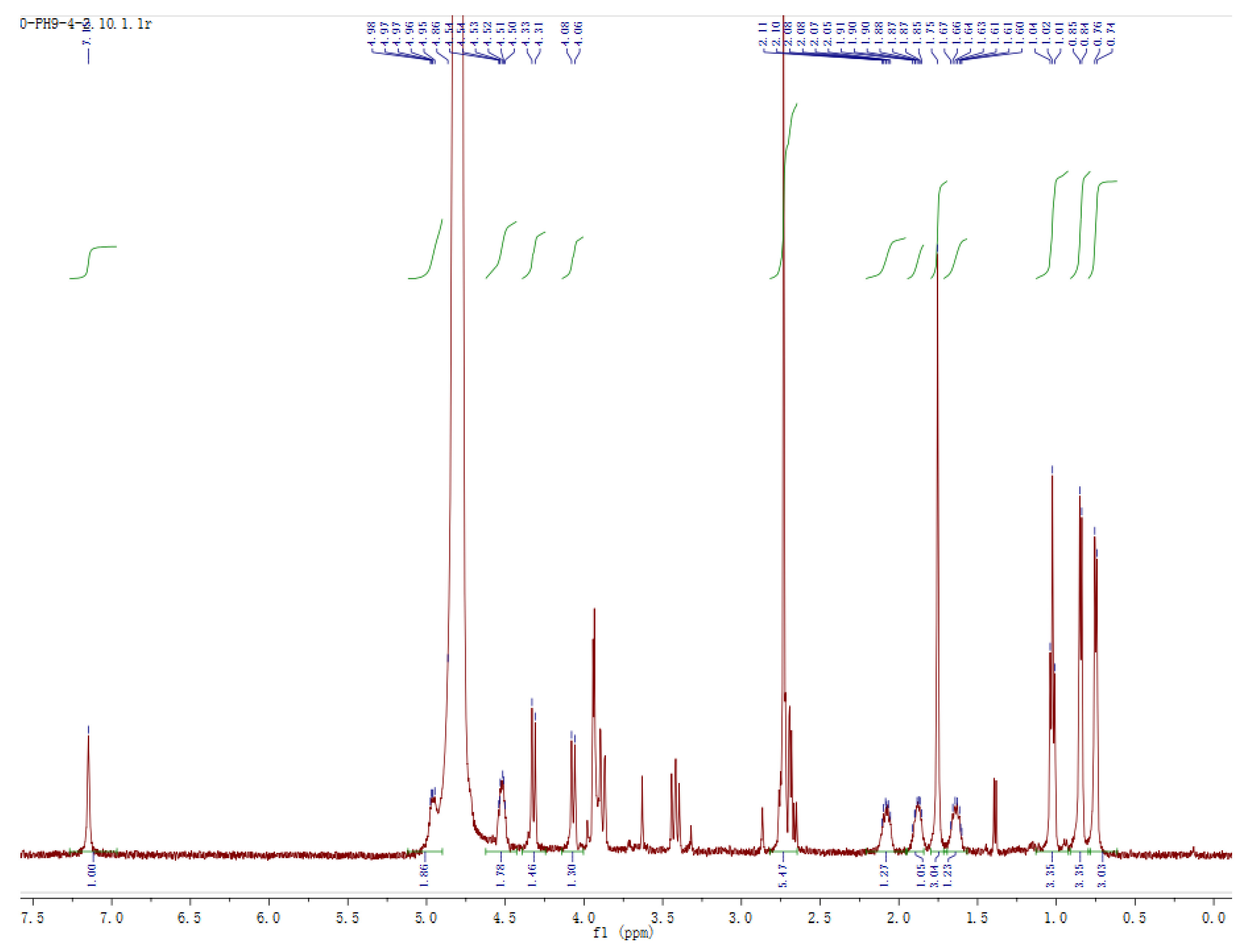
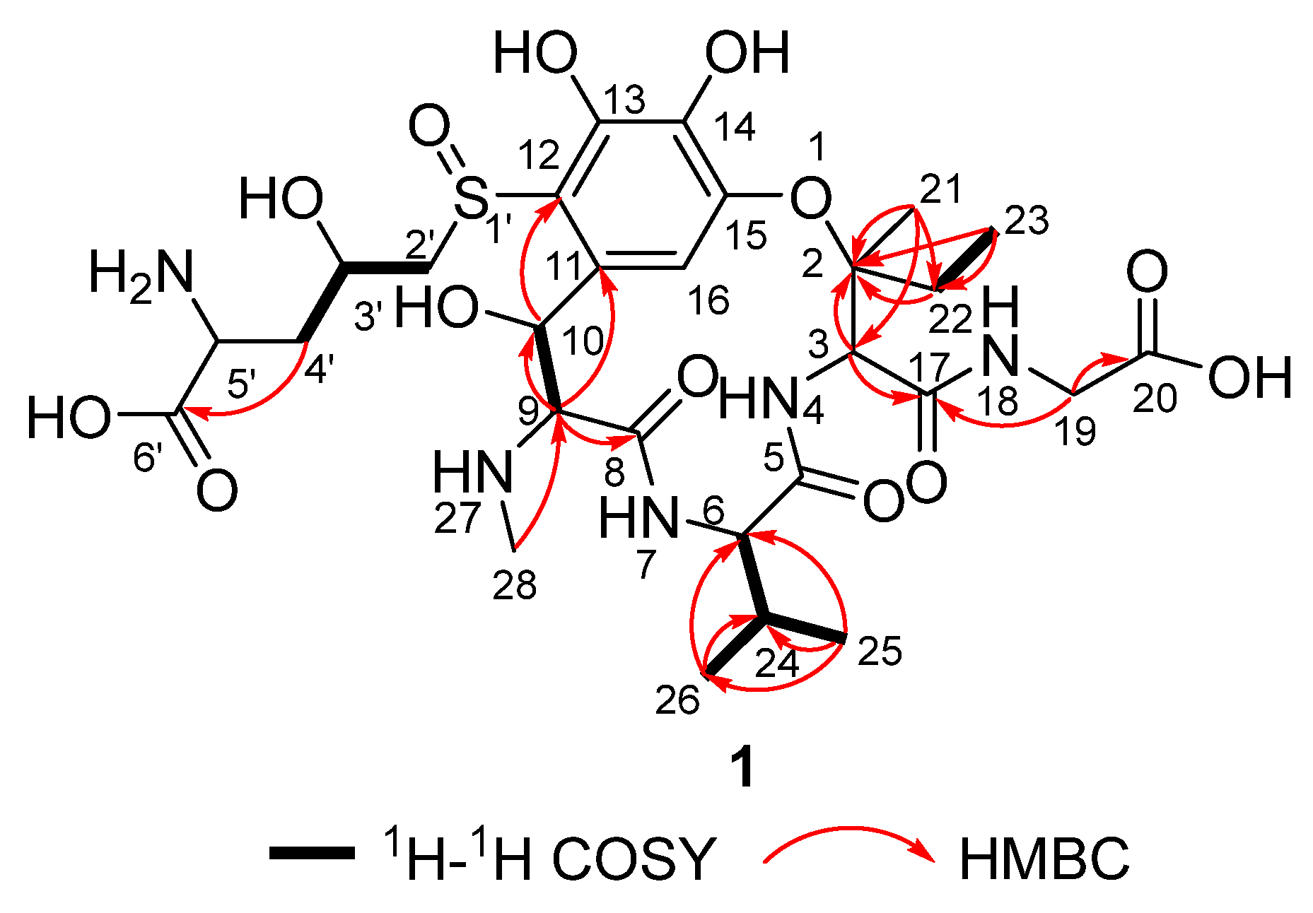
3.2. Structural Identification of Transformed Product 1

Compound 1, which was isolated as a colorless amorphous powder, exhibited prominent pseudomolecular ion peaks at m/z 690.2292 [M+H]+ (calcd for C28H44N5O13S+, 690.2240) and m/z 688.2064 [M-H] (calcd for C28H42N5O13S, 688.2500) in the HRESIMS spectra (Figure 3), indicating a molecular formula of C28H43N5O13S, with 10 degrees of unsaturation. However, based on the fact that the compound 1 had a higher ionization efficiency in the positive ion mode, the positive ion mode spectrum (Figure 3A) was better than that of the negative ion mode (Figure 3B).
The 1H NMR spectrum of 1 is shown in Figure 4. Analysis of its 1H NMR data (Table 1) and HSQC spectrum (Figure S1) revealed the presence of one aromatic proton (δH 7.16), six heteroatomic substituted methine groups (δH 4.96, 4.82, 4.52, 4.33, 4.08, 3.86–3.96), two heteroatomic substituted methylene groups (δH 3.95; 3.86–3.96, 3.43), five methyl groups (δH 2.74, 1.76, 1.03, 0.85, 0.75), and several aliphatic proton signals.
The 1H-1H COSY spectrum and HMBC spectrum of 1 are shown in Figures S2 and S3, respectively. The key 1H-1H COSY and selected HMBC correlations of 1 are shown in Figure 5. They were very similar to those of ustiloxin A [3], and ustiloxins A1 and A2 [16]; the main difference was that the proton at C-13 of ustiloxin A is replaced by a hydroxyl group in 1 (Figure 1). This was confirmed by the disappearance of the aromatic proton signal (δH 7.61) at C-13 (Table 1), corresponding to the molecular formulae of 1 and ustiloxin A. Considering incubation of ustiloxin A in fungal CFE, the absolute configuration of 1 was assumed to be the same as that of ustiloxin A. Thus, the structure of 1 was determined (Figure 1), and 1 was designated as 13-hydroxy ustiloxin A.

3.3. Cytotoxic and Phytotoxic Activities of Ustiloxin A and 13-Hydroxy Ustiloxin A (1)

Ustiloxin A and its transformed product 13-hydroxy ustiloxins A (1) were assessed for their cytotoxic activity on five human cancer cell lines (Table 2). The median inhibitory concentration (IC50) value of ustiloxin A was smaller than that of 13-hydroxy ustiloxin A (1), which indicated that the cytotoxic activity of the transformed product was weaker than that of the ustiloxin A.
Ustiloxin A and its transformed product 13-hydroxy ustiloxins A (1) were also assessed for their inhibitory activities on the radicle and germ elongation of rice seeds. It was found that both ustiloxin A and 13-hydroxy ustiloxin A (1) had no inhibitions on the germ elongation. However, they had obvious inhibitory activities on the radicle elongation of rice seeds. The IC50 values of ustiloxin A, 13-hydroxy ustiloxin A (1), and glyphosate (the positive control) were 70.5, 380.4, and 61.5 μg/mL, respectively. The IC50 value of ustiloxin A was much smaller than that of 13-hydroxy ustiloxin A (1), which indicated that the phytotoxic activity of the transformed product 13-hydroxy ustiloxin A (1) was much weaker than that of ustiloxin A.

4. Discussion

The detoxification of mycotoxins generally includes physical, chemical, and biological strategies [22,23,24,25,26]. Compared with physical and chemical strategies, biological strategies of mycotoxin detoxification with mild and environmentally friendly conditions have been recognized as the promising solution for decontamination of mycotoxins [26,27]. The biotransformation of mycotoxins by living organisms (i.e., bacteria, fungi, plants, and animals) and their synthesized enzymes is a complex biochemical process with their efficiency, specificity, and stereo selectivity. They are able to metabolize, destroy, or deactivate mycotoxins into stable, less toxic, or even nontoxic products [28,29].
Hydroxylation of mycotoxins is an oxidation-reduction process that introduces a hydroxyl group (-OH) into the molecule. Regio- and stereoselective introduction of hydroxyl groups at the various positions of the molecule are often facilitated by the enzymes called hydroxylases. Hydroxylation often increases the polarity of mycotoxins, and reduces their toxicity [19,30]. For examples, zearalenone (ZEN) was converted into two detoxified monohydroxylated metabolites, 13-hydroxy-ZEN and 15-hydroxy-ZEN, with aromatic hydroxylation by human hepatic microsomes [31]. Fusaric acid is a host non-specific phytotoxin produced by the fungi from the genus Fusarium [32,33]. Fusaric acid was converted into the detoxified product 8-hydroxyfusaric acid with hydroxylation by the fungus Mucor rouxii [33]. In the present study, an additional transformed product, 13-hydroxy ustiloxins A (1) of ustiloxin A, was identified by using the CFE (pH 9.0) of plant endophytic fungus P. setifera Nitaf10 in addition to the two main products previously identified, ustiloxins A1 and A2. It was proposed to proceed via hydroxylation by the catalysis of hydroxylase. Biotransformation reaction of ustiloxin A in the CFE of P. setifera Nitaf10 is shown in Figure 1. The cytotoxic and phytotoxic activities of the transformed product 13-hydroxy ustiloxin A (1) were significantly reduced compared to the precursor ustiloxin A. It indicated that the additional aromatic hydroxylation decreased the cytotoxicity of ustiloxin A. The other toxic activities such as animal toxic activities need to be further investigated. It should be an efficient strategy to detoxify ustiloxin A through hydroxylation by using the CFE of P. setifera Nitaf10. The hydroxylase produced by P. setifera Nitaf10 requires purification for verifying this conversion reaction.

5. Conclusions

In this study, an additional transformed product of ustiloxin A was identified by using the CFE of plant endophytic fungus P. setifera Nitaf10 prepared with 5 mmol/L of pH 9.0 CBS, and was structurally identified as a novel ustiloxin analog named 13-hydroxy ustiloxin A (1) by spectroscopic analyses. Biotransformation reaction of ustiloxin A was speculated as hydroxylation under the catalysis of hydroxylase. The cytotoxic and phytotoxic activities of 13-hydroxy ustiloxin A (1) were obviously weaker than those of the precursor ustiloxin A, which indicated that ustiloxin A can be detoxified through hydroxylation by P. setifera Nitaf10. Detoxification of ustiloxin A through hydroxylation should be an efficient strategy. However, the gene clone of hydroxylase and heterologous expression as well as increasing hydroxylase activity and conversion efficiency to accelerate the application of ustiloxin A detoxification need to be investigated in detail.

Supplementary Materials

The following supporting information can be downloaded at: https://www.mdpi.com/article/10.3390/microbiolres16050093/s1, Figure S1: HSQC spectrum of 13-hydroxy ustiloxin A; Figure S2: 1H-1H COSY spectrum of 13-hydroxy ustiloxin A; Figure S3: HMBC spectrum of 13-hydroxy ustiloxin A.

Author Contributions

Conceptualization, L.Z.; methodology, P.L., X.H. and G.G.; validation, P.L., G.G. and D.L.; formal analysis, P.L., G.G. and X.H.; investigation, P.L., G.G. and X.H.; data curation, P.L., G.G. and X.H.; writing—original draft preparation, P.L., G.G. and L.Z.; writing—review and editing, D.L.; project administration, L.Z.; funding acquisition, L.Z. All authors have read and agreed to the published version of the manuscript.

Funding

This research was funded by the National Key Research and Development Program of China (2023YFD1700700 and 2017YFC1600905).

Institutional Review Board Statement

Not applicable.

Informed Consent Statement

Not applicable.

Data Availability Statement

The original contributions presented in this study are included in this article/Supplementary Materials. Further inquiries can be directed to the corresponding author.

Acknowledgments

Jungui Dai of the Institute of Materia Medica, Chinese Academy of Medical Sciences and Peking Medical College, China was appreciated for the cytotoxic activity evaluation. The seeds of rice variety Danluo No. 2 was kindly provided by Jiankun Miao of the Institute of Plant Protection, Liaoning Academy of Agricultural Sciences, China.

Conflicts of Interest

The authors declare no conflicts of interest.

References

  1. Sun, W.; Fan, J.; Fang, A.; Li, Y.; Tariqjaveed, M.; Li, D.; Hu, D.; Wang, W.-M. Ustilaginoidea virens: Insights into an emerging rice pathogen. Annu. Rev. Phytopathol. 2020, 58, 363–385. [Google Scholar] [CrossRef] [PubMed]
  2. Fan, J.; Yang, J.; Wang, Y.; Li, G.; Li, Y.; Huang, F.; Wang, W. Current understanding on Villosiclava virens, a unique flower-infecting fungus causing rice false smut disease. Mol. Plant Pathol. 2016, 17, 1321–1330. [Google Scholar] [CrossRef] [PubMed]
  3. Koiso, Y.; Li, Y.; Iwasaki, S.; Hanaoka, K.; Kobayashi, T.; Fujita, Y.; Yaegashi, H.; Sato, Z. Ustiloxins, antimitotic cyclic peptides from false smut balls on rice panicles caused by Ustilaginoidea virens. J. Antibiot. 1994, 47, 765–773. [Google Scholar] [CrossRef] [PubMed]
  4. Wang, X.; Wang, J.; Lai, D.; Wang, W.; Dai, J.; Zhou, L.; Liu, Y. Ustiloxin G, a new cyclopeptide mycotoxin from rice false smut balls. Toxins 2017, 9, 54. [Google Scholar] [CrossRef]
  5. Lin, X.; Bian, Y.; Mou, R.; Cao, Z.; Cao, Z.; Zhu, Z.; Chen, M. Isolation, identification, and characterization of Ustilaginoidea virens from rice false smut balls with high ustilotoxin production potential. J. Basic Microbiol. 2018, 58, 670–678. [Google Scholar] [CrossRef]
  6. Wang, X.; Fu, X.; Lin, F.; Sun, W.; Meng, J.; Wang, A.; Lai, D.; Zhou, L.; Liu, Y. The contents of ustiloxins A and B along with their distribution in rice false smut balls. Toxins 2016, 8, 262. [Google Scholar] [CrossRef]
  7. Nakamura, K.-I.; Izumiyama, N.; Ohtsubo, K.-I.; Koiso, Y.; Iwasaki, S.; Sonoda, R.; Fujita, Y.; Yaegashi, H.; Sato, Z. “Lupinosis”-like lesions in mice caused by ustiloxin, produced by Ustilaginoidea virens: A morphological study. Nat. Toxins 1994, 2, 22–28. [Google Scholar] [CrossRef]
  8. Zhang, G.; Li, H.; Liu, S.; Zhou, X.; Lu, M.; Tang, L.; Sun, L. Water extract of rice false smut balls activates Nrf2/HO-1 and apoptosis pathways, causing liver injury. Rice Sci. 2023, 30, 473–485. [Google Scholar]
  9. Zhang, Y.; Xu, Q.; Sun, Q.; Kong, R.; Liu, H.; Yi, X.; Liang, Z.; Letcher, R.J.; Liu, C. Ustiloxin A inhibits proliferation of renal tubular epithelial cells in vitro and induces renal injury in mice by disrupting structure and respiratory function of mitochondria. J. Hazard. Mater. 2023, 448, 130791. [Google Scholar] [CrossRef]
  10. Sun, Q.; Liu, H.; Zhang, Y.; Kong, R.; Yi, X.; Liu, C. Detection of ustiloxin A in urine by ultra-high-performance liquid chromatography-tandem mass spectrometry coupled with two-step solid-phase extraction. J. Chromatogr. B 2021, 1181, 122916. [Google Scholar] [CrossRef]
  11. Sun, Q.; Liu, H.; Zhang, Y.; Yi, X.; Kong, R.; Cheng, S.; Man, J.; Zheng, L.; Huang, J.; Su, G.; et al. Global distribution of ustiloxins in rice and their male-biased hepatotoxicity. Environ. Pollut. 2022, 301, 118992. [Google Scholar] [CrossRef] [PubMed]
  12. Zhang, G.; Zhou, X.; Liu, S.; Ma, Y.; Li, H.; Du, Y.; Cao, Z.; Sun, L. Full-length transcriptomics study of ustiloxins-induced hepatocyte injury. Toxicon 2024, 238, 107604. [Google Scholar] [CrossRef]
  13. Koiso, Y.; Natori, M.; Iwasaki, S.; Sato, S.; Sonoda, R.; Fujita, Y.; Yaegashi, H.; Sato, Z. Ustiloxin: A phytotoxin and a mycotoxin from false smuth balls on rice panicles. Tetrahedron Lett. 1992, 33, 4157–4160. [Google Scholar] [CrossRef]
  14. Abbas, H.K.; Shier, W.T.; Cartwright, R.D.; Sciumbato, G.L. Ustilaginoidea virens infection of rice in Arkansas: Toxicity of false smut galls, their extracts and the ustiloxin fraction. Am. J. Plant Sci. 2014, 5, 3166–3176. [Google Scholar] [CrossRef]
  15. Fu, R.; Wang, J.; Chen, C.; Gong, X.; Lu, D. Effect of crude toxins of Ustilaginoidea virens on rice seed germination. Afr. J. Microbiol. Res. 2017, 11, 1267–1273. [Google Scholar]
  16. Li, P.; Gu, G.; Hou, X.; Xu, D.; Dai, J.; Kuang, Y.; Wang, M.; Lai, D.; Zhou, L. Detoxification of ustiloxin A through oxidative deamination and decarboxylation by endophytic fungus Petriella setifera. Toxins 2025, 17, 48. [Google Scholar] [CrossRef]
  17. Rustamova, N.; Huang, G.; Isokov, M.; Movlanov, J.; Farid, R.; Buston, I.; Xiang, H.; Davranov, K.; Yili, A. Modification of natural compounds through biotransformation process by microorganisms and their pharmacological properties. Fitoterapia 2024, 79, 106227. [Google Scholar] [CrossRef]
  18. Loi, M.; Francesca, F.; Liuzzi, V.C.; Logrieco, A.F.; Mule, G. Mycotoxin biotransformation by native and commercial enzymes: Present and future perspectives. Toxins 2017, 9, 111. [Google Scholar] [CrossRef]
  19. Li, P.; Su, R.; Yin, R.; Lai, D.; Wang, M.; Liu, Y.; Zhou, L. Detoxification of mycotoxins through biotransformation. Toxins 2020, 12, 121. [Google Scholar] [CrossRef]
  20. Adegoke, T.V.; Yang, B.; Xing, F.; Tian, X.; Wang, G.; Tai, B.; Si, P.; Hussain, S.; Jahn, I. Microbial enzymes involved in the biotransformation of major mycotoxins. J. Agric. Food Chem. 2023, 71, 35–51. [Google Scholar] [CrossRef]
  21. Zhou, K.; Wang, W.; Peng, Y.; Yu, R.; Yue, Y.; Lai, D.; Zhou, L. Endophytic fungi from Nicotiana tabacum L. and their antibacterial activity. Nat. Prod. Res. Dev. 2015, 27, 1847–1852. [Google Scholar]
  22. Freitas-Silva, O.; Venancio, A. Ozone applications to prevent and degrade mycotoxin: A review. Drug Metab. Rev. 2010, 42, 612–620. [Google Scholar] [CrossRef] [PubMed]
  23. McCormick, S.P. Microbial detoxification of mycotoxins. J. Chem. Ecol. 2013, 39, 907–918. [Google Scholar] [CrossRef]
  24. Hathout, A.S.; Aly, S.E. Biological detoxification of mycotoxins: A review. Ann. Microbiol. 2014, 64, 905–919. [Google Scholar] [CrossRef]
  25. Diao, E.; Li, X.; Zhang, Z.; Ma, W.; Ji, N.; Dong, H. Ultraviolet irradiation detoxification of aflatoxins: A review. Trends Food Sci. Technol. 2015, 42, 64–69. [Google Scholar] [CrossRef]
  26. Ji, C.; Fan, Y.; Zhao, L. Review on biological degradation of mycotoxins. Anim. Nutr. 2016, 2, 127–133. [Google Scholar] [CrossRef]
  27. Zhu, Y.; Hassan, Y.I.; Lepp, D.; Shao, S.; Zhou, T. Strategies and methodologies for developing microbial detoxification systems to mitigate mycotoxins. Toxins 2017, 9, 130. [Google Scholar] [CrossRef]
  28. Wang, N.; Wu, W.; Pan, J.; Long, M. Detoxification strategies for zearalenone using microorganism: A review. Microorgansims 2019, 7, 208. [Google Scholar] [CrossRef]
  29. Dos Santos, V.H.P.; Neto, D.M.C.; Lacerda, V.; Borges, W.D.; Silva, E.D. Fungal biotransformation: An efficient approach for stereoselective chemical reactions. Curr. Org. Chem. 2020, 24, 2902–2953. [Google Scholar] [CrossRef]
  30. Birolli, W.G.; Lima, R.N.; Porto, A.L.M. Applications of marine-derived microorganisms and their Enzymes in biocatalysis and biotransformation, the underexplored potentials. Front. Microbiol. 2019, 10, 1453. [Google Scholar] [CrossRef]
  31. Pfeiffer, E.; Hildebrand, A.; Damm, G.; Rapp, A.; Cramer, B.; Uumpf, H.-U.; Metzler, M. Aromatic hydroxylation is a major metabolic pathway of the mycotoxin zearalenone in vitro. Mol. Nutr. Food Res. 2009, 53, 1123–1133. [Google Scholar] [CrossRef] [PubMed]
  32. Xu, D.; Xue, M.; Shen, Z.; Jia, X.; Hou, X.; Lai, D.; Zhou, L. Phytotoxic secondary metabolites from fungi. Toxins 2021, 13, 261. [Google Scholar] [CrossRef] [PubMed]
  33. Crutcher, F.K.; Puckhaber, L.S.; Bell, A.A.; Liu, J.; Duke, S.E.; Stipanovic, R.D.; Nichols, R.L. Detoxification of fusaric acid by the soil microbe Mucor rouxii. J. Agric. Food Chem. 2017, 65, 4989–4992. [Google Scholar] [CrossRef] [PubMed]
Figure 1. Ustiloxin A was converted to 13-hydroxy ustiloxin A (1) in the CFE (pH 9.0) of P. setifera Nitaf10.
Microbiolres 16 00093 g001
Figure 2. Incubation of ustiloxin A with the CFE containing intracellular enzymes of P. setifera Nitaf10 in 5 mmol/L of either pH 7.0 PBS or pH 9.0 CBS for 36 h detected by HPLC analyses at 210 nm. (A) Ustiloxin A; (B) incubation of ustiloxin A with the CFE (pH 7.0); (C) incubation of ustiloxin A with the CFE (pH 9.0); (D) UV spectrum of ustiloxin A (peak a); (E) UV spectrum of peak d (1). Peaks b and c in Figure 2B were previously identified as ustiloxins A1 and A2, respectively [16].
Microbiolres 16 00093 g002
Figure 3. The HRESIMS spectra of 1. (A) The positive ion mode of HRESIMS; (B) the negative ion mode of HRESIMS.
Microbiolres 16 00093 g003aMicrobiolres 16 00093 g003b
Figure 4. The 1H NMR spectrum of 1 (D2O, 500 MHz).
Microbiolres 16 00093 g004
Figure 5. Key 1H-1H COSY and selected HMBC (H→C) correlations of 1.
Microbiolres 16 00093 g005
Table 1. 1H NMR (500 MHz) data of 1 in D2O.
PositionCompound (δH, Mult., J in Hz)
Ustiloxin A a13-Hydroxy Ustiloxin A (1)
2
34.83 s4.82 s
5
64.13 d (10.0)4.08 d (10.0)
8
94.28 d (10.0)4.33 d (10.0)
104.96 d (10.0)4.96 d (10.0)
11
12
137.61 s
14
15
167.11 s7.16 s
17
193.79 s3.95 s
20
211.77 s1.76 s
222.24 dd (14.2, 7.2); 1.73 dd (14.2, 7.2)2.08 dd (14.4, 7.2); 1.65 dd (14.4, 7.2)
231.09 dd (7.2, 7.2)1.03 t (7.2)
241.92 dd (10.0, 7.0)1.89 m
250.80 d (7.0)0.75 d (6.6)
260.88 d (7.0)0.85 d (6.6)
282.77 s2.74 s
2′3.33 dd (13.6, 10.0);
3.04 dd (13.6, 3.0)
3.86–3.96 overlapped;
3.43 dd (14.0, 10.0)
3′4.39 m4.52 m
4′2.22 ddd (15.0, 10.0, 8.0); 2.12 ddd (15.0, 8.0, 3.0)2.72 m
5′4.01 dd (8.0, 8.0)3.86–3.96 overlapped
6′
a The values of reference [3].
Table 2. Cytotoxic activity of ustiloxin A and its transformed product 13-hydroxy ustiloxins A (1) on human cancer cells.
CompoundIC50 (μmol/L)
HCT-8PANC-1HGC-27HepG2PC9
Ustiloxin A2.813.593.6211.941.85
13-Hydroxy ustiloxin A12.085.3618.4220.715.62
Taxol (CK+)4.18 × 10−31.53 × 10−31.76 × 10−34.50 × 10−32.51 × 10⁻³
Note: HCT-8, colon cancer cell line; PANC-1, pancreatic cancer cell line; HGC-27, gastric cancer cell line; HepG2, liver hepatocellular carcinoma cell line; PC9, lung cancer cell line; CK+ represents positive control.
Disclaimer/Publisher’s Note: The statements, opinions and data contained in all publications are solely those of the individual author(s) and contributor(s) and not of MDPI and/or the editor(s). MDPI and/or the editor(s) disclaim responsibility for any injury to people or property resulting from any ideas, methods, instructions or products referred to in the content.

